

The DOI matches its Crossref record; 75 of the 76 references it cites were found in a registry or an index.
Fibroblasts and myofibroblasts in wound healing
Open accessAbstract
(Myo)fibroblasts are key players for maintaining skin homeostasis and for orchestrating physiological tissue repair. (Myo)fibroblasts are embedded in a sophisticated extracellular matrix (ECM) that they secrete, and a complex and interactive dialogue exists between (myo)fibroblasts and their microenvironment. In addition to the secretion of the ECM, (myo)fibroblasts, by secreting matrix metalloproteinases and tissue inhibitors of metalloproteinases, are able to remodel this ECM. (Myo)fibroblasts and their microenvironment form an evolving network during tissue repair, with reciprocal actions leading to cell differentiation, proliferation, quiescence, or apoptosis, and actions on growth factor bioavailability by binding, sequestration, and activation. In addition, the (myo)fibroblast phenotype is regulated by mechanical stresses to which they are subjected and thus by mechanical signaling. In pathological situations (excessive scarring or fibrosis), or during aging, this dialogue between the (myo)fibroblasts and their microenvironment may be altered or disrupted, leading to repair defects or to injuries with damaged and/or cosmetic skin alterations such as wrinkle development. The intimate dialogue between the (myo)fibroblasts and their microenvironment therefore represents a fascinating domain that must be better understood in order not only to characterize new therapeutic targets and drugs able to prevent or treat pathological developments but also to interfere with skin alterations observed during normal aging or premature aging induced by a deleterious environment.
Overview
The claims and statistics of this paper haven’t been checked yet.
Run the check — free, no account needed. It reads the open-access text and checks the sources it cites.
Integrity signals
Registry
Cites
Statistics
Figures
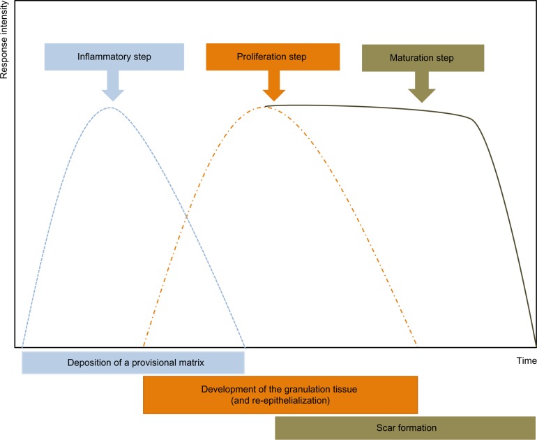
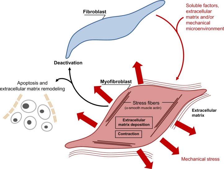
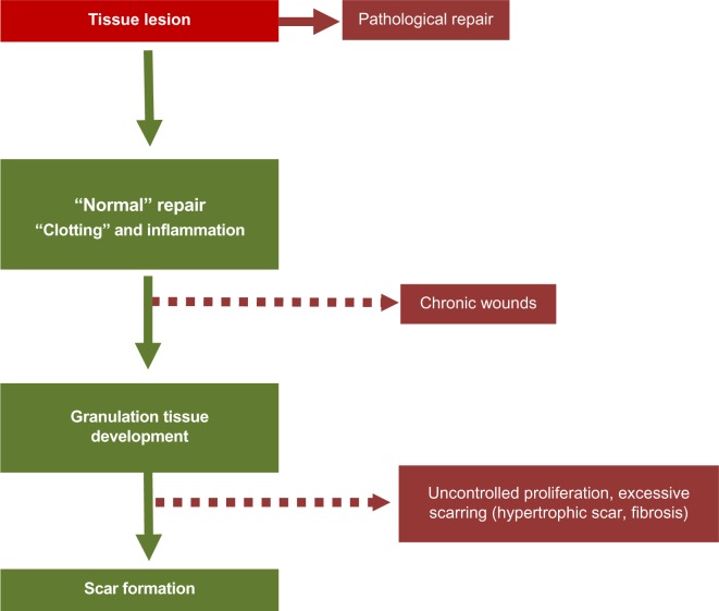
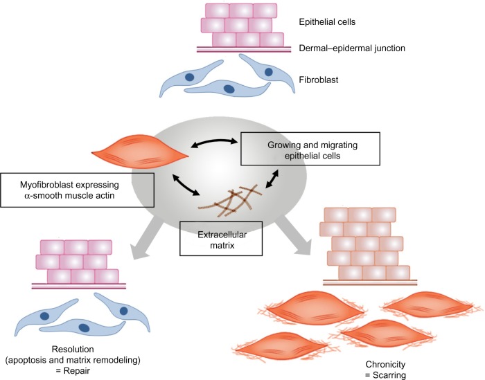
Verification
Claims the paper makes
The claims and statistics of this paper haven’t been checked yet.
Run the check — free, no account needed. It reads the open-access text and checks the sources it cites.
Claims other papers make about this one
Each is checked against this paper’s own text: does it say what the citing paper says it does?
In the citing paper
“In addition to contraction of wound beds and generation of collagen, myofibroblasts contribute to remodeling by release of MMPs that degrade collagen laid down during granulation tissue formation [96, 97].”
From Immunology of Wound Healing (2018) · cites this paper as [96]
Self-check
Percentages, sums and reported differences re-computed from the paper’s own numbers.
The self-check needs the paper’s full text.
Citations
Cites
Multilineage potential of adult human mesenchymal stem cells
Pittenger, M. F., Mackay, A. M., Beck, S. C., Jaiswal, R. K., Douglas, R., Mosca, J. D., Moorman, M. A., Simonetti, D. W., Craig, S., & Marshak, D. R. (1999) · Science · doi:10.1126/science.284.5411.143 · Cited by 21,052
Singer, A. J., & Clark, R. A. F. (1999) · New England Journal of Medicine · doi:10.1056/nejm199909023411006 · Cited by 6,106
Visse, R., & Nagase, H. (2003) · Circulation Research · doi:10.1161/01.res.0000070112.80711.3d · Cited by 4,575
Kalluri, R., & Zeisberg, M. (2006) · Nature Reviews Cancer · doi:10.1038/nrc1877 · Cited by 4,538
Myofibroblasts and mechano-regulation of connective tissue remodelling
Tomasek, J. J., Gabbiani, G., Hinz, B., Chaponnier, C., & Brown, R. A. (2002) · Nature Reviews Molecular Cell Biology · doi:10.1038/nrm809 · Cited by 4,290
Effects of substrate stiffness on cell morphology, cytoskeletal structure, and adhesion
Yeung, T., Georges, P. C., Flanagan, L. A., Marg, B., Ortiz, M., Funaki, M., Zahir, N., Ming, W., Weaver, V., & Janmey, P. A. (2005) · Cell Motility and the Cytoskeleton · doi:10.1002/cm.20041 · Cited by 2,325
TGF-β signaling and the fibrotic response
Leask, A., & Abraham, D. J. (2004) · The FASEB Journal · doi:10.1096/fj.03-1273rev · Cited by 2,319
Endothelial/Pericyte Interactions
Armulik, A., Abramsson, A., & Betsholtz, C. (2005) · Circulation Research · doi:10.1161/01.res.0000182903.16652.d7 · Cited by 2,048
Formation and function of the myofibroblast during tissue repair
Hinz, B. (2007) · Journal of Investigative Dermatology · doi:10.1038/sj.jid.5700613 · Cited by 1,658
Presence of modified fibroblasts in granulation tissue and their possible role in wound contraction
Gabbiani, G., Ryan, G. B., & Majno, G. (1971) · Experientia · doi:10.1007/bf02147594 · Cited by 1,485
Cited by
Sorted by citations
Items 101–200 of 1,000
Current concepts in the physiology of adult wound healing
Thiruvoth, F. M., Mohapatra, D. P., Sivakumar, D. K., Chittoria, R. K., & Nandhagopal, V. (2015) · Plastic and Aesthetic Research · doi:10.4103/2347-9264.158851 · Cited by 115
Wound healing related agents: Ongoing research and perspectives
Kaplani, K., Koutsi, S., Armenis, V., Skondra, F. G., Karantzelis, N., Tsaniras, S. C., & Taraviras, S. L. (2018) · Advanced Drug Delivery Reviews · doi:10.1016/j.addr.2018.02.007 · Cited by 110
Veerasubramanian, P. K., Ponrasu, T., Kannan, R., Chakraborty, S., Ramachandran, B., Suguna, L., & Muthuvijayan, V. (2018) · Colloids and Surfaces B Biointerfaces · doi:10.1016/j.colsurfb.2018.02.022 · Cited by 102
Role of photobiomodulation on the activation of the Smad pathway via TGF-β in wound healing
Mokoena, D. R., Kumar, S. S. D., Houreld, N. N., & Abrahamse, H. (2018) · Journal of Photochemistry and Photobiology B Biology · doi:10.1016/j.jphotobiol.2018.10.011 · Cited by 104
Li, Q., Zhao, H., Chen, W., Huang, P., & Bi, J. (2019) · The International Journal of Biochemistry & Cell Biology · doi:10.1016/j.biocel.2019.105570 · Cited by 101
Kay, E. J., Koulouras, G., & Zanivan, S. R. (2021) · Frontiers in Oncology · doi:10.3389/fonc.2021.719922 · Cited by 105
Levesque, C., & Nelson, P. S. (2017) · Cold Spring Harbor Perspectives in Medicine · doi:10.1101/cshperspect.a030510 · Cited by 101
Schanuel, F. S., Santos, K. S. R., Monte‐Alto‐Costa, A., & de Oliveira, M. G. (2015) · Colloids and Surfaces B Biointerfaces · doi:10.1016/j.colsurfb.2015.04.007 · Cited by 96
Corrêa, F. R. S., Schanuel, F. S., Moura‐Nunes, N., Monte‐Alto‐Costa, A., & Daleprane, J. B. (2016) · Biomedicine & Pharmacotherapy · doi:10.1016/j.biopha.2016.12.018 · Cited by 99
Beyond photoaging: additional factors involved in the process of skin aging
Addor, F. A. S. (2018) · Clinical Cosmetic and Investigational Dermatology · doi:10.2147/ccid.s177448 · Cited by 98
Keloid pathophysiology: fibroblast or inflammatory disorders?
Nangole, F. W., & Agak, G. W. (2019) · JPRAS Open · doi:10.1016/j.jpra.2019.09.004 · Cited by 101
Reprogramming of profibrotic macrophages for treatment of bleomycin‐induced pulmonary fibrosis
Zhang, F., Ayaub, E. A., Wang, B., Puchulu‐Campanella, E., Li, Y., Hettiarachchi, S. U., Lindeman, S. D., Luo, Q., Rout, S., Srinivasarao, M., et al. (2020) · EMBO Molecular Medicine · doi:10.15252/emmm.202012034 · Cited by 96
Cancer-associated fibroblasts mediate cancer progression and remodel the tumouroid stroma
Pape, J., Magdeldin, T., Stamati, K., Nyga, A., Loizidou, M., Emberton, M., & Cheema, U. (2020) · British Journal of Cancer · doi:10.1038/s41416-020-0973-9 · Cited by 95
Lassance, L., Marino, G. K., Medeiros, C. S., Thangavadivel, S., & Wilson, S. E. (2018) · Experimental Eye Research · doi:10.1016/j.exer.2018.02.018 · Cited by 92
Molecular Mechanisms and Cellular Contribution from Lung Fibrosis to Lung Cancer Development
Samarelli, A. V., Masciale, V., Aramini, B., Coló, G. P., Tonelli, R., Marchioni, A., Bruzzi, G., Gozzi, F., Andrisani, D., Castaniere, I., et al. (2021) · International Journal of Molecular Sciences · doi:10.3390/ijms222212179 · Cited by 94
Gao, Y. H., Sun, Y., Yang, H., Qiu, P., Cong, Z., Zou, Y., Song, L., Guo, J., & Anastassiades, T. (2019) · International Journal of Molecular Sciences · doi:10.3390/ijms20153722 · Cited by 95
Mihalko, E. P., Huang, K., Sproul, E. P., Cheng, K., & Brown, A. C. (2018) · ACS Nano · doi:10.1021/acsnano.8b01977 · Cited by 89
Role of matrix metalloproteinases in diabetic foot ulcers: Potential therapeutic targets
Fu, K., Zheng, X., Chen, Y., Wu, L., Yang, Z., Chen, X., & Song, W. (2022) · Frontiers in Pharmacology · doi:10.3389/fphar.2022.1050630 · Cited by 89
Song, L., Zhang, W., Tang, S., Luo, S., Xiong, P., Liu, J., Hu, H., Chen, Y., Jia, B., Yan, Q., et al. (2023) · Biomedicine & Pharmacotherapy · doi:10.1016/j.biopha.2023.116039 · Cited by 93
Myofibroblast progeny in wound biology and wound healing studies
Monika, P., Waiker, P. V., Chandraprabha, M. N., Rangarajan, A., & Murthy, K. N. C. (2021) · Wound Repair and Regeneration · doi:10.1111/wrr.12937 · Cited by 90
Mechanoregulation of Wound Healing and Skin Homeostasis
Rosińćzuk, J., Taradaj, J., Dymarek, R., & Sopel, M. (2016) · BioMed Research International · doi:10.1155/2016/3943481 · Cited by 91
Electrospun Biomaterials in the Treatment and Prevention of Scars in Skin Wound Healing
Mulholland, E. J. (2020) · Frontiers in Bioengineering and Biotechnology · doi:10.3389/fbioe.2020.00481 · Cited by 89
Post-intubation subglottic stenosis: aetiology at the cellular and molecular level
Dorris, E., Russell, J., & Murphy, M. (2021) · European Respiratory Review · doi:10.1183/16000617.0218-2020 · Cited by 91
Modulation of Wound Healing and Scar Formation by MG53 Protein-mediated Cell Membrane Repair
Li, H., Duann, P., Lin, P., Zhao, L., Fan, Z., Tan, T., Zhou, X., Sun, M., Fu, M., Orange, M., et al. (2015) · Journal of Biological Chemistry · doi:10.1074/jbc.m115.680074 · Cited by 86
Cerium oxide nanoparticles in wound care: a review of mechanisms and therapeutic applications
Chen, S., Wang, Y., Bao, S., Li, Y., Fu, X., Yu, Y., Lyu, H., Pang, H., Guo, S., Zhang, H., et al. (2024) · Frontiers in Bioengineering and Biotechnology · doi:10.3389/fbioe.2024.1404651 · Cited by 88
Epidermal SIRT1 regulates inflammation, cell migration, and wound healing
Qiang, L., Sample, A., Liu, H., Wu, X., & He, Y. (2017) · Scientific Reports · doi:10.1038/s41598-017-14371-3 · Cited by 84
Wound healing in post-smolt Atlantic salmon (Salmo salar L.)
Sveen, L. R., Timmerhaus, G., Krasnov, A., Takle, H., Handeland, S. O., & Ytteborg, E. (2019) · Scientific Reports · doi:10.1038/s41598-019-39080-x · Cited by 86
Yu, Y., Mei, L., Shi, Y., Zhang, X., Cheng, K., Cao, F., Zhang, L., Jia, X., Li, X.-M., & Xu, Z. (2020) · Journal of Materials Chemistry B · doi:10.1039/c9tb02300c · Cited by 83
Irreversibility of Pulmonary Fibrosis
Yu, Q. Y., & Tang, X. X. (2022) · Aging and Disease · doi:10.14336/ad.2021.0730 · Cited by 83
The Role of Serotonin during Skin Healing in Post-Thermal Injury
Sadiq, A., Shah, A., Jeschke, M. G., Belo, C., Hayat, M. Q., Murad, S., & Amini‐Nik, S. (2018) · International Journal of Molecular Sciences · doi:10.3390/ijms19041034 · Cited by 81
Stem Cells and Tissue Engineering
Strong, A. L., Neumeister, M. W., & Lévi, B. (2017) · Clinics in Plastic Surgery · doi:10.1016/j.cps.2017.02.020 · Cited by 85
Chen, X., Zhang, M., Chen, S., Wang, X., Tian, Z., Chen, Y., Xu, P., Zhang, L., Zhang, L., & Zhang, L. (2017) · Cell Transplantation · doi:10.1177/0963689717721216 · Cited by 80
Human Adipose Mesenchymal Stem Cell-Derived Exosomes: A Key Player in Wound Healing
Heo, J. S., Kim, S., Yang, C. E., Choi, Y., Song, S. Y., & Kim, H. O. (2021) · Tissue Engineering and Regenerative Medicine · doi:10.1007/s13770-020-00316-x · Cited by 80
Kalva, S. N., Augustine, R., Mamun, A. A., Dalvi, Y. B., Vijay, N., & Hasan, A. (2021) · Journal of Drug Delivery Science and Technology · doi:10.1016/j.jddst.2021.102500 · Cited by 80
Qin, X., He, J., Wang, X., Wang, J., Yang, R., & Chen, X. (2023) · Frontiers in Immunology · doi:10.3389/fimmu.2023.1256687 · Cited by 80
Electrical stimulation: a novel therapeutic strategy to heal biological wounds
Preetam, S., Ghosh, A., Mishra, R., Pandey, A., Roy, D. S., Rustagi, S., & Malik, S. (2024) · RSC Advances · doi:10.1039/d4ra04258a · Cited by 83
Mohanty, C., & Pradhan, J. (2020) · Materials Science and Engineering C · doi:10.1016/j.msec.2020.110751 · Cited by 78
Basalova, N., Sagaradze, G., Arbatskiy, M., Evtushenko, E., Kulebyakin, K., Grigorieva, O., Akopyan, Z., Kalinina, N., & Efimenko, A. (2020) · Cells · doi:10.3390/cells9051272 · Cited by 78
Scarring Skin: Mechanisms and Therapies
Lin, X., & Lai, Y. (2024) · International Journal of Molecular Sciences · doi:10.3390/ijms25031458 · Cited by 79
PKCδ inhibition normalizes the wound-healing capacity of diabetic human fibroblasts
Khamaisi, M., Katagiri, S., Keenan, H. A., Park, K., Maeda, Y., Li, Q., Qi, W., Thomou, T., Eschuk, D., Tellechea, A., et al. (2016) · Journal of Clinical Investigation · doi:10.1172/jci82788 · Cited by 83
TGFβ-induced fibroblast activation requires persistent and targeted HDAC-mediated gene repression
Jones, D. L., Haak, A. J., Caporarello, N., Choi, K. M., Ye, Z., Yan, H., Varelas, X., Ordog, T., Ligresti, G., & Tschumperlin, D. J. (2019) · Journal of Cell Science · doi:10.1242/jcs.233486 · Cited by 77
LeBert, D. C., Squirrell, J. M., Freisinger, C., Rindy, J., Golenberg, N., Frecentese, G., Gibson, A. L. F., Eliceiri, K. W., & Huttenlocher, A. (2018) · eLife · doi:10.7554/elife.30703 · Cited by 77
Reproducible strategy for excisional skin-wound-healing studies in mice
Yampolsky, M., Bachelet, I., & Fuchs, Y. (2023) · Nature Protocols · doi:10.1038/s41596-023-00899-4 · Cited by 79
MSC-released TGF-ß regulate a-SMA expression of myofibroblast during wound healing
Putra, A. B., Alif, I., Hamra, N. F., Santosa, O., Kustiyah, A. R., Muhar, A. M., & Lukman, K. (2020) · Journal of Stem Cells and Regenerative Medicine · doi:10.46582/jsrm.1602011 · Cited by 81
Khan, I., Rahman, S. U., Tang, E., Engel, K. W., Hall, B., Kulkarni, A. B., & Arany, P. R. (2021) · Scientific Reports · doi:10.1038/s41598-021-92650-w · Cited by 81
Rameshbabu, A. P., Bankoti, K., Datta, S., Subramani, E., Apoorva, A., Ghosh, P., Maity, P. P., Manchikanti, P., Chaudhury, K., & Dhara, S. (2018) · ACS Applied Materials & Interfaces · doi:10.1021/acsami.7b19007 · Cited by 76
Best, K. T., Nichols, A. E. C., Knapp, E. K., Hammert, W. C., Ketonis, C., Jonason, J. H., Awad, H. A., & Loiselle, A. E. (2020) · Science Signaling · doi:10.1126/scisignal.abb7209 · Cited by 75
Assar, D. H., El‐Habashi, N., Mokhbatly, A.-A. A., Ragab, A. E., Elbialy, Z. I., Rizk, S. A., Albalawi, A. E., Althobaiti, N. A., Jaouni, S. A., & Atiba, A. S. (2021) · Biomedicine & Pharmacotherapy · doi:10.1016/j.biopha.2021.112151 · Cited by 74
Dang, L. H., Nguyen, H. T., Tran, H. L. B., Doan, V. N., & Trân, N. Q. (2018) · Journal of Healthcare Engineering · doi:10.1155/2018/5754890 · Cited by 73
Heterogeneity of the NIH3T3 Fibroblast Cell Line
Rahimi, A. M., Cai, M., & Hoyer‐Fender, S. (2022) · Cells · doi:10.3390/cells11172677 · Cited by 79
Morbidelli, L., Genah, S., & Cialdai, F. (2021) · Frontiers in Bioengineering and Biotechnology · doi:10.3389/fbioe.2021.720091 · Cited by 74
Liao, H.-T., Lai, Y.-T., Kuo, C., & Chen, J. (2020) · Materials Science and Engineering C · doi:10.1016/j.msec.2020.111689 · Cited by 72
Ibañez, R. I. R., do Amaral, R. J. F. C., Reis, R. L., Marques, A. P., Murphy, C. M., & O’Brien, F. J. (2021) · Polymers · doi:10.3390/polym13152510 · Cited by 71
Immunomodulatory Properties of Host Defence Peptides in Skin Wound Healing
Petkovic, M., Mouritzen, M. V., Mojsoska, B., & Jenssen, H. (2021) · Biomolecules · doi:10.3390/biom11070952 · Cited by 69
Tryptophan metabolites kynurenine and serotonin regulate fibroblast activation and fibrosis
Dolivo, D., Larson, S. A., & Dominko, T. (2018) · Cellular and Molecular Life Sciences · doi:10.1007/s00018-018-2880-2 · Cited by 71
IPF pathogenesis is dependent upon TGFβ induction of IGF‐1
Hernandez, D. M., Kang, J. H., Choudhury, M., Andrianifahanana, M., Yin, X., LIMPER, A. H., & Leof, E. B. (2020) · The FASEB Journal · doi:10.1096/fj.201901719rr · Cited by 68
Berry-Kilgour, C., Cabral, J. D., & Wise, L. M. (2020) · Advances in Wound Care · doi:10.1089/wound.2020.1183 · Cited by 68
Fine Regulation during Wound Healing by Mast Cells, a Physiological Role Not Yet Clarified
Bacci, S. (2022) · International Journal of Molecular Sciences · doi:10.3390/ijms23031820 · Cited by 66
Wang, Q., Liu, C., An, J., Liu, J., Wang, Y., & Cai, Y. (2025) · Frontiers in Endocrinology · doi:10.3389/fendo.2025.1657928 · Cited by 69
BMP1-like proteinases are essential to the structure and wound healing of skin
Muir, A. M., Massoudi, D., Nguyen, N. T., Keene, D. R., Lee, S., Birk, D. E., Davidson, J. M., Marinkovich, M. P., & Greenspan, D. S. (2016) · Matrix Biology · doi:10.1016/j.matbio.2016.06.004 · Cited by 66
Marchioni, A., Tonelli, R., Andreani, A., Cappiello, G. F., Fermi, M., Trentacosti, F., Castaniere, I., Fantini, R., Tabbì, L., Andrisani, D., et al. (2022) · International Journal of Molecular Sciences · doi:10.3390/ijms23052421 · Cited by 65
Skin immunity in wound healing and cancer
Jakovija, A., & Chtanova, T. (2023) · Frontiers in Immunology · doi:10.3389/fimmu.2023.1060258 · Cited by 70
Garg, S. S., Dubey, R., Sharma, S., Vyas, A., & Gupta, J. (2023) · International Journal of Biological Macromolecules · doi:10.1016/j.ijbiomac.2023.125636 · Cited by 64
Mignon, C., Uzunbajakava, N. E., Castellano‐Pellicena, I., Botchkareva, N. V., & Tobin, D. J. (2018) · Lasers in Surgery and Medicine · doi:10.1002/lsm.22823 · Cited by 65
Norton, K., Jin, K., & Popel, A. S. (2018) · Journal of Theoretical Biology · doi:10.1016/j.jtbi.2018.05.003 · Cited by 66
Isolation and Characterization of Mouse Fibroblasts
Edelman, B., & Redente, E. F. (2018) · Methods in molecular biology · doi:10.1007/978-1-4939-8570-8_5 · Cited by 64
Zhang, D. X., Vu, L. T., Ismail, N. N., Le, M. T. N., & Grimson, A. (2021) · Seminars in Cancer Biology · doi:10.1016/j.semcancer.2021.01.007 · Cited by 64
GÜZEL, S., Özay, Y., Kumaş, M., Uzun, Ç., Özkorkmaz, E. G., Yıldırım, Z., Ülger, M., Güler, G., Çelık, A., Çamlıca, Y., et al. (2019) · Biomedicine & Pharmacotherapy · doi:10.1016/j.biopha.2019.01.038 · Cited by 63
The wound healing effect of four types of beta-glucan
Seo, G., Hyun, C. L., Choi, S., Kim, Y., & Cho, M. (2019) · Applied Biological Chemistry · doi:10.1186/s13765-019-0428-2 · Cited by 66
Matrisome Properties of Scaffolds Direct Fibroblasts in Idiopathic Pulmonary Fibrosis
Elowsson, L., Löfdahl, A., Åhrman, E., Müller, C., Notermans, T., Michaliková, B., Rosmark, O., Zhou, X.-H., Dellgren, G., Silverborn, M., et al. (2019) · International Journal of Molecular Sciences · doi:10.3390/ijms20164013 · Cited by 64
Pereira, D., & Sequeira, I. (2021) · Frontiers in Cell and Developmental Biology · doi:10.3389/fcell.2021.682143 · Cited by 65
Anti-inflammatory and wound healing properties of lactic acid bacteria and its peptides
Saravanan, P., Pooja, R., Balachander, N., Kesav Ram Singh, K., Silpa, S., & Rupachandra, S. (2023) · Folia Microbiologica · doi:10.1007/s12223-022-01030-y · Cited by 64
Geometry and network connectivity govern the mechanics of stress fibers
Kassianidou, E., Brand, C. A., Schwarz, U. S., & Kumar, S. (2017) · Proceedings of the National Academy of Sciences · doi:10.1073/pnas.1606649114 · Cited by 62
Fibroblast heterogeneity: Keystone of tissue homeostasis and pathology in inflammation and ageing
Gauthier, V., Kyriazi, M., Nefla, M., Pucino, V., Raza, K., Buckley, C. D., & Alsaleh, G. (2023) · Frontiers in Immunology · doi:10.3389/fimmu.2023.1137659 · Cited by 63
Bromma, K., Cicon, L., Beckham, W., & Chithrani, D. B. (2020) · Scientific Reports · doi:10.1038/s41598-020-68994-0 · Cited by 61
Seo, G., Lim, Y., Koh, D., Huh, J., Hyun, C. L., Kim, Y., & Cho, M. (2017) · Experimental & Molecular Medicine · doi:10.1038/emm.2016.167 · Cited by 61
Corneal myofibroblasts inhibit regenerating nerves during wound healing
Jeon, K., Hindman, H. B., Bubel, T. M., McDaniel, T., DeMagistris, M., Callan, C. L., & Huxlin, K. R. (2018) · Scientific Reports · doi:10.1038/s41598-018-30964-y · Cited by 60
Induction of Fibroblast Senescence During Mouse Corneal Wound Healing
Wang, X., Qu, M., Li, J., Danielson, P., Yang, L., & Zhou, Q. (2019) · Investigative Ophthalmology & Visual Science · doi:10.1167/iovs.19-26983 · Cited by 64
Chaudhary, A., Bag, S., Banerjee, P., & Chatterjee, J. (2019) · Journal of Traditional and Complementary Medicine · doi:10.1016/j.jtcme.2019.10.002 · Cited by 61
Li, C., An, Y., Sun, Y., Yang, F., Xu, Q., & Wang, Z. (2022) · Stem Cell Reviews and Reports · doi:10.1007/s12015-022-10378-0 · Cited by 60
Stoppel, W. L., Gao, A. E., Greaney, A. M., Partlow, B. P., Bretherton, R. C., Kaplan, D. L., & Black, L. D. (2016) · Journal of Biomedical Materials Research Part A · doi:10.1002/jbm.a.35850 · Cited by 59
Xu, K., An, N., Zhang, H., Zhang, Q., Zhang, K., Hu, X., Wu, Y., Wu, F., Xiao, J., Zhang, H., et al. (2019) · Journal of Drug Delivery Science and Technology · doi:10.1016/j.jddst.2019.101405 · Cited by 58
Cellular crosstalk in fibrosis: Insights into macrophage and fibroblast dynamics
Froom, Z. S. C. S., Callaghan, N. I., & Huyer, L. D. (2025) · Journal of Biological Chemistry · doi:10.1016/j.jbc.2025.110203 · Cited by 64
Kilvaer, T. K., Rakaee, M., Hellevik, T., Östman, A., Strell, C., Bremnes, R. M., Busund, L., Dønnem, T., & Martínez-Zubiaurre, I. (2018) · PLoS ONE · doi:10.1371/journal.pone.0192157 · Cited by 58
Quirós, M., & Nusrat, A. (2018) · Annual Review of Physiology · doi:10.1146/annurev-physiol-020518-114504 · Cited by 59
Merkt, W., Zhou, Y., Han, H., & Lagares, D. (2021) · Wound Repair and Regeneration · doi:10.1111/wrr.12952 · Cited by 62
The role of cytokines in wound healing: from mechanistic insights to therapeutic applications
Wong, R. S.-Y., Tan, T., Pang, A. S., & Srinivasan, D. K. (2025) · Exploration of Immunology · doi:10.37349/ei.2025.1003183 · Cited by 62
Schnieder, J., Mamazhakypov, A., Birnhuber, A., Wilhelm, J., Kwapiszewska, G., Ruppert, C., Markart, P., Wujak, L. A., Rubio, K., Barreto, G., et al. (2019) · Matrix Biology · doi:10.1016/j.matbio.2019.12.001 · Cited by 58
An, R., Zhang, Y., Qiao, Y., Song, L., Wang, H., & Dong, X. (2020) · Stem Cell Research & Therapy · doi:10.1186/s13287-020-01621-x · Cited by 56
Mu, R., de Souza, S. C., Liao, Z., Dong, L., & Wang, C. (2022) · Advanced Drug Delivery Reviews · doi:10.1016/j.addr.2022.114298 · Cited by 57
Smart bandage: A device for wound monitoring and targeted treatment
Hossain, M. I., Zahid, M. S., Chowdhury, M. A., Hossain, M. M. M., Hossain, N., Islam, M. A., & Mobarak, M. H. (2023) · Results in Chemistry · doi:10.1016/j.rechem.2023.101292 · Cited by 56
Advancements in 3D skin bioprinting: processes, bioinks, applications and sensor integration
Derman, İ. D., Rivera, T., Garriga-Cerda, L., Singh, Y. P., Saini, S., Abaci, H. E., & Ozbolat, I. T. (2024) · International Journal of Extreme Manufacturing · doi:10.1088/2631-7990/ad878c · Cited by 55
Skin telocytes versus fibroblasts: two distinct dermal cell populations
Kang, Y., Zhu, Z., Zheng, Y., Wan, W., Manole, C. G., & Zhang, Q. (2015) · Journal of Cellular and Molecular Medicine · doi:10.1111/jcmm.12671 · Cited by 55
Lebonvallet, N., Laverdet, B., Misery, L., Desmoulière, A., & Girard, D. (2018) · Experimental Dermatology · doi:10.1111/exd.13681 · Cited by 54
Self-citationMultifunctional Natural Polymer Nanoparticles as Antifibrotic Gene Carriers for CKD Therapy
Midgley, A. C., Wei, Y., Zhu, D., Gao, F., Yan, H., Khalique, A., Luo, W., Jiang, H., Liu, X., Guo, J., et al. (2020) · Journal of the American Society of Nephrology · doi:10.1681/asn.2019111160 · Cited by 53
Transforming growth factor-β signalling pathway in tendon healing
Li, Y., Liu, X., Liu, X., Peng, Y., Zhu, B., Guo, S., Wang, C., Wang, D., & Li, S. (2022) · Growth Factors · doi:10.1080/08977194.2022.2082294 · Cited by 53
Staruch, R. M. T., Glass, G. E., Rickard, R. F., Hettiaratchy, S. P., & Butler, P. E. M. (2016) · Tissue Engineering Part B Reviews · doi:10.1089/ten.teb.2016.0305 · Cited by 53
Multifunctional alginate/polydeoxyribonucleotide hydrogels for promoting diabetic wound healing
Dananjaya, S. H. S., Bandara, N., Molagoda, I. M. N., Sandamalika, W. M. G., Kim, D., Ganepola, N., Attanayake, A. P., & Choi, D. (2023) · International Journal of Biological Macromolecules · doi:10.1016/j.ijbiomac.2023.128367 · Cited by 52
Nhi, T. T., Khon, H. C., Nguyen, T. T. H., Bao, B. C., Quyen, T. N., Toi, V. V., & Nguyen, T.-H. (2016) · Journal of Materials Science Materials in Medicine · doi:10.1007/s10856-016-5768-4 · Cited by 52
Mohtashami, M., Mohamadi, M., Azimi–Nezhad, M., Saeidi, J., Nia, F. F., & Ghasemi, A. (2020) · Biotechnology and Applied Biochemistry · doi:10.1002/bab.2064 · Cited by 53
Metadata
- Authors
- Desmouliere, A., Darby, I. A., Laverdet, B., & Bonté, F.
- Affiliations
- RMIT University (AU)
- Université de Limoges (FR)
- LVMH (France) (FR)
- Venue
- Clinical, Cosmetic and Investigational Dermatology
- Publisher
- Informa UK Limited
- Published
- 2014-11
- Volume
- 7
- Pages
- 301
- ISSN
- 1178-7015 (ISSN-L)
- PMC
- PMC4226391
- OpenAlex
- W2023805473
- Cited by
- 1,039
- References
- 76
- Citations by year
- 2026: 43 · 2025: 98 · 2024: 103 · 2023: 115 · 2022: 121 · 2021: 123 · 2020: 117 · 2019: 108 · 2018: 91 · 2017: 61 · 2016: 37 · 2015: 21
- FWCI
- 4.89
- Type
- journal article
- Language
- en
- Open access
- gold
- doi.org
- Licence
- Locations
- Clinical Cosmetic and Investigational Dermatology (publishedVersion, cc-by-nc)
- Europe PMC (PubMed Central) (publishedVersion, cc-by-nc)
- PubMed Central (publishedVersion, cc-by-nc)
- PubMed (publishedVersion)
- RMIT Research Repository (RMIT University Library) (submittedVersion)
- Dove Medical Press (Taylor and Francis Group) (submittedVersion, cc-by-nc)
- Figshare (submittedVersion)
- Topics
- Health Sciences › Medicine › Surgery › Wound Healing and Treatments
- Health Sciences › Medicine › Dermatology › Dermatologic Treatments and Research
- Health Sciences › Medicine › Hepatology › Liver physiology and pathology
- Keywords
- fibroblasts, myofibroblasts, wound healing, skin homeostasis, extracellular matrix, matrix metalloproteinases, tissue inhibitors of metalloproteinases, extracellular matrix remodeling, growth factor bioavailability, mechanical signaling, fibrosis, scar formation, skin aging, wrinkle formation, cell-microenvironment interactions
About these checks
Registry, retraction, Cites and statistics results are computed, not judged, with one exception: a printed reference that no index lists is read once by a model before it counts as a citation error. Claims are extracted by a model and kept only when their quote appears verbatim in the text; each cited source is then checked, with a model’s help, for whether it says what the claim relies on. Those verdicts can be wrong — read the claim’s quote and the cited source before relying on one.
Statistics and arithmetic are recomputed, not judged.
Confidence bands are shown only for fields calibrated on our annotated test set. None is calibrated yet, so no band is shown.
What counts as a claim ›How we measure confidence ›How we check this ›
Are you an author, or do you see a problem with this page? Request a review
