

The DOI matches its Crossref record; 75 of the 76 references it cites were found in a registry or an index.
Fibroblasts and myofibroblasts in wound healing
Open accessAbstract
(Myo)fibroblasts are key players for maintaining skin homeostasis and for orchestrating physiological tissue repair. (Myo)fibroblasts are embedded in a sophisticated extracellular matrix (ECM) that they secrete, and a complex and interactive dialogue exists between (myo)fibroblasts and their microenvironment. In addition to the secretion of the ECM, (myo)fibroblasts, by secreting matrix metalloproteinases and tissue inhibitors of metalloproteinases, are able to remodel this ECM. (Myo)fibroblasts and their microenvironment form an evolving network during tissue repair, with reciprocal actions leading to cell differentiation, proliferation, quiescence, or apoptosis, and actions on growth factor bioavailability by binding, sequestration, and activation. In addition, the (myo)fibroblast phenotype is regulated by mechanical stresses to which they are subjected and thus by mechanical signaling. In pathological situations (excessive scarring or fibrosis), or during aging, this dialogue between the (myo)fibroblasts and their microenvironment may be altered or disrupted, leading to repair defects or to injuries with damaged and/or cosmetic skin alterations such as wrinkle development. The intimate dialogue between the (myo)fibroblasts and their microenvironment therefore represents a fascinating domain that must be better understood in order not only to characterize new therapeutic targets and drugs able to prevent or treat pathological developments but also to interfere with skin alterations observed during normal aging or premature aging induced by a deleterious environment.
Overview
The claims and statistics of this paper haven’t been checked yet.
Run the check — free, no account needed. It reads the open-access text and checks the sources it cites.
Integrity signals
Registry
Cites
Statistics
Figures
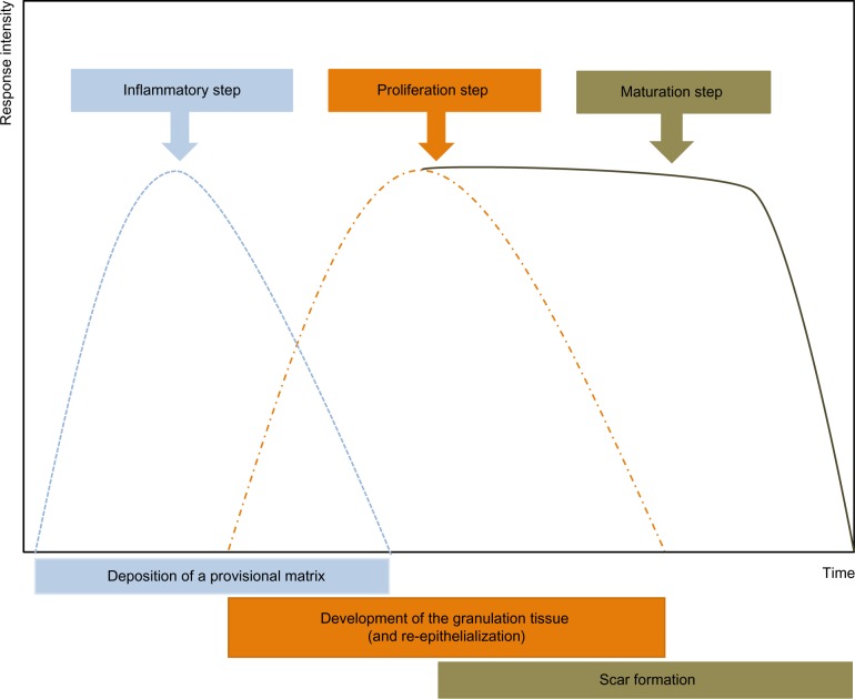
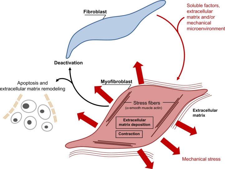
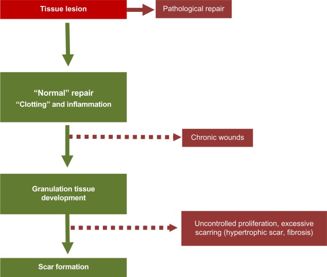
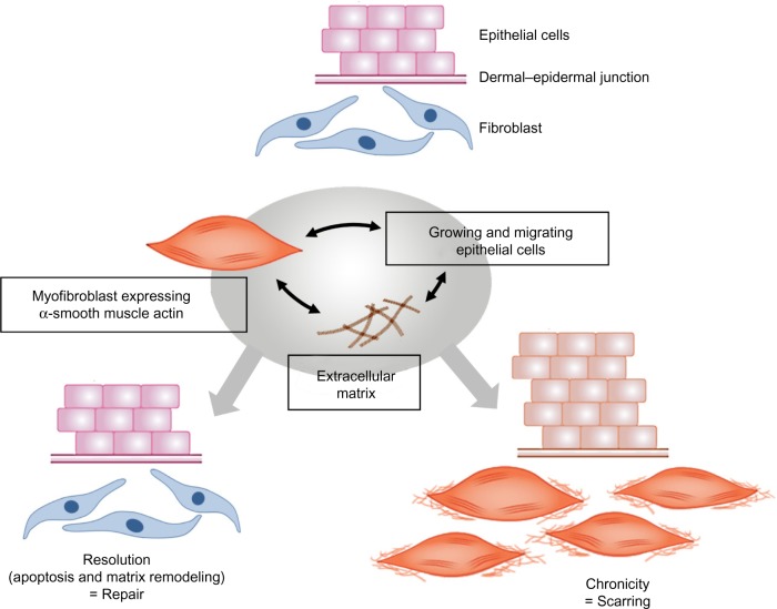
Verification
Claims the paper makes
The claims and statistics of this paper haven’t been checked yet.
Run the check — free, no account needed. It reads the open-access text and checks the sources it cites.
Claims other papers make about this one
Each is checked against this paper’s own text: does it say what the citing paper says it does?
In the citing paper
“In addition to contraction of wound beds and generation of collagen, myofibroblasts contribute to remodeling by release of MMPs that degrade collagen laid down during granulation tissue formation [96, 97].”
From Immunology of Wound Healing (2018) · cites this paper as [96]
Self-check
Percentages, sums and reported differences re-computed from the paper’s own numbers.
The self-check needs the paper’s full text.
Citations
Cites
Multilineage potential of adult human mesenchymal stem cells
Pittenger, M. F., Mackay, A. M., Beck, S. C., Jaiswal, R. K., Douglas, R., Mosca, J. D., Moorman, M. A., Simonetti, D. W., Craig, S., & Marshak, D. R. (1999) · Science · doi:10.1126/science.284.5411.143 · Cited by 21,052
Singer, A. J., & Clark, R. A. F. (1999) · New England Journal of Medicine · doi:10.1056/nejm199909023411006 · Cited by 6,106
Visse, R., & Nagase, H. (2003) · Circulation Research · doi:10.1161/01.res.0000070112.80711.3d · Cited by 4,575
Kalluri, R., & Zeisberg, M. (2006) · Nature Reviews Cancer · doi:10.1038/nrc1877 · Cited by 4,538
Myofibroblasts and mechano-regulation of connective tissue remodelling
Tomasek, J. J., Gabbiani, G., Hinz, B., Chaponnier, C., & Brown, R. A. (2002) · Nature Reviews Molecular Cell Biology · doi:10.1038/nrm809 · Cited by 4,290
Effects of substrate stiffness on cell morphology, cytoskeletal structure, and adhesion
Yeung, T., Georges, P. C., Flanagan, L. A., Marg, B., Ortiz, M., Funaki, M., Zahir, N., Ming, W., Weaver, V., & Janmey, P. A. (2005) · Cell Motility and the Cytoskeleton · doi:10.1002/cm.20041 · Cited by 2,325
TGF-β signaling and the fibrotic response
Leask, A., & Abraham, D. J. (2004) · The FASEB Journal · doi:10.1096/fj.03-1273rev · Cited by 2,319
Endothelial/Pericyte Interactions
Armulik, A., Abramsson, A., & Betsholtz, C. (2005) · Circulation Research · doi:10.1161/01.res.0000182903.16652.d7 · Cited by 2,048
Formation and function of the myofibroblast during tissue repair
Hinz, B. (2007) · Journal of Investigative Dermatology · doi:10.1038/sj.jid.5700613 · Cited by 1,658
Presence of modified fibroblasts in granulation tissue and their possible role in wound contraction
Gabbiani, G., Ryan, G. B., & Majno, G. (1971) · Experientia · doi:10.1007/bf02147594 · Cited by 1,485
Cited by
Sorted by citations
Items 401–500 of 1,000
Higashijima, F., Hasegawa, M., Yoshimoto, T., Kobayashi, Y., Wakuta, M., & Kimura, K. (2023) · Frontiers in Ophthalmology · doi:10.3389/fopht.2022.1060087 · Cited by 24
Rahul, V. G., Ellur, G., Gaber, A. A., Govindappa, P. K., & Elfar, J. C. (2024) · Cell Death Discovery · doi:10.1038/s41420-024-02199-6 · Cited by 23
Ucci, S., Caradonna, E., Aliberti, A., & Cusano, A. (2025) · Frontiers in Bioengineering and Biotechnology · doi:10.3389/fbioe.2025.1675619 · Cited by 23
Scarano, A., Lorusso, F., Postiglione, F., Mastrangelo, F., & Petrini, M. (2020) · Journal of Oral and Maxillofacial Surgery · doi:10.1016/j.joms.2020.09.008 · Cited by 23
An overview of bio-actuation in collagen hydrogels: a mechanobiological phenomenon
Hameed, P., & Manivasagam, G. (2021) · Biophysical Reviews · doi:10.1007/s12551-021-00804-x · Cited by 26
Protective effects of intranasal curcumin on silica-induced lung damage
Kumari, S., & Singh, R. (2022) · Cytokine · doi:10.1016/j.cyto.2022.155949 · Cited by 23
Radstake, W. E., Gautam, K., Miranda, S., Vermeesen, R., Tabury, K., Rehnberg, E., Buset, J., Janssen, A., Leysen, L., Neefs, M., et al. (2023) · Cells · doi:10.3390/cells12020246 · Cited by 23
Heterotypic signaling of cancer-associated fibroblasts in shaping the cancer cell drug resistance
Butti, R., Khaladkar, A., Bhardwaj, P., & Prakasam, G. (2023) · Cancer Drug Resistance · doi:10.20517/cdr.2022.72 · Cited by 23
Gravitational effects on fibroblasts’ function in relation to wound healing
Radstake, W. E., Gautam, K., Miranda, S., Rompay, C. V., Vermeesen, R., Tabury, K., Verslegers, M., Dowson, A., Gorissen, J., van Loon, J. J. W. A., et al. (2023) · npj Microgravity · doi:10.1038/s41526-023-00286-z · Cited by 23
Du, H., Jiang, L. H., Geng, W., Li, J., Zhang, R., Dang, J.-G., Shu, M., & Li, L. (2017) · BioMed Research International · doi:10.1155/2017/2578017 · Cited by 23
Asai, E., Yamamoto, M., Ueda, K., & Waguri, S. (2018) · FUKUSHIMA JOURNAL OF MEDICAL SCIENCE · doi:10.5387/fms.2016-13 · Cited by 23
Farrell, E. C., Aliabouzar, M., Quesada, C., Baker, B. M., Franceschi, R. T., Putnam, A. J., & Fabiilli, M. L. (2021) · Acta Biomaterialia · doi:10.1016/j.actbio.2021.11.020 · Cited by 23
Iosăgeanu, A., Ștefan, L. M., Crăciunescu, O., & Cîmpean, A. (2024) · Life · doi:10.3390/life14091187 · Cited by 23
Pathogenesis, attenuation, and treatment strategies for keloid management
Mishra, T., & Wairkar, S. (2025) · Tissue and Cell · doi:10.1016/j.tice.2025.102800 · Cited by 22
Sterile inflammation in the pathogenesis of maturation failure of arteriovenous fistula
Nguyen, M., Thankam, F. G., & Agrawal, D. K. (2021) · Journal of Molecular Medicine · doi:10.1007/s00109-021-02056-4 · Cited by 22
Li, Y., Yu, Z., Zhao, D., & Han, D. (2021) · Life Science · doi:10.1016/j.lfs.2021.119483 · Cited by 20
Cellular Heterogeneity and Plasticity of Skin Epithelial Cells in Wound Healing and Tumorigenesis
Wang, J., He, J., Zhu, M., Han, Y. Y., Yang, R., Liu, H., Xu, X., & Chen, X. (2022) · Stem Cell Reviews and Reports · doi:10.1007/s12015-021-10295-8 · Cited by 22
Dehydrozingerone promotes healing of diabetic foot ulcers: a molecular insight
Begum, F., Manandhar, S., Kumar, G., Keni, R., Sankhe, R., Gurram, P. C., Beegum, F., Meka, S. T., Nandakumar, K., & Shenoy, R. R. (2022) · Journal of Cell Communication and Signaling · doi:10.1007/s12079-022-00703-0 · Cited by 22
Tang, R., Shang, L., Scumpia, P. O., & Carlo, D. D. (2023) · Advanced Healthcare Materials · doi:10.1002/adhm.202302477 · Cited by 22
Metalloproteinases as Biomarkers and Sociomarkers in Human Health and Disease
Costa, D., Scalise, E., Ielapi, N., Bracale, U. M., Andreucci, M., & Serra, R. (2024) · Biomolecules · doi:10.3390/biom14010096 · Cited by 23
Heat delays skin wound healing in mice
Silva, M. A. D. S., Trajano, E. T. L., Schanuel, F. S., & Monte‐Alto‐Costa, A. (2016) · Experimental Biology and Medicine · doi:10.1177/1535370216675066 · Cited by 22
Effects of nicotine on corneal wound healing following acute alkali burn
Kim, J., Lim, C. W., & Kim, B. (2017) · PLoS ONE · doi:10.1371/journal.pone.0179982 · Cited by 22
Hanumantharao, S. N., Que, C., & Rao, S. (2019) · Materialia · doi:10.1016/j.mtla.2019.100296 · Cited by 22
Mendes, L. G., Ferreira, F. V., Sielski, M. S., Livi, S., Rocco, S. A., Sforça, M. L., Burga-Sánchez, J., Vicente, C. P., & Mei, L. H. I. (2021) · ACS Applied Bio Materials · doi:10.1021/acsabm.1c00402 · Cited by 22
Yan, L., Fan, D., Yang, J., Wang, J., Hu, X., Zhang, X., Huang, Y., Wang, H., Yin, W., Cai, X., et al. (2025) · Burns & Trauma · doi:10.1093/burnst/tkaf035 · Cited by 21
Sulfasalazine Resolves Joint Stiffness in a Rabbit Model of Arthrofibrosis
Atluri, K., Brouillette, M. J., Seol, D., Khorsand, B., Sander, E. A., Salem, A. K., Fredericks, D. C., Petersen, E. B., Smith, S. M., Fowler, T. P., et al. (2019) · Journal of Orthopaedic Research® · doi:10.1002/jor.24499 · Cited by 21
Cell-specific drug targeting in the lung
Abed, S., Turner, R. A., Serniuck, N. J., Tat, V., Naiel, S., Hayat, A. I., Mekhael, O., Vierhout, M., Ask, K., & Rullo, A. F. (2021) · Biochemical Pharmacology · doi:10.1016/j.bcp.2021.114577 · Cited by 21
Tang, Z., Wang, X., Yang, J., Song, X., Huang, Y., Chen, C., Yang, H., Fu, Z., Gong, X., & Chen, G. (2021) · ACS Biomaterials Science & Engineering · doi:10.1021/acsbiomaterials.0c01647 · Cited by 21
Nichols, A. E. C., Muscat, S. N., Miller, S. E., Green, L. J., Richards, M. S., & Loiselle, A. E. (2021) · The FASEB Journal · doi:10.1096/fj.202100405r · Cited by 21
Alhussan, A., Palmerley, N., Smazynski, J., Karasinska, J. M., Renouf, D. J., Schaeffer, D. F., Beckham, W., Alexander, A., & Chithrani, D. B. (2022) · Cancers · doi:10.3390/cancers14153586 · Cited by 21
EGCG Restores Keratinocyte Autophagy to Promote Diabetic Wound Healing through the AMPK/ULK1 Pathway
Tian, C., Feng, Y., Chen, T., Zhang, Z., He, X., Jiang, L., & Liu, M. (2023) · Frontiers in Bioscience-Landmark · doi:10.31083/j.fbl2812324 · Cited by 21
Wei, S., Li, Z., Xia, H., Wang, Z., Deng, J., Lu, L., Huang, R., Ye, T., Huang, Y., & Yang, Y. (2024) · International Journal of Biological Macromolecules · doi:10.1016/j.ijbiomac.2024.131723 · Cited by 21
Kouhbananinejad, S. M., Derakhshani, A., Vahidi, R., Dabiri, S., Fatemi, A., Armin, F., & Farsinejad, A. (2019) · Biomaterials Science · doi:10.1039/c8bm01377b · Cited by 21
Xu, N., Chen, S., Qu, G., Li, X., Lin, W., Xue, X., Lin, Y., Zheng, Q., & Wei, Y. (2017) · PubMed Central · Cited by 21
Skin and Wound Healing: Conventional Dosage versus Nanobased Emulsions Forms
Silva, J. V. V. D., Pereira, J. O. D. S. L., do Carmo, F. A., & Patricio, B. F. D. C. (2025) · ACS Omega · doi:10.1021/acsomega.5c00455 · Cited by 20
Petryk, N. M., Thai, N. L. B., Saldanha, L. V., Sutherland, S. T., & Monroe, M. B. B. (2025) · ACS Applied Materials & Interfaces · doi:10.1021/acsami.5c02532 · Cited by 20
Calcium Phosphate Ceramics Can Prevent Bisphosphonate-Related Osteonecrosis of the Jaw
Paulo, S., Laranjo, M., Paula, A. B., Abrantes, A. M., Martins, J. F. B., Marto, C. M., Coelho, A. S. E. D. C., Casalta‐Lopes, J., Carvalho, L., Carrilho, E., et al. (2020) · Materials · doi:10.3390/ma13081955 · Cited by 20
Wang, R. L., Chen, B., Wei, H., Yan, W., Wu, Y., Cao, W. F., Zhang, B., Liu, F., Tian, H., Chen, D., et al. (2022) · Materials Today Bio · doi:10.1016/j.mtbio.2022.100260 · Cited by 20
Signaling Pathways Associated with Chronic Wound Progression: A Systems Biology Approach
Basu, P., & Martins‐Green, M. M. (2022) · Antioxidants · doi:10.3390/antiox11081506 · Cited by 21
Saúde-Conde, R., Öztürk, A. A., Stosic, K., Senar, O. A., Navez, J., Bouchart, C., Arsenijevic, T., Flamen, P., & Laethem, J. V. (2024) · Biomedicines · doi:10.3390/biomedicines12030591 · Cited by 20
Kurian, A. G., Mandakhbayar, N., Singh, R. K., Lee, J., & Kim, H. (2024) · ACS Applied Materials & Interfaces · doi:10.1021/acsami.4c05636 · Cited by 20
Görtz, G.-E., Moshkelgosha, S., Jesenek, C., Edelmann, B., Horstmann, M., Banga, J. P., Eckstein, A. K., & Berchner‐Pfannschmidt, U. (2016) · Endocrinology · doi:10.1210/en.2016-1304 · Cited by 20
Seo, G., Hyun, C. L., Koh, D., Park, S., Lim, Y., Kim, Y., & Cho, M. (2018) · International Journal of Molecular Sciences · doi:10.3390/ijms19041164 · Cited by 20
Phosphorylation of the glucocorticoid receptor alters SMAD signaling in vocal fold fibroblasts
Mukudai, S., Hiwatashi, N., Bing, R., Garabedian, M. J., & Branski, R. C. (2018) · The Laryngoscope · doi:10.1002/lary.27570 · Cited by 21
Coupling synthetic biology and programmable materials to construct complex tissue ecosystems
Millar‐Haskell, C. S., Dang, A. M., & Gleghorn, J. P. (2019) · MRS Communications · doi:10.1557/mrc.2019.69 · Cited by 22
Larson, S. A., Dolivo, D., & Dominko, T. (2019) · Cell Biology International · doi:10.1002/cbin.11220 · Cited by 20
Luo, X., Gao, Z. ‐., Lin, S., Tong, M. ‐., Liu, L., Lin, L., Ke, W., & Yang, T. (2019) · Journal of the European Academy of Dermatology and Venereology · doi:10.1111/jdv.16162 · Cited by 20
Seyedian, R., Isavi, F., Najafiasl, M., & Zaeri, S. (2021) · Journal of Drug Delivery Science and Technology · doi:10.1016/j.jddst.2021.102528 · Cited by 20
Microplasma Treatment versus Negative Pressure Therapy for Promoting Wound Healing in Diabetic Mice
Shao, P., Liao, J., Wu, S., Chen, Y.-H., & Wong, T. (2021) · International Journal of Molecular Sciences · doi:10.3390/ijms221910266 · Cited by 20
Dual effects of bisphenol A on wound healing, involvement of estrogen receptor β
Wen, X., Zhu, M., Li, Z., Li, T., & Xu, X. (2022) · Ecotoxicology and Environmental Safety · doi:10.1016/j.ecoenv.2022.113207 · Cited by 20
5-Aminolaevulinic Acid-Based Photodynamic Therapy Restrains Pathological Hyperplasia of Fibroblasts
Wang, X., Cao, P., Liu, J., Du, P. F., Wang, Z., Chen, W., Liu, C., & Wu, Y. (2017) · Medical Science Monitor · doi:10.12659/msm.898221 · Cited by 20
Harman, R. M., Bihun, I. V., & de Walle, G. R. V. (2017) · Wound Repair and Regeneration · doi:10.1111/wrr.12515 · Cited by 20
Self-assembled fibrinogen nanofibers support fibroblast adhesion and prevent E. coli infiltration
Suter, N., Joshi, A., Wunsch, T., Graupner, N., Stapelfeldt, K., Radmacher, M., Müssig, J., & Brüggemann, D. (2021) · Materials Science and Engineering C · doi:10.1016/j.msec.2021.112156 · Cited by 19
Therapeutic downregulation of neuronal PAS domain 2 (Npas2) promotes surgical skin wound healing
Shibuya, Y., Hokugo, A., Okawa, H., Kondo, T., Khalil, D., Wang, L., Roca, Y., Clements, A., Sasaki, H., Berry, E., et al. (2022) · eLife · doi:10.7554/elife.71074 · Cited by 20
Role of MMP3 and fibroblast-MMP14 in skin homeostasis and repair
Kümper, M., Zamek, J., Steinkamp, J., Pach, E., Mauch, C., & Zigrino, P. (2022) · European Journal of Cell Biology · doi:10.1016/j.ejcb.2022.151276 · Cited by 20
The role of tumor microenvironment reprogramming in primary liver cancer chemotherapy resistance
Zhao, C., Liu, S., Gao, F., Zou, Y., Ren, Z., & Yu, Z. (2022) · Frontiers in Oncology · doi:10.3389/fonc.2022.1008902 · Cited by 19
3D bioprinting: opportunities for wound dressing development
Wang, X., Wang, Y., Teng, Y., Shi, J., Yang, X., Ding, Z., Guo, X., Hou, S., & Lv, Q. (2023) · Biomedical Materials · doi:10.1088/1748-605x/ace228 · Cited by 19
A novel lncRNA-mediated epigenetic regulatory mechanism in periodontitis
Zhu, Z. X., Liu, Y., Wang, J., Xie, Y., Li, R. Y., Ma, Q., Tu, Q., Melhem, N. A., Couldwell, S., El-Araby, R. E., et al. (2023) · International Journal of Biological Sciences · doi:10.7150/ijbs.87977 · Cited by 19
Greener healing: sustainable nanotechnology for advanced wound care
Sharda, D., Attri, K., & Choudhury, D. (2024) · Discover Nano · doi:10.1186/s11671-024-04061-1 · Cited by 22
Evaluation of a Hyaluronic Acid-Based Biomaterial to Enhance Wound Healing in the Equine Distal Limb
Dahlgren, L. A., Milton, S. C., Boswell, S. G., Werre, S. R., Brewster, C. C., Jones, C. S., & Crisman, M. V. (2016) · Journal of Equine Veterinary Science · doi:10.1016/j.jevs.2016.05.011 · Cited by 19
Bayrami, Z., Hajiaghaee, R., Khalighi‐Sigaroodi, F., Rahimi, R., Farzaei, M. H., Hodjat, M., Baeeri, M., Rahimifard, M., Nigjeh, M. N., & Abdollahi, M. R. (2018) · Journal of Ethnopharmacology · doi:10.1016/j.jep.2018.08.002 · Cited by 19
Azmi, L., Shukla, I., Goutam, A., Allauddin, Rao, C. V., Jawaid, T., Kamal, M., Awaad, A. S., Alqasoumi, S. I., & Alkhamees, O. A. (2019) · Saudi Pharmaceutical Journal · doi:10.1016/j.jsps.2019.01.017 · Cited by 20
Arif, S., Larochelle, S., & Moulin, V. J. (2020) · Journal of Cell Communication and Signaling · doi:10.1007/s12079-020-00572-5 · Cited by 19
Arfian, N., Suzuki, Y., Hartopo, A. B., Anggorowati, N., Nugrahaningsih, D. A. A., & Emoto, N. (2020) · Life Sciences · doi:10.1016/j.lfs.2020.118223 · Cited by 19
Sodium alginate-based composites as a collagen substitute for skin bioengineering
Solovieva, E. V., Teterina, A. Y., Klein, O. I., Комлев, В. С., Alekseev, A. A., & Panteleyev, A. A. (2020) · Biomedical Materials · doi:10.1088/1748-605x/abb524 · Cited by 19
Narayanan, K. B., Park, G. T., & Han, S. S. (2020) · Food Control · doi:10.1016/j.foodcont.2020.107695 · Cited by 19
Gellan gum-gelatin viscoelastic hydrogels as scaffolds to promote fibroblast differentiation
Xu, Z., Zhang, L., Bentil, S. A., & Bratlie, K. M. (2021) · Materials Science and Engineering C · doi:10.1016/j.msec.2021.112370 · Cited by 19
Mitochondrial homeostasis: Exploring their impact on skin disease pathogenesis
Jamil, H. A., & Karim, N. A. (2025) · Biomedicine & Pharmacotherapy · doi:10.1016/j.biopha.2025.118349 · Cited by 19
Evaluation of native and non‐native biomaterials for engineering human skin tissue
Catarino, C. M., Kaiser, K., Baltazar, T., Catarino, L. M., Brewer, J. R., & Karande, P. (2022) · Bioengineering & Translational Medicine · doi:10.1002/btm2.10297 · Cited by 18
Investigations on the Wound Healing Potential of Tilapia Piscidin (TP)2-5 and TP2-6
Liu, C.-W., Hsieh, C.-Y., & Chen, J. (2022) · Marine Drugs · doi:10.3390/md20030205 · Cited by 18
Mechanobiology in the Comorbidities of Ehlers Danlos Syndrome
Royer, S. P., & Han, S. J. (2022) · Frontiers in Cell and Developmental Biology · doi:10.3389/fcell.2022.874840 · Cited by 19
Advancement and Future Perspective of FAPI PET/CT In Gynecological Malignancies
Dendl, K., Koerber, S. A., Tamburini, K., Mori, Y., Cardinale, J., Haberkorn, U., & Giesel, F. L. (2022) · Seminars in Nuclear Medicine · doi:10.1053/j.semnuclmed.2022.04.002 · Cited by 18
Emerging role of BMPs/BMPR2 signaling pathway in treatment for pulmonary fibrosis
Ye, Q., Taleb, S. J., Zhao, J., & Zhao, Y. (2024) · Biomedicine & Pharmacotherapy · doi:10.1016/j.biopha.2024.117178 · Cited by 18
Silveira, L. L., Sarandy, M. M., Novaes, R. D., Santos, M. M., & Gonçalves, R. V. (2024) · International Journal of Molecular Sciences · doi:10.3390/ijms25168992 · Cited by 18
Ohta, S., Matsuura, M., Kawashima, Y., Cai, X., Taniguchi, M., Okochi, H., Asano, Y., Sato, S., & Ito, T. (2019) · Colloids and Surfaces B Biointerfaces · doi:10.1016/j.colsurfb.2019.04.028 · Cited by 18
Injectable Scaffolds Enriched with Silver to Inhibit Bacterial Invasion in Tissue Regeneration
Ceresa, C., Fracchia, L., Marchetti, A., Rinaldi, M., & Bosetti, M. (2019) · Materials · doi:10.3390/ma12121931 · Cited by 19
Razali, R. A., Lokanathan, Y., Yazid, M. D., Ansari, A. S., Saim, A. B., & Idrus, R. B. H. (2019) · International Journal of Molecular Sciences · doi:10.3390/ijms20143492 · Cited by 18
Zerdoum, A. B., Fowler, E. W., & Jia, X. (2019) · ACS Biomaterials Science & Engineering · doi:10.1021/acsbiomaterials.9b00425 · Cited by 20
Zooming in across the Skin: A Macro-to-Molecular Panorama
Deniz, A. A. H., Abdik, E. A., Abdik, H., Aydın, S., Şahin, F., & Taşlı, P. N. (2019) · Advances in experimental medicine and biology · doi:10.1007/5584_2019_442 · Cited by 19
Silva, R. S. D. L., Pessoa, D. R., Mariano, R. R., Castro, A. B. S., de Oliveira, R. A., & Ferraresi, C. (2020) · Photochemistry and Photobiology · doi:10.1111/php.13262 · Cited by 18
Wang, C., Wu, T., Liu, G., Cheng, R., Fei, J., Song, X., Chai, Y., Fan, C., Liu, X., Cui, W., et al. (2020) · Chemical Engineering Journal · doi:10.1016/j.cej.2020.125414 · Cited by 18
Cheng, W.-S., Chen, C., Chen, J., Lin, L.-T., Pao, S., Chen, Y., & Lu, D. (2020) · Molecules · doi:10.3390/molecules25194422 · Cited by 18
Arbab, S., Ullah, H., Muhammad, N., Wang, W., & Zhang, J. (2024) · Frontiers in Bioengineering and Biotechnology · doi:10.3389/fbioe.2024.1488748 · Cited by 17
Actions of thrombin in the interstitium
de Ridder, G. G., Lundblad, R. L., & Pizzo, S. V. (2015) · Journal of Thrombosis and Haemostasis · doi:10.1111/jth.13191 · Cited by 17
Tahri, S., Maarof, M., Masri, S., Man, R. C., Masmoudi, H., & Fauzi, M. B. (2023) · Frontiers in Bioengineering and Biotechnology · doi:10.3389/fbioe.2023.1200618 · Cited by 17
Hu, J., Wang, Z., Hu, H., Zhao, J., Li, H., Zhang, X., Bi, J., & Li, J. (2023) · Acta Biomaterialia · doi:10.1016/j.actbio.2023.10.009 · Cited by 17
Yahaya, E. S., Cordier, W., Steenkamp, P. A., & Steenkamp, V. (2018) · South African Journal of Botany · doi:10.1016/j.sajb.2018.06.003 · Cited by 17
Qian, W., Li, N., Cao, Q., & Fan, J. (2018) · Journal of Cellular Physiology · doi:10.1002/jcp.26877 · Cited by 17
Boncler, M. A., Lukasiak, M., Dastych, J., Golański, J., & Watała, C. W. (2018) · Basic & Clinical Pharmacology & Toxicology · doi:10.1111/bcpt.13117 · Cited by 17
Semaphorin 3A potentiates the profibrotic effects of transforming growth factor-β1 in the cornea
Jeon, K., Nehrke, K. W., & Huxlin, K. R. (2019) · Biochemical and Biophysical Research Communications · doi:10.1016/j.bbrc.2019.10.107 · Cited by 17
The Gastrointestinal Tumor Microenvironment: An Updated Biological and Clinical Perspective
Batista, S., Gregório, A. C., Otake, A. H., Couto, N., & Costa‐Silva, B. (2019) · Journal of Oncology · doi:10.1155/2019/6240505 · Cited by 17
Zehra, M., Curry, J. C., Pillai, S. S., Lakhani, H. V., Edwards, C. E., & Sodhi, K. (2020) · International Journal of Molecular Sciences · doi:10.3390/ijms21134737 · Cited by 17
Chellini, F., Tani, A., Zecchi‐Orlandini, S., Giannelli, M., & Sassoli, C. (2020) · Applied Sciences · doi:10.3390/app10217878 · Cited by 17
Alhussan, A., Bromma, K., Bozdoğan, E. P. D., Metcalfe, A. L., Karasinska, J. M., Beckham, W., Alexander, A., Renouf, D. J., Schaeffer, D. F., & Chithrani, D. B. (2021) · Current Oncology · doi:10.3390/curroncol28030183 · Cited by 17
Davis, A. M., Rapley, A., Dawson, W. O., Young, L. S., & Morris, M. A. (2021) · Pathogens · doi:10.3390/pathogens10080982 · Cited by 17
Kafili, G., Tamjid, E., & Simchi, A. (2024) · Scientific Reports · doi:10.1038/s41598-024-80973-3 · Cited by 16
Shimura, T., Takahashi, Y., Saito, C., Maida, R., Sasatani, M., Kunoh, T., & Ushiyama, A. (2025) · Scientific Reports · doi:10.1038/s41598-025-04845-0 · Cited by 16
Yan, Q., Shi, S., Yang, G., Wan, S., Li, M., & Li, M. (2023) · ACS Biomaterials Science & Engineering · doi:10.1021/acsbiomaterials.3c00508 · Cited by 17
Neural dependency in wound healing and regeneration
Noble, A., Qubrosi, R., Cariba, S., Favaro, K. J., & Payne, S. L. (2023) · Developmental Dynamics · doi:10.1002/dvdy.650 · Cited by 18
Effect of earthworm active protein on fibroblast proliferation and its mechanism
Song, S., Wang, Y., Ji, K., Liang, H., & Ji, A. (2015) · Pharmaceutical Biology · doi:10.3109/13880209.2015.1073333 · Cited by 16
Metadata
- Authors
- Desmouliere, A., Darby, I. A., Laverdet, B., & Bonté, F.
- Affiliations
- RMIT University (AU)
- Université de Limoges (FR)
- LVMH (France) (FR)
- Venue
- Clinical, Cosmetic and Investigational Dermatology
- Publisher
- Informa UK Limited
- Published
- 2014-11
- Volume
- 7
- Pages
- 301
- ISSN
- 1178-7015 (ISSN-L)
- PMC
- PMC4226391
- OpenAlex
- W2023805473
- Cited by
- 1,039
- References
- 76
- Citations by year
- 2026: 43 · 2025: 98 · 2024: 103 · 2023: 115 · 2022: 121 · 2021: 123 · 2020: 117 · 2019: 108 · 2018: 91 · 2017: 61 · 2016: 37 · 2015: 21
- FWCI
- 4.89
- Type
- journal article
- Language
- en
- Open access
- gold
- doi.org
- Licence
- Locations
- Clinical Cosmetic and Investigational Dermatology (publishedVersion, cc-by-nc)
- Europe PMC (PubMed Central) (publishedVersion, cc-by-nc)
- PubMed Central (publishedVersion, cc-by-nc)
- PubMed (publishedVersion)
- RMIT Research Repository (RMIT University Library) (submittedVersion)
- Dove Medical Press (Taylor and Francis Group) (submittedVersion, cc-by-nc)
- Figshare (submittedVersion)
- Topics
- Health Sciences › Medicine › Surgery › Wound Healing and Treatments
- Health Sciences › Medicine › Dermatology › Dermatologic Treatments and Research
- Health Sciences › Medicine › Hepatology › Liver physiology and pathology
- Keywords
- fibroblasts, myofibroblasts, wound healing, skin homeostasis, extracellular matrix, matrix metalloproteinases, tissue inhibitors of metalloproteinases, extracellular matrix remodeling, growth factor bioavailability, mechanical signaling, fibrosis, scar formation, skin aging, wrinkle formation, cell-microenvironment interactions
About these checks
Registry, retraction, Cites and statistics results are computed, not judged, with one exception: a printed reference that no index lists is read once by a model before it counts as a citation error. Claims are extracted by a model and kept only when their quote appears verbatim in the text; each cited source is then checked, with a model’s help, for whether it says what the claim relies on. Those verdicts can be wrong — read the claim’s quote and the cited source before relying on one.
Statistics and arithmetic are recomputed, not judged.
Confidence bands are shown only for fields calibrated on our annotated test set. None is calibrated yet, so no band is shown.
What counts as a claim ›How we measure confidence ›How we check this ›
Are you an author, or do you see a problem with this page? Request a review
