

The DOI matches its Crossref record; 75 of the 76 references it cites were found in a registry or an index.
Fibroblasts and myofibroblasts in wound healing
Open accessAbstract
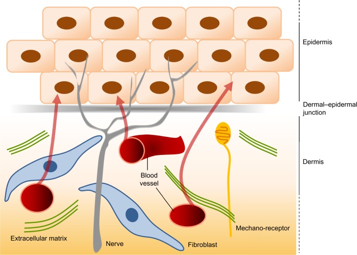
(Myo)fibroblasts are key players for maintaining skin homeostasis and for orchestrating physiological tissue repair. (Myo)fibroblasts are embedded in a sophisticated extracellular matrix (ECM) that they secrete, and a complex and interactive dialogue exists between (myo)fibroblasts and their microenvironment. In addition to the secretion of the ECM, (myo)fibroblasts, by secreting matrix metalloproteinases and tissue inhibitors of metalloproteinases, are able to remodel this ECM. (Myo)fibroblasts and their microenvironment form an evolving network during tissue repair, with reciprocal actions leading to cell differentiation, proliferation, quiescence, or apoptosis, and actions on growth factor bioavailability by binding, sequestration, and activation. In addition, the (myo)fibroblast phenotype is regulated by mechanical stresses to which they are subjected and thus by mechanical signaling. In pathological situations (excessive scarring or fibrosis), or during aging, this dialogue between the (myo)fibroblasts and their microenvironment may be altered or disrupted, leading to repair defects or to injuries with damaged and/or cosmetic skin alterations such as wrinkle development. The intimate dialogue between the (myo)fibroblasts and their microenvironment therefore represents a fascinating domain that must be better understood in order not only to characterize new therapeutic targets and drugs able to prevent or treat pathological developments but also to interfere with skin alterations observed during normal aging or premature aging induced by a deleterious environment.
Overview
The claims and statistics of this paper haven’t been checked yet.
Run the check — free, no account needed. It reads the open-access text and checks the sources it cites.
Integrity signals
Registry
Cites
Statistics
Figures
Verification
Claims the paper makes
The claims and statistics of this paper haven’t been checked yet.
Run the check — free, no account needed. It reads the open-access text and checks the sources it cites.
Claims other papers make about this one
Each is checked against this paper’s own text: does it say what the citing paper says it does?
In the citing paper
“In addition to contraction of wound beds and generation of collagen, myofibroblasts contribute to remodeling by release of MMPs that degrade collagen laid down during granulation tissue formation [96, 97].”
From Immunology of Wound Healing (2018) · cites this paper as [96]
Self-check
Percentages, sums and reported differences re-computed from the paper’s own numbers.
The self-check needs the paper’s full text.
Citations
Cites
Multilineage potential of adult human mesenchymal stem cells
Pittenger, M. F., Mackay, A. M., Beck, S. C., Jaiswal, R. K., Douglas, R., Mosca, J. D., Moorman, M. A., Simonetti, D. W., Craig, S., & Marshak, D. R. (1999) · Science · doi:10.1126/science.284.5411.143 · Cited by 21,052
Singer, A. J., & Clark, R. A. F. (1999) · New England Journal of Medicine · doi:10.1056/nejm199909023411006 · Cited by 6,106
Visse, R., & Nagase, H. (2003) · Circulation Research · doi:10.1161/01.res.0000070112.80711.3d · Cited by 4,575
Kalluri, R., & Zeisberg, M. (2006) · Nature Reviews Cancer · doi:10.1038/nrc1877 · Cited by 4,538
Myofibroblasts and mechano-regulation of connective tissue remodelling
Tomasek, J. J., Gabbiani, G., Hinz, B., Chaponnier, C., & Brown, R. A. (2002) · Nature Reviews Molecular Cell Biology · doi:10.1038/nrm809 · Cited by 4,290
Effects of substrate stiffness on cell morphology, cytoskeletal structure, and adhesion
Yeung, T., Georges, P. C., Flanagan, L. A., Marg, B., Ortiz, M., Funaki, M., Zahir, N., Ming, W., Weaver, V., & Janmey, P. A. (2005) · Cell Motility and the Cytoskeleton · doi:10.1002/cm.20041 · Cited by 2,325
TGF-β signaling and the fibrotic response
Leask, A., & Abraham, D. J. (2004) · The FASEB Journal · doi:10.1096/fj.03-1273rev · Cited by 2,319
Endothelial/Pericyte Interactions
Armulik, A., Abramsson, A., & Betsholtz, C. (2005) · Circulation Research · doi:10.1161/01.res.0000182903.16652.d7 · Cited by 2,048
Formation and function of the myofibroblast during tissue repair
Hinz, B. (2007) · Journal of Investigative Dermatology · doi:10.1038/sj.jid.5700613 · Cited by 1,658
Presence of modified fibroblasts in granulation tissue and their possible role in wound contraction
Gabbiani, G., Ryan, G. B., & Majno, G. (1971) · Experientia · doi:10.1007/bf02147594 · Cited by 1,485
Cited by
Sorted by citations
The biology and function of fibroblasts in cancer
Kalluri, R. (2016) · Nature Reviews Cancer · doi:10.1038/nrc.2016.73 · Cited by 4,366
Wound Healing: A Cellular Perspective
Rodrigues, M., Kosaric, N., Bonham, C. A., & Gurtner, G. C. (2018) · Physiological Reviews · doi:10.1152/physrev.00067.2017 · Cited by 3,211
Wound healing: cellular mechanisms and pathological outcomes
Wilkinson, H. N., & Hardman, M. J. (2020) · Open Biology · doi:10.1098/rsob.200223 · Cited by 1,975
Ellis, S., Lin, E. J., & Tartar, D. (2018) · Current Dermatology Reports · doi:10.1007/s13671-018-0234-9 · Cited by 625
Innovative Treatment Strategies to Accelerate Wound Healing: Trajectory and Recent Advancements
Kolimi, P., Narala, S., Nyavanandi, D., Youssef, A. A. A., & Dudhipala, N. R. (2022) · Cells · doi:10.3390/cells11152439 · Cited by 658
Preventing Engrailed-1 activation in fibroblasts yields wound regeneration without scarring
Mascharak, S., desJardins-Park, H. E., Davitt, M. F., Griffin, M., Borrelli, M. R., Moore, A. L., Chen, K., Duoto, B. A., Chinta, M. S., Foster, D. S., et al. (2021) · Science · doi:10.1126/science.aba2374 · Cited by 630
Evasion of apoptosis by myofibroblasts: a hallmark of fibrotic diseases
Hinz, B., & Lagares, D. (2019) · Nature Reviews Rheumatology · doi:10.1038/s41584-019-0324-5 · Cited by 642
Jahromi, M. A. M., Zangabad, P. S., Basri, S. M. M., Zangabad, K. S., Ghamarypour, A., Aref, A. R., Karimi, M., & Hamblin, M. R. (2017) · Advanced Drug Delivery Reviews · doi:10.1016/j.addr.2017.08.001 · Cited by 566
Skin tissue engineering: wound healing based on stem-cell-based therapeutic strategies
Dehkordi, A. N., Mirahmadi, F., Chehelgerdi, M., & Dehkordi, S. R. (2019) · Stem Cell Research & Therapy · doi:10.1186/s13287-019-1212-2 · Cited by 561
Myofibroblast proliferation and heterogeneity are supported by macrophages during skin repair
Shook, B. A., Wasko, R. R., Rivera-Gonzalez, G. C., Salazar-Gatzimas, E., López-Giráldez, F., Dash, B. C., Muñoz-Rojas, A. R., Aultman, K. D., Zwick, R. K., Lei, V., et al. (2018) · Science · doi:10.1126/science.aar2971 · Cited by 519
Scleral hypoxia is a target for myopia control
Wu, H., Chen, W., Zhao, F., Zhou, Q., Reinach, P. S., Deng, L., Ma, L., Luo, S., Srinivasalu, N., Pan, M., et al. (2018) · Proceedings of the National Academy of Sciences · doi:10.1073/pnas.1721443115 · Cited by 507
Cancer-Associated Fibroblasts Build and Secure the Tumor Microenvironment
Liu, T., Zhou, L., Li, D., Andl, T., & Zhang, Y. (2019) · Frontiers in Cell and Developmental Biology · doi:10.3389/fcell.2019.00060 · Cited by 446
Shiekh, P. A., Singh, A., & Kumar, A. (2020) · Biomaterials · doi:10.1016/j.biomaterials.2020.120020 · Cited by 443
Scaffolds for Wound Healing Applications
Neguț, I., Dorcioman, G., & Grumezescu, V. (2020) · Polymers · doi:10.3390/polym12092010 · Cited by 436
He, J., Liang, Y., Shi, M., & Guo, B. (2019) · Chemical Engineering Journal · doi:10.1016/j.cej.2019.123464 · Cited by 423
Myofibroblast transdifferentiation: The dark force in ocular wound healing and fibrosis
Shu, D. Y., & Lovicu, F. J. (2017) · Progress in Retinal and Eye Research · doi:10.1016/j.preteyeres.2017.08.001 · Cited by 394
Alteration of the Antitumor Immune Response by Cancer-Associated Fibroblasts
Ziani, L., Chouaïb, S., & Thiery, J. (2018) · Frontiers in Immunology · doi:10.3389/fimmu.2018.00414 · Cited by 391
Role of fibroblasts in wound healing and tissue remodeling on Earth and in space
Cialdai, F., Risaliti, C., & Monici, M. (2022) · Frontiers in Bioengineering and Biotechnology · doi:10.3389/fbioe.2022.958381 · Cited by 390
Wang, L., Hu, L., Zhou, X., Xiong, Z., Zhang, C., Shehada, H. M. A., Hu, B., Song, J., & Chen, L. (2017) · Scientific Reports · doi:10.1038/s41598-017-12919-x · Cited by 377
Cancer-associated fibroblasts and their influence on tumor immunity and immunotherapy
Barrett, R. L., & Puré, E. (2020) · eLife · doi:10.7554/elife.57243 · Cited by 357
Ayuk, S. M., Abrahamse, H., & Houreld, N. N. (2016) · Journal of Photochemistry and Photobiology B Biology · doi:10.1016/j.jphotobiol.2016.05.027 · Cited by 346
Yoshida, G. J. (2020) · Journal of Experimental & Clinical Cancer Research · doi:10.1186/s13046-020-01611-0 · Cited by 328
Smart Bandages: The Future of Wound Care
Derakhshandeh, H., Kashaf, S. S., Aghabaglou, F., Ghanavati, I., & Tamayol, A. (2018) · Trends in biotechnology · doi:10.1016/j.tibtech.2018.07.007 · Cited by 320
Casado‐Díaz, A., Quesada‐Gómez, J. M., & Dorado, G. (2020) · Frontiers in Bioengineering and Biotechnology · doi:10.3389/fbioe.2020.00146 · Cited by 325
Fibronectin in development and wound healing
Patten, J., & Wang, K. (2020) · Advanced Drug Delivery Reviews · doi:10.1016/j.addr.2020.09.005 · Cited by 317
TGF ‐β as a driver of fibrosis: physiological roles and therapeutic opportunities
Budi, E. H., Schaub, J. R., Decaris, M. L., Turner, S., & Derynck, R. (2021) · The Journal of Pathology · doi:10.1002/path.5680 · Cited by 310
C-Reactive Protein and Cancer—Diagnostic and Therapeutic Insights
Hart, P. C., Rajab, I. M., Alebraheem, M., & Potempa, L. A. (2020) · Frontiers in Immunology · doi:10.3389/fimmu.2020.595835 · Cited by 291
The Functional Role of Extracellular Matrix Proteins in Cancer
Popova, N. V., & Jücker, M. (2022) · Cancers · doi:10.3390/cancers14010238 · Cited by 300
Chronopoulos, A., Robinson, B. K., Sarper, M., Cortés, E., Auernheimer, V., Lachowski, D., Attwood, S. J., García, R., Ghassemi, S., Fabry, B., et al. (2016) · Nature Communications · doi:10.1038/ncomms12630 · Cited by 286
The therapeutic effects of 4-phenylbutyric acid in maintaining proteostasis
Kolb, P. S., Ayaub, E. A., Zhou, W., Yum, V., Dickhout, J. G., & Ask, K. (2015) · The International Journal of Biochemistry & Cell Biology · doi:10.1016/j.biocel.2015.01.015 · Cited by 271
Oral wound healing models and emerging regenerative therapies
Toma, A. I., Fuller, J. M., Willett, N. J., & Goudy, S. L. (2021) · Translational research · doi:10.1016/j.trsl.2021.06.003 · Cited by 263
The cellular basis of fibrotic tendon healing: challenges and opportunities
Nichols, A. E. C., Best, K. T., & Loiselle, A. E. (2019) · Translational research · doi:10.1016/j.trsl.2019.02.002 · Cited by 251
Huang, J., Heng, S., Zhang, W., Liu, Y., Xia, T., Ji, C., & Zhang, L.-J. (2022) · Seminars in Cell & Developmental Biology · doi:10.1016/j.semcdb.2022.02.027 · Cited by 238
Qiang, L., Yang, S., Cui, Y., & He, Y. (2020) · Autophagy · doi:10.1080/15548627.2020.1816342 · Cited by 238
The IL-4/IL-13 axis in skin fibrosis and scarring: mechanistic concepts and therapeutic targets
Nguyen, J. K., Austin, E., Huang, A., Mamalis, A., & Jagdeo, J. R. (2019) · Archives of Dermatological Research · doi:10.1007/s00403-019-01972-3 · Cited by 243
Bioglass Activated Skin Tissue Engineering Constructs for Wound Healing
Yu, H., Peng, J., Xu, Y., Chang, J., & Li, H. (2015) · ACS Applied Materials & Interfaces · doi:10.1021/acsami.5b09853 · Cited by 233
Amirian, J., Zeng, Y., Shekh, M. I., Sharma, G., Stadler, F. J., Song, J., Du, B., & Zhu, Y. (2020) · Carbohydrate Polymers · doi:10.1016/j.carbpol.2020.117005 · Cited by 225
Metabolic Cooperation and Competition in the Tumor Microenvironment: Implications for Therapy
Gupta, S., Roy, A., & Dwarakanath, B. S. R. (2017) · Frontiers in Oncology · doi:10.3389/fonc.2017.00068 · Cited by 225
Instructive microenvironments in skin wound healing: Biomaterials as signal releasing platforms
Castaño, Ó., Pérez‐Amodio, S., Navarro‐Requena, C., Mateos‐Timoneda, M. A., & Engel, E. (2018) · Advanced Drug Delivery Reviews · doi:10.1016/j.addr.2018.03.012 · Cited by 217
A Systematic Review on Classification, Identification, and Healing Process of Burn Wound Healing
Abazari, M., Ghaffari, A., Rashidzadeh, H., Badeleh, S. M., & Maleki, Y. (2020) · The International Journal of Lower Extremity Wounds · doi:10.1177/1534734620924857 · Cited by 216
Bioadhesives for internal medical applications: A review
Zhu, W., Chuah, Y. J., & Wang, D. (2018) · Acta Biomaterialia · doi:10.1016/j.actbio.2018.04.034 · Cited by 217
Jiang, S., Lyu, C., Zhao, P., Li, W., Kong, W., Huang, C., Genin, G. M., & Du, Y. (2019) · Nature Communications · doi:10.1038/s41467-019-11397-1 · Cited by 210
The tension biology of wound healing
Harn, H. I., Ogawa, R., Hsu, C., Hughes, M. W., Tang, M., & Chuong, C. (2017) · Experimental Dermatology · doi:10.1111/exd.13460 · Cited by 213
Loh, E. Y. X., Mohamad, N., Fauzi, M. B., Ng, M. H., Ng, S. F., & Amin, M. C. I. M. (2018) · Scientific Reports · doi:10.1038/s41598-018-21174-7 · Cited by 208
Targeting the renin-angiotensin-aldosterone system in fibrosis
Alqudah, M., Hale, T. M., & Czubryt, M. P. (2020) · Matrix Biology · doi:10.1016/j.matbio.2020.04.005 · Cited by 208
Dermal fibroblast in cutaneous development and healing
Thulabandu, V., Chen, D., & Atit, R. P. (2017) · Wiley Interdisciplinary Reviews Developmental Biology · doi:10.1002/wdev.307 · Cited by 213
Cancer-associated fibroblasts: Origin, function, imaging, and therapeutic targeting
Rimal, R., Desai, P. B., Daware, R., Hosseinnejad, A., Prakash, J., Lammers, T., & Singh, S. (2022) · Advanced Drug Delivery Reviews · doi:10.1016/j.addr.2022.114504 · Cited by 210
Advanced strategies to thwart foreign body response to implantable devices
Capuani, S., Malgir, G., Chua, C. Y. X., & Grattoni, A. (2022) · Bioengineering & Translational Medicine · doi:10.1002/btm2.10300 · Cited by 204
Savcı, Y., Kırbaş, O. K., Bozkurt, B. T., Abdik, E. A., Taşlı, P. N., Şahin, F., & Abdik, H. (2021) · Food & Function · doi:10.1039/d0fo02953j · Cited by 203
A Comprehensive Review of Natural Compounds for Wound Healing: Targeting Bioactivity Perspective
Trinh, T. X., Long, N. V., Anh, L. T. V., Nga, P. T., Giang, N. N., Chien, P. N., Nam, S., & Heo, C. Y. (2022) · International Journal of Molecular Sciences · doi:10.3390/ijms23179573 · Cited by 202
Keloids and Hypertrophic Scars: A Spectrum of Clinical Challenges
Trace, A. P., Enos, C., Mantel, A., & Harvey, V. M. (2016) · American Journal of Clinical Dermatology · doi:10.1007/s40257-016-0175-7 · Cited by 189
Naturally-derived electrospun wound dressings for target delivery of bio-active agents
Fahimirad, S., & Ajalloueian, F. (2019) · International Journal of Pharmaceutics · doi:10.1016/j.ijpharm.2019.05.053 · Cited by 191
Dermal adipose tissue has high plasticity and undergoes reversible dedifferentiation in mice
Zhang, Z., Shao, M., Hepler, C., Zi, Z., Zhao, S., An, Y., Zhu, Y., Ghaben, A. L., Wang, M.-Y., Li, N., et al. (2019) · Journal of Clinical Investigation · doi:10.1172/jci130239 · Cited by 191
Silk Fibroin Biomaterials and Their Beneficial Role in Skin Wound Healing
Mazurek, Ł., Szudzik, M., Rybka, M., & Konop, M. (2022) · Biomolecules · doi:10.3390/biom12121852 · Cited by 191
Development and use of biomaterials as wound healing therapies
Murray, R. Z., West, Z. E., Cowin, A. J., & Farrugia, B. L. (2019) · Burns & Trauma · doi:10.1186/s41038-018-0139-7 · Cited by 189
Direct conversion of injury-site myeloid cells to fibroblast-like cells of granulation tissue
Sinha, M., Sen, C. K., Singh, K., Das, A., Ghatak, S., Rhea, B., Blackstone, B. N., Powell, H. M., Khanna, S., & Roy, S. (2018) · Nature Communications · doi:10.1038/s41467-018-03208-w · Cited by 182
An Updated Review of Hypertrophic Scarring
Mony, M. P., Harmon, K. A., Hess, R., Dorafshar, A. H., & Shafikhani, S. H. (2023) · Cells · doi:10.3390/cells12050678 · Cited by 183
Functional electrospun fibers for the treatment of human skin wounds
Wang, J., & Windbergs, M. (2017) · European Journal of Pharmaceutics and Biopharmaceutics · doi:10.1016/j.ejpb.2017.07.001 · Cited by 176
He, J., Fang, B., Shan, S., Xie, Y., Wang, C., Zhang, Y., Zhang, X., & Li, Q. (2021) · Cell Death and Disease · doi:10.1038/s41419-021-03481-6 · Cited by 178
Topical gel-based biomaterials for the treatment of diabetic foot ulcers
Bardill, J. R., Laughter, M. R., Stager, M. A., Liechty, K. W., Krebs, M. D., & Zgheib, C. (2021) · Acta Biomaterialia · doi:10.1016/j.actbio.2021.10.045 · Cited by 171
Fibroblast senescence in the pathology of idiopathic pulmonary fibrosis
Waters, D. W., Blokland, K. E. C., Pathinayake, P. S., Burgess, J. K., Mutsaers, S. E., Prêle, C. M., Schuliga, M. J., Grainge, C., & Knight, D. A. (2018) · American Journal of Physiology-Lung Cellular and Molecular Physiology · doi:10.1152/ajplung.00037.2018 · Cited by 171
Jiang, T., Wang, Z., & Sun, J. (2020) · Stem Cell Research & Therapy · doi:10.1186/s13287-020-01723-6 · Cited by 167
FAP and FAPI-PET/CT in Malignant and Non-Malignant Diseases: A Perfect Symbiosis?
Dendl, K., Koerber, S. A., Kratochwil, C., Cardinale, J., Finck, R., Dabir, M., Novruzov, E., Watabe, T., Kramer, V., Choyke, P. L., et al. (2021) · Cancers · doi:10.3390/cancers13194946 · Cited by 164
Epithelial–mesenchymal transition in tissue repair and degeneration
Youssef, K. K., & Nieto, M. A. (2024) · Nature Reviews Molecular Cell Biology · doi:10.1038/s41580-024-00733-z · Cited by 165
Dai, X., Liu, J., Zheng, H., Wichmann, J., Hopfner, Ú., Sudhop, S., Prein, C., Shen, Y., Machens, H.-G., & Schilling, A. F. (2017) · NPG Asia Materials · doi:10.1038/am.2017.31 · Cited by 161
Wound Healing Effect of Kaempferol in Diabetic and Nondiabetic Rats
Özay, Y., Güzel, S., Yumrutaş, Ö., Pehlivanoğlu, B., Erdoğdu, İ. H., Yildirim, Z., Türk, B. A., & Darcan, S. (2019) · Journal of Surgical Research · doi:10.1016/j.jss.2018.08.009 · Cited by 158
Corneal injury: Clinical and molecular aspects
Barrientez, B., Nicholas, S. E., Whelchel, A. E., Sharif, R., Hjortdal, J., & Karamichos, D. (2019) · Experimental Eye Research · doi:10.1016/j.exer.2019.107709 · Cited by 156
Role of the Skin Immune System in Wound Healing
Cioce, A., Cavani, A., Cattani, C., & Scopelliti, F. (2024) · Cells · doi:10.3390/cells13070624 · Cited by 164
Microneedle arrays for the treatment of chronic wounds
Barnum, L., Samandari, M., Schmidt, T. A., & Tamayol, A. (2020) · Expert Opinion on Drug Delivery · doi:10.1080/17425247.2020.1819787 · Cited by 155
Yen, Y., Pu, C., Liu, C., Chen, Y., Chen, Y., Liang, C., Hsieh, J., Huang, H., & Chen, Y. (2018) · International Wound Journal · doi:10.1111/iwj.12904 · Cited by 154
Xu, Z., Li, Z., Jiang, S., & Bratlie, K. M. (2018) · ACS Omega · doi:10.1021/acsomega.8b00683 · Cited by 151
Gushiken, L. F. S., Beserra, F. P., Hussni, M. F., Gonzaga, M. T., Ribeiro, V. P., de Souza, P. F., Campos, J. C. L., Massaro, T. N. C., Hussni, C. A., Takahira, R. K., et al. (2022) · Oxidative Medicine and Cellular Longevity · doi:10.1155/2022/9004014 · Cited by 150
Ponrasu, T., Kannan, R., Ramachandran, B., Ganeshkumar, M., Suguna, L., & Muthuvijayan, V. (2018) · Journal of Colloid and Interface Science · doi:10.1016/j.jcis.2018.01.110 · Cited by 147
Mechanisms of organ fibrosis: Emerging concepts and implications for novel treatment strategies
Lurje, I., Gaisa, N. T., Weiskirchen, R., & Tacke, F. (2023) · Molecular Aspects of Medicine · doi:10.1016/j.mam.2023.101191 · Cited by 152
The Tumor Microenvironment in Tumorigenesis and Therapy Resistance Revisited
Dzobo, K., Senthebane, D. A., & Dandara, C. (2023) · Cancers · doi:10.3390/cancers15020376 · Cited by 146
Xiao, S., Xiao, C., Miao, Y., Wang, J., Chen, R., Fan, Z., & Hu, Z. (2021) · Stem Cell Research & Therapy · doi:10.1186/s13287-021-02333-6 · Cited by 145
Almasian, A., Najafi, F., Eftekhari, M., Ardekani, M. R. S., Sharifzadeh, M. S., & Khanavi, M. (2020) · Materials Science and Engineering C · doi:10.1016/j.msec.2020.111039 · Cited by 139
Liu, X., Chen, W., Zeng, Q., Ma, B., Li, Z., Meng, T., Chen, J., Yu, N., Zhou, Z., & Long, X. (2021) · Journal of Investigative Dermatology · doi:10.1016/j.jid.2021.06.010 · Cited by 140
Williams, D. F. (2016) · ACS Biomaterials Science & Engineering · doi:10.1021/acsbiomaterials.6b00607 · Cited by 139
Emerging Innovative Wound Dressings
Aljghami, M. E., Saboor, S., & Amini‐Nik, S. (2018) · Annals of Biomedical Engineering · doi:10.1007/s10439-018-02186-w · Cited by 136
Fibroblasts in the Tumor Microenvironment
Truffi, M., Sorrentino, L., & Corsi, F. (2020) · Advances in experimental medicine and biology · doi:10.1007/978-3-030-37184-5_2 · Cited by 141
Wang, X., Jiao, Y., Pan, Y., Zhang, L., Gong, H., Qi, Y., Wang, M., Gong, H., Shao, M., Wang, X., et al. (2019) · Stem Cells International · doi:10.1155/2019/2402916 · Cited by 136
Update on management of keloid and hypertrophic scars: A systemic review
Elsaie, M. L. (2021) · Journal of Cosmetic Dermatology · doi:10.1111/jocd.14310 · Cited by 128
Yoshida, G. J., Azuma, A., Miura, Y., & Orimo, A. (2019) · International Journal of Molecular Sciences · doi:10.3390/ijms20092256 · Cited by 131
PEG grafted chitosan scaffold for dual growth factor delivery for enhanced wound healing
Vijayan, A., Sabareeswaran, A., & Kumar, G. S. V. (2019) · Scientific Reports · doi:10.1038/s41598-019-55214-7 · Cited by 127
Mohamad, N., Loh, E. Y. X., Fauzi, M. B., Ng, M. H., & Amin, M. C. I. M. (2018) · Drug Delivery and Translational Research · doi:10.1007/s13346-017-0475-3 · Cited by 127
Quercetin promotes cutaneous wound healing in mice through Wnt/β-catenin signaling pathway
Mi, Y., Zhong, L., Lu, S., Hu, P., Pan, Y., Ma, X., Yan, B., Wei, Z., & Yang, G. (2022) · Journal of Ethnopharmacology · doi:10.1016/j.jep.2022.115066 · Cited by 127
Beneficial Effects of Green Tea EGCG on Skin Wound Healing: A Comprehensive Review
Xu, F.-W., Lv, Y.-L., Zhong, Y., Xue, Y., Wang, Y., Zhang, L., Hu, X. M., & Tan, W. (2021) · Molecules · doi:10.3390/molecules26206123 · Cited by 127
Čoma, M., Fröhlichová, L., Urban, L., Zajíček, R., Urban, T., Szabó, P., Novak, S., Fetissov, V., Dvořánková, B., Smetana, K., et al. (2021) · International Journal of Molecular Sciences · doi:10.3390/ijms22020897 · Cited by 124
The Role of the Lysyl Oxidases in Tissue Repair and Remodeling: A Concise Review
Cai, L., Xiong, X., Kong, X., & Xie, J. (2017) · Tissue Engineering and Regenerative Medicine · doi:10.1007/s13770-016-0007-0 · Cited by 124
Cellular Signalling and Photobiomodulation in Chronic Wound Repair
Leyane, T. S., Jere, S. W., & Houreld, N. N. (2021) · International Journal of Molecular Sciences · doi:10.3390/ijms222011223 · Cited by 127
Cancer-associated fibroblasts: key determinants of tumor immunity and immunotherapy
Barrett, R., & Puré, E. (2020) · Current Opinion in Immunology · doi:10.1016/j.coi.2020.03.004 · Cited by 119
A biomechanical perspective on stress fiber structure and function
Kassianidou, E., & Kumar, S. (2015) · Biochimica et Biophysica Acta (BBA) - Molecular Cell Research · doi:10.1016/j.bbamcr.2015.04.006 · Cited by 114
Evaluating polymeric biomaterials to improve next generation wound dressing design
Hodge, J., Zamierowski, D. S., Robinson, J. L., & Mellott, A. J. (2022) · Biomaterials Research · doi:10.1186/s40824-022-00291-5 · Cited by 117
Pei, W., Zhong, C., Yang, X., Shu, F., Xiao, S., Gong, T., Luo, P., Li, L., Chen, Z., Zheng, Y., et al. (2020) · Burns & Trauma · doi:10.1093/burnst/tkaa020 · Cited by 111
Role of Fibroblasts and Myofibroblasts on the Pathogenesis and Treatment of Pelvic Organ Prolapse
Güler, Z., & Roovers, J. (2022) · Biomolecules · doi:10.3390/biom12010094 · Cited by 116
Differential contribution of pancreatic fibroblast subsets to the pancreatic cancer stroma
Garcia, P. E., Adoumie, M., Kim, E. C., Zhang, Y., Scales, M. K., El-Tawil, Y. S., Shaikh, A. Z., Wen, H.-J., Bednar, F., Allen, B. L., et al. (2020) · Cellular and Molecular Gastroenterology and Hepatology · doi:10.1016/j.jcmgh.2020.05.004 · Cited by 110
Elbialy, Z. I., Atiba, A. S., Abdelnaby, A., Al‐Hawary, I. I., Elsheshtawy, A., El‐Serehy, H. A., Abdel‐Daim, M. M., Fadl, S. E., & Assar, D. H. (2020) · BMC Veterinary Research · doi:10.1186/s12917-020-02566-2 · Cited by 110
The Spectrum of Scarring in Craniofacial Wound Repair
desJardins-Park, H. E., Mascharak, S., Chinta, M. S., Wan, D. C., & Longaker, M. T. (2019) · Frontiers in Physiology · doi:10.3389/fphys.2019.00322 · Cited by 114
Zhou, X., Ni, Y., Liang, X., Lin, Y., An, B., He, X., & Zhao, X. (2022) · Frontiers in Immunology · doi:10.3389/fimmu.2022.915094 · Cited by 111
Metadata
- Authors
- Desmouliere, A., Darby, I. A., Laverdet, B., & Bonté, F.
- Affiliations
- RMIT University (AU)
- Université de Limoges (FR)
- LVMH (France) (FR)
- Venue
- Clinical, Cosmetic and Investigational Dermatology
- Publisher
- Informa UK Limited
- Published
- 2014-11
- Volume
- 7
- Pages
- 301
- ISSN
- 1178-7015 (ISSN-L)
- PMC
- PMC4226391
- OpenAlex
- W2023805473
- Cited by
- 1,039
- References
- 76
- Citations by year
- 2026: 43 · 2025: 98 · 2024: 103 · 2023: 115 · 2022: 121 · 2021: 123 · 2020: 117 · 2019: 108 · 2018: 91 · 2017: 61 · 2016: 37 · 2015: 21
- FWCI
- 4.89
- Type
- journal article
- Language
- en
- Open access
- gold
- doi.org
- Licence
- Locations
- Clinical Cosmetic and Investigational Dermatology (publishedVersion, cc-by-nc)
- Europe PMC (PubMed Central) (publishedVersion, cc-by-nc)
- PubMed Central (publishedVersion, cc-by-nc)
- PubMed (publishedVersion)
- RMIT Research Repository (RMIT University Library) (submittedVersion)
- Dove Medical Press (Taylor and Francis Group) (submittedVersion, cc-by-nc)
- Figshare (submittedVersion)
- Topics
- Health Sciences › Medicine › Surgery › Wound Healing and Treatments
- Health Sciences › Medicine › Dermatology › Dermatologic Treatments and Research
- Health Sciences › Medicine › Hepatology › Liver physiology and pathology
- Keywords
- fibroblasts, myofibroblasts, wound healing, skin homeostasis, extracellular matrix, matrix metalloproteinases, tissue inhibitors of metalloproteinases, extracellular matrix remodeling, growth factor bioavailability, mechanical signaling, fibrosis, scar formation, skin aging, wrinkle formation, cell-microenvironment interactions
About these checks
Registry, retraction, Cites and statistics results are computed, not judged, with one exception: a printed reference that no index lists is read once by a model before it counts as a citation error. Claims are extracted by a model and kept only when their quote appears verbatim in the text; each cited source is then checked, with a model’s help, for whether it says what the claim relies on. Those verdicts can be wrong — read the claim’s quote and the cited source before relying on one.
Statistics and arithmetic are recomputed, not judged.
Confidence bands are shown only for fields calibrated on our annotated test set. None is calibrated yet, so no band is shown.
What counts as a claim ›How we measure confidence ›How we check this ›
Are you an author, or do you see a problem with this page? Request a review
