

The DOI matches its Crossref record; 1 of the 1 references it cites were found in a registry or an index.
Targeted anticancer potential against glioma cells of thymoquinone delivered by mesoporous silica core-shell nanoformulations with pH-dependent release
Overview
The claims and statistics of this paper haven’t been checked yet.
Run the check — free, no account needed. It reads the open-access text and checks the sources it cites.
Integrity signals
Registry
Cites
Statistics
Figures
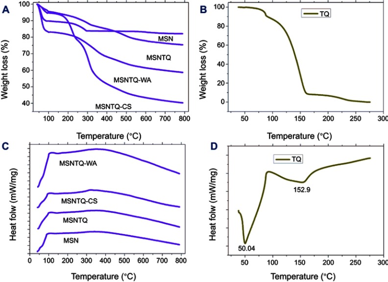
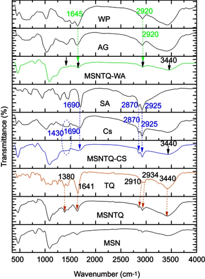
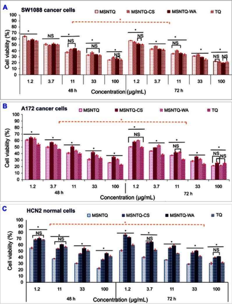
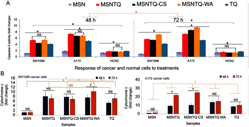
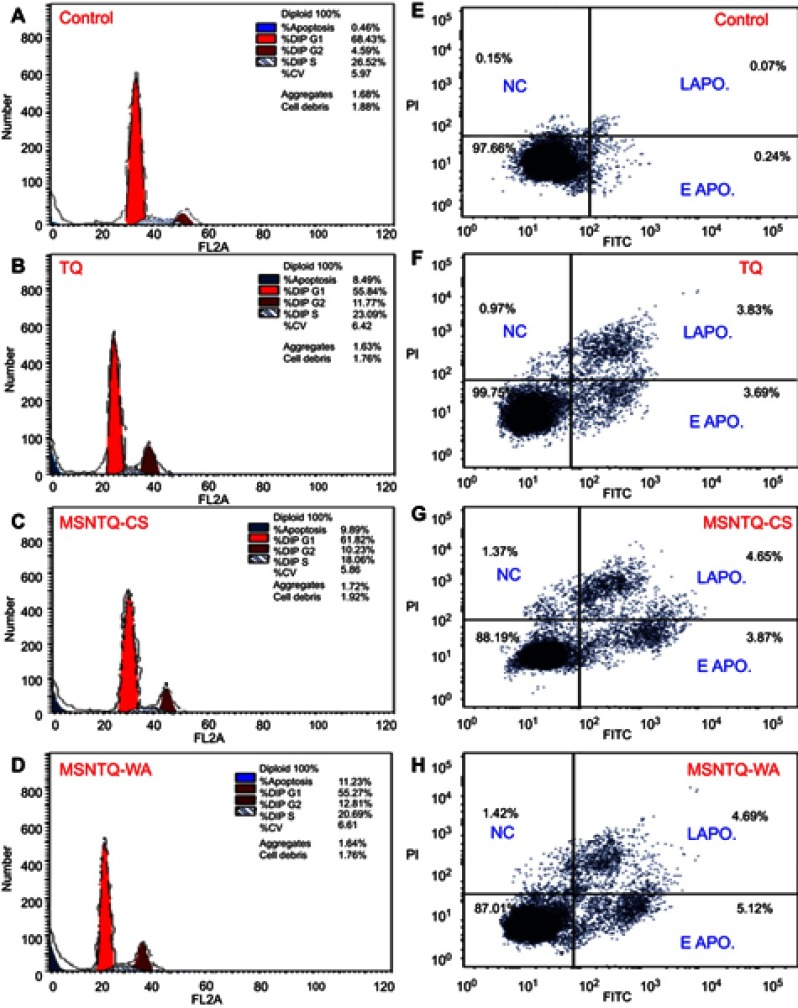
Verification
Claims the paper makes
The claims and statistics of this paper haven’t been checked yet.
Run the check — free, no account needed. It reads the open-access text and checks the sources it cites.
Claims other papers make about this one
Each is checked against this paper’s own text: does it say what the citing paper says it does?
In the citing paper
“This observation indicates successful preparation, in agreement with previous results [26,27,37,38].”
From Targeted Nano-Drug Delivery of Colchicine against Colon Cancer Cells by Means of Mesoporous Silica Nanoparticles (2020) · cites this paper as [27]
In the citing paper
“MSNs have been investigated as a drug delivery carrier for several drugs and biomolecules [27,31,32].”
From Targeted Nano-Drug Delivery of Colchicine against Colon Cancer Cells by Means of Mesoporous Silica Nanoparticles (2020) · cites this paper as [27]
In the citing paper
“Therefore, cancer-targeting delivery systems with different nano-platforms have gained attention [25,26,27].”
From Targeted Nano-Drug Delivery of Colchicine against Colon Cancer Cells by Means of Mesoporous Silica Nanoparticles (2020) · cites this paper as [27]
In the citing paper
“The polymeric coating controls the release of guest molecules from MSNs depending on time, pH, and other factors [27,31,46].”
From Targeted Nano-Drug Delivery of Colchicine against Colon Cancer Cells by Means of Mesoporous Silica Nanoparticles (2020) · cites this paper as [27]
Self-check
Percentages, sums and reported differences re-computed from the paper’s own numbers.
The self-check needs the paper’s full text.
Citations
Cites
Mo, J., He, L., Ma, B., & Chen, T. (2016) · ACS Applied Materials & Interfaces · doi:10.1021/acsami.5b11730 · Cited by 159
Research works printed but not linked: 86
- [1]Ostrom QT, Gittleman H, Farah P, et al. CBTRUS statistical report: primary brain and central nervous system tumors diagnosed in the United States in 2006-2010. Neuro-Oncology. 2013; 15(Suppl 2): ii1– ii56. doi: 10.1093/neuonc/not151 24137015 PMC3798196 · DOI
- [2]Wen PY, Kesari S. Malignant gliomas in adults. N Engl J Med. 2008; 359(5): 492– 507. doi: 10.1056/NEJMra0708126 18669428 · DOI · Paper page
- [3]Arko L, Katsyv I, Park GE, Luan WP, Park JK. Experimental approaches for the treatment of malignant gliomas. Pharmacol Ther. 2010; 128(1): 1– 36. doi: 10.1016/j.pharmthera.2010.04.015 20546782 PMC2939300 · DOI
- [4]Stupp R, Mason WP, van Den Bent MJ, et al. Radiotherapy plus concomitant and adjuvant temozolomide for glioblastoma. N Engl J Med. 2005; 352(10): 987– 996. doi: 10.1056/NEJMoa043330 15758009 · DOI · Paper page
- [5]Hou LC, Veeravagu A, Hsu AR, Tse VC. Recurrent glioblastoma multiforme: a review of natural history and management options. Neurosurg Focus. 2006; 20(4): E5. doi: 10.3171/foc.2006.20.4.2 16709036 · DOI
- [6]Kim SS, Harford JB, Pirollo KF, Chang EH. Effective treatment of glioblastoma requires crossing the blood-brain barrier and targeting tumors including cancer stem cells: the promise of nanomedicine. Biochem Biophys Res Commun. 2015; 468(3): 485– 489. doi: 10.1016/j.bbrc.2015.06.137 26116770 PMC4690805 · DOI
- [7]Nicholas MK, Lukas R, Bangalore S. Chemotherapy for malignant glioma. Expert Rev Neurother. 2005; 5(6 Suppl): S41– S49. doi: 10.1586/14737175.5.6.S41 16274270 · DOI
- [8]Perry J, Laperriere N, Zuraw L, Chambers A, Spithoff K, Cairncross JG. Adjuvant chemotherapy for adults with malignant glioma: a systematic review. Can J Neurol Sci. 2007; 34(4): 402– 410. 18062446
- [9]Schneider-Stock R, Fakhoury IH, Zaki AM, El-Baba CO, Gali-Muhtasib HU. Thymoquinone: fifty years of success in the battle against cancer models. Drug Discov Today. 2014; 19(1): 18– 30. doi: 10.1016/j.drudis.2013.08.021 24001594 · DOI
- [10]Goyal SN, Prajapati CP, Gore PR, et al. Therapeutic potential and pharmaceutical development of thymoquinone: a multitargeted molecule of natural origin. Front Pharmacol. 2017; 8: 656. doi: 10.3389/fphar.2017.00656 28983249 PMC5613109 · DOI
- [11]Gali-Muhtasib H, Ocker M, Kuester D, et al. Thymoquinone reduces mouse colon tumor cell invasion and inhibits tumor growth in murine colon cancer models. J Cell Mol Med. 2008; 12(1): 330– 342. doi: 10.1111/j.1582-4934.2007.00095.x 18366456 PMC3823493 · DOI
- [12]Jafri SH, Glass J, Shi R, Zhang S, Prince M, Kleiner-Hancock H. Thymoquinone and cisplatin as a therapeutic combination in lung cancer: in vitro and in vivo. J Exp Clin Cancer Res. 2010; 29: 87. doi: 10.1186/1756-9966-29-87 20594324 PMC2909169 · DOI
- [13]El-Mahdy MA, Zhu Q, Wang QE, Wani G, Wani AA. Thymoquinone induces apoptosis through activation of caspase-8 and mitochondrial events in p53-null myeloblastic leukemia HL-60 cells. Int J Cancer. 2005; 117(3): 409– 417. doi: 10.1002/ijc.21205 15906362 · DOI
- [14]Arafa El SA, Zhu Q, Shah ZI, et al. Thymoquinone up-regulates PTEN expression and induces apoptosis in doxorubicin-resistant human breast cancer cells. Mutat Res. 2011; 706(1–2): 28– 35. doi: 10.1016/j.mrfmmm.2010.10.007 21040738 PMC3037029 · DOI
- [15]Asaduzzaman Khan M, Tania M, Fu S, Fu J. Thymoquinone, as an anticancer molecule: from basic research to clinical investigation. Oncotarget. 2017; 8(31): 51907– 51919. doi: 10.18632/oncotarget.17206 28881699 PMC5584300 · DOI
- [16]Gurung RL, Lim SN, Khaw AK, et al. Thymoquinone induces telomere shortening, DNA damage and apoptosis in human glioblastoma cells. PLoS One. 2010; 5(8): e12124. doi: 10.1371/journal.pone.0012124 20711342 PMC2920825 · DOI
- [17]Kolli-Bouhafs K, Boukhari A, Abusnina A, et al. Thymoquinone reduces migration and invasion of human glioblastoma cells associated with FAK, MMP-2 and MMP-9 down-regulation. Invest New Drugs. 2012; 30(6): 2121– 2131. doi: 10.1007/s10637-011-9777-3 22170088 · DOI
- [18]Racoma IO, Meisen WH, Wang QE, Kaur B, Wani AA. Thymoquinone inhibits autophagy and induces cathepsin-mediated, caspase-independent cell death in glioblastoma cells. PLoS One. 2013; 8(9): e72882. doi: 10.1371/journal.pone.0072882 24039814 PMC3767730 · DOI
- [19]Pazhouhi M, Sariri R, Rabzia A, Khazaei M. Thymoquinone synergistically potentiates temozolomide cytotoxicity through the inhibition of autophagy in U87MG cell line. Iran J Basic Med Sci. 2016; 19(8): 890– 898. 27746872 PMC5048126
- [20]Elmaci I, Altinoz MA. Thymoquinone: an edible redox-active quinone for the pharmacotherapy of neurodegenerative conditions and glial brain tumors. A short review. Biomed Pharmacother. 2016; 83: 635– 640. doi: 10.1016/j.biopha.2016.07.018 27459120 · DOI
- [21]Chowdhury FA, Hossain MK, Mostofa AGM, Akbor MM, Bin Sayeed MS. Therapeutic potential of thymoquinone in glioblastoma treatment: targeting major gliomagenesis signaling pathways. Biomed Res Int. 2018; 2018: 4010629. doi: 10.1155/2018/4010629 29651429 PMC5831880 · DOI
- [22]Jakaria M, Cho D-Y, Ezazul Haque M, et al. Neuropharmacological potential and delivery prospects of thymoquinone for neurological disorders. Oxid Med Cell Longev. 2018; 2018: 17. doi: 10.1155/2018/1209801 PMC5883931 29743967 · DOI
- [23]Banerjee I, De K, Mukherjee D, et al. Paclitaxel-loaded solid lipid nanoparticles modified with Tyr-3-octreotide for enhanced anti-angiogenic and anti-glioma therapy. Acta Biomater. 2016; 38: 69– 81. doi: 10.1016/j.actbio.2016.04.026 27109765 · DOI
- [24]Orunoğlu M, Kaffashi A, Pehlivan SB, et al. Effects of curcumin-loaded PLGA nanoparticles on the RG2 rat glioma model. Mater Sci Eng C. 2017; 78: 32– 38. doi: 10.1016/j.msec.2017.03.292 28575990 · DOI
- [25]Lee ES, Gao Z, Bae YH. Recent progress in tumor pH targeting nanotechnology. J Controlled Release. 2008; 132(3): 164– 170. doi: 10.1016/j.jconrel.2008.05.003 PMC2695946 18571265 · DOI
- [26]Liu J, Huang Y, Kumar A, et al. pH-Sensitive nano-systems for drug delivery in cancer therapy. Biotechnol Adv. 2014; 32(4): 693– 710. doi: 10.1016/j.biotechadv.2013.11.009 24309541 · DOI · Paper page
- [27]Wirries A, Breyer S, Quint K, Schobert R, Ocker M. Thymoquinone hydrazone derivatives cause cell cycle arrest in p53-competent colorectal cancer cells. Exp Ther Med. 2010; 1(2): 369– 375. doi: 10.3892/etm_00000058 22993551 PMC3445902 · DOI
- [28]Odeh F, Ismail SI, Abu-Dahab R, Mahmoud IS, Al Bawab A. Thymoquinone in liposomes: a study of loading efficiency and biological activity towards breast cancer. Drug Deliv. 2012; 19(8): 371– 377. doi: 10.3109/10717544.2012.727500 23043626 · DOI
- [29]Ng WK, Saiful Yazan L, Yap LH, Wan Nor Hafiza WAG, How CW, Abdullah R. Thymoquinone-loaded nanostructured lipid carrier exhibited cytotoxicity towards breast cancer cell lines (MDA-MB-231 and MCF-7) and cervical cancer cell lines (HeLa and SiHa). Biomed Res Int. 2015; 2015: 10. doi: 10.1155/2015/263131 PMC4303008 25632388 · DOI
- [30]Dehghani H, Hashemi M, Entezari M, Mohsenifar A. The comparison of anticancer activity of thymoquinone and nanothymoquinone on human breast adenocarcinoma. IJPR. 2015; 14(2): 539– 546. 25901162 PMC4403071
- [31]Vallet-Regi M, Rámila A, Del Real RP, Pérez-Pariente J. A new property of MCM-41: drug delivery system. Chem Mater. 2001; 13(2): 308– 311. doi: 10.1021/cm0011559 · DOI
- [32]Kwon S, Singh RK, Perez RA, Abou Neel EA, Kim H-W, Chrzanowski W. Silica-based mesoporous nanoparticles for controlled drug delivery. J Tissue Eng. 2013; 4: 2041731413503357. doi: 10.1177/2041731413503357 24020012 PMC3764983 · DOI
- [33]Wang Y, Zhao Q, Han N, et al. Mesoporous silica nanoparticles in drug delivery and biomedical applications. Nanomedicine. 2015; 11(2): 313– 327. doi: 10.1016/j.nano.2014.09.014 25461284 · DOI · Paper page
- [34]Watermann A, Brieger J. Mesoporous silica nanoparticles as drug delivery vehicles in cancer. Nanomaterials (Basel, Switzerland). 2017; 7(7). doi: 10.3390/nano7120458 PMC5535255 28737672 · DOI
- [35]Baghirov H, Karaman D, Viitala T, et al. Feasibility study of the permeability and uptake of mesoporous silica nanoparticles across the blood-brain barrier. PLoS One. 2016; 11(8): e0160705. doi: 10.1371/journal.pone.0160705 27547955 PMC4993362 · DOI
- [36]Tamba BI, Streinu V, Foltea G, et al. Tailored surface silica nanoparticles for blood-brain barrier penetration: preparation and in vivo investigation. Arab J Chem. 2018; 11(6): 981– 990. doi: 10.1016/j.arabjc.2018.03.019 · DOI
- [37]Shi J, Hou S, Huang J, et al. An MSN-PEG-IP drug delivery system and IL13Rα2 as targeted therapy for glioma. Nanoscale. 2017; 9(26): 8970– 8981. doi: 10.1039/c6nr08786h 28443896 · DOI
- [38]Zhang H, Zhang W, Zhou Y, Jiang Y, Li S. Dual functional mesoporous silicon nanoparticles enhance the radiosensitivity of VPA in glioblastoma. Transl Oncol. 2017; 10(2): 229– 240. doi: 10.1016/j.tranon.2016.12.011 28193559 PMC5304234 · DOI
- [39]You Y, Yang L, He L, Chen T. Tailored mesoporous silica nanosystem with enhanced permeability of the blood–brain barrier to antagonize glioblastoma. J Mat Chem B. 2016; 4(36): 5980– 5990. doi: 10.1039/C6TB01329E 32263487 · DOI
- [41]Song Y, Du D, Li L, Xu J, Dutta P, Lin Y. In vitro study of receptor-mediated silica nanoparticles delivery across blood-brain barrier. ACS Appl Mater Interfaces. 2017; 9(24): 20410– 20416. doi: 10.1021/acsami.7b03504 28541655 PMC5533093 · DOI
- [42]Salmani JM, Asghar S, Lv H, Zhou J. Aqueous solubility and degradation kinetics of the phytochemical anticancer thymoquinone; probing the effects of solvents, pH and light. Molecules. 2014; 19(5): 5925– 5939. doi: 10.3390/molecules19055925 24815311 PMC6270770 · DOI
- [43]El-Toni AM, Khan A, Ibrahim MA, et al. Synthesis of double mesoporous core-shell silica spheres with tunable core porosity and their drug release and cancer cell apoptosis properties. J Colloid Interface Sci. 2012; 378(1): 83– 92. doi: 10.1016/j.jcis.2012.04.006 22551476 · DOI
- [44]Shen D, Yang J, Li X, et al. Biphase stratification approach to three-dimensional dendritic biodegradable mesoporous silica nanospheres. Nano Lett. 2014; 14(2): 923– 932. doi: 10.1021/nl404316v 24467566 · DOI
- [45]Hu F-Q, Zhao M-D, Yuan H, You J, Du Y-Z, Zeng S. A novel chitosan oligosaccharide–stearic acid micelles for gene delivery: properties and in vitro transfection studies. Int J Pharm. 2006; 315(1): 158– 166. doi: 10.1016/j.ijpharm.2006.02.026 16632285 · DOI
- [46]Yuan H, Bao X, Du Y-Z, You J, Hu F-Q. Preparation and evaluation of SiO2-deposited stearic acid-g-chitosan nanoparticles for doxorubicin delivery. Int J Nanomedicine. 2012; 7: 5119– 5128. doi: 10.2147/IJN.S35575 23055724 PMC3463401 · DOI
- [47]Yang T-S, Liu -T-T, Lin IH. Functionalities of chitosan conjugated with stearic acid and gallic acid and application of the modified chitosan in stabilizing labile aroma compounds in an oil-in-water emulsion. Food Chem. 2017; 228: 541– 549. doi: 10.1016/j.foodchem.2017.02.035 28317761 · DOI
- [48]Klein M, Aserin A, Ben Ishai P, Garti N. Interactions between whey protein isolate and gum Arabic. Colloids Surf B. 2010; 79(2): 377– 383. doi: 10.1016/j.colsurfb.2010.04.021 20488681 · DOI
- [49]Franz TJ. Percutaneous absorption on the relevance of in vitro data. J Invest Dermatol. 1975; 64(3): 190– 195. 123263
- [50]Salama RO, Traini D, Chan HK, Young PM. Preparation and characterisation of controlled release co-spray dried drug-polymer microparticles for inhalation 2: evaluation of in vitro release profiling methodologies for controlled release respiratory aerosols. Eur J Pharm Biopharm. 2008; 70(1): 145– 152. doi: 10.1016/j.ejpb.2008.04.009 18534832 · DOI
- [51]Ammar HO, Ghorab MM, Mahmoud AA, Higazy IM. Lamotrigine loaded poly-varepsilon-(d,l-lactide-co-caprolactone) nanoparticles as brain delivery system. Eur J Pharm Sci. 2018; 115: 77– 87. doi: 10.1016/j.ejps.2018.01.028 29341900 · DOI
- [52]Mosmann T. Rapid colorimetric assay for cellular growth and survival: application to proliferation and cytotoxicity assays. J Immunol Methods. 1983; 65(1–2): 55– 63. 6606682
- [53]Yergeau E, Arbour M, Brousseau R, et al. Microarray and real-time PCR analyses of the responses of high-arctic soil bacteria to hydrocarbon pollution and bioremediation treatments. Appl Environ Microbiol. 2009; 75(19): 6258– 6267. doi: 10.1128/AEM.01029-09 19684169 PMC2753079 · DOI
- [54]Qu F, Zhu G, Huang S, et al. Controlled release of Captopril by regulating the pore size and morphology of ordered mesoporous silica. Microporous Mesoporous Mater. 2006; 92(1–3): 1– 9. doi: 10.1016/j.micromeso.2005.12.004 · DOI
- [55]AbouAitah K, Farghali A, Swiderska-Sroda A, Lojkowski W, Razin A, Khedr M. pH-controlled release system for curcumin based on functionalized dendritic mesoporous silica nanoparticles. J Nanomed Nanotechnol. 2016; 7: 1. doi:10.4172/2157-7439.1000351 · DOI
- [56]AbouAitah K, Farghali A, Swiderska-Sroda A, Lojkowski W, Razin A. Mesoporous silica materials in drug delivery system: pH/glutathione-responsive release of poorly water-soluble pro-drug quercetin from two and three-dimensional pore-structure nanoparticles. J Nanomed Nanotechnol. 2016; 7: 1. doi: 10.4172/2157-7439.1000360 · DOI · Paper page
- [57]Sahoo B, Devi KSP, Sahu SK, et al. Facile preparation of multifunctional hollow silica nanoparticles and their cancer specific targeting effect. Biomater Sci. 2013; 1(6): 647– 657. doi: 10.1039/c3bm00007a 32481837 · DOI
- [58]Möller K, Kobler J, Bein T. Colloidal suspensions of nanometer-sized mesoporous silica. Adv Funct Mater. 2007; 17(4): 605– 612
- [59]Poudel BK, Soe ZC, Ruttala HB, et al. In situ fabrication of mesoporous silica-coated silver-gold hollow nanoshell for remotely controllable chemo-photothermal therapy via phase-change molecule as gatekeepers. Int J Pharm. 2018; 548(1): 92– 103. doi: 10.1016/j.ijpharm.2018.06.056 29959089 · DOI
- [60]Sun Y, Sun Y-L, Wang L, Ma J, Yang Y-W, Gao H. Nanoassembles constructed from mesoporous silica nanoparticles and surface-coated multilayer polyelectrolytes for controlled drug delivery. Microporous Mesoporous Mater. 2014; 185: 245– 253. doi: 10.1016/j.micromeso.2013.11.020 · DOI
- [61]Khan KA. The concept of dissolution efficiency. J Pharm Pharmacol. 1975; 27(1): 48– 49. 235616
- [62]Rinaki E, Dokoumetzidis A, Macheras P. The mean dissolution time depends on the dose/solubility ratio. Pharm Res. 2003; 20(3): 406– 408. 12669960
- [63]Korsmeyer RW, Gurny R, Doelker E, Buri P, Peppas NA. Mechanisms of solute release from porous hydrophilic polymers. Int J Pharm. 1983; 15(1): 25– 35. doi: 10.1016/0378-5173(83)90064-9 6644570 · DOI · Paper page
- [64]Karasulu HY, Ertan G, Kose T. Modeling of theophylline release from different geometrical erodible tablets. Eur J Pharm Biopharm. 2000; 49(2): 177– 182. doi: 10.1016/S0939-6411(99)00082-X 10704902 · DOI
- [65]Surekha R, Aishwarya V, Sumathi T. Thymoquinone loaded solid lipid nanoparticle: formulation, characterization and in-vitro cell viability assay. Int J Pharm Bio Sci. 2014; 6: 449– 464.
- [66]Ritger PL, Peppas NA. A simple equation for description of solute release II. Fickian and anomalous release from swellable devices. J Controlled Release. 1987; 5(1): 37– 42. doi: 10.1016/0168-3659(87)90035-6 25356469 · DOI
- [67]Wang C, Fu X, Yang L. Water-soluble chitosan nanoparticles as a novel carrier system for protein delivery. Chin Sci Bull. 2007; 52(7): 883– 889. doi: 10.1007/s11434-007-0127-y · DOI
- [68]Barahuie F, Dorniani D, Saifullah B, et al. Sustained release of anticancer agent phytic acid from its chitosan-coated magnetic nanoparticles for drug-delivery system. Int J Nanomedicine. 2017; 12: 2361– 2372. doi: 10.2147/IJN.S126245 28392693 PMC5376211 · DOI
- [69]Williams PA, Phillips GO. 11 - Gum arabic In: Phillips GO, Williams PA, editors. Handbook of Hydrocolloids. 2nd ed. Cambridge: Woodhead Publishing; 2009: 252– 273.
- [70]Cornelsen PA, Quintanilha RC, Vidotti M, Gorin PA, Simas-Tosin FF, Riegel-Vidotti IC. Native and structurally modified gum arabic: exploring the effect of the gum’s microstructure in obtaining electroactive nanoparticles. Carbohydr Polym. 2015; 119: 35– 43. doi: 10.1016/j.carbpol.2014.11.020 25563942 · DOI
- [71]Gunasekaran S, Ko S, Xiao L. Use of whey proteins for encapsulation and controlled delivery applications. J Food Eng. 2007; 83(1): 31– 40. doi: 10.1016/j.jfoodeng.2006.11.001 · DOI
- [72]Abu-Dahab R, Odeh F, Ismail SI, Azzam H, Al Bawab A. Preparation, characterization and antiproliferative activity of thymoquinone-β-cyclodextrin self assembling nanoparticles. Die Pharm-Int J Pharm Sci. 2013; 68(12): 939– 944. 24400439
- [73]Bhattacharya S, Ahir M, Patra P, et al. PEGylated-thymoquinone-nanoparticle mediated retardation of breast cancer cell migration by deregulation of cytoskeletal actin polymerization through miR-34a. Biomaterials. 2015; 51: 91– 107. doi: 10.1016/j.biomaterials.2015.01.007 25771001 · DOI
- [74]AbouAitah K, Swiderska-Sroda A, Farghali AA, et al. Folic acid-conjugated mesoporous silica particles as nanocarriers of natural prodrugs for cancer targeting and antioxidant action. Oncotarget. 2018; 9(41): 26466– 26490. doi: 10.18632/oncotarget.25470 29899871 PMC5995188 · DOI · Paper page
- [75]Choi JY, Ramasamy T, Kim SY, et al. PEGylated lipid bilayer-supported mesoporous silica nanoparticle composite for synergistic co-delivery of axitinib and celastrol in multi-targeted cancer therapy. Acta Biomater. 2016; 39: 94– 105. doi: 10.1016/j.actbio.2016.05.012 27163403 · DOI
- [76]Park EJ, Chauhan AK, Min KJ, Park DC, Kwon TK. Thymoquinone induces apoptosis through downregulation of c-FLIP and Bcl-2 in renal carcinoma Caki cells. Oncol Rep. 2016; 36(4): 2261– 2267. doi: 10.3892/or.2016.5019 27573448 · DOI
- [77]Zhang M, Du H, Huang Z, et al. Thymoquinone induces apoptosis in bladder cancer cell via endoplasmic reticulum stress-dependent mitochondrial pathway. Chem Biol Interact. 2018; 292: 65– 75. doi: 10.1016/j.cbi.2018.06.013 29981725 · DOI
- [78]Gali-Muhtasib H, Diab-Assaf M, Boltze C, et al. Thymoquinone extracted from black seed triggers apoptotic cell death in human colorectal cancer cells via a p53-dependent mechanism. Int J Oncol. 2004; 25(4): 857– 866. 15375533
- [79]Xu D, Ma Y, Zhao B, et al. Thymoquinone induces G2/M arrest, inactivates PI3K/Akt and nuclear factor-kappaB pathways in human cholangiocarcinomas both in vitro and in vivo. Oncol Rep. 2014; 31(5): 2063– 2070. doi: 10.3892/or.2014.3059 24603952 · DOI
- [80]Alhosin M, Ibrahim A, Boukhari A, et al. Anti-neoplastic agent thymoquinone induces degradation of alpha and beta tubulin proteins in human cancer cells without affecting their level in normal human fibroblasts. Invest New Drugs. 2012; 30(5): 1813– 1819. doi: 10.1007/s10637-011-9734-1 21881916 · DOI
- [81]Paramasivam A, Sambantham S, Shabnam J, et al. Anti-cancer effects of thymoquinone in mouse neuroblastoma (Neuro-2a) cells through caspase-3 activation with down-regulation of XIAP. Toxicol Lett. 2012; 213(2): 151– 159. doi: 10.1016/j.toxlet.2012.06.011 22732633 · DOI
- [82]Rajput S, Kumar BN, Dey KK, Pal I, Parekh A, Mandal M. Molecular targeting of Akt by thymoquinone promotes G(1) arrest through translation inhibition of cyclin D1 and induces apoptosis in breast cancer cells. Life Sci. 2013; 93(21): 783– 790. doi: 10.1016/j.lfs.2013.09.009 24044882 · DOI
- [83]Xie Y-T, Du Y-Z, Yuan H, Hu F-Q. Brain-targeting study of stearic acid-grafted chitosan micelle drug-delivery system. Int J Nanomedicine. 2012; 7: 3235– 3244. doi: 10.2147/IJN.S32701 22802685 PMC3396390 · DOI
- [84]Ha H-K, Kim JW, Lee M-R, Jun W, Lee W-J. Cellular uptake and cytotoxicity of β-lactoglobulin nanoparticles: the effects of particle size and surface charge. Asian-Aust J Anim Sci. 2015; 28(3): 420– 427. doi: 10.5713/ajas.14.0761 PMC4341088 25656189 · DOI
- [85]Bounous G. Whey protein concentrate (WPC) and glutathione modulation in cancer treatment. Anticancer Res. 2000; 20(6c): 4785– 4792. 11205219
- [86]Traverso N, Ricciarelli R, Nitti M, et al. Role of glutathione in cancer progression and chemoresistance. Oxid Med Cell Longev. 2013; 2013: 10. doi: 10.1155/2013/972913 PMC3673338 23766865 · DOI · Paper page
- [87]Ramasamy T, Ruttala HB, Gupta B, et al. Smart chemistry-based nanosized drug delivery systems for systemic applications: a comprehensive review. J Controlled Release. 2017; 258: 226– 253. doi: 10.1016/j.jconrel.2017.04.043 28472638 · DOI
Cited by
Sorted by citations
AbouAitah, K., Hassan, H. A., Swiderska-Sroda, A., Gohar, L., Shaker, O. G., Wojnarowicz, J., Opalinska, A., Smalc-Koziorowska, J., Gierlotka, S., & Lojkowski, W. (2020) · Cancers · doi:10.3390/cancers12010144 · Cited by 105
Self-citation
Metadata
- Authors
- Shahein, S. A., Aboul-Enein, A. M., Higazy, I. M., Abou-Elella, F., Lojkowski, W., Ahmed, E. R., Mousa, S. A., & AbouAitah, K.
- Venue
- International Journal of Nanomedicine
- Publisher
- Informa UK Limited
- Published
- 2019-07
- Volume
- Volume 14
- Pages
- 5503-5526
- ISSN
- 1178-2013
- PMC
- PMC6650459
- Cited by
- 52
- References
- 1
- Language
- en
About these checks
Registry, retraction, Cites and statistics results are computed, not judged, with one exception: a printed reference that no index lists is read once by a model before it counts as a citation error. Claims are extracted by a model and kept only when their quote appears verbatim in the text; each cited source is then checked, with a model’s help, for whether it says what the claim relies on. Those verdicts can be wrong — read the claim’s quote and the cited source before relying on one.
Statistics and arithmetic are recomputed, not judged.
Confidence bands are shown only for fields calibrated on our annotated test set. None is calibrated yet, so no band is shown.
What counts as a claim ›How we measure confidence ›How we check this ›
Are you an author, or do you see a problem with this page? Request a review
