

The DOI matches its Crossref record; 67 of the 68 references it cites were found in a registry or an index.
Cx3cr1- deficient microglia exhibit a premature aging transcriptome
Open accessAbstract
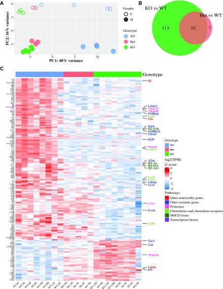
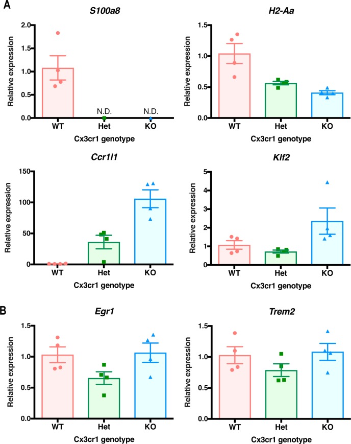
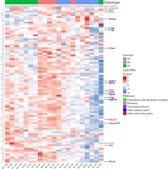
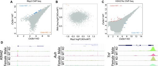
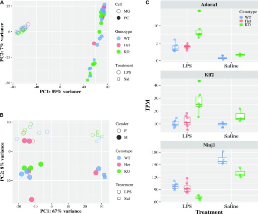
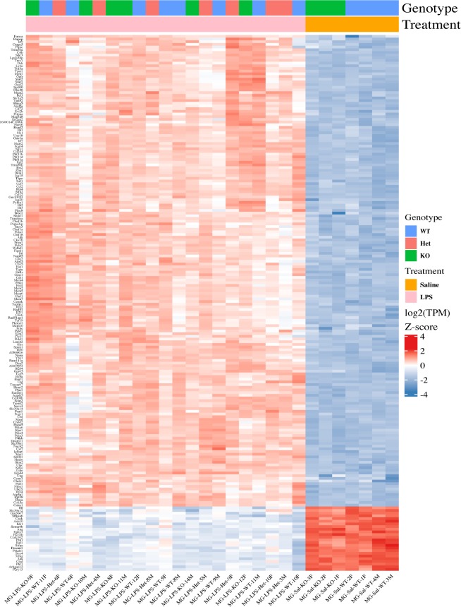
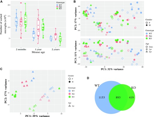
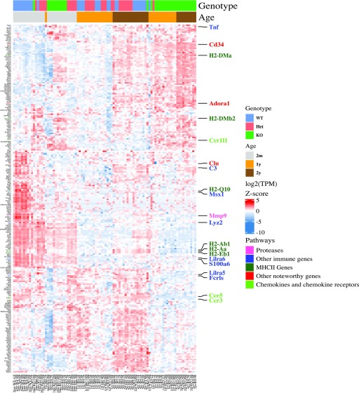
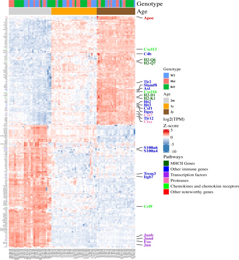
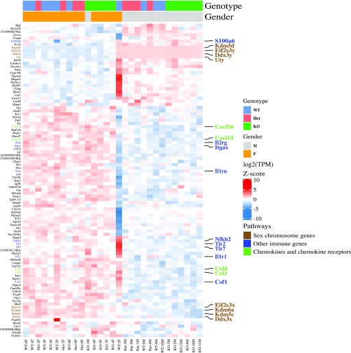
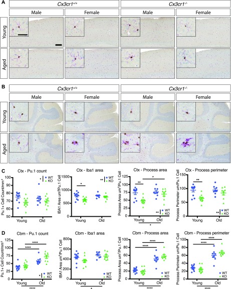
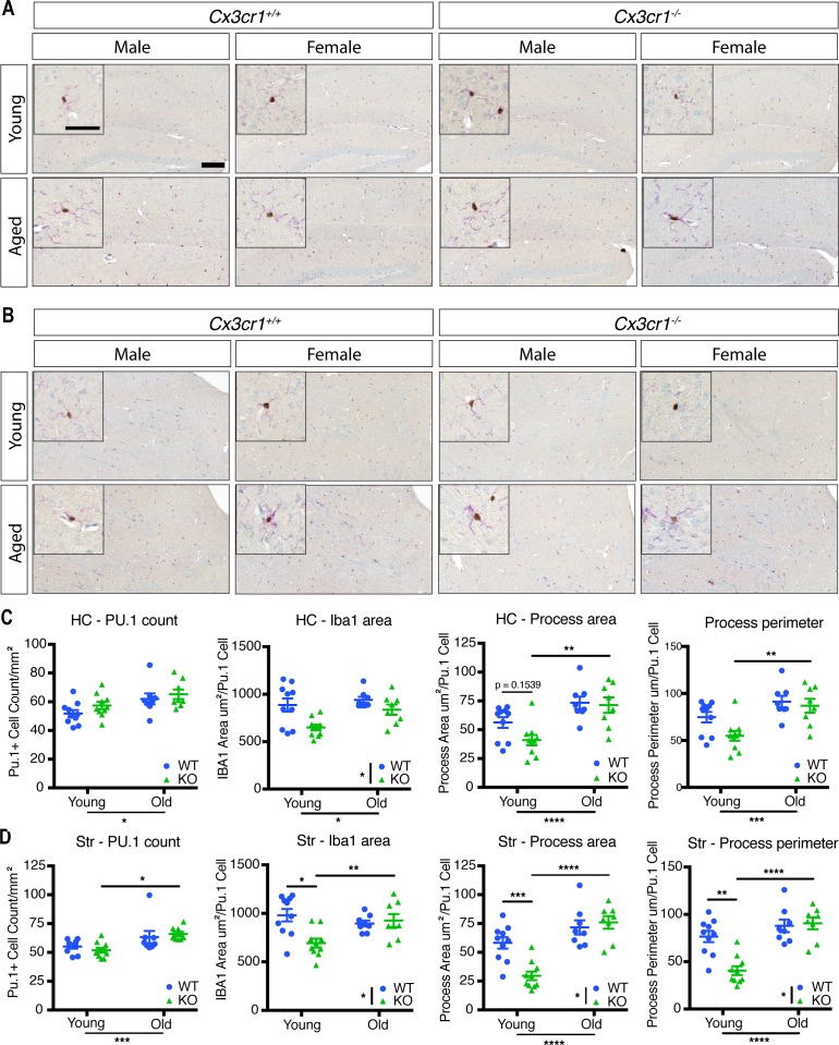
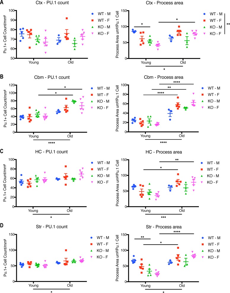
CX3CR1, one of the highest expressed genes in microglia in mice and humans, is implicated in numerous microglial functions. However, the molecular mechanisms underlying Cx3cr1 signaling are not well understood. Here, we analyzed transcriptomes of Cx3cr1- deficient microglia under varying conditions by RNA-sequencing (RNA-seq). In 2-mo-old mice, Cx3cr1 deletion resulted in the down-regulation of a subset of immune-related genes, without substantial epigenetic changes in markers of active chromatin. Surprisingly, Cx3cr1- deficient microglia from young mice exhibited a transcriptome consistent with that of aged Cx3cr1- sufficient animals, suggesting a premature aging transcriptomic signature. Immunohistochemical analysis of microglia in young and aged mice revealed that loss of Cx3cr1 modulates microglial morphology in a comparable fashion. Our results suggest that CX3CR1 may regulate microglial function in part by modulating the expression levels of a subset of inflammatory genes during chronological aging, making Cx3cr1 -deficient mice useful for studying aged microglia.
Overview
The claims and statistics of this paper haven’t been checked yet.
Run the check — free, no account needed. It reads the open-access text and checks the sources it cites.
Integrity signals
Registry
Cites
Statistics
Figures
Verification
Claims the paper makes
The claims and statistics of this paper haven’t been checked yet.
Run the check — free, no account needed. It reads the open-access text and checks the sources it cites.
Claims other papers make about this one
Each is checked against this paper’s own text: does it say what the citing paper says it does?
In the citing paper
“NOX1 expression may be an artefact of the Cx3cr1-EGFP reporter line, since Cx3cr1 is a signature microglial gene and haploinsufficiency has wide-reaching effects on gene expression of microglia, specifically encouraging a prematurely aged transcriptome [78,79,80].”
From ROS Generation in Microglia: Understanding Oxidative Stress and Inflammation in Neurodegenerative Disease (2020) · cites this paper as [78]
In the citing paper
“This expression profile concurs with other studies of DAM [10,37,78,124,194].”
From ROS Generation in Microglia: Understanding Oxidative Stress and Inflammation in Neurodegenerative Disease (2020) · cites this paper as [78]
Self-check
Percentages, sums and reported differences re-computed from the paper’s own numbers.
The self-check needs the paper’s full text.
Citations
Cites
Moderated estimation of fold change and dispersion for RNA-seq data with DESeq2
Love, M. I., Huber, W., & Anders, S. (2014) · Genome Biology · doi:10.1186/s13059-014-0550-8 · Cited by 95,106 · cited 3×
“DESeq2 (Love et al, 2014) was used to identify differential chromatin binding between Cx3cr1 -KO and -WT mice.”
Fast gapped-read alignment with Bowtie 2
Langmead, B., & Salzberg, S. L. (2012) · Nature Methods · doi:10.1038/nmeth.1923 · Cited by 56,400 · cited 1×
“The raw Rbp2 or H3K27Ac ChIP sequences were mapped to mm10 genome by Bowtie 2 (Langmead & Salzberg, 2013).”
STAR: ultrafast universal RNA-seq aligner
Dobin, A., Davis, C. A., Schlesinger, F., Drenkow, J., Zaleski, C., Jha, S., Batut, P., Chaisson, M., & Gingeras, T. R. (2013) · Bioinformatics · doi:10.1093/bioinformatics/bts635 · Cited by 51,709 · cited 2×
“Raw count fi les were aligned to mouse genome build mm10 using STAR and transcripts quanti fi ed with RSEM; in general, the uniquemappingratewas>80%.”
RSEM: accurate transcript quantification from RNA-Seq data with or without a reference genome
Li, B., & Dewey, C. N. (2011) · BMC Bioinformatics · doi:10.1186/1471-2105-12-323 · Cited by 22,179
Heinz, S., Benner, C., Spann, N., Bertolino, E., Lin, Y. C., Laslo, P., Cheng, J. X., Murre, C., Singh, H., & Glass, C. K. (2010) · Molecular Cell · doi:10.1016/j.molcel.2010.05.004 · Cited by 13,382 · cited 2×
“ChIP-seq library preparation, sequencing, and data analysis ChIP-seq libraries were prepared from recovered DNA by blunting, A-tailing, and adapter ligation using barcoded adapters (NextFlex; Bioo Scienti fi c) as previously described (Heinz et al, 2010; Gosselin et al, 2014).”
Self-citationComplex heatmaps reveal patterns and correlations in multidimensional genomic data
Gu, Z., Eils, R., & Schlesner, M. (2016) · Bioinformatics · doi:10.1093/bioinformatics/btw313 · Cited by 10,037 · cited 1×
“Heatmap visualizations of the DEGs were made by using custom R scripts based on ComplexHeatmap (Gu et al, 2016).”
A Unique Microglia Type Associated with Restricting Development of Alzheimer’s Disease
Keren-Shaul, H., Spinrad, A., Weiner, A., Matcovitch-Natan, O., Dvir-Szternfeld, R., Ulland, T. K., David, E., Baruch, K., Lara-Astaiso, D., Toth, B., et al. (2017) · Cell · doi:10.1016/j.cell.2017.05.018 · Cited by 5,493 · cited 1×
“In multiple conditions, mouse microglia demonstrate a core conserved reactive-state transcriptome, which is extensively modi fi ed to yield disease-speci fi c transcriptomes (Chiu et al, 2013; Keren-Shaul et al, 2017; Friedman et al, 2018).”
Zhang, Y., Chen, K., Sloan, S. A., Bennett, M. L., Scholze, A. R., O'Keeffe, S., Phatnani, H. P., Guarnieri, P., Caneda, C., Ruderisch, N., et al. (2014) · The Journal of Neuroscience · doi:10.1523/jneurosci.1860-14.2014 · Cited by 4,875 · cited 1×
“In WT mice, the microglial expression pro fi le obtained here is consistent with the published microglial transcriptomes (Table S1) (Hickman et al, 2013; Butovsky et al, 2014; Zhang et al, 2014).”
Synaptic Pruning by Microglia Is Necessary for Normal Brain Development
Paolicelli, R. C., Bolasco, G., Pagani, F., Maggi, L., Scianni, M., Panzanelli, P., Giustetto, M., Ferreira, T. A., Guiducci, E., Dumas, L., et al. (2011) · Science · doi:10.1126/science.1202529 · Cited by 4,244 · cited 1×
“…Cx3cr1 expression show altered brain development during embryogenesis, dysregulated neuronal fi ring in the fi rst few weeks after birth, and abnormal results in tests designed to mimic sociopsychological behaviors (Paolicelli et al, 2011; Rogers et al, 2011; Zhan et al, 2014; Bolós et al, 2017).”
Mutation of the mouse klotho gene leads to a syndrome resembling ageing
Kuro-o, M., Matsumura, Y., Aizawa, H., Kawaguchi, H., Suga, T., Utsugi, T., Ohyama, Y., Kurabayashi, M., Kaname, T., Kume, E., et al. (1997) · Nature · doi:10.1038/36285 · Cited by 3,837 · cited 1×
“Indeed, Kl (Klotho) is down-regulated in Cx3cr1 -de fi cient microglia, and Klotho protein has been linked to aging and homeostatic support of brain function (Kuro-o et al, 1997).”
Cited by
Sorted by citations
Simpson, D. S. A., & Oliver, P. L. (2020) · Antioxidants · doi:10.3390/antiox9080743 · Cited by 1,044
“This expression profile concurs with other studies of DAM [10,37,78,124,194].”
BackgroundResultMicroglia regulation of synaptic plasticity and learning and memory
Zhou, M., Cornell, J., Salinas, S., & Huang, H.-Y. (2022) · Neural Regeneration Research · doi:10.4103/1673-5374.322423 · Cited by 548
“CX3CR1/CX3CL1 signaling contributes to changes in microglia morphology and expression of immune proteins such as chemokines and chemokine receptors (Gyoneva et al., 2019).”
BackgroundMicroglia Phenotypes in Aging and Neurodegenerative Diseases
Wendimu, M. Y., & Hooks, S. B. (2022) · Cells · doi:10.3390/cells11132091 · Cited by 336
Graves, J. S., Krysko, K. M., Hua, L. H., Absinta, M., Franklin, R. J. M., & Segal, B. M. (2023) · The Lancet Neurology · doi:10.1016/s1474-4422(22)00184-3 · Cited by 260
Microglia in Alzheimer’s Disease in the Context of Tau Pathology
Perea, J. R., Bolós, M., & Ávila, J. (2020) · Biomolecules · doi:10.3390/biom10101439 · Cited by 134
“The first one determined that Cx3cr1−/− microglia present a lower phagocytic and migratory capacity per se [242], while the second revealed that Cx3cr1−/− microglia of young animals are very similar to the wild type (WT) microglia of older animals [243].”
ResultMills, S. A., Jobling, A. I., Dixon, M., Bui, B. V., Vessey, K. A., Phipps, J. A., Greferath, U., Venables, G., Wong, V. H. Y., Wong, C. H. Y., et al. (2021) · Proceedings of the National Academy of Sciences · doi:10.1073/pnas.2112561118 · Cited by 122
“03), reflecting the literature showing Cx3cr1 to have a more activated inflammatory profile (29).”
BackgroundPuntambekar, S. S., Moutinho, M., Lin, P. B.-C., Jadhav, V., Tumbleson-Brink, D., Balaji, A., Benito, M. A., Xu, G., Oblak, A., Lasagna-Reeves, C. A., et al. (2022) · Molecular Neurodegeneration · doi:10.1186/s13024-022-00545-9 · Cited by 113
“Similarly, sc-RNA seq studies that have demonstrated that transcriptomic differences observed in FACS purified, CD11b+CD45+ microglia from 2 month-old B6;Cx3cr1−/− mice are not evident in aged microglia from 12 and 24 month-old Cx3cr1 deficient mice as compared to cells isolated from agematched B6;Cx3cr1+/+ animals [3…”
BackgroundMicroglial morphometric analysis: so many options, so little consistency
Reddaway, J., Richardson, P., Bevan, R. J., Stoneman, J., & Palombo, M. (2023) · Frontiers in Neuroinformatics · doi:10.3389/fninf.2023.1211188 · Cited by 131
Augusto‐Oliveira, M., Arrifano, G. D. P., Delage, C. I., Tremblay, M., Lopez, M. E. C., & Verkhratsky, A. (2021) · Biological reviews/Biological reviews of the Cambridge Philosophical Society · doi:10.1111/brv.12797 · Cited by 114
BIN1 is a key regulator of proinflammatory and neurodegeneration-related activation in microglia
Sudwarts, A., Ramesha, S., Gao, T., Ponnusamy, M., Wang, S., Hansen, M. T., Kozlova, A., Bitarafan, S., Kumar, P., Beaulieu‐Abdelahad, D., et al. (2022) · Molecular Neurodegeneration · doi:10.1186/s13024-022-00535-x · Cited by 110
“A limitation of our in vivo model system relates to the inherent Cx3cr1 haploinsufficiency in Cx3cr1CreER mice which by itself is known to impact microglia via premature upregulation of an aging transcriptomic phenotype, inclusive of some disease-associated microglial genes observed in AD pathology [83].”
BackgroundWhite matter microglia heterogeneity in the CNS
Amor, S., McNamara, N. B., Gerrits, E., Marzin, M. C., Kooistra, S. M., Miron, V. E., & Nutma, E. (2022) · Acta Neuropathologica · doi:10.1007/s00401-021-02389-x · Cited by 89
Microglia as hunters or gatherers of brain synapses
Pereira‐Iglesias, M., Maldonado-Teixidó, J., Melero, A., Piriz, J., Galea, E., Ransohoff, R. M., & Sierra, A. (2024) · Nature Neuroscience · doi:10.1038/s41593-024-01818-w · Cited by 83
“However, these phagocytosis-related proteins affect multiple microglial functions beyond phagocytosis, including alterations in their physiological functions, transcriptome and inflammatory responses 52–57 , and their targeted deletion does not allow discriminating between culling and scavenging.”
Self-citationBedolla, A., Wegman, E., Weed, M., Stevens, M. K., Ware, K., Paranjpe, A., Alkhimovitch, A., Ifergan, I., Таранов, А., Peter, J. D., et al. (2024) · Nature Communications · doi:10.1038/s41467-024-49596-0 · Cited by 85
Groh, J., Feng, R., Yuan, X., Liu, L., Klein, D., Hutahaean, G., Butz, E. S., Wang, Z., Steinbrecher, L., Neher, J. J., et al. (2025) · Nature Neuroscience · doi:10.1038/s41593-025-01955-w · Cited by 81
“The rationale for using CX3CR1-deficient mice is based on the crucial role of the chemokine fractalkine (CX3CL1) and its receptor, CX3CR1, in neuroinflammation associated with neurodegenerative and demyelinating conditions 26,27 .”
Campagno, K. E., Lu, W., Jassim, A. H., Albalawi, F., Cenaj, A., Tso, H., Clark, S. P., Sripinun, P., Gómez, N. M., & Mitchell, C. H. (2021) · Journal of Neuroinflammation · doi:10.1186/s12974-021-02251-7 · Cited by 68
Snijders, G. J. L., Sneeboer, M. A. M., Fernández-Andreu, A., Udine, E., (NBB-Psy), P. D. P. O. T. N. B. B., Boks, M. P., Ormel, P. R., van Berlekom, A. B., van Mierlo, H. C., Böttcher, C., et al. (2020) · Molecular Psychiatry · doi:10.1038/s41380-020-00896-z · Cited by 66
Nebeling, F. C., Poll, S., Justus, L. C., Mittag, M., Steffen, J., Keppler, K., & Fuhrmann, M. (2023) · eLife · doi:10.7554/elife.83176 · Cited by 67
Neuroinflammation in retinitis pigmentosa: Therapies targeting the innate immune system
Zhao, L., Hou, C., & Yan, N. (2022) · Frontiers in Immunology · doi:10.3389/fimmu.2022.1059947 · Cited by 65
Harley, S. B. R., Willis, E. F., Shaikh, S., Blackmore, D. G., Sah, P., Ruitenberg, M. J., Bartlett, P. F., & Vukovic, J. (2021) · Journal of Neuroscience · doi:10.1523/jneurosci.2539-20.2021 · Cited by 54
“(2013) demonstrated that microglia regulate synapse formation and elimination during motor learning, with microglial brain-derived neurotrophic factor (BDNF; Bennett et al., 2016; Gyoneva et al., 2019) proposed as the main molecular driver of these effects.”
BackgroundFractalkine/CX3CR1-Dependent Modulation of Synaptic and Network Plasticity in Health and Disease
Camacho-Hernández, N. P., & Peña‐Ortega, F. (2023) · Neural Plasticity · doi:10.1155/2023/4637073 · Cited by 52
“Thus, microglia synapse engulfment is deteriorated in CX3CR1-deficient mice [58], which has been linked to a higher number of dendritic spines and greater PSD95 immunoreactivity, due to a deficit in synaptic pruning in the developing hippocampus [56, 194] and which correlates with alterations in synaptic transmission…”
BackgroundTargeting innate immunity to protect and cure Alzheimer’s disease: opportunities and pitfalls
Cisbani, G., & Rivest, S. (2021) · Molecular Psychiatry · doi:10.1038/s41380-021-01083-4 · Cited by 44
“CX3CR1 is a unique fracktalkine receptor that is expressed in microglia and circulating monocytes and it is necessary to maintain healthy homoeostatic functions of microglia [141].”
BackgroundZhao, Z., Zhang, D., Yang, F., Xu, M., Zhao, S., Pan, T., Liu, C., Liu, Y., Wu, Q., Tu, Q., et al. (2022) · Cell Research · doi:10.1038/s41422-022-00635-9 · Cited by 48
Microglia Drive Pockets of Neuroinflammation in Middle Age
Moca, E. N., Lecca, D., Hope, K. T., Etienne, F., Schaler, A. W., Espinoza, K., Chappell, M. S., Gray, D. T., Tweedie, D., Sidhu, S., et al. (2022) · The Journal of Neuroscience · doi:10.1523/jneurosci.1922-21.2022 · Cited by 42
Sadeghi, I., Gispert, J. D., Palumbo, E., Muñoz-Aguirre, M., Wucher, V., D’Argenio, V., Santpere, G., Navarro, A., Guigó, R., & Vilor‐Tejedor, N. (2022) · Computational and Structural Biotechnology Journal · doi:10.1016/j.csbj.2022.08.037 · Cited by 42
“across brain regions suggests shared perturbation of several mechanisms such as activation of microglia [77], inflammatory mechanisms, synapse development, and synaptic plasticity [54] across conditions.”
BackgroundSaitoh, B.-Y., Tanaka, E., Yamamoto, N., Kruining, D. V., Iinuma, K., Nakamuta, Y., Yamaguchi, H., Yamasaki, R., Matsumoto, K., & Kira, J.-I. (2021) · Brain, Behavior, and Immunity · doi:10.1016/j.bbi.2021.04.008 · Cited by 47
Carrier, M., Šimončičová, E., St‐Pierre, M., McKee, C., & Tremblay, M. (2021) · Frontiers in Molecular Neuroscience · doi:10.3389/fnmol.2021.749737 · Cited by 37
Cytokine Signalling at the Microglial Penta-Partite Synapse
Aramideh, J. A., Vidal-Itriago, A., Morsch, M., & Graeber, M. B. (2021) · International Journal of Molecular Sciences · doi:10.3390/ijms222413186 · Cited by 35
“CXCR4/CXCR7 Human, mice microglia [67,68] Rat, mice neuron [69–73]”
BackgroundGlial cells: an important switch for the vascular function of the central nervous system
Gao, L., Pan, X., Zhang, J. H., & Xia, Y. (2023) · Frontiers in Cellular Neuroscience · doi:10.3389/fncel.2023.1166770 · Cited by 35
“CX3CR1 can modulate the expression of a subset of inflammatory genes to regulate the phenotype change between activated and resting microglia (Gyoneva et al., 2019).”
BackgroundAtkinson, J., Jerome, A. D., Sas, A., Munie, A. N., Wang, C., Ma, A., Arnold, W. D., & Segal, B. M. (2022) · JCI Insight · doi:10.1172/jci.insight.158153 · Cited by 36
“Neuronal 225 maintenance of microglial homeostasis via CX3CL1/CX3CR1 and CD200/CD200R interactions wanes 226 with aging (20-22).”
BackgroundDysfunctional microglia and tau pathology in Alzheimer’s disease
Ayyubova, G. (2022) · Reviews in the Neurosciences · doi:10.1515/revneuro-2022-0087 · Cited by 36
Sarma, J. D., Burrows, A. C., Rayman, P. A., Hwang, M., Kundu, S., Sharma, N., Bergmann, C. C., & Sen, G. C. (2020) · PLoS Pathogens · doi:10.1371/journal.ppat.1009034 · Cited by 34
“Despite numerous studies comparing WT, CX3CR1-/-, and CX3CR1-/+ mice, there is surprisingly little information on regulation of CX3CR1 or CX3CL1 expression at the transcriptional, translational or posttranslational levels [60].”
BackgroundMacLean, M., Juranek, J., Cuddapah, S., López-Díez, R., Ruiz, H. H., Hu, J., Frye, L., Li, H., Gugger, P. F., & Schmidt, A. M. (2021) · Journal of Neuroinflammation · doi:10.1186/s12974-021-02191-2 · Cited by 33
Biological aging in multiple sclerosis
Zhang, Y., Atkinson, J., Burd, C. E., Graves, J., & Segal, B. M. (2023) · Multiple Sclerosis Journal · doi:10.1177/13524585231204122 · Cited by 34
Berki, P., Cserép, C., Környei, Z., Pósfai, B., Szabadits, E., Domonkos, A., Kellermayer, A., Nyerges, M., Wei, X., Módy, I., et al. (2024) · Nature Communications · doi:10.1038/s41467-024-49773-1 · Cited by 35
Microglia Susceptibility to Free Bilirubin Is Age-Dependent
Vaz, A. R., Falcão, A. S., Scarpa, E., Semproni, C., & Brites, D. M. T. O. (2020) · Frontiers in Pharmacology · doi:10.3389/fphar.2020.01012 · Cited by 28
“In fact, both protective and detrimental effects have been reported for CX3CR1 deletion (Gyoneva et al., 2019) and the consequences of low and high CX3CR1 expression in 2DIV and 16DIV Bf-treated microglia should be later explored in co-cultures of neurons and microglia.”
BackgroundCooper, O., Hallett, P. J., & Ole Isacson, A. (2022) · FEBS Journal · doi:10.1111/febs.16638 · Cited by 26
“1) [28 – 31].”
Metabolic factors in the regulation of hypothalamic innate immune responses in obesity
Folick, A., Cheang, R. T., Valdearcos, M., & Koliwad, S. K. (2022) · Experimental & Molecular Medicine · doi:10.1038/s12276-021-00666-z · Cited by 26
Church, K. A., Rodriguez, D., Mendiola, A. S., Vanegas, D., Gutiérrez, I. L., Tamayo, I. M., Amadu, A., Velazquez, P., Cardona, S. M., Gyoneva, S., et al. (2023) · Frontiers in Immunology · doi:10.3389/fimmu.2023.1130735 · Cited by 22
Self-citationAugusto‐Oliveira, M., Arrifano, G. D. P., Leal-Nazaré, C. G., Chaves-Filho, A. J. M., Santos-Sacramento, L., Lopes-Araújo, A., Tremblay, M., & Lopez, M. E. C. (2025) · Neuroscience & Biobehavioral Reviews · doi:10.1016/j.neubiorev.2025.106091 · Cited by 28
Zhang, W., Karschnia, P., von Mücke-Heim, I.-A., Mulazzani, M., Zhou, X., Blobner, J., Mueller, N., Teske, N., Dede, S., Xu, T., et al. (2021) · Neoplasia · doi:10.1016/j.neo.2021.09.001 · Cited by 21
Salcido, F. A. M., Torres-Flores, M. I., Ordaz, B., & Peña‐Ortega, F. (2022) · Glia · doi:10.1002/glia.24185 · Cited by 21
Injury primes mutation-bearing astrocytes for dedifferentiation in later life
Ragdale, H. S., Clements, M., Tang, W., Deltcheva, E. M., Andreassi, C., Lai, A. G., Chang, W. H., Pandrea, M., Andrew, I., Gamé, L., et al. (2023) · Current Biology · doi:10.1016/j.cub.2023.02.013 · Cited by 20
“A similar increase was also observed in published datasets of microglia activation following LPS treatment 36 or upon aging, 37 consistent with EGFR signaling potentially responding to neuroinflammation (Figures S3K and S3L).”
Zhou, K., Han, J., Lund, H., Boggavarapu, N. R., Lauschke, V. M., Goto, S., Cheng, H., Wang, Y., Tachi, A., Xie, C., et al. (2022) · Journal of Neuroinflammation · doi:10.1186/s12974-022-02381-6 · Cited by 20
Immune Regulation of Adult Neurogenic Niches in Health and Disease
Chintamen, S., Imessadouene, F., & Kernie, S. G. (2021) · Frontiers in Cellular Neuroscience · doi:10.3389/fncel.2020.571071 · Cited by 22
“Decreased levels of CX3CR1 are associated with premature aging in microglia and result in elevated cytokines (Vukovic et al., 2012; Gyoneva et al., 2019).”
BackgroundQuickomics: exploring omics data in an intuitive, interactive and informative manner
Gao, B., Zhu, J., Negi, S., Zhang, X., Gyoneva, S., Casey, F., Wei, R., & Zhang, B. (2021) · Bioinformatics · doi:10.1093/bioinformatics/btab255 · Cited by 17
Self-citationLe, L., Feidler, A. M., Rodríguez, L., Cealie, M. Y., Plunk, E. C., Li, H., Kara-Pabani, K., Lamantia, C. E., O’Banion, M. K., & Majewska, A. K. (2023) · Brain Behavior and Immunity · doi:10.1016/j.bbi.2025.04.022 · Cited by 19
Sepulveda, J., Kim, J. Y., Binder, J., Vicini, S., & Rebeck, G. W. (2024) · Molecular Neurodegeneration · doi:10.1186/s13024-024-00714-y · Cited by 19
“In addition, recent work [97] suggests that the loss of a single copy of CX3CR1 leads to alteration in microglial transcriptomics compared to wild-type animals.”
BackgroundDu, X., Peñalva, R. G., Little, K., Kissenpfennig, A., Chen, M., & Xu, H. (2021) · Molecular Neurodegeneration · doi:10.1186/s13024-021-00432-9 · Cited by 16
Schlecht, A., Thien, A., Wolf, J., Prinz, G., Agostini, H. T., Schlunck, G., Wieghofer, P., Boneva, S., & Lange, C. (2021) · International Journal of Molecular Sciences · doi:10.3390/ijms222413318 · Cited by 16
“Despite the fact that young mice deficient in Cx3cr1 were found to have a transcription signature that was comparable to aged mice, we used mice heterozygous for Cx3cr1 throughout our study to avoid possible effects mediated by Cx3cr1 expression differences [58].”
MethodNheu, D., Ellen, O., Ye, S., Öztürk, E., Pagnin, M., Kertadjaja, S., Theotokis, P., Grigoriadis, N., McLean, C., & Petratos, S. (2022) · Cells · doi:10.3390/cells11233768 · Cited by 16
Carrier, M., Hui, C. W., Watters, V., Šimončičová, E., Picard, K., Ibáñez, F. G., Vernoux, N., Droit, A., Desjardins, M., & Tremblay, M. (2023) · Brain Behavior and Immunity · doi:10.1016/j.bbi.2023.11.025 · Cited by 15
Var, S. R., Strell, P., Johnson, S. T., Roman, A., Vasilakos, Z., & Low, W. C. (2023) · Cell Transplantation · doi:10.1177/09636897231171001 · Cited by 15
Cerebellar microglia: On the edge between neuroinflammation and neuroregulation
Dukhinova, M., Guo, J., Shen, E.-W., Liu, W., Huang, W., Shen, Y., & Wang, L. (2024) · Neural Regeneration Research · doi:10.4103/nrr.nrr-d-24-00550 · Cited by 15
“Additionally, male microglia do not exhibit the phenomenon of age-related clustering in the cerebellum observed in females (Gyoneva et al., 2019).”
Le, L., Eliseeva, S. I., Plunk, E. C., Kara-Pabani, K., Li, H., Yarovinsky, F., & Majewska, A. K. (2025) · Cell Reports · doi:10.1016/j.celrep.2024.115176 · Cited by 13
Respiratory syncytial virus glycoprotein G impedes CX3CR1-activation by CX3CL1 and monocyte function
Meineke, R., Agac, A., Knittler, M.-C., Ludlow, M., Osterhaus, A. D. M. E., & Rimmelzwaan, G. F. (2024) · npj Viruses · doi:10.1038/s44298-024-00075-9 · Cited by 11
Sirerol-Piquer, M. S., Pérez‐Villalba, A., Duart-Abadía, P., Belenguer, G., Gómez‐Pinedo, U., Blasco-Chamarro, L., Carrillo-Barberà, P., Pérez‐Cañamás, A., Navarro, V., Dehay, B., et al. (2025) · Molecular Neurodegeneration · doi:10.1186/s13024-025-00816-1 · Cited by 12
“It has been reported that Cx3cr1-deficient microglia from Cx3cr1 eGFP/eGFP young mice exhibit a transcriptome consistent with that of aged Cx3cr1-sufficient animals, suggesting a premature aging transcriptomic signature [57].”
Stables, J., Pal, R., Bradford, B. M., Carter‐Cusack, D., Taylor, I., Pridans, C., Khan, N., Woodruff, T. M., Irvine, K. M., SUMMERS, K. M., et al. (2024) · Neurobiology of Disease · doi:10.1016/j.nbd.2024.106743 · Cited by 9
Targeting CX3CR1 Suppresses the Fanconi Anemia DNA Repair Pathway and Synergizes with Platinum
Lehto, J., Ninou, A. H., Chioureas, D., Jonkers, J., & Gustafsson, N. (2021) · Cancers · doi:10.3390/cancers13061442 · Cited by 7
“In addition, the receptor is also associated with inflammation, neurodegenerative diseases [15], and aging [16], and the CX3CR1 inhibitor KAND567 [17] is under clinical trials for hyperinflammatory conditions (EudraCT: 2020-002322-85).”
BackgroundWang, D., Hu, H., Ding, H., Zhao, H., Tian, F., & Chi, Q. (2022) · Cancer Biomarkers · doi:10.3233/cbm-210538 · Cited by 8
Murray, C. J., Tunderman, E. D., Vecchiarelli, H. A., Ibáñez, F. G., & Tremblay, M. (2025) · Glial health research. · doi:10.1016/j.ghres.2025.100007 · Cited by 9
Bui, V. T., Wu, K., Chen, C., Nguyen, A. T., Huang, W., Lee, L., Chen, W., Huang, C. F., & Shiao, Y. (2024) · CNS Neuroscience & Therapeutics · doi:10.1111/cns.70137 · Cited by 7
“Deleting of CX3CR1 on microglia decreases microglial ramification and maturation in young mice [43].”
Pissarek, M. (2020) · Pharmaceutics · doi:10.3390/pharmaceutics12100925 · Cited by 6
“Moreover, recently, CX3CR1-deficient microglia is presumed to exhibit a premature aging transcriptome and a reduced inflammatory response [89].”
BackgroundPaul, D., & Basavan, D. (2022) · Future Journal of Pharmaceutical Sciences · doi:10.1186/s43094-022-00446-0 · Cited by 5
“Similarly, absence of CX3CR1 expression will decline neurogenesis in the hippocampus and olfactory bulb of adults, and will adversely affect integration of newborn neurons [9].”
BackgroundExtracellular matrix and microglial regulation of cognitive agingPreprint
Gray, D. T., Gutierrez, A., Jami‐Alahmadi, Y., Pandey, V., Pan, L., Zhang, Y., Wohlschlegel, J. A., McDevitt, R. A., & Biase, L. M. D. (2024) · bioRxiv (Cold Spring Harbor Laboratory) · doi:10.1101/2024.01.04.574215 · Cited by 5
Augusto‐Oliveira, M., Arrifano, G. D. P., Leal-Nazaré, C. G., Soares-Silva, I., Lopes-Araújo, A., Santos-Sacramento, L., & Lopez, M. E. C. (2025) · Molecular Neurobiology · doi:10.1007/s12035-025-05311-8 · Cited by 4
Midbrain extracellular matrix and microglia are associated with cognition in aging mice
Gray, D. T., Gutierrez, A., Jami‐Alahmadi, Y., Pandey, V., Pan, L., Zhang, Y., Wohlschlegel, J. A., McDevitt, R. A., & Biase, L. M. D. (2025) · Nature Communications · doi:10.1038/s41467-025-66434-z · Cited by 6
“Publicly available bulk RNA-seq datasets of isolated microglia from Cx3cr1 knockout (Cx3cr1 − / − ), Cx3Cr1 heterozygous (Cx3cr1 +/ − ), and WT micewereretrievedfromGyonevaetal.,2019 43 .”
InfluentialRNASequest: An End-to-End Reproducible RNAseq Data Analysis and Publishing Framework
Zhu, J., Sun, Y. H., Ouyang, Z., Li, K., Negi, S., Piya, S., Hu, W., Zavodszky, M. I., Yalamanchili, H. B., Chen, Y., et al. (2023) · Journal of Molecular Biology · doi:10.1016/j.jmb.2023.168017 · Cited by 4
Self-citationConstitutive expression of CX3CR1-BAC-Cre introduces minimal off-target effects in microglia
Mroue-Ruiz, F. H., Desai, B., Garvin, M. M., Shehu, J. H., Kamau, F., Kar, U., & Bolton, J. L. (2026) · Biochemical and Biophysical Research Communications · doi:10.1016/j.bbrc.2026.153657 · Cited by 3
Exploring CX3CR1 as a prognostic biomarker and immunotherapeutic target in sarcoma
Li, T., Li, X., Kang, P., & Zhao, J. (2025) · Translational Oncology · doi:10.1016/j.tranon.2025.102283 · Cited by 3
Lowery, R. L., Latchney, S. E., Peer, R. P., Lamantia, C. E., Opanashuk, L. A., McCall, M. N., & Majewska, A. K. (2020) · Neuroscience Letters · doi:10.1016/j.neulet.2020.135516 · Cited by 3
“A moderate LPS dose has been demonstrated to induce a shift in the microglial transcriptome towards an inflammatory profile without approaching septic levels [25].”
BackgroundMethodCpeb1 remodels cell type–specific translational program to promote fear extinction
Zhang, J., Yang, C., Liu, Z., Wu, S., Li, Z.-G., Zhang, L. Y., Fan, H.-W., Guo, Z., Man, H., Li, X., et al. (2025) · Science Advances · doi:10.1126/sciadv.adr8687 · Cited by 2
“Cx3cr1 is a chemokine receptor expressed on microglial cells in the brain, and disruption of Cx3cr1 is accompanied by dysfunction of microglial status and function ( 39 ).”
InfluentialRadiation Retinopathy: Microangiopathy-Inflammation-Neurodegeneration
Davids, A., Pompös, I.-M., Kociok, N., Heufelder, J., Skosyrski, S., Reichhart, N., Joussen, A. M., & Wolf, S. A. (2025) · Cells · doi:10.3390/cells14040298 · Cited by 2
Flow Cytometry Identification of Cell Compartments in the Murine Brain
Moffet, J. J. D., Moore, Z., Oliver, S. J., Towers, T., Jenkins, M. R., Freytag, S., Whittle, J. R., & Best, S. A. (2023) · Methods in molecular biology · doi:10.1007/978-1-0716-3331-1_14 · Cited by 2
Microglia and border-associated macrophages in the central nervous system
Zanluqui, N. G., Polonio, C. M., de Oliveira, M. G., de Oliveira, L. G., Faria, L. C., & Peron, J. P. S. (2022) · Elsevier eBooks · doi:10.1016/b978-0-12-821385-8.00013-5 · Cited by 1
Majewska, A. K., Le, L., Feidler, A. M., Li, H., Kara-Pabani, K., Lamantia, C. E., & O’Banion, M. K. (2024) · Research Square · doi:10.21203/rs.3.rs-3976896/v1 · Cited by 1
“It is worth noting that for all in vivo imaging experiments, we used mice with only one functional copy of CX3CR1, which can alter microglial transcriptomic pro�les 50 and exert a complex effect on amyloid pathology progression, slowing down disease onset but facilitating progression 51,52 .”
MethodLe, L., Eliseeva, S. I., Kara-Pabani, K., Li, H., Yarovinsky, F., & Majewska, A. K. (2024) · SSRN Electronic Journal · doi:10.2139/ssrn.4795163
Microglia conduct the symphony of white matter degeneration in aging through T cell recruitment
Monteiro, J. K., & Miron, V. E. (2025) · Nature Neuroscience · doi:10.1038/s41593-025-01929-y
Ananathanarayana, V., Upadhyay, R., Illanad, G. H., Krishnan, D., Bochageri, N. P., Sevanan, M., Bagewadi, Z., Sivasamy, R., & Chinnathambi, S. (2025) · Advances in immunology · doi:10.1016/bs.ai.2025.11.002
Campagno, K. E., Lu, W., Jassim, A. H., Albalawi, F., Cenaj, A., Tso, H.-Y., Clark, S. P., Sripinun, P., Gómez, N., & Mitchell, C. H. (2021) · Figshare · doi:10.6084/m9.figshare.16650524
Du, X., Peñalva, R. G., Little, K., Kissenpfennig, A., Chen, M., & Xu, H. (2021) · Figshare · doi:10.6084/m9.figshare.14059735
Campagno, K. E., Lu, W., Jassim, A. H., Albalawi, F., Cenaj, A., Tso, H.-Y., Clark, S. P., Sripinun, P., Gómez, N., & Mitchell, C. H. (2021) · Figshare · doi:10.6084/m9.figshare.16650518
Du, X., Peñalva, R. G., Little, K., Kissenpfennig, A., Chen, M., & Xu, H. (2021) · Figshare · doi:10.6084/m9.figshare.14059744
Du, X., Peñalva, R. G., Little, K., Kissenpfennig, A., Chen, M., & Xu, H. (2021) · Figshare · doi:10.6084/m9.figshare.14059741
Du, X., Peñalva, R. G., Little, K., Kissenpfennig, A., Chen, M., & Xu, H. (2021) · Figshare · doi:10.6084/m9.figshare.14059747
Du, X., Peñalva, R. G., Little, K., Kissenpfennig, A., Chen, M., & Xu, H. (2021) · Figshare · doi:10.6084/m9.figshare.14059738
Zhou, K., Han, J., Lund, H., Boggavarapu, N. R., Lauschke, V. M., Goto, S., Cheng, H., Wang, Y., Tachi, A., Xie, C., et al. (2022) · Figshare · doi:10.6084/m9.figshare.18864770
Zhou, K., Han, J., Lund, H., Boggavarapu, N. R., Lauschke, V. M., Goto, S., Cheng, H., Wang, Y., Tachi, A., Xie, C., et al. (2022) · Figshare · doi:10.6084/m9.figshare.18864767
Puntambekar, S. S., Moutinho, M., Lin, P. B., Jadhav, V., Tumbleson, D. M., Balaji, A., Benito, M. A., Xu, G., Oblak, A. L., Lasagna‐Reeves, C. A., et al. (2022) · Figshare · doi:10.6084/m9.figshare.20175603
Puntambekar, S. S., Moutinho, M., Lin, P. B., Jadhav, V., Tumbleson, D. M., Balaji, A., Benito, M. A., Xu, G., Oblak, A. L., Lasagna‐Reeves, C. A., et al. (2022) · Figshare · doi:10.6084/m9.figshare.20175579
Du, X., Peñalva, R. G., Little, K., Kissenpfennig, A., Chen, M., & Xu, H. (2021) · Figshare · doi:10.6084/m9.figshare.14059750
Puntambekar, S. S., Moutinho, M., Lin, P. B., Jadhav, V., Tumbleson, D. M., Balaji, A., Benito, M. A., Xu, G., Oblak, A. L., Lasagna‐Reeves, C. A., et al. (2022) · Figshare · doi:10.6084/m9.figshare.20175606
Female-specific Cx3cr1-driven regulation of ALS and Alzheimer’s risk genes in tauopathy
Sudwarts, A., & Thinakaran, G. (2026) · Molecular Neurodegeneration Advances · doi:10.1186/s44477-026-00022-3
“Interestingly, others have reported that Cx3cr1 ablation induces an accelerated ageing, proinflammatory transcriptome [7].”
Microglia detection and phagocytosis of dying neurons is regulated by CX3CR1Preprint
Barasa, M. N., Pietramale, A. N., & Hill, R. (2026) · bioRxiv (Cold Spring Harbor Laboratory) · doi:10.64898/2026.04.02.716180
“Similarly, Cx3cr1 -/- microglia have an increased pro-inflammatory signature 21,47,48 , exhibit shorter and less ramified processes 48,49 , and show increased phagocytic activity 21– 23 , all indicative of a change in microglial state.”
Djuretić, J., Ivanović, J., Jezdić, K., Todorović, V., Bufan, B., Santrač, A., & Aranđelović, J. (2026) · npj Aging · doi:10.1038/s41514-026-00364-y
Iba1 deficiency impairs microglial synaptic remodeling and neuronal survival after axonal injury
Sekiguchi, K., Shoji, H., Shindo, T., Sasabe, J., Tokuyasu, D., Nakahara, J., Miyakawa, T., & Ito, D. (2026) · Journal of Neuroinflammation · doi:10.1186/s12974-026-03848-6 · Cited by 1
“Although these molecules are involved in apoptotic cell phagocytosis [43–46], they also exert multifaceted effects on microglial physiology, transcriptomic profiles, and inflammatory responses [47–52].”
Microglial pruning of extinction-ensemble synapses preserves fear memoryPreprint
Wang, Y., Cao, Y., Liu, T., Shi, T.-T., Zhao, Y., Jin, Z., Yang, X., Wang, S. Y., Ruan, J.-Z., Zhang, F., et al. (2026) · bioRxiv (Cold Spring Harbor Laboratory) · doi:10.64898/2026.05.05.722833
Zhang, B., Zhao, R., Li, X.-Y., Wei, Z.-H., Wang, D.-M., & Lu, M.-H. (2026) · Experimental Neurology · doi:10.1016/j.expneurol.2026.115845
Self-citationMicroglial CX3CR1 signaling mediates stress-induced pain behavior in mice
Fülöp, B., Király, Á., Petrák, R., Müller, J., Biró-Sütő, T., Kormos, V., Tékus, V., Rozmer, K., Dénes, Á., Borbély, É., et al. (2026) · Frontiers in Immunology · doi:10.3389/fimmu.2026.1869876
Schwab, E., Tewelde, E., Chen, L., & Benayoun, B. A. (2026) · BMC Biology · doi:10.1186/s12915-026-02672-x · Cited by 1
Distinct Transposable Element Transcript Patterns in Microglia Across Aging and Alzheimer's Disease
Grant, R. A., Doser, R. L., & LaRocca, T. J. (2026) · Aging Cell · doi:10.1111/acel.70653
“…controls (total RNA, GSE125050 (Srinivasan et al. 2020)), Mouse Dataset 1: microglia isolated from WT mice across lifespan (total RNA, GSE131869 (Gyoneva et al. 2019)), Mouse Dataset 2: microglia isolated from rTg4510 mice (total RNA, GSE123467 (Wang et al. 2018)), Mouse Dataset 3: microglia…”
Metadata
- Authors
- Gyoneva, S., Hosur, R., Gosselin, D., Zhang, B., Ouyang, Z., Cotleur, A. C., Peterson, M., Allaire, N., Challa, R., Cullen, P., Roberts, C., Miao, K., Reynolds, T. L., Glass, C. K., Burkly, L., & Ransohoff, R. M.
- Affiliations
- Biogen (United States) (US)
- University of California San Diego (US)
- Corresponding author
- Gyoneva, Stefka
- Ransohoff, Richard M
- ORCID
- Venue
- Life Science Alliance
- Publisher
- Life Science Alliance, LLC
- Published
- 2019-12
- Volume
- 2
- Issue
- 6
- Pages
- e201900453
- ISSN
- 2575-1077 (ISSN-L)
- PMC
- PMC6892408
- OpenAlex
- W2994105471
- Cited by
- 103
- References
- 68
- Citations by year
- 2026: 13 · 2025: 12 · 2024: 11 · 2023: 12 · 2022: 23 · 2021: 25 · 2020: 7
- FWCI
- 6.06
- Type
- journal article
- Language
- en
- Open access
- gold
- doi.org
- Licence
- Locations
- Life Science Alliance (publishedVersion, cc-by)
- PubMed Central (publishedVersion, cc-by)
- PubMed (publishedVersion)
- DOAJ (DOAJ: Directory of Open Access Journals) (submittedVersion, cc-by-sa)
- eScholarship (California Digital Library) (submittedVersion)
- eScholarship (California Digital Library) (submittedVersion, cc-by)
- Funders
- National Institute of Neurological Disorders and Stroke (R01NS096170)
- Topics
- Life Sciences › Neuroscience › Neurology › Neuroinflammation and Neurodegeneration Mechanisms
- Life Sciences › Biochemistry, Genetics and Molecular Biology › Aging › Genetics, Aging, and Longevity in Model Organisms
- Life Sciences › Neuroscience › Developmental Neuroscience › Neurogenesis and neuroplasticity mechanisms
- Keywords
- CX3CR1, microglia, premature aging, transcriptome, microglial aging, microglial morphology, inflammatory gene expression, active chromatin, neuroinflammation
- MeSH terms
- CX3C Chemokine Receptor 1, Genetic Profile, Animals, Disease Models, Animal, Female, Inflammation, Lipopolysaccharides, Male, Mice, Inbred C57BL, Mice, Transgenic, Neurons, Signal Transduction, Gene Deletion, Microglia, Mice, Knockout, Aging, Premature, Receptors, Chemokine, Mice, Transcriptome
About these checks
Registry, retraction, Cites and statistics results are computed, not judged, with one exception: a printed reference that no index lists is read once by a model before it counts as a citation error. Claims are extracted by a model and kept only when their quote appears verbatim in the text; each cited source is then checked, with a model’s help, for whether it says what the claim relies on. Those verdicts can be wrong — read the claim’s quote and the cited source before relying on one.
Statistics and arithmetic are recomputed, not judged.
Confidence bands are shown only for fields calibrated on our annotated test set. None is calibrated yet, so no band is shown.
What counts as a claim ›How we measure confidence ›How we check this ›
Are you an author, or do you see a problem with this page? Request a review
