

The DOI matches its Crossref record; 119 of the 120 references it cites were found in a registry or an index.
T Cell Dysfunction and Exhaustion in Cancer
Open accessAbstract
Tumor immunotherapy is a promising therapeutic strategy for patients with advanced cancers. T cells are key mediators of antitumor function that specifically recognize and react to tumor-expressing antigens and have proven critical for cancer immunotherapy. However, T cells are not as effective against cancer as expected. This is partly because T cells enter a dysfunctional or exhausted state, which is characterized by sustained expression of inhibitory receptors and a transcriptional state distinct from that of functional effector or memory T cells. T cell dysfunction induces the out of control of tumors. Recently, T cell dysfunction has been investigated in many experimental and clinical settings. The molecular definition of T cell dysfunction and the underlying causes of the T cell dysfunction has been advanced regardless of the fact that the pathways involved are not well elucidated, which proposing promising therapeutic opportunities in clinic. In this review, we will discuss the recent advances in the molecular mechanisms that affect TME and induce T cell dysfunction, and the development of promising immunotherapies to counteract the mechanisms of tumor-induced T cell dysfunction. Better understanding these underlying mechanisms may lead to new strategies to improve the clinical outcome of patients with cancer.
Overview
The claims and statistics of this paper haven’t been checked yet.
Run the check — free, no account needed. It reads the open-access text and checks the sources it cites.
Integrity signals
Registry
Cites
Statistics
Figures
Verification
Claims the paper makes
The claims and statistics of this paper haven’t been checked yet.
Run the check — free, no account needed. It reads the open-access text and checks the sources it cites.
Claims other papers make about this one
Each is checked against this paper’s own text: does it say what the citing paper says it does?
In the citing paper
“T cell senescence is irreversible, as opposed to their anergy and exhaustion, both of which are considered reversible [31, 38–40].”
From Immunosenescence: a key player in cancer development (2020) · cites this paper as [40]
Self-check
Percentages, sums and reported differences re-computed from the paper’s own numbers.
The self-check needs the paper’s full text.
Citations
Cites
Macrophage plasticity and polarization: in vivo veritas
Sica, A., & Mantovani, A. (2012) · Journal of Clinical Investigation · doi:10.1172/jci59643 · Cited by 6,335 · cited 1×
“Similarly, TCF-1 is also required for the stemlike functions of TCF1+PD-1+ TILs, which were detected in the blood of patients with melanoma and who had responded to checkpoint blockade (Siddiqui et al., 2019).”
BackgroundChimeric Antigen Receptor T Cells for Sustained Remissions in Leukemia
Maude, S. L., Frey, N., Shaw, P. A., Aplenc, R., Barrett, D. M., Bunin, N. J., Chew, A., Gonzalez, V. E., Zheng, Z., Lacey, S. F., et al. (2014) · New England Journal of Medicine · doi:10.1056/nejmoa1407222 · Cited by 5,413 · cited 1×
“A clinical trial showed MDSC frequencies as potential biomarkers and reported on their correlation with clinical outcomes of melanoma patients treated with ipilimumab (Meyer et al., 2014).”
BackgroundMeijer, L., Nelson, D. J., Riazanski, V., Gabdoulkhakova, A. G., Héry-Arnaud, G., Berre, R. L., Loaëc, N., Oumata, N., Galons, H., Nowak, E., et al. (2016) · Journal of Innate Immunity · doi:10.1159/000444256 · Cited by 5,008
Gut microbiome modulates response to anti-PD-1 immunotherapy in melanoma patients
Gopalakrishnan, V., Spencer, C. N., Nezi, L., Reuben, A., Andrews, M. C., Karpinets, T. V., Prieto, P. A., Vicente, D., Hoffman, K., Wei, S. C., et al. (2018) · Science · doi:10.1126/science.aan4236 · Cited by 4,812
Self-citationMolecular and cellular insights into T cell exhaustion
Wherry, E. J., & Kurachi, M. (2015) · Nature Reviews Immunology · doi:10.1038/nri3862 · Cited by 4,652 · cited 1×
“In addition, NO alone can suppress CD8+ T cells by inhibiting the phosphorylation and activation of JAK3 and STAT5 transcription factors, as well as inducing T-cell apoptosis (Wu et al., 2015).”
BackgroundWherry, E. J. (2011) · Nature Immunology · doi:10.1038/ni.2035 · Cited by 4,062 · cited 1×
“However, when antigen-experienced T cells are chronically exposed to the same antigen, substantial alterations in T cell activation and differentiation may occur, leading to T cell “dysfunction” or “exhaustion” (Wherry, 2011; Schietinger and Greenberg, 2014).”
BackgroundMetabolic Competition in the Tumor Microenvironment Is a Driver of Cancer Progression
Chang, C.-H., Qiu, J., O’Sullivan, D., Buck, M. D., Noguchi, T., Curtis, J. D., Chen, Q., Gindin, M., Gubin, M. M., van der Windt, G. J. W., et al. (2015) · Cell · doi:10.1016/j.cell.2015.08.016 · Cited by 3,331 · cited 3×
“The metabolic program is a set of biochemical reactions that allows T cells to acquire and utilize nutrients necessary for their survival, proliferation, and functions (Chang et al., 2015).”
BackgroundInfluentialAn immunogenic personal neoantigen vaccine for patients with melanoma
Ott, P. A., Hu, Z., Keskin, D. B., Shukla, S. A., Sun, J., Bozym, D. J., Zhang, W., Luoma, A., Giobbie-Hurder, A., Peter, L., et al. (2017) · Nature · doi:10.1038/nature22991 · Cited by 2,936
Delivery technologies for cancer immunotherapy
Riley, R., June, C. H., Langer, R., & Mitchell, M. J. (2019) · Nature Reviews Drug Discovery · doi:10.1038/s41573-018-0006-z · Cited by 2,678 · cited 1×
“Indeed, expression of ARG-1 has been reported to decrease CD3ζ-chain biosynthesis, thus impairing T cell function (Rodriguez et al., 2002).”
BackgroundMiller, B. C., Sen, D. R., Al Abosy, R., Bi, K., Virkud, Y. V., LaFleur, M. W., Yates, K. B., Lako, A., Felt, K., Naik, G. S., et al. (2019) · Nature Immunology · doi:10.1038/s41590-019-0312-6 · Cited by 2,087 · cited 1×
“Interestingly, inhibition of EZH2 in ovarian and colorectal cancer patients resulted in increased of CXCL9 and CXCL10 production and augmented the infiltration of T cells that eliminate tumors (Nagarsheth et al., 2016; Jones et al., 2018).”
BackgroundO’Rourke, D. M., Nasrallah, M. P., Desai, A. S., Melenhorst, J. J., Mansfield, K. G., Morrissette, J. J. D., Martinez‐Lage, M., Brem, S. S., Maloney, E., Shen, A., et al. (2017) · Science Translational Medicine · doi:10.1126/scitranslmed.aaa0984 · Cited by 1,847
Regression of Glioblastoma after Chimeric Antigen Receptor T-Cell Therapy
Brown, C. E., Alizadeh, D., Starr, R., Weng, L., Wagner, J. R., Naranjo, A., Ostberg, J. R., Blanchard, M. S., Kilpatrick, J., Simpson, J., et al. (2016) · New England Journal of Medicine · doi:10.1056/nejmoa1610497 · Cited by 1,842 · cited 1×
“overall survival of patients with glioma (Brown et al., 2016; Ahmed et al., 2017; O’Rourke et al., 2017; Migliorini et al., 2018).”
BackgroundChronic inflammation and cytokines in the tumor microenvironment
Landskron, G., De la Fuente, M., Thuwajit, P., Thuwajit, C., & Hermoso, M. A. (2014) · Journal of Immunology Research · doi:10.1155/2014/149185 · Cited by 1,765
Siddiqui, I., Schaeuble, K., Chennupati, V., Fuertes Marraco, S. A., Calderon-Copete, S., Pais Ferreira, D., Carmona, S. J., Scarpellino, L., Gfeller, D., Pradervand, S., et al. (2019) · Immunity · doi:10.1016/j.immuni.2018.12.021 · Cited by 1,679 · cited 1×
“Importantly, FoxO1 sustains PD-1 expression, which promotes the differentiation of terminally exhausted T cells (Staron et al., 2014).”
BackgroundGlobal characterization of T cells in non-small-cell lung cancer by single-cell sequencing
Guo, X., Zhang, Y., Zheng, L., Zheng, C., Song, J., Zhang, Q., Kang, B., Liu, Z., Jin, L., Xing, R., et al. (2018) · Nature Medicine · doi:10.1038/s41591-018-0045-3 · Cited by 1,651
Self-citationTOX transcriptionally and epigenetically programs CD8+ T cell exhaustion
Khan, O., Giles, J. R., McDonald, S., Manne, S., Ngiow, S. F., Patel, K. P., Werner, M. T., Huang, A. C., Alexander, K. A., Wu, J. E., et al. (2019) · Nature · doi:10.1038/s41586-019-1325-x · Cited by 1,646
Fueling Immunity: Insights into Metabolism and Lymphocyte Function
Pearce, E. L., Poffenberger, M. C., Chang, C.-H., & Jones, R. G. (2013) · Science · doi:10.1126/science.1242454 · Cited by 1,363 · cited 1×
“In addition, factors beyond tumor genomics influence cancer development and therapeutic responses, including host factors such as the gastrointestinal (gut) microbiome and obesity (Gopalakrishnan et al., 2018; Popovic et al., 2018; Wang et al., 2019e).”
BackgroundThommen, D. S., & Schumacher, T. N. (2018) · Cancer Cell · doi:10.1016/j.ccell.2018.03.012 · Cited by 1,351
Epigenetic stability of exhausted T cells limits durability of reinvigoration by PD-1 blockade
Pauken, K. E., Sammons, M. A., Odorizzi, P. M., Manne, S., Godec, J., Khan, O., Drake, A. M., Chen, Z., Sen, D. R., Kurachi, M., et al. (2016) · Science · doi:10.1126/science.aaf2807 · Cited by 1,348
Fourcade, J., Sun, Z., Benallaoua, M., Guillaume, P., Luescher, I. F., Sander, C., Kirkwood, J. M., Kuchroo, V., & Zarour, H. M. (2010) · Journal of Experimental Medicine · doi:10.1084/jem.20100637 · Cited by 1,307 · cited 1×
“However, activated and functional CD8+ T cells can also overexpress PD-1 in cancer patients (Fourcade et al., 2010), and not all PD-1+ cells might respond equally to anti-PD-1 therapy (Thommen et al., 2018).”
BackgroundT-cell exhaustion in the tumor microenvironment
Jiang, Y., Li, Y., & Zhu, B. (2015) · Cell Death and Disease · doi:10.1038/cddis.2015.162 · Cited by 1,197 · cited 2×
“Recent studies have shown that the exhaustion and functional impairment of T cells in the TME is a defining feature of many cancers (Jiang et al., 2015).”
BackgroundThommen, D. S., Koelzer, V. H., Herzig, P., Roller, A., Trefny, M., Dimeloe, S., Kiialainen, A., Hanhart, J., Schill, C., Hess, C., et al. (2018) · Nature Medicine · doi:10.1038/s41591-018-0057-z · Cited by 1,161 · cited 1×
“Some studies suggest that the efficacy of immunotherapy is limited by the generation of dysfunctional T cells in the tumor microenvironment (TME) (Thommen and Schumacher, 2018; Vodnala et al., 2019).”
BackgroundTOX is a critical regulator of tumour-specific T cell differentiation
Scott, A. C., Dündar, F., Zumbo, P., Chandran, S. S., Klebanoff, C. A., Shakiba, M., Trivedi, P., Menocal, L., Appleby, H., Camara, S., et al. (2019) · Nature · doi:10.1038/s41586-019-1324-y · Cited by 1,106 · cited 1×
“Emerging evidence indicates that epigenetic states and chromatin landscapes are closely associated with the functional state of dysfunctional or exhausted CD8+ T cells, which are abnormally expressed PD-1 (Pauken et al., 2016; Sen et al., 2016; Kartikasari et al., 2018).”
BackgroundThe Principles of Engineering Immune Cells to Treat Cancer
Lim, W. A., & June, C. H. (2017) · Cell · doi:10.1016/j.cell.2017.01.016 · Cited by 1,103 · cited 2×
“Additionally, these infused CAR-T cells express high levels of PD-1, T-cell immunoglobulin domain and mucin domain protein 3 (TIM3) and CTLA-4, indicating that they become exhausted or dysfunctional cells (Lim and June, 2017).”
BackgroundLi, H., van der Leun, A. M., Yofe, I., Lubling, Y., Gelbard-Solodkin, D., van Akkooi, A. C. J., van den Braber, M., Rozeman, E. A., Haanen, J. B. A. G., Blank, C. U., et al. (2019) · Cell · doi:10.1016/j.cell.2018.11.043 · Cited by 1,101
Targeting TGF-β Signaling in Cancer
Colak, S., & ten Dijke, P. (2017) · Trends in cancer · doi:10.1016/j.trecan.2016.11.008 · Cited by 1,003
The epigenetic landscape of T cell exhaustion
Sen, D. R., Kaminski, J., Barnitz, R. A., Kurachi, M., Gerdemann, U., Yates, K. B., Tsao, H.-W., Godec, J., LaFleur, M. W., Brown, F. D., et al. (2016) · Science · doi:10.1126/science.aae0491 · Cited by 986
TOX reinforces the phenotype and longevity of exhausted T cells in chronic viral infection
Alfei, F., Kanev, K., Hofmann, M., Wu, M., Ghoneim, H. E., Roelli, P., Utzschneider, D. T., von Hoesslin, M., Cullen, J. G., Fan, Y., et al. (2019) · Nature · doi:10.1038/s41586-019-1326-9 · Cited by 930 · cited 1×
“Recently, the high-mobility group (HMG)-box transcription factor TOX was reported as a critical regulator in the progression of T cell dysfunction and the maintenance of exhausted T cells during chronic infection (Alfei et al., 2019).”
BackgroundHER2-Specific Chimeric Antigen Receptor–Modified Virus-Specific T Cells for Progressive Glioblastoma
Ahmed, N. M., Brawley, V. S., Hegde, M. G., Bielamowicz, K., Kalra, M., Landi, D. B., Robertson, C. S., Gray, T., Diouf, O., Wakefield, A., et al. (2017) · JAMA Oncology · doi:10.1001/jamaoncol.2017.0184 · Cited by 884
Self-citationNR4A transcription factors limit CAR T cell function in solid tumours
Chen, J., López-Moyado, I. F., Seo, H., Lio, C.-W. J., Hempleman, L. J., Sekiya, T., Yoshimura, A., Scott-Browne, J. P., & Rao, A. (2019) · Nature · doi:10.1038/s41586-019-0985-x · Cited by 874
Wang, Z., Aguilar, E. G., Luna, J. I., Dunai, C., Khuat, L. T., Le, C. T., Mirsoian, A., Minnar, C. M., Stoffel, K. M., Sturgill, I. R., et al. (2019) · Nature Medicine · doi:10.1038/s41591-018-0221-5 · Cited by 865 · cited 1×
“However, when antigen-experienced T cells are chronically exposed to the same antigen, substantial alterations in T cell activation and differentiation may occur, leading to T cell “dysfunction” or “exhaustion” (Wherry, 2011; Schietinger and Greenberg, 2014).”
BackgroundMartinez, M. C., & Moon, E. K. (2019) · Frontiers in Immunology · doi:10.3389/fimmu.2019.00128 · Cited by 865 · cited 2×
“These encouraging results demonstrate that T cell-based cancer therapies offer great promise in inducing complete responses in patients against several types of cancer (Maude et al., 2014; Yee et al., 2015).”
BackgroundResultThe Transcription Factor NFAT Promotes Exhaustion of Activated CD8 + T Cells
Martinez, G. J., Pereira, R. M., Äijö, T., Kim, E. Y., Marangoni, F., Pipkin, M. E., Togher, S., Heissmeyer, V., Zhang, Y. C., Crotty, S., et al. (2015) · Immunity · doi:10.1016/j.immuni.2015.01.006 · Cited by 857
Self-citationTumor-associated macrophages: from basic research to clinical application
Yang, L., & Zhang, Y. (2017) · Journal of Hematology & Oncology · doi:10.1186/s13045-017-0430-2 · Cited by 847 · cited 2×
“Li and colleagues demonstrated that TAMs-secreted IL-10 promotes cancer stem cell-like properties and tumor growth in NSCLC; High levels of IL-10 are associated with a poor prognosis of NSCLC patients (Yang and Zhang, 2017).”
BackgroundSelf-citationSeo, H., Chen, J., González-Avalos, E., Samaniego-Castruita, D., Das, A., Wang, Y. H., López-Moyado, I. F., Georges, R. O., Zhang, W., Onodera, A., et al. (2019) · Proceedings of the National Academy of Sciences · doi:10.1073/pnas.1905675116 · Cited by 808
De Novo Epigenetic Programs Inhibit PD-1 Blockade-Mediated T Cell Rejuvenation
Ghoneim, H. E., Fan, Y., Moustaki, A., Abdelsamed, H. A., Dash, P., Dogra, P., Carter, R., Awad, W., Neale, G. A. M., Thomas, P. G., et al. (2017) · Cell · doi:10.1016/j.cell.2017.06.007 · Cited by 808
TIGIT and PD-1 impair tumor antigen–specific CD8+ T cells in melanoma patients
Chauvin, J.-M., Pagliano, O., Fourcade, J. J., Sun, Z., Wang, H., Sander, C., Kirkwood, J. M., Chen, T.-H. T., Maurer, M. F., Korman, A. J., et al. (2015) · Journal of Clinical Investigation · doi:10.1172/jci80445 · Cited by 805 · cited 3×
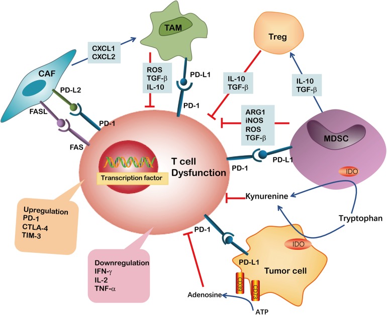
“In addition, these dysfunctional CD8 T cells show impaired production of effector cytokines, such as IL-2, IFN-γ and tumor necrosis factor-α (TNF-α) (Chauvin et al., 2015; Wang et al., 2017).”
BackgroundLi, J., Byrne, K. T., Yan, F., Yamazoe, T., Chen, Z., Baslan, T., Richman, L. P., Lin, J., Sun, Y. H., Rech, A. J., et al. (2018) · Immunity · doi:10.1016/j.immuni.2018.06.006 · Cited by 746
Schietinger, A., Philip, M., Krisnawan, V. E., Chiu, E. Y., Delrow, J. J., Basom, R. S., Lauer, P., Brockstedt, D. G., Knoblaugh, S. E., Hämmerling, G. J., et al. (2016) · Immunity · doi:10.1016/j.immuni.2016.07.011 · Cited by 720
TCF-1-centered transcriptional network drives an effector versus exhausted CD8 T cell-fate decision
Chen, Z., Ji, Z., Ngiow, S. F., Manne, S., Cai, Z., Huang, A. C., Johnson, J., Staupe, R. P., Bengsch, B., Xu, C., et al. (2019) · Immunity · doi:10.1016/j.immuni.2019.09.013 · Cited by 709
Tolerance and exhaustion: defining mechanisms of T cell dysfunction
Schietinger, A., & Greenberg, P. D. (2013) · Trends in Immunology · doi:10.1016/j.it.2013.10.001 · Cited by 704
CD8 T Cell Exhaustion in Chronic Infection and Cancer: Opportunities for Interventions
Hashimoto, M., Kamphorst, A. O., Im, S. J., Kissick, H. T., Pillai, R. N., Ramalingam, S. S., Araki, K., & Ahmed, R. (2018) · Annual Review of Medicine · doi:10.1146/annurev-med-012017-043208 · Cited by 679
Yu, J., Du, W., Fang, Y., Wang, Y., Li, H., Cao, S., Yu, W., Shen, C., Liu, J., & Ren, X. (2013) · The Journal of Immunology · doi:10.4049/jimmunol.1201449 · Cited by 625
Molecular regulation of effector and memory T cell differentiation
Chang, J. T., Wherry, E. J., & Goldrath, A. W. (2014) · Nature Immunology · doi:10.1038/ni.3031 · Cited by 604 · cited 2×
“Following the peak of effector cell expansion and the clearance of the specific antigen, most effector T cells die, with the exception of a small number of memory T cells that survive and provide longterm protection against the antigen (Chang et al., 2014).”
BackgroundLakins, M. A., Ghorani, E., Munir, H., Martins, C. P., & Shields, J. D. (2018) · Nature Communications · doi:10.1038/s41467-018-03347-0 · Cited by 578 · cited 1×
“One of the underlying mechanisms was the upregulation of FAS/FASL and PD-1/PDL2 on CD8+ T cells and CAFs, respectively, which drives the dysfunction of tumor-specific CD8+ T cells (Lakins et al., 2018).”
BackgroundImmune-mediated antitumor effect by type 2 diabetes drug, metformin
Eikawa, S., Nishida, M., Mizukami, S., Yamazaki, C., Nakayama, E., & Udono, H. (2015) · Proceedings of the National Academy of Sciences · doi:10.1073/pnas.1417636112 · Cited by 563
Meyer, C., Cagnon, L., Costa-Nunes, C. M., Baumgaertner, P., Montandon, N., Leyvraz, L., Michielin, O., Romano, E., & Speiser, D. E. (2014) · Cancer Immunology Immunotherapy · doi:10.1007/s00262-013-1508-5 · Cited by 560
T cell stemness and dysfunction in tumors are triggered by a common mechanism
Vodnala, S. K., Eil, R., Kishton, R. J., Sukumar, M., Yamamoto, T. N., Ha, N.-H., Lee, P.-H., Shin, M., Patel, S. J., Yu, Z., et al. (2019) · Science · doi:10.1126/science.aau0135 · Cited by 552 · cited 1×
“Combination treatments aimed at PD1 inhibition and activation of GITR, decrease CD8+ T cell dysfunction and induce a highly proliferative precursor effector memory T cell phenotype (Wang et al., 2018).”
BackgroundMetabolic Regulation of T Cell Longevity and Function in Tumor Immunotherapy
Kishton, R. J., Sukumar, M., & Restifo, N. P. (2017) · Cell Metabolism · doi:10.1016/j.cmet.2017.06.016 · Cited by 548
Regulation of T Cell Receptor CD3ζ Chain Expression byl-Arginine
Rodrı́guez, P. C., Zea, A. H., Culotta, K. S., Zabaleta, J., Ochoa, J. B., & Ochoa, A. C. (2002) · Journal of Biological Chemistry · doi:10.1074/jbc.m110675200 · Cited by 537 · cited 1×
“Indeed, expression of ARG-1 has been reported to decrease CD3ζ-chain biosynthesis, thus impairing T cell function (Rodriguez et al., 2002).”
BackgroundMyeloid-derived suppressor cells: more mechanisms for inhibiting antitumor immunity
Ostrand‐Rosenberg, S. (2010) · Cancer Immunology Immunotherapy · doi:10.1007/s00262-010-0855-8 · Cited by 534
Genome-wide analysis identifies NR4A1 as a key mediator of T cell dysfunction
Liu, X., Wang, Y., Lu, H., Li, J., Yan, X., Xiao, M., Hao, J., Alekseev, A., Khong, H., Chen, T., et al. (2019) · Nature · doi:10.1038/s41586-019-0979-8 · Cited by 533 · cited 1×
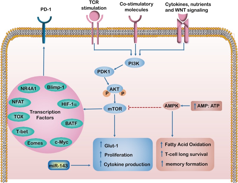
“This results in maintaining long-term T cell survival and memory formation (Ma et al., 2017).”
BackgroundQuigley, M. M., Pereyra, F., Nilsson, B., Porichis, F., Fonseca, C., Eichbaum, Q. G., Jülg, B. D., Jesneck, J. L., Brosnahan, K., Imam, S., et al. (2010) · Nature Medicine · doi:10.1038/nm.2232 · Cited by 527 · cited 1×
“Intriguingly, BATF, a transcription factor of the AP-1 family, was found to impair T cell proliferation and cytokine secretion during HIV infection in a pathway downstream of PD-1 (Quigley et al., 2010).”
BackgroundChen, Y., Zhang, S., Wang, Q., & Zhang, X. (2017) · Journal of Hematology & Oncology · doi:10.1186/s13045-017-0408-0 · Cited by 487
Reversing T-cell Dysfunction and Exhaustion in Cancer
Zarour, H. M. (2016) · Clinical Cancer Research · doi:10.1158/1078-0432.ccr-15-1849 · Cited by 462 · cited 2×
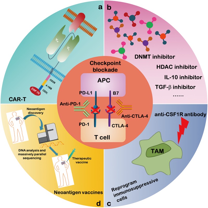
“Thus, a combined treatment of IL-10 or TGF-β inhibitor(s) with ICB represents a promising strategy for immunotherapy strategy (Zarour, 2016).”
BackgroundMethodFates of CD8+ T cells in Tumor Microenvironment
Maimela, N. R., Liu, S., & Zhang, Y. (2019) · Computational and Structural Biotechnology Journal · doi:10.1016/j.csbj.2018.11.004 · Cited by 459 · cited 1×
“In addition, tumor cells and immunosuppressive cells within TME produced highly reactive soluble oxygen and toxic metabolites, which inhibit T cell responses (Maimela et al., 2019).”
BackgroundSelf-citationBET Bromodomain Inhibition Promotes Anti-tumor Immunity by Suppressing PD-L1 Expression
Zhu, H., Bengsch, F., Svoronos, N., Rutkowski, M. R., Bitler, B. G., Allegrezza, M. J., Yokoyama, Y., Kossenkov, A. V., Bradner, J. E., Conejo-Garcia, J. R., et al. (2016) · Cell Reports · doi:10.1016/j.celrep.2016.08.032 · Cited by 437 · cited 1×
“Meanwhile, treatment with JQ1-α specific inhibitor of the histone acetylation reader bromodomain-containing protein 4 (BRD4)results in decreased PD-L1 expression on tumor cells and macrophages, which is correlated with an increase in antitumor T cell activity (Zhu et al., 2016).”
BackgroundArmored CAR T cells enhance antitumor efficacy and overcome the tumor microenvironment
Yeku, O., Purdon, T. J., Koneru, M., Spriggs, D. R., & Brentjens, R. J. (2017) · Scientific Reports · doi:10.1038/s41598-017-10940-8 · Cited by 420 · cited 1×
“In a mouse model of ovarian cancer, it was shown that tumor rejection by CAR-T cells required the presence of M1 macrophages, suggesting that tumor-reactive T cells were not sufficient to completely eliminate the tumor on their own (Yeku et al., 2017).”
BackgroundZhao, E., Maj, T., Kryczek, I., Li, W., Wu, K., Zhao, L., Wei, S., Crespo, J., Wan, S., Vatan, L., et al. (2016) · Nature Immunology · doi:10.1038/ni.3313 · Cited by 405 · cited 4×
“The most widely studied histone lysine methylation sites in T cells are histone 3 lysine 4 (H3K4), and H3K27.”
BackgroundInfluentialSelf-citationStaron, M. M., Gray, S. M., Marshall, H. D., Parish, I. A., Chen, J. H., Perry, C. J., Cui, G., Li, M. O., & Kaech, S. M. (2014) · Immunity · doi:10.1016/j.immuni.2014.10.013 · Cited by 402 · cited 1×
“Addition of HDACi and DNMTi 5-azacytidine (AZA) enhances the modulation of the immune microenvironment, specifically increasing T cell activation and reducing the percentage of macrophage in vivo (Stone et al., 2017).”
BackgroundJeannet, G., Boudousquié, C., Gardiol, N., Kang, J., Huelsken, J., & Held, W. (2010) · Proceedings of the National Academy of Sciences · doi:10.1073/pnas.0914127107 · Cited by 360 · cited 1×
“TCF-1 has been implicated in the formation of memory precursor T cells mediated by Wnt signaling pathway (Jeannet et al., 2010).”
BackgroundEmerging concepts of T cell metabolism as a target of immunotherapy
Chang, C., & Pearce, E. L. (2016) · Nature Immunology · doi:10.1038/ni.3415 · Cited by 345 · cited 1×
“, AMPK, Myc, HIF-1α and mTOR) that control T cell differentiation have been highlighted as a novel therapeutic targets for immune modulation (Chang and Pearce, 2016).”
BackgroundT Cell Dysfunction in Cancer Immunity and Immunotherapy
Xia, A., Zhang, Y., Jiang, X., Yin, T., & Lu, X. (2019) · Frontiers in Immunology · doi:10.3389/fimmu.2019.01719 · Cited by 344 · cited 1×
“Meanwhile, the TME contains a variety of cell types and cytokines (Table 1) that take part in tumor progression, which could contribute to T cell dysfunction (Xia et al., 2019).”
BackgroundSelf-citationVerma, V., Shrimali, R. K., Ahmad, S., Dai, W., Wang, H., Lu, S., Nandre, R., Gaur, P., Lopez, J., Sade-Feldman, M., et al. (2019) · Nature Immunology · doi:10.1038/s41590-019-0441-y · Cited by 333 · cited 1×
“Some studies suggest that the efficacy of immunotherapy is limited by the generation of dysfunctional T cells in the tumor microenvironment (TME) (Thommen and Schumacher, 2018; Vodnala et al., 2019).”
BackgroundReactive Oxygen Species Regulate T Cell Immune Response in the Tumor Microenvironment
Chen, X., Song, M., Zhang, B., & Zhang, Y. (2016) · Oxidative Medicine and Cellular Longevity · doi:10.1155/2016/1580967 · Cited by 326 · cited 1×
“ROS produced by MDSCs and other cells may interact with T cells and cause oxidative stress which may induce CD8 T cell hypo-responsiveness in cancer (Chen et al., 2016).”
BackgroundSelf-citationChimeric antigen receptor T cells: a novel therapy for solid tumors
Yu, S., Li, A., Liu, Q., Li, T., Yuan, X., Han, X., & Wu, K. (2017) · Journal of Hematology & Oncology · doi:10.1186/s13045-017-0444-9 · Cited by 314 · cited 1×
“However, despite extensive research, the efficacy of CAR-T cell therapy on controlling solid tumors is limited effects due to the influence of TME (Yu et al., 2017; Li et al., 2018d; Yan et al., 2019).”
BackgroundLi, J., Wang, L., Chen, X., Li, L., Li, Y., Ping, Y., Huang, L., Yue, D., Zhang, Z., Wang, F., et al. (2017) · OncoImmunology · doi:10.1080/2162402x.2017.1320011 · Cited by 299 · cited 1×
“Moreover, TGF-β stimulates CD39 and CD73 expression, thereby inhibiting autologous CD8+ T cell proliferation and function (Li et al., 2017).”
BackgroundSelf-citationStone, M. L., Chiappinelli, K. B., Li, H., Murphy, L. M., Travers, M. E., Topper, M. J., Mathios, D., Lim, M. K., Shih, I., Wang, T., et al. (2017) · Proceedings of the National Academy of Sciences · doi:10.1073/pnas.1712514114 · Cited by 297
Wang, X., He, Q., Shen, H., Xia, A., Tian, W., Yu, W., & Sun, B. (2019) · Journal of Hepatology · doi:10.1016/j.jhep.2019.05.015 · Cited by 287
Reprogramming the tumor microenvironment: tumor-induced immunosuppressive factors paralyze T cells
Wu, A. A., Drake, V. E., Huang, H.-S., Chiu, S., & Zheng, L. (2015) · OncoImmunology · doi:10.1080/2162402x.2015.1016700 · Cited by 281 · cited 1×
“In addition, NO alone can suppress CD8 T cells by inhibiting the phosphorylation and activation of JAK3 and STAT5 transcription factors, as well as inducing T-cell apoptosis (Wu et al., 2015).”
BackgroundNagarsheth, N., Peng, D., Kryczek, I., Wu, K., Li, W., Zhao, E., Zhao, L., Wei, S., Frankel, T. L., Vatan, L., et al. (2015) · Cancer Research · doi:10.1158/0008-5472.can-15-1938 · Cited by 277 · cited 16×
“The ARG1, iNOS, TGF-β and ROS are secreted by MDSCs or TAMs and induce CD8+ T cell dysfunction.”
InfluentialLi, L., Wang, L., Li, J., Fan, Z., Li, Y., Zhang, Z., Zhang, C., Yue, D., Qin, G., Zhang, T., et al. (2018) · Cancer Research · doi:10.1158/0008-5472.can-17-2460 · Cited by 267
Self-citationBoyiadzis, M. M., Dhodapkar, M. V., Brentjens, R. J., Kochenderfer, J. N., Neelapu, S. S., Maus, M. V., Porter, D. L., Maloney, D. G., Grupp, S. A., Mackall, C. L., et al. (2018) · Journal for ImmunoTherapy of Cancer · doi:10.1186/s40425-018-0460-5 · Cited by 255 · cited 2×
“…neoantigen vaccines and checkpoint blockade therapies using anti-programed cell death 1 (PD-1) and anti-PD-ligand 1 (PD-L1) have shown potent therapeutic effects in patients with advanced cancer (Chapuis et al., 2016b; Yee and Lizee, 2017; Boyiadzis et al., 2018; Yee, 2018; Riley et al., 2019).”
BackgroundGenetic and Epigenetic Regulation of PD-1 Expression
Bally, A. P. R., Austin, J. W., & Boss, J. M. (2016) · The Journal of Immunology · doi:10.4049/jimmunol.1502643 · Cited by 254 · cited 1×
“When TCR stimulation and IL-6 or IL-12 treatment were combined, both H3K4me1 and H3K27 acetylation were contributed to increased PD-1 expression (Bally et al., 2016).”
BackgroundLi, J., Li, W., Huang, K., Zhang, Y., Kupfer, G. M., & Zhao, Q. (2018) · Journal of Hematology & Oncology · doi:10.1186/s13045-018-0568-6 · Cited by 251
Self-citationHigh Levels of Eomes Promote Exhaustion of Anti-tumor CD8+ T Cells
Li, J., He, Y., Hao, J., Ni, L., & Dong, C. (2018) · Frontiers in Immunology · doi:10.3389/fimmu.2018.02981 · Cited by 229
Zhen, Z., Tang, W., Wang, M., Zhou, S., Wang, H., Wu, Z., Hao, Z., Li, Z., Liu, L., & Xie, J. (2016) · Nano Letters · doi:10.1021/acs.nanolett.6b04150 · Cited by 222 · cited 1×
“They can also polarize these recruited myeloid cells toward the M2 phenotype (Zhen et al., 2017).”
BackgroundBrooks, D. G., Ha, S., Elsaesser, H. J., Sharpe, A. H., Freeman, G. J., & Oldstone, M. B. A. (2008) · Proceedings of the National Academy of Sciences · doi:10.1073/pnas.0811139106 · Cited by 220 · cited 1×
“Thus, IL-10 and PD-1 pathways act synergistically through distinct pathways to suppress T cell survival and function (Brooks et al., 2008).”
BackgroundChimeric antigen receptor T-cell therapies for multiple myeloma
Mikkilineni, L., & Kochenderfer, J. N. (2017) · Blood · doi:10.1182/blood-2017-06-793869 · Cited by 216 · cited 1×
“For example, treatment with anti-PD-1/PD-L1 and anti-CTLA4 immune checkpoint inhibitors reinvigorate dysfunctional TILs and augment their anti-tumor effects (Wherry and Kurachi, 2015; Zarour, 2016; Miller et al., 2019; Wang et al., 2019a).”
MethodLi, J., Lee, Y., Li, Y., Jiang, Y. Y., Lu, H., Zang, W., Zhao, X., Liu, L., Chen, Y., Tan, H., et al. (2018) · Immunity · doi:10.1016/j.immuni.2018.03.018 · Cited by 213
Ma, R., Ji, T., Zhang, Y., Dong, W., Chen, X., Xu, P., Chen, D., Liang, X., Yin, X., Liu, Y., et al. (2017) · Nature Cell Biology · doi:10.1038/s41556-017-0002-2 · Cited by 211 · cited 1×
“A previous study demonstrated that the abrogation of phosphoenolpyruvate carboxykinase (Pck1)–glycogen–pentose phosphate pathway (PPP) decreases GSH/GSSG ratios and increases levels of ROS levels, leading to impairment of memory CD8+ T cell formation (Ma et al., 2018).”
BackgroundSelf-citationmTOR Regulation of Glycolytic Metabolism in T Cells
Salmond, R. J. (2018) · Frontiers in Cell and Developmental Biology · doi:10.3389/fcell.2018.00122 · Cited by 211 · cited 1×
“However, when antigen-experienced T cells are chronically exposed to the same antigen, substantial alterations in T cell activation and differentiation may occur, leading to T cell “dysfunction” or “exhaustion” (Wherry, 2011; Schietinger and Greenberg, 2014).”
BackgroundEmerging strategies for combination checkpoint modulators in cancer immunotherapy
Popović, A., Jaffee, E. M., & Zaidi, N. (2018) · Journal of Clinical Investigation · doi:10.1172/jci120775 · Cited by 209 · cited 1×
“Intriguingly, BATF, a transcription factor of the AP-1 family, was found to impair T cell proliferation and cytokine secretion during HIV infection in a pathway downstream of PD-1 (Quigley et al., 2010).”
BackgroundMetabolic Barriers to T Cell Function in Tumors
Sugiura, A., & Rathmell, J. C. (2018) · The Journal of Immunology · doi:10.4049/jimmunol.1701041 · Cited by 208 · cited 2×
“However, activated and functional CD8+ T cells can also overexpress PD-1 in cancer patients (Fourcade et al., 2010), and not all PD-1+ cells might respond equally to anti-PD-1 therapy (Thommen et al., 2018).”
BackgroundCassetta, L., & Kitamura, T. (2018) · Frontiers in Cell and Developmental Biology · doi:10.3389/fcell.2018.00038 · Cited by 200 · cited 1×
“An anti-CSF1R antibody was shown to reprogram TAM polarization and improve the responses to ICB therapy in pancreatic cancer (Cassetta and Kitamura, 2018).”
BackgroundBenner, B. N., Scarberry, L., Suarez‐Kelly, L. P., Duggan, M. C., Campbell, A. R., Smith, E., Lapurga, G., Jiang, K., Butchar, J. P., Tridandapani, S., et al. (2019) · Journal for ImmunoTherapy of Cancer · doi:10.1186/s40425-019-0622-0 · Cited by 188 · cited 15×
“The ARG1, iNOS, TGF-β and ROS are secreted by MDSCs or TAMs and induce CD8+ T cell dysfunction.”
BackgroundInfluentialWang, D., Yang, L., Yue, D., Cao, L., Li, L., Wang, D., Ping, Y., Shen, Z., Zheng, Y., Wang, L., et al. (2019) · Cancer Letters · doi:10.1016/j.canlet.2019.03.040 · Cited by 183
Self-citationWang, B., Zhang, W., Jankovic, V., Golubov, J., Poon, P., Oswald, E. M., Gurer, C., Wei, J., Ramos, I., Wu, Q., et al. (2018) · Science Immunology · doi:10.1126/sciimmunol.aat7061 · Cited by 167
The role of AMPK in T cell metabolism and function
Ma, E. H., Poffenberger, M. C., Wong, A., & Jones, R. G. (2017) · Current Opinion in Immunology · doi:10.1016/j.coi.2017.04.004 · Cited by 161
Targeting T Cell Metabolism for Improvement of Cancer Immunotherapy
Bourgeois, T. L., Strauss, L., Aksoylar, H., Daneshmandi, S., Seth, P., Patsoukis, N., & Boussiotis, V. A. (2018) · Frontiers in Oncology · doi:10.3389/fonc.2018.00237 · Cited by 157
CAR T-Cell Therapies in Glioblastoma: A First Look
Migliorini, D., Dietrich, P.-Y., Stupp, R., Linette, G. P., Posey, A. D., & June, C. H. (2017) · Clinical Cancer Research · doi:10.1158/1078-0432.ccr-17-2871 · Cited by 137
He, S., Liu, Y., Meng, L., Sun, H., Wang, Y., Ji, Y., Purushe, J., Chen, P., Li, C., Madžo, J., et al. (2017) · Nature Communications · doi:10.1038/s41467-017-02187-8 · Cited by 128
Self-citationChapuis, A. G., Roberts, I. M., Thompson, J. A., Margolin, K. A., Bhatia, S., Lee, S. M., Sloan, H. L., Lai, I. P., Farrar, E. A., Wagener, F. D., et al. (2016) · Journal of Clinical Oncology · doi:10.1200/jco.2015.65.5142 · Cited by 122 · cited 2×
“…neoantigen vaccines and checkpoint blockade therapies using anti-programed cell death 1 (PD-1) and anti-PD-ligand 1 (PD-L1) have shown potent therapeutic effects in patients with advanced cancer (Chapuis et al., 2016b; Yee and Lizee, 2017; Boyiadzis et al., 2018; Yee, 2018; Riley et al., 2019).”
BackgroundHistone Methyltransferase EZH2: A Therapeutic Target for Ovarian Cancer
Jones, B. A., Varambally, S., & Arend, R. C. (2018) · Molecular Cancer Therapeutics · doi:10.1158/1535-7163.mct-17-0437 · Cited by 118 · cited 1×
“Interestingly, inhibition of EZH2 in ovarian and colorectal cancer patients resulted in increased of CXCL9 and CXCL10 production and augmented the infiltration of T cells that eliminate tumors (Nagarsheth et al., 2016; Jones et al., 2018).”
BackgroundYan, Z.-X., Li, L., Wang, W., OuYang, B.-S., Cheng, S., Wang, L., Wu, W., Xu, P.-P., Muftuoglu, M., Hao, M., et al. (2019) · Clinical Cancer Research · doi:10.1158/1078-0432.ccr-19-0101 · Cited by 115 · cited 1×
“However, despite extensive research, the efficacy of CAR-T cell therapy on controlling solid tumors is limited effects due to the influence of TME (Yu et al., 2017; Li et al., 2018d; Yan et al., 2019).”
BackgroundAmbade, A., Satishchandran, A., Saha, B., Gyöngyösi, B., Lowe, P., Kodys, K., Catalano, D., & Szabó, G. (2016) · OncoImmunology · doi:10.1080/2162402x.2016.1221557 · Cited by 101 · cited 1×
“They also have strong microbicidal and tumoricidal activity (Sica and Mantovani, 2012; Ambade et al., 2016).”
BackgroundBlimp-1–mediated CD4 T cell exhaustion causes CD8 T cell dysfunction during chronic toxoplasmosis
Hwang, S., Cobb, D. A., Bhadra, R., Youngblood, B., & Khan, I. A. (2016) · The Journal of Experimental Medicine · doi:10.1084/jem.20151995 · Cited by 98 · cited 1×
“Similarly, Blimp-1 was found to drive T cells toward a dysfunctional phenotype during chronic LCMV infection (Hwang et al., 2016).”
BackgroundLi, Y., Wang, F., Wang, L., Huang, L. J., Wang, J., Zhang, B., & Zhang, Y. (2015) · Oncotarget · doi:10.18632/oncotarget.3547 · Cited by 95 · cited 2×
“This effect primarily relies on activation of toll like receptor 4 (TLR4) (Yang et al., 2015).”
BackgroundSelf-citationCombination regimens with PD-1/PD-L1 immune checkpoint inhibitors for gastrointestinal malignancies
Wang, D., Lin, J., Yang, X., Long, J., Bai, Y., Yang, X., Mao, Y., Sang, X., Seery, S., & Zhao, H. (2019) · Journal of Hematology & Oncology · doi:10.1186/s13045-019-0730-9 · Cited by 89
Ji, Y., Fioravanti, J., Zhu, W., Wang, H., Wu, T., Hu, J., Lacey, N. E., Gautam, S., Gall, J. B. L., Yang, X., et al. (2019) · Nature Communications · doi:10.1038/s41467-019-09882-8 · Cited by 88
Self-citationMolecular and clinical characterization of CD163 expression via large-scale analysis in glioma
Liu, S., Zhang, C., Maimela, N. R., Li, Y., Zhang, Z., Yü, P., Huang, L. J., & Zhang, Y. (2019) · OncoImmunology · doi:10.1080/2162402x.2019.1601478 · Cited by 88
Self-citationChapuis, A. G., Lee, S. M., Thompson, J. A., Roberts, I. M., Margolin, K. A., Bhatia, S., Sloan, H. L., Lai, I. P., Wagener, F. D., Shibuya, K. C., et al. (2016) · The Journal of Experimental Medicine · doi:10.1084/jem.20152021 · Cited by 88
Suppression of T cells by myeloid-derived suppressor cells in cancer
Chen, J., Ye, Y., Liu, P., Yu, W., Wei, F., Li, H., & Yu, J. (2016) · Human Immunology · doi:10.1016/j.humimm.2016.12.001 · Cited by 87
miR-143 Regulates Memory T Cell Differentiation by Reprogramming T Cell Metabolism
Zhang, T., Zhang, Z., Li, F., Ping, Y., Qin, G., Zhang, C., & Zhang, Y. (2018) · The Journal of Immunology · doi:10.4049/jimmunol.1800230 · Cited by 81 · cited 1×
“Our previous work demonstrated that miR-143 enhances the anti-tumor effects of CD8+ T cells by promoting memory T cell differentiation and metabolism reprograming by inhibiting of glycolysis targeting glucose transporter-1 (Glut-1) (Zhang et al., 2018).”
BackgroundSelf-citationIsolation of Mouse and Human Tumor-Associated Macrophages
Cassetta, L., Noy, R., Swierczak, A., Sugano, G., Smith, H. O., Wiechmann, L., & Pollard, J. W. (2016) · Advances in experimental medicine and biology · doi:10.1007/978-3-319-26666-4_12 · Cited by 77 · cited 15×
“The ARG1, iNOS, TGF-β and ROS are secreted by MDSCs or TAMs and induce CD8+ T cell dysfunction.”
BackgroundInfluentialBohrer, L. R., Chen, D., Hallstrom, T. C., & Huang, H. (2010) · Endocrinology · doi:10.1210/en.2010-0436 · Cited by 76 · cited 1×
“EZH2 represses the expression of tumor suppressor genes in various cancer cells, thereby promoting cell invasion and driving tumor progression (Bohrer et al., 2010; Hayashi et al., 2011).”
BackgroundEngineering Chimeric Antigen Receptor T-Cells for Racing in Solid Tumors: Don’t Forget the Fuel
Irving, M. B., de Silly, R. V., Scholten, K. B. J., Dilek, N., & Coukos, G. (2017) · Frontiers in Immunology · doi:10.3389/fimmu.2017.00267 · Cited by 76
Tick-TOX, it’s time for T cell exhaustion
Mann, T. H., & Kaech, S. M. (2019) · Nature Immunology · doi:10.1038/s41590-019-0478-y · Cited by 70 · cited 1×
“The Transcription factors such as HIF-1α, NR4A1, TOX, Eomes, T-bet, Blimp-1, NFAT and BATF regulate PD-1 expression and have been implicated in T cell exhaustion and dysfunction. exhaustion) in both CD4+ and CD8+ T cells, if it does not bind AP-1 transcription factors (Martinez et al., 2015).”
BackgroundPersonalized cancer neoantigen vaccines come of age
Chu, Y., Liu, Q., Wei, J., & Liu, B. (2018) · Theranostics · doi:10.7150/thno.24387 · Cited by 63
NFκB–Pim-1–Eomesodermin axis is critical for maintaining CD8 T-cell memory quality
Knudson, K. M., Pritzl, C. J., Saxena, V., Altman, A., Daniëls, M. A., & Teixeiro, E. (2017) · Proceedings of the National Academy of Sciences · doi:10.1073/pnas.1608448114 · Cited by 62 · cited 1×
“In contrast, Eomes is highly expressed in memory T cells and is considered important for the maintenance of memory T cells (Knudson et al., 2017).”
BackgroundMolecular Dissection of CD8 + T-Cell Dysfunction
Wang, C., Singer, M., & Anderson, A. C. (2017) · Trends in Immunology · doi:10.1016/j.it.2017.05.008 · Cited by 60
Adoptive T cell therapy: points to consider
Yee, C. (2018) · Current Opinion in Immunology · doi:10.1016/j.coi.2018.04.007 · Cited by 58 · cited 2×
“…neoantigen vaccines and checkpoint blockade therapies using anti-programed cell death 1 (PD-1) and anti-PD-ligand 1 (PD-L1) have shown potent therapeutic effects in patients with advanced cancer (Chapuis et al., 2016b; Yee and Lizee, 2017; Boyiadzis et al., 2018; Yee, 2018; Riley et al., 2019).”
BackgroundTherapeutic Cancer Vaccines—T Cell Responses and Epigenetic Modulation
Kartikasari, A. E. R., Prakash, M. D., Cox, M., Wilson, K., Boer, J. C., Cauchi, J. A., & Plebanski, M. (2019) · Frontiers in Immunology · doi:10.3389/fimmu.2018.03109 · Cited by 54
Li, L., Ma, Y., & Xu, Y. (2019) · International Immunopharmacology · doi:10.1016/j.intimp.2018.12.051 · Cited by 51
Wang, X., Brea, L. T., & Yu, J. (2019) · PubMed Central · Cited by 43
Regulation of T cell differentiation and function by epigenetic modification enzymes
Liu, H., Li, P., Wei, Z., Zhang, C., Xia, M., Du, Q., Chen, Y., Liu, N., Li, H., & Yang, X. (2019) · Seminars in Immunopathology · doi:10.1007/s00281-019-00731-w · Cited by 39
Hayashi, S., Kumai, T., Matsuda, Y., Aoki, N., Sato, K., Kimura, S., Kitada, M., Tateno, M., Celis, E., & Kobayashi, H. (2011) · Journal of Translational Medicine · doi:10.1186/1479-5876-9-191 · Cited by 34
Yee, C., Lizee, G. A., & Schueneman, A. J. (2015) · The Cancer Journal · doi:10.1097/ppo.0000000000000158 · Cited by 30 · cited 1×
“These encouraging results demonstrate that T cell-based cancer therapies offer great promise in inducing complete responses in patients against several types of cancer (Maude et al., 2014; Yee et al., 2015).”
ResultYee, C., & Lizee, G. A. (2017) · The Cancer Journal · doi:10.1097/ppo.0000000000000255 · Cited by 14 · cited 1×
“In addition, neoantigen vaccines and checkpoint blockade therapies using anti-programed cell death 1 (PD-1) and anti-PD-ligand 1 (PD-L1) have shown potent therapeutic effects in patients with advanced cancer (Chapuis et al., 2016b; Yee and Lizee, 2017; Boyiadzis et al., 2018; Yee, 2018; Riley et al., 2019).”
Background
Cited by
Sorted by citations
The foundations of immune checkpoint blockade and the ipilimumab approval decennial
Korman, A. J., Garrett-Thomson, S. C., & Lönberg, N. (2021) · Nature Reviews Drug Discovery · doi:10.1038/s41573-021-00345-8 · Cited by 572
Immunosenescence: a key player in cancer development
Lian, J., Yue, Y., Yu, W., & Zhang, Y. (2020) · Journal of Hematology & Oncology · doi:10.1186/s13045-020-00986-z · Cited by 510
Self-citationResistance Mechanisms of Anti-PD1/PDL1 Therapy in Solid Tumors
Lei, Q., Wang, D., Sun, K., Wang, L., & Zhang, Y. (2020) · Frontiers in Cell and Developmental Biology · doi:10.3389/fcell.2020.00672 · Cited by 431
Self-citationImmune evasion in cancer: mechanisms and cutting-edge therapeutic approaches
Tufail, M., Jiang, C., & Li, N. (2025) · Signal Transduction and Targeted Therapy · doi:10.1038/s41392-025-02280-1 · Cited by 454
Bao, H., Cao, J., Chen, M., Min, C., Chen, W., Chen, X., Chen, Y., Chen, Y., Chen, Y., Chen, Z., et al. (2023) · Science China Life Sciences · doi:10.1007/s11427-023-2305-0 · Cited by 402
Self-citation
Metadata
- Authors
- Zhang, Z., Liu, S., Zhang, B., Qiao, L., Zhang, Y., & Zhang, Y.
- Affiliations
- First Affiliated Hospital of Zhengzhou University (CN)
- Northwestern University (US)
- Loyola University Chicago (US)
- Venue
- Frontiers in Cell and Developmental Biology
- Publisher
- Frontiers Media SA
- Published
- 2020-02-11
- Volume
- 8
- Pages
- 17-17
- ISSN
- 2296-634X (ISSN-L)
- PMC
- PMC7027373
- OpenAlex
- W3006138994
- Cited by
- 385
- References
- 120
- Citations by year
- 2026: 39 · 2025: 52 · 2024: 70 · 2023: 64 · 2022: 65 · 2021: 60 · 2020: 34
- FWCI
- 5.99
- Type
- journal article
- Language
- en
- Open access
- gold
- doi.org
- Licence
- Locations
- Frontiers in Cell and Developmental Biology (publishedVersion, cc-by)
- PubMed Central (publishedVersion, cc-by)
- PubMed (publishedVersion)
- DOAJ (DOAJ: Directory of Open Access Journals) (submittedVersion, cc-by-sa)
- Funders
- National Natural Science Foundation of China (U1804281, 81771781, 81702810, 81773060)
- Topics
- Health Sciences › Medicine › Oncology › Cancer Immunotherapy and Biomarkers
- Life Sciences › Immunology and Microbiology › Immunology › T-cell and B-cell Immunology
- Life Sciences › Immunology and Microbiology › Immunology › Immunotherapy and Immune Responses
- Keywords
- T cell dysfunction, T cell exhaustion, cancer immunotherapy, tumor microenvironment, inhibitory receptors, antitumor immunity, immunotherapy resistance
About these checks
Registry, retraction, Cites and statistics results are computed, not judged, with one exception: a printed reference that no index lists is read once by a model before it counts as a citation error. Claims are extracted by a model and kept only when their quote appears verbatim in the text; each cited source is then checked, with a model’s help, for whether it says what the claim relies on. Those verdicts can be wrong — read the claim’s quote and the cited source before relying on one.
Statistics and arithmetic are recomputed, not judged.
Confidence bands are shown only for fields calibrated on our annotated test set. None is calibrated yet, so no band is shown.
What counts as a claim ›How we measure confidence ›How we check this ›
Are you an author, or do you see a problem with this page? Request a review
