

The DOI matches its Crossref record; 169 of the 170 references it cites were found in a registry or an index, and 1 are not checked yet.
Important Shapeshifter: Mechanisms Allowing Astrocytes to Respond to the Changing Nervous System During Development, Injury and Disease
Open accessAbstract
Astrocytes are the most prevalent glial cells in the brain. Historically considered as "merely supporting" neurons, recent research has shown that astrocytes actively participate in a large variety of central nervous system (CNS) functions including synaptogenesis, neuronal transmission and synaptic plasticity. During disease and injury, astrocytes efficiently protect neurons by various means, notably by sealing them off from neurotoxic factors and repairing the blood-brain barrier. Their ramified morphology allows them to perform diverse tasks by interacting with synapses, blood vessels and other glial cells. In this review article, we provide an overview of how astrocytes acquire their complex morphology during development. We then move from the developing to the mature brain, and review current research on perisynaptic astrocytic processes, with a particular focus on how astrocytes engage synapses and modulate their formation and activity. Comprehensive changes have been reported in astrocyte cell shape in many CNS pathologies. Factors influencing these morphological changes are summarized in the context of brain pathologies, such as traumatic injury and degenerative conditions. We provide insight into the molecular, cellular and cytoskeletal machinery behind these shape changes which drive the dynamic remodeling in astrocyte morphology during injury and the development of pathologies.
Overview
The claims and statistics of this paper haven’t been checked yet.
Run the check — free, no account needed. It reads the open-access text and checks the sources it cites.
Integrity signals
Registry
Cites
Statistics
Figures
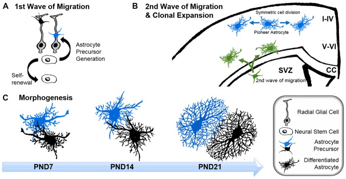
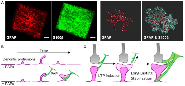
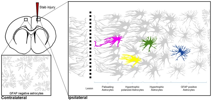
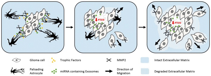
Verification
Claims the paper makes
The claims and statistics of this paper haven’t been checked yet.
Run the check — free, no account needed. It reads the open-access text and checks the sources it cites.
Claims other papers make about this one
Each is checked against this paper’s own text: does it say what the citing paper says it does?
The cited source says what the claim attributes to it.
In the citing paper
“Thorough visualization is necessary because astrocytes undergo distinct morphological changes other than hypertrophy in pathological contexts, including elongation, process extension toward injury site, and some three-dimensional (3D) domain overlap26.”
From Reactive astrocyte nomenclature, definitions, and future directions (2021) · cites this paper as [26]
Self-check
Percentages, sums and reported differences re-computed from the paper’s own numbers.
The self-check needs the paper’s full text.
Citations
Cites
Resting microglial cells are highly dynamic surveillants of brain parenchyma in vivo
Nimmerjahn, A., Kirchhoff, F., & Helmchen, F. (2005) · Science · doi:10.1126/science.1110647 · Cited by 5,827
Preparation of separate astroglial and oligodendroglial cell cultures from rat cerebral tissue.
McCarthy, K. D., & de Vellis, J. (1980) · The Journal of Cell Biology · doi:10.1083/jcb.85.3.890 · Cited by 3,825
Daneman, R., & Prat, A. (2015) · Cold Spring Harbor Perspectives in Biology · doi:10.1101/cshperspect.a020412 · Cited by 3,437
Molecular dissection of reactive astrogliosis and glial scar formation
Sofroniew, M. V. (2009) · Trends in Neurosciences · doi:10.1016/j.tins.2009.08.002 · Cited by 2,594
Tripartite synapses: glia, the unacknowledged partner
Araque, A., Parpura, V., Sanzgiri, R. P., & Haydon, P. G. (1999) · Trends in Neurosciences · doi:10.1016/s0166-2236(98)01349-6 · Cited by 2,529
Astrocyte scar formation aids central nervous system axon regeneration
Anderson, M. A., Burda, J. E., Ren, Y., Ao, Y., O’Shea, T. M., Kawaguchi, R., Coppola, G., Khakh, B. S., Deming, T. J., & Sofroniew, M. V. (2016) · Nature · doi:10.1038/nature17623 · Cited by 1,851
Synaptic scaling mediated by glial TNF-α
Stellwagen, D., & Malenka, R. C. (2006) · Nature · doi:10.1038/nature04671 · Cited by 1,715
Thrombospondins are astrocyte-secreted proteins that promote CNS synaptogenesis
Christopherson, K. S., Ullian, E. M., Stokes, C. C. A., Mullowney, C. E., Hell, J. W., Agah, A., Lawler, J., Mosher, D. F., Bornstein, P., & Barres, B. A. (2005) · Cell · doi:10.1016/j.cell.2004.12.020 · Cited by 1,688
Reactive Astrocytes Protect Tissue and Preserve Function after Spinal Cord Injury
Faulkner, J. R., Herrmann, J. E., Woo, M. J., Tansey, K. E., Doan, N., & Sofroniew, M. V. (2004) · Journal of Neuroscience · doi:10.1523/jneurosci.3547-03.2004 · Cited by 1,652
Protoplasmic Astrocytes in CA1 Stratum Radiatum Occupy Separate Anatomical Domains
Bushong, E. A., Martone, M. E., Jones, Y. Z., & Ellisman, M. H. (2002) · The Journal of Neuroscience · doi:10.1523/jneurosci.22-01-00183.2002 · Cited by 1,578
Research works printed but not linked: 1
- Ramón y Cajal, 1913, Un nuevo proceder para la impregnación de la neuroglía, Bol. Soc. Esp. Bio., 2, 104
Cited by
Sorted by citations
Reactive astrocyte nomenclature, definitions, and future directions
Escartin, C., Galea, E., Lakatos, A., O’Callaghan, J. P., Petzold, G. C., Serrano-Pozo, A., Steinhäuser, C., Volterra, A., Carmignoto, G., Agarwal, A., et al. (2021) · Nature Neuroscience · doi:10.1038/s41593-020-00783-4 · Cited by 2,513
Copper Dyshomeostasis in Neurodegenerative Diseases—Therapeutic Implications
Gromadzka, G., Tarnacka, B., Flaga, A., & Adamczyk, A. (2020) · International Journal of Molecular Sciences · doi:10.3390/ijms21239259 · Cited by 347
Blood–Brain Barrier Dynamics to Maintain Brain Homeostasis
Segarra, M., Aburto, M. R., & Acker‐Palmer, A. (2021) · Trends in Neurosciences · doi:10.1016/j.tins.2020.12.002 · Cited by 316
Questions and (some) answers on reactive astrocytes
Escartin, C., Guillemaud, O., & Carrillo‐de Sauvage, M. (2019) · Glia · doi:10.1002/glia.23687 · Cited by 275
Fatigue, Sleep, and Autoimmune and Related Disorders
Zielinski, M. R., Systrom, D. M., & Rose, N. R. (2019) · Frontiers in Immunology · doi:10.3389/fimmu.2019.01827 · Cited by 281
Goshi, N., Morgan, R. K., Lein, P. J., & Şeker, E. (2020) · Journal of Neuroinflammation · doi:10.1186/s12974-020-01819-z · Cited by 248
Astrocytic processes: from tripartite synapses to the active milieu
Semyanov, A. V., & Verkhratsky, A. (2021) · Trends in Neurosciences · doi:10.1016/j.tins.2021.07.006 · Cited by 243
A Developmental Analysis of Juxtavascular Microglia Dynamics and Interactions with the Vasculature
Mondo, E., Becker, S. C., Kautzman, A. G., Schifferer, M., Baer, C. E., Chen, J., Huang, E. J., Simons, M., & Schafer, D. P. (2020) · Journal of Neuroscience · doi:10.1523/jneurosci.3006-19.2020 · Cited by 153
The role of astrocyte structural plasticity in regulating neural circuit function and behavior
Lawal, O., Ulloa Severino, F. P., & Eroglu, C. (2022) · Glia · doi:10.1002/glia.24191 · Cited by 142
APOE4 drives inflammation in human astrocytes via TAGLN3 repression and NF-κB activation
Arnaud, L., Benech, P., Greetham, L., Stephan, D., Jimenez, A., Jullien, N., García‐González, L., Tsvetkov, P. O., Devred, F., Sancho-Martinez, I., et al. (2022) · Cell Reports · doi:10.1016/j.celrep.2022.111200 · Cited by 130
Baxter, P. S., Dando, O., Emelianova, K., He, X., McKay, S., Hardingham, G. E., & Qiu, J. (2021) · Cell Reports · doi:10.1016/j.celrep.2021.108882 · Cited by 122
Hsu, C., Xu, J., Brinkhof, B., Wang, H., Cui, Z., Huang, W. E., & Ye, H. (2020) · Proceedings of the National Academy of Sciences · doi:10.1073/pnas.2001906117 · Cited by 118
Emerging evidence for astrocyte dysfunction in schizophrenia
Figueiredo, E. C. D. O., Calì, C., Petrelli, F., & Bezzi, P. (2022) · Glia · doi:10.1002/glia.24221 · Cited by 87
Li, J., Jaiswal, M. K., Chien, J.-F., Kozlenkov, A., Jung, J., Zhou, P., Gardashli, M., Pregent, L., Engelberg-Cook, E., Dickson, D. W., et al. (2023) · Nature Communications · doi:10.1038/s41467-023-41033-y · Cited by 81
Extracellular Vesicle-Mediated Bilateral Communication between Glioblastoma and Astrocytes
Nieland, L., Morsett, L. M., Broekman, M. L. D., Breakefield, X. O., & Abels, E. R. (2020) · Trends in Neurosciences · doi:10.1016/j.tins.2020.10.014 · Cited by 77
Civita, P., Leite, D. M., & Pilkington, G. J. (2019) · International Journal of Molecular Sciences · doi:10.3390/ijms20236017 · Cited by 74
Yue, Q., & Hoi, M. P. M. (2023) · Neural Regeneration Research · doi:10.4103/1673-5374.367832 · Cited by 78
Neuron-astrocyte omnidirectional signaling in neurological health and disease
Pathak, D., & Sriram, K. (2023) · Frontiers in Molecular Neuroscience · doi:10.3389/fnmol.2023.1169320 · Cited by 72
Neuromodulation of Glial Function During Neurodegeneration
Stevenson, R., Samokhina, E., Rossetti, I., Morley, J. W., & Buskila, Y. (2020) · Frontiers in Cellular Neuroscience · doi:10.3389/fncel.2020.00278 · Cited by 72
Astrocyte–synapse interactions and cell adhesion molecules
Saint‐Martin, M., & Goda, Y. (2022) · FEBS Journal · doi:10.1111/febs.16540 · Cited by 70
Organizing principles of astrocytic nanoarchitecture in the mouse cerebral cortex
Salmon, C. K., Syed, T. A., Kacerovsky, J. B., Alivodej, N., Schober, A. L., Sloan, T. F. W., Pratte, M. T., Rosen, M. P., Green, M., Chirgwin-Dasgupta, A., et al. (2023) · Current Biology · doi:10.1016/j.cub.2023.01.043 · Cited by 70
DNA methylation controls stemness of astrocytes in health and ischaemia
Kremer, L. P. M., Cerrizuela, S., El‐Sammak, H., Shukairi, M. E. A., Ellinger, T., Straub, J., Korkmaz, A., Volk, K., Brunken, J., Kleber, S., et al. (2024) · Nature · doi:10.1038/s41586-024-07898-9 · Cited by 61
Zhang, H., Zhang, X., Chai, Y., Wang, Y., Zhang, J., & Chen, X. (2025) · Frontiers in Immunology · doi:10.3389/fimmu.2025.1584577 · Cited by 64
Bentsen, M. A., Rausch, D. M., Mirzadeh, Z., Muta, K., Scarlett, J. M., Brown, J. M., Herranz‐Pérez, V., Baquero, A. F., Thompson, J. J., Alonge, K. M., et al. (2020) · Nature Communications · doi:10.1038/s41467-020-17720-5 · Cited by 51
Schiweck, J., Murk, K., Ledderose, J. M. T., Münster‐Wandowski, A., Ornaghi, M., Vida, I., & Eickholt, B. J. (2021) · Nature Communications · doi:10.1038/s41467-021-21662-x · Cited by 57
Self-citationGlial Dysfunction in MeCP2 Deficiency Models: Implications for Rett Syndrome
Kahanovitch, U., Patterson, K. C., Hernandez, R. D., & Olsen, M. L. (2019) · International Journal of Molecular Sciences · doi:10.3390/ijms20153813 · Cited by 53
Generation of Human iPSC-Derived Astrocytes with a mature star-shaped phenotype for CNS modeling
Voulgaris, D., Nikolakopoulou, P., & Herland, A. (2022) · Stem Cell Reviews and Reports · doi:10.1007/s12015-022-10376-2 · Cited by 46
In vivo spatiotemporal dynamics of astrocyte reactivity following neural electrode implantation
Savya, S. P., Li, F., Lam, S., Wellman, S. M., Stieger, K. C., Chen, K., Eles, J. R., & Kozai, T. D. Y. (2022) · Biomaterials · doi:10.1016/j.biomaterials.2022.121784 · Cited by 46
Contribution of astrocytes to metabolic dysfunction in the Alzheimer’s disease brain
Zulfiqar, S., Garg, P., & Nieweg, K. (2019) · Biological Chemistry · doi:10.1515/hsz-2019-0140 · Cited by 46
Astrocytes in rapid ketamine antidepressant action
Stenovec, M., Li, B., Verkhratsky, A., & Zorec, R. (2020) · Neuropharmacology · doi:10.1016/j.neuropharm.2020.108158 · Cited by 52
Increased connectivity of hiPSC-derived neural networks in multiphase granular hydrogel scaffolds
Hsu, C., George, J. H., Waller, S., Besnard, C., Nagel, D. A., Hill, E. J., Coleman, M. D., Korsunsky, A. M., Cui, Z., & Ye, H. (2021) · Bioactive Materials · doi:10.1016/j.bioactmat.2021.07.008 · Cited by 44
Astroglia in Autism Spectrum Disorder
Gzieło, K., & Nikiforuk, A. (2021) · International Journal of Molecular Sciences · doi:10.3390/ijms222111544 · Cited by 46
Astrocytic Factors Controlling Synaptogenesis: A Team Play
Fossati, G., Matteoli, M., & Menna, E. (2020) · Cells · doi:10.3390/cells9102173 · Cited by 46
Astroglial Hmgb1 regulates postnatal astrocyte morphogenesis and cerebrovascular maturation
Andrade, M. F., Comin, C. H., Dyken, P. C. V., Ouellette, J., Raman‐Nair, J., Blakeley, N., Liu, Q. Y., Leclerc, S., Pan, Y., Liu, Z., et al. (2023) · Nature Communications · doi:10.1038/s41467-023-40682-3 · Cited by 42
Morphological and functional alterations of astrocytes responding to traumatic brain injury
Cheng, X., Wang, J., Sun, X., Shao, L., Guo, Z., & Li, Y. (2019) · Journal of Integrative Neuroscience · doi:10.31083/j.jin.2019.02.110 · Cited by 41
Circadian rhythms and glial cells of the central nervous system
Brécier, A., Li, V. W., Smith, C. S., Halievski, K., & Ghasemlou, N. (2022) · Biological reviews/Biological reviews of the Cambridge Philosophical Society · doi:10.1111/brv.12917 · Cited by 40
Utilising Induced Pluripotent Stem Cells in Neurodegenerative Disease Research: Focus on Glia
Albert, K., Niskanen, J., Kälvälä, S., & Lehtonen, Š. (2021) · International Journal of Molecular Sciences · doi:10.3390/ijms22094334 · Cited by 40
Benchmarking brain organoid recapitulation of fetal corticogenesis
Cheroni, C., Trattaro, S., Caporale, N., Tobon, A., Tenderini, E., Sebastiani, S., Troglio, F., Gabriele, M., Bressan, R. B., Pollard, S. M., et al. (2022) · Translational Psychiatry · doi:10.1038/s41398-022-02279-0 · Cited by 43
Sun, J., Osenberg, S., Irwin, A., Ma, L.-H., Lee, N., Xiang, Y., Li, F., Wan, Y.-W., Park, I.-H., Maletic-Savatic, M., et al. (2023) · Cell Reports · doi:10.1016/j.celrep.2022.111942 · Cited by 37
Amro, Z., Yool, A. J., & Collins‐Praino, L. E. (2021) · Brain Behavior & Immunity - Health · doi:10.1016/j.bbih.2021.100242 · Cited by 39
Budgett, R. F., Bakker, G., Sergeev, E., Bennett, K. A., & Bradley, S. J. (2022) · Frontiers in Pharmacology · doi:10.3389/fphar.2022.893422 · Cited by 38
García‐León, J. A., Cáceres-Palomo, L., Sánchez‐Mejías, E., Mejias‐Ortega, M., Nuñez‐Diaz, C., Fernandez-Valenzuela, J. J., Sanchez-Varo, R. M., Dávila, J. C., Vitórica, J., & Gutiérrez, A. (2020) · International Journal of Molecular Sciences · doi:10.3390/ijms21186867 · Cited by 35
Cervelli, M., Averna, M., Vergani, L., Pedrazzi, M., Amato, S., Fiorucci, C., Rossi, M. N., Maura, G., Mariottini, P., Cervetto, C., et al. (2022) · Biomedicines · doi:10.3390/biomedicines10071756 · Cited by 34
Profilin Isoforms in Health and Disease – All the Same but Different
Murk, K., Ornaghi, M., & Schiweck, J. (2021) · Frontiers in Cell and Developmental Biology · doi:10.3389/fcell.2021.681122 · Cited by 38
Self-citationThe microglia-blood vessel interactions in the developing brain
Hattori, Y. (2022) · Neuroscience Research · doi:10.1016/j.neures.2022.09.006 · Cited by 37
SOX2 is essential for astrocyte maturation and its deletion leads to hyperactive behavior in mice
Wang, Y., Zhang, S., Lan, Z., Doan, V., Kim, B., Liu, S., Zhu, M., Hull, V. L., Rihani, S., Zhang, C., et al. (2022) · Cell Reports · doi:10.1016/j.celrep.2022.111842 · Cited by 32
Doust, Y. V., Rowe, R. K., Adelson, P. D., Lifshitz, J., & Ziebell, J. M. (2021) · Frontiers in Neurology · doi:10.3389/fneur.2021.722526 · Cited by 33
Diverged morphology changes of astrocytic and neuronal primary cilia under reactive insults
Sterpka, A., Yang, J., Strobel, M., Zhou, Y., Pauplis, C., & Chen, X. (2020) · Molecular Brain · doi:10.1186/s13041-020-00571-y · Cited by 30
Kayasandık, C. B., Ru, W., & Demetrio Labate, A. (2020) · Scientific Reports · doi:10.1038/s41598-020-61953-9 · Cited by 31
Inflammation Drives Alzheimer's Disease: Emphasis on 5-lipoxygenase Pathways
Siddiqui, A., Akhtar, S., Shah, Z., Othman, I., & Kumari, Y. (2020) · Current Neuropharmacology · doi:10.2174/1570159x18666200924122732 · Cited by 30
Roemeling, C. A. V., Patel, J. A., Carpenter, S. L., Yegorov, O., Yang, C., Bhatia, A., Doonan, B. P., Russell, R. A., Trivedi, V., Klippel, K., et al. (2024) · Nature Communications · doi:10.1038/s41467-024-49989-1 · Cited by 33
Novgorodov, S., Voltin, J. R., Wang, W., Tomlinson, S., Riley, C. L., & Gudz, T. I. (2019) · Journal of Lipid Research · doi:10.1194/jlr.m091132 · Cited by 20
Shi, W., Dong, P., Kuss, M. A., Gu, L., Kievit, F. M., Kim, H. J., & Duan, B. (2021) · Advanced Healthcare Materials · doi:10.1002/adhm.202100180 · Cited by 36
Effects of radiation on endothelial barrier and vascular integrity
Bouten, R. M., Young, E. F., Selwyn, R. G., Iacono, D., Rittase, W. B., & Day, R. M. (2021) · Elsevier eBooks · doi:10.1016/b978-0-12-818561-2.00007-2 · Cited by 30
Astrocytes regulate neuronal network activity by mediating synapse remodeling
Kim, N., & Chung, W. (2022) · Neuroscience Research · doi:10.1016/j.neures.2022.09.007 · Cited by 31
Astrocyte diversity and subtypes: aligning transcriptomics with multimodal perspectives
Hennes, M., Richter, M. L., Fischer-Sternjak, J., & Götz, M. (2025) · EMBO Reports · doi:10.1038/s44319-025-00529-y · Cited by 35
Martínez‐Lozada, Z., & Robinson, M. B. (2020) · Neurochemistry International · doi:10.1016/j.neuint.2020.104787 · Cited by 26
Vibrational resonance in a neuron–astrocyte coupled model
Çalım, A., Longtin, A., & Uzuntarla, M. (2021) · Philosophical Transactions of the Royal Society A Mathematical Physical and Engineering Sciences · doi:10.1098/rsta.2020.0267 · Cited by 26
Arizono, M., & Nägerl, U. V. (2021) · Glia · doi:10.1002/glia.24103 · Cited by 28
The Blood–Brain Barrier: Much More Than a Selective Access to the Brain
Sánchez-Cano, F., Hernández‐Kelly, L. C., & Ortega, A. (2021) · Neurotoxicity Research · doi:10.1007/s12640-021-00431-0 · Cited by 30
Lana, D., Traini, C., Bulli, I., Sarti, G., Magni, G., Attorre, S., Giovannini, M. G., & Vannucchi, M. G. (2024) · Frontiers in Pharmacology · doi:10.3389/fphar.2024.1451114 · Cited by 27
Glial Patchwork: Oligodendrocyte Progenitor Cells and Astrocytes Blanket the Central Nervous System
Barber, H. M., Ali, M., & Kucenas, S. (2022) · Frontiers in Cellular Neuroscience · doi:10.3389/fncel.2021.803057 · Cited by 20
Astrocyte 3D culture and bioprinting using peptide functionalized hyaluronan hydrogels
Matthiesen, I., Jury, M., Boroojeni, F. R., Ludwig, S. L., Holzreuter, M. A., Buchmann, S., Träger, A., Selegård, R., Winkler, T. E., Aili, D., et al. (2023) · Science and Technology of Advanced Materials · doi:10.1080/14686996.2023.2165871 · Cited by 25
Retinal astrocyte morphology predicts integration of vascular and neuronal architecture
Holden, J. M., Wareham, L. K., & Calkins, D. J. (2023) · Frontiers in Neuroscience · doi:10.3389/fnins.2023.1244679 · Cited by 27
Reactive Astrocytosis in a Mouse Model of Chronic Polyamine Catabolism Activation
Cervetto, C., Averna, M., Vergani, L., Pedrazzi, M., Amato, S., Pelassa, S., Giuliani, S., Baldini, F., Maura, G., Mariottini, P., et al. (2021) · Biomolecules · doi:10.3390/biom11091274 · Cited by 23
An, J., Yang, H., Yang, E., Chung, S., Kim, D.-Y., Jou, I., Park, S. M., Kim, B. G., Chwae, Y., & Joe, E.-H. (2020) · Glia · doi:10.1002/glia.23947 · Cited by 23
The Diverse Roles of Reactive Astrocytes in the Pathogenesis of Amyotrophic Lateral Sclerosis
Yang, K., Liu, Y., & Zhang, M. (2024) · Brain Sciences · doi:10.3390/brainsci14020158 · Cited by 29
Marcoli, M., Cervetto, C., Amato, S., Fiorucci, C., Maura, G., Mariottini, P., & Cervelli, M. (2022) · Biomolecules · doi:10.3390/biom12020204 · Cited by 22
Optogenetic Modulation of Neural Progenitor Cells Improves Neuroregenerative Potential
Giraldo, E., Palmero-Canton, D., Martínez-Rojas, B., Sánchez-Martín, M. D. M., & Moreno‐Manzano, V. (2020) · International Journal of Molecular Sciences · doi:10.3390/ijms22010365 · Cited by 20
Torregrosa, T., Lehman, S., Hana, S., Marsh, G., Xu, S., Koszka, K., Mastrangelo, N., McCampbell, A., Henderson, C. E., & Lo, S. (2021) · Gene Therapy · doi:10.1038/s41434-021-00223-3 · Cited by 24
Yawoot, N., Sengking, J., Wicha, P., Govitrapong, P., Tocharus, C., & Tocharus, J. (2021) · Journal of Cellular Physiology · doi:10.1002/jcp.30649 · Cited by 20
Visualizing Astrocyte Morphology Using Lucifer Yellow Iontophoresis
Moye, S. L., Castro, B. D., Gangwani, M. R., & Khakh, B. S. (2019) · Journal of Visualized Experiments · doi:10.3791/60225 · Cited by 21
Hoeferlin, G. F., Grabinski, S. E., Druschel, L. N., Duncan, J. L., Burkhart, G., Weagraff, G. R., Lee, A. H., Hong, C., Bambroo, M., Olivares, H., et al. (2025) · Nature Communications · doi:10.1038/s41467-025-56979-4 · Cited by 21
Are cytoskeleton changes observed in astrocytes functionally linked to aging?
Villablanca, C., Vidal, R. L., & González-Billault, C. E. (2023) · Brain Research Bulletin · doi:10.1016/j.brainresbull.2023.03.007 · Cited by 19
Jiang, N., Cupolillo, D., Grosjean, N., Muller, E., Deforges, S., Mulle, C., & Amédée, T. (2021) · Neurobiology of Disease · doi:10.1016/j.nbd.2021.105345 · Cited by 17
Sethi, P., Virmani, G., Gupta, K., Thumu, S. C. R., Ramanan, N., & Marathe, S. V. (2021) · Journal of Cell Science · doi:10.1242/jcs.258430 · Cited by 17
Ketamine Alters Functional Plasticity of Astroglia: An Implication for Antidepressant Effect
Stenovec, M. (2021) · Life · doi:10.3390/life11060573 · Cited by 18
Hypergravity Attenuates Reactivity in Primary Murine Astrocytes
Lichterfeld, Y., Kalinski, L., Schunk, S., Schmakeit, T., Feles, S., Frett, T., Herrmann, H. O., Hemmersbach, R., & Liemersdorf, C. (2022) · Biomedicines · doi:10.3390/biomedicines10081966 · Cited by 17
O’Neill, K. M., Saracino, E., Barile, B., Mennona, N. J., Mola, M. G., Pathak, S., Posati, T., Zamboni, R., Nicchia, G. P., Benfenati, V., et al. (2023) · Advanced Biology · doi:10.1002/adbi.202200269 · Cited by 16
Espírito‐Santo, S., Coutinho, V. G., Dezonne, R. S., Stipursky, J., dos Santos‐Rodrigues, A., Batista, C., Paes‐de‐Carvalho, R., Fuss, B., & Gomes, F. C. A. (2021) · Glia · doi:10.1002/glia.23971 · Cited by 19
Putative Roles of Astrocytes in General Anesthesia
Mulkey, D. K., Olsen, M. L., Ou, M., Cleary, C. M., & Du, G. (2021) · Current Neuropharmacology · doi:10.2174/1570159x19666210215120755 · Cited by 19
Expression of synaptic proteins in the hippocampus is modulated by neonatal oxytocin treatment
Filová, B., Reichova, A., Zatkova, M., Srančíková, A., Bukatová, S., Bačová, Z., & Bakoš, J. (2020) · Neuroscience Letters · doi:10.1016/j.neulet.2020.134912 · Cited by 18
Slota, J. A., Medina, S. J., Frost, K. L., & Booth, S. A. (2022) · Frontiers in Neuroscience · doi:10.3389/fnins.2022.918811 · Cited by 16
Axon–glia interactions in the ascending auditory system
Kohrman, D. C., Borges, B. C., Cassinotti, L. R., Ji, L., & Corfas, G. (2021) · Developmental Neurobiology · doi:10.1002/dneu.22813 · Cited by 16
Automated detection of GFAP-labeled astrocytes in micrographs using YOLOv5
Huang, Y., Kruyer, A., Syed, S., Kayasandık, C. B., Papadakis, M., & Demetrio Labate, A. (2022) · Scientific Reports · doi:10.1038/s41598-022-26698-7 · Cited by 18
Gordillo-Sampedro, S., Antounians, L., Wei, W., Mufteev, M., Lendemeijer, B., Kushner, S. A., de Vrij, F. M. S., Zani, A., & Ellis, J. (2024) · Molecular and Cellular Neuroscience · doi:10.1016/j.mcn.2024.103933 · Cited by 15
Hofmann, G., Hasser, E. M., & Kline, D. D. (2021) · American Journal of Physiology-Regulatory, Integrative and Comparative Physiology · doi:10.1152/ajpregu.00019.2021 · Cited by 15
Jo, J., Woo, J., Cristobal, C. D., Choi, J. M., Wang, C., Ye, Q., Smith, J. A., Ung, K., Liu, G., Cortes, D., et al. (2021) · EMBO Reports · doi:10.15252/embr.202153200 · Cited by 13
Astrocyte arborization enhances Ca2+ but not cAMP signaling plasticity
Pirnat, S., Božić, M., Dolanc, D., Horvat, A., Verdev, P. T., Vardjan, N., Verkhratsky, A., Zorec, R., & Stenovec, M. (2021) · Glia · doi:10.1002/glia.24076 · Cited by 13
Antonovaité, N., van Wageningen, T. A., Paardekam, E. J., van Dam, A., & Iannuzzi, D. (2020) · Journal of the mechanical behavior of biomedical materials/Journal of mechanical behavior of biomedical materials · doi:10.1016/j.jmbbm.2020.103783 · Cited by 14
Astrocyte-driven vasoconstriction impairs glymphatic clearance in a human tauopathy-on-chip model
Park, R., Peng, Y., Yslas, A. R., & Lee, E. (2025) · APL Bioengineering · doi:10.1063/5.0261875 · Cited by 12
Crosstalk between Blood Vessels and Glia during the Central Nervous System Development
Tabata, H. (2022) · Life · doi:10.3390/life12111761 · Cited by 14
Dozio, V., Daali, Y., Desmeules, J. A., & Sanchez, J. (2020) · Journal of Neuroscience Research · doi:10.1002/jnr.24731 · Cited by 14
Albizzati, E., Florio, E., Miramondi, F., Sormonta, I., Landsberger, N., & Frasca, A. (2022) · Frontiers in Neuroscience · doi:10.3389/fnins.2022.823060 · Cited by 12
Welby, E., & Ebert, A. D. (2023) · Glia · doi:10.1002/glia.24340 · Cited by 12
Meller, S., Käufer, C., Gailus, B., Brandt, C., & Löscher, W. (2021) · Neurobiology of Disease · doi:10.1016/j.nbd.2021.105446 · Cited by 12
Zhang, W., Ye, F., Xiong, J., He, F., Li, Y., Yin, F., Peng, J., & Wang, X. (2022) · The FASEB Journal · doi:10.1096/fj.202200110rr · Cited by 13
Bondi, H., Chiazza, F., Masante, I., Bortolotto, V., Canonico, P. L., & Grilli, M. (2022) · Neurobiology of Aging · doi:10.1016/j.neurobiolaging.2022.12.010 · Cited by 14
In vitro model of traumatic brain injury to screen neuro-regenerative biomaterials
Basit, R. H., Tzerakis, N., Jenkins, S. I., & Chari, D. M. (2021) · Materials Science and Engineering C · doi:10.1016/j.msec.2021.112253 · Cited by 12
Liu, Y., Hong, W., Gong, P., Qi, G., Wang, X., Kang, S., Tang, H., & Qin, S. (2022) · Glia · doi:10.1002/glia.24298 · Cited by 11
Metadata
- Authors
- Schiweck, J., Eickholt, B. J., & Murk, K.
- Affiliations
- Charité – Universitätsmedizin Berlin (DE)
- Corresponding author
- Eickholt, Britta J.
- Murk, Kai
- Venue
- Frontiers in Cellular Neuroscience
- Publisher
- Frontiers Media SA
- Published
- 2018-08-21
- Volume
- 12
- Pages
- 261-261
- ISSN
- 1662-5102 (ISSN-L)
- PMC
- PMC6111612
- OpenAlex
- W2888094777
- Cited by
- 223
- References
- 169
- Citations by year
- 2026: 14 · 2025: 28 · 2024: 25 · 2023: 23 · 2022: 38 · 2021: 44 · 2020: 33 · 2019: 18
- FWCI
- 10.82
- Type
- journal article
- Language
- en
- Open access
- gold
- doi.org
- Locations
- Frontiers in Cellular Neuroscience (publishedVersion, cc-by)
- Europe PMC (PubMed Central) (publishedVersion, cc-by)
- PubMed Central (publishedVersion, cc-by)
- PubMed (publishedVersion)
- DOAJ (DOAJ: Directory of Open Access Journals) (submittedVersion)
- Refubium (Universitätsbibliothek der Freien Universität Berlin) (submittedVersion, cc-by)
- Universitätsbibliothek der FU Berlin Hochschulschriftenstelle u. Dokumentenserver (cc-by)
- Funders
- Deutsche Forschungsgemeinschaft (SFB958, TRR168, 285933818)
- Topics
- Life Sciences › Neuroscience › Cellular and Molecular Neuroscience › Neuroscience and Neuropharmacology Research
- Life Sciences › Neuroscience › Neurology › Neuroinflammation and Neurodegeneration Mechanisms
- Life Sciences › Neuroscience › Developmental Neuroscience › Neurogenesis and neuroplasticity mechanisms
- Keywords
- astrocytes, astrocyte morphology, synaptogenesis, synaptic plasticity, neural transmission, astrocyte development, traumatic brain injury, neurodegenerative diseases, blood-brain barrier, cytoskeleton, glial cells
About these checks
Registry, retraction, Cites and statistics results are computed, not judged, with one exception: a printed reference that no index lists is read once by a model before it counts as a citation error. Claims are extracted by a model and kept only when their quote appears verbatim in the text; each cited source is then checked, with a model’s help, for whether it says what the claim relies on. Those verdicts can be wrong — read the claim’s quote and the cited source before relying on one.
Statistics and arithmetic are recomputed, not judged.
Confidence bands are shown only for fields calibrated on our annotated test set. None is calibrated yet, so no band is shown.
What counts as a claim ›How we measure confidence ›How we check this ›
Are you an author, or do you see a problem with this page? Request a review
