

The DOI matches its Crossref record; 376 of the 376 references it cites were found in a registry or an index.
Phosphoinositides: Roles in the Development of Microglial-Mediated Neuroinflammation and Neurodegeneration
Open accessAbstract
Microglia are increasingly recognized as vital players in the pathology of a variety of neurodegenerative conditions including Alzheimer’s (AD) and Parkinson’s (PD) disease. While microglia have a protective role in the brain, their dysfunction can lead to neuroinflammation and contributes to disease progression. Also, a growing body of literature highlights the seven phosphoinositides, or PIPs, as key players in the regulation of microglial-mediated neuroinflammation. These small signaling lipids are phosphorylated derivates of phosphatidylinositol, are enriched in the brain, and have well-established roles in both homeostasis and disease.Disrupted PIP levels and signaling has been detected in a variety of dementias. Moreover, many known AD disease modifiers identified via genetic studies are expressed in microglia and are involved in phospholipid metabolism. One of these, the enzyme PLCγ2 that hydrolyzes the PIP species PI(4,5)P 2 , displays altered expression in AD and PD and is currently being investigated as a potential therapeutic target.Perhaps unsurprisingly, neurodegenerative conditions exhibiting PIP dyshomeostasis also tend to show alterations in aspects of microglial function regulated by these lipids. In particular, phosphoinositides regulate the activities of proteins and enzymes required for endocytosis, toll-like receptor signaling, purinergic signaling, chemotaxis, and migration, all of which are affected in a variety of neurodegenerative conditions. These functions are crucial to allow microglia to adequately survey the brain and respond appropriately to invading pathogens and other abnormalities, including misfolded proteins. AD and PD therapies are being developed to target many of the above pathways, and although not yet investigated, simultaneous PIP manipulation might enhance the beneficial effects observed. Currently, only limited therapeutics are available for dementia, and although these show some benefits for symptom severity and progression, they are far from curative. Given the importance of microglia and PIPs in dementia development, this review summarizes current research and asks whether we can exploit this information to design more targeted, or perhaps combined, dementia therapeutics. More work is needed to fully characterize the pathways discussed in this review, but given the strength of the current literature, insights in this area could be invaluable for the future of neurodegenerative disease research.
Overview
The claims and statistics of this paper haven’t been checked yet.
Run the check — free, no account needed. It reads the open-access text and checks the sources it cites.
Integrity signals
Registry
Cites
Statistics
Figures
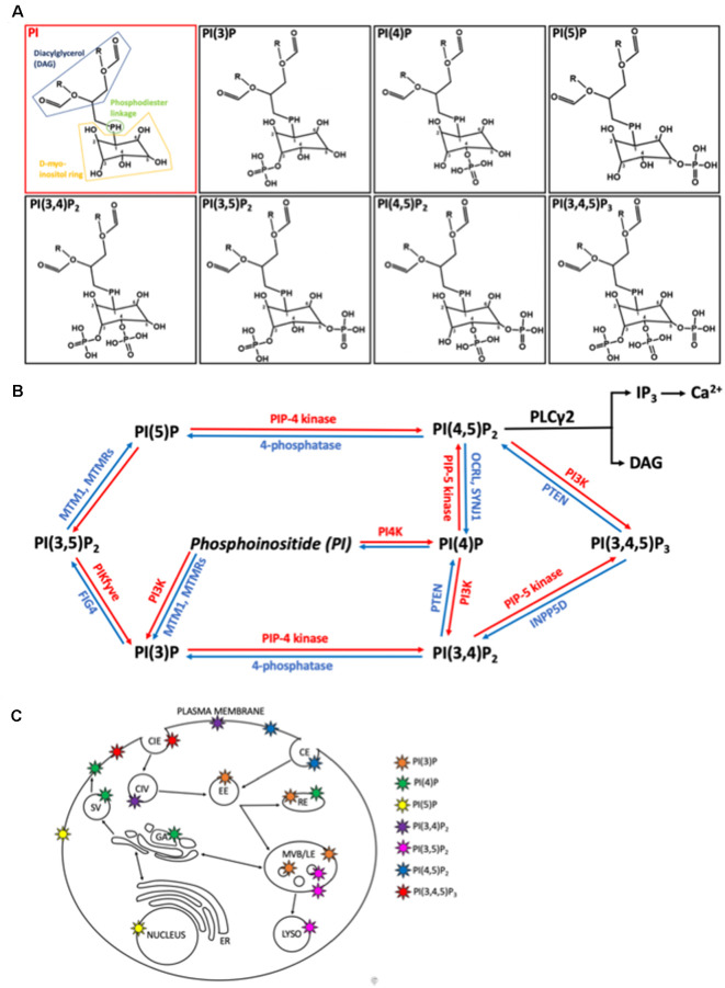
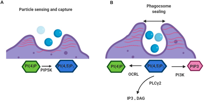
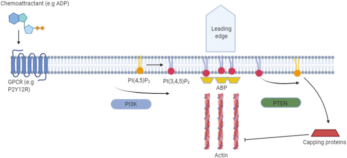
Verification
Claims the paper makes
The claims and statistics of this paper haven’t been checked yet.
Run the check — free, no account needed. It reads the open-access text and checks the sources it cites.
Claims other papers make about this one
Each is checked against this paper’s own text: does it say what the citing paper says it does?
The source backs part of the claim, not all of it — or backs it for a different population, condition or comparison.
In the citing paper
“PI(3,4,5)P3 can be converted back to PI(4,5)P2 by the protein phosphatase and tensin homology (PTEN) or converted into PI(3,4)P2 by SHIP1, which negatively regulates PI3K/Akt signaling (Fig. 2) [89–92].”
From Microglial function, INPP5D/SHIP1 signaling, and NLRP3 inflammasome activation: implications for Alzheimer’s disease (2023) · cites this paper as [90]
Experimental — accuracy not yet measuredThe cited source says what the claim attributes to it.
In the citing paper
“SHIP1 is a phosphatase involved in phosphoinositide signaling.”
From Microglial function, INPP5D/SHIP1 signaling, and NLRP3 inflammasome activation: implications for Alzheimer’s disease (2023) · cites this paper as [90]
Self-check
Percentages, sums and reported differences re-computed from the paper’s own numbers.
The self-check needs the paper’s full text.
Citations
Cites
The role of pattern-recognition receptors in innate immunity: update on Toll-like receptors
Kawai, T., & Akira, S. (2010) · Nature Immunology · doi:10.1038/ni.1863 · Cited by 9,304
MACDONALD, M. (1993) · Cell · doi:10.1016/0092-8674(93)90585-e · Cited by 8,366
Takeda, K., Kaisho, T., & Akira, S. (2003) · Annual Review of Immunology · doi:10.1146/annurev.immunol.21.120601.141126 · Cited by 6,143
Resting microglial cells are highly dynamic surveillants of brain parenchyma in vivo
Nimmerjahn, A., Kirchhoff, F., & Helmchen, F. (2005) · Science · doi:10.1126/science.1110647 · Cited by 5,827
DeJesus-Hernandez, M., Mackenzie, I. R., Boeve, B. F., Boxer, A. L., Baker, M., Rutherford, N. J., Nicholson, A. M., Finch, N. A., Flynn, H., Adamson, J., et al. (2011) · Neuron · doi:10.1016/j.neuron.2011.09.011 · Cited by 4,936 · cited 2×
“The most common cause (of both ALS and FTD) in North America and Europe is a hexanucleotide GGGGCC expansion in the c9orf72 gene (Dejesus-Hernandez et al., 2011; Renton et al., 2011).”
BackgroundA Hexanucleotide Repeat Expansion in C9ORF72 Is the Cause of Chromosome 9p21-Linked ALS-FTD
Renton, A. E., Majounie, E., Waite, A., Simón-Sánchez, J., Rollinson, S., Gibbs, J. R., Schymick, J. C., Laaksovirta, H., van Swieten, J. C., Myllykangas, L., et al. (2011) · Neuron · doi:10.1016/j.neuron.2011.09.010 · Cited by 4,409
Meta-Analysis of 74,046 Individuals Identifies 11 New Susceptibility Loci for Alzheimer’s Disease
(EADI), E. A. D. I., Lambert, J.-C., (GERAD), G. A. E. R. I. A. D., Alzheimer's Disease Genetic Consortium (ADGC), (CHARGE), C. F. H. A. A. R. I. G. E., Ibrahim-Verbaas, C. A., Harold, D., Naj, A. C., Sims, R., Bellenguez, C., et al. (2013) · Nature Genetics · doi:10.1038/ng.2802 · Cited by 4,336
Cell Migration: A Physically Integrated Molecular Process
Lauffenburger, D. A., & Horwitz, A. F. (1996) · Cell · doi:10.1016/s0092-8674(00)81280-5 · Cited by 3,915
ATP mediates rapid microglial response to local brain injury in vivo
Davalos, D., Grutzendler, J., Yang, G., Kim, J. V., Zuo, Y., Jung, S., Littman, D. R., Dustin, M. L., & Gan, W.-B. (2005) · Nature Neuroscience · doi:10.1038/nn1472 · Cited by 3,913
Ageing as a risk factor for neurodegenerative disease
Hou, Y., Dan, X., Babbar, M., Wei, Y., Hasselbalch, S. G., Croteau, D. L., & Bohr, V. A. (2019) · Nature Reviews Neurology · doi:10.1038/s41582-019-0244-7 · Cited by 3,310
Research works printed but not linked: 2
- Prince, 2013, Alzheimer’s Disease International. Policy Brief for Heads of Government: The Global Impact of Dementia 2013–2050
- 2015, The Global Impact of Dementia: An Analysis of Prevalence, Incidence, Cost and Trends.
Cited by
Sorted by citations
Terzioglu, G., & Young-Pearse, T. L. (2023) · Molecular Neurodegeneration · doi:10.1186/s13024-023-00674-9 · Cited by 91
Ma, C., Wu, Y., Song, L., Yin, L., Yin, J.-J., Wang, Q., Yu, J., & Xiao, B. (2022) · Neural Regeneration Research · doi:10.4103/1673-5374.355747 · Cited by 37
Lipid mediated brain disorders: A perspective
Singh, A., Kukal, S., Kanojia, N., Singh, M., Saso, L., Kukreti, S., & Kukreti, R. (2023) · Prostaglandins & Other Lipid Mediators · doi:10.1016/j.prostaglandins.2023.106737 · Cited by 27
He, G., He, Y., Ni, H., Wang, K., Zhu, Y., & Bao, Y. (2023) · European Journal of Histochemistry · doi:10.4081/ejh.2023.3766 · Cited by 23
Phosphoinositide signalling in cell motility and adhesion
Hou, X., Ren, C., Jin, J., Chen, Y., Lyu, X.-Y., Bi, K., Carrillo, N. D., Cryns, V. L., Anderson, R. A., Sun, J., et al. (2025) · Nature Cell Biology · doi:10.1038/s41556-025-01647-4 · Cited by 22
Metadata
- Authors
- Ernest James Phillips, T., & Maguire, E.
- Affiliations
- Cardiff University (GB)
- Corresponding author
- Maguire, Emily
- ORCID
- Maguire, Emily
- Venue
- Frontiers in Cellular Neuroscience
- Publisher
- Frontiers Media SA
- Published
- 2021-03-26
- Volume
- 15
- Pages
- 652593-652593
- ISSN
- 1662-5102 (ISSN-L)
- PMC
- PMC8032904
- OpenAlex
- W3140288526
- Cited by
- 33
- References
- 376
- Citations by year
- 2026: 7 · 2025: 7 · 2024: 6 · 2023: 8 · 2022: 2 · 2021: 3
- FWCI
- 0.56
- Type
- journal article
- Language
- en
- Open access
- gold
- doi.org
- Licence
- Locations
- Frontiers in Cellular Neuroscience (publishedVersion, cc-by)
- PubMed Central (publishedVersion, cc-by)
- PubMed (publishedVersion)
- ORCA Online Research @Cardiff (Cardiff University) (acceptedVersion)
- DOAJ (DOAJ: Directory of Open Access Journals) (submittedVersion, cc-by-sa)
- Funders
- Alzheimer's Society
- Cardiff University
- UK Dementia Research Institute
- Medical Research Council
- Topics
- Life Sciences › Neuroscience › Neurology › Neuroinflammation and Neurodegeneration Mechanisms
- Life Sciences › Neuroscience › Cellular and Molecular Neuroscience › Alzheimer's disease research and treatments
- Life Sciences › Neuroscience › Neurology › Neurological Disease Mechanisms and Treatments
- Keywords
- phosphoinositides, microglia, neuroinflammation, neurodegeneration, Alzheimer's disease, Parkinson's disease, PLCγ2, phosphatidylinositol 4,5-bisphosphate, phospholipid metabolism, endocytosis, Toll-like receptor signaling, purinergic signaling, chemotaxis, microglial migration, microglial dysfunction
About these checks
Registry, retraction, Cites and statistics results are computed, not judged, with one exception: a printed reference that no index lists is read once by a model before it counts as a citation error. Claims are extracted by a model and kept only when their quote appears verbatim in the text; each cited source is then checked, with a model’s help, for whether it says what the claim relies on. Those verdicts can be wrong — read the claim’s quote and the cited source before relying on one.
Statistics and arithmetic are recomputed, not judged.
Confidence bands are shown only for fields calibrated on our annotated test set. None is calibrated yet, so no band is shown.
What counts as a claim ›How we measure confidence ›How we check this ›
Are you an author, or do you see a problem with this page? Request a review
