

The DOI matches its Crossref record; 184 of the 191 references it cites were found in a registry or an index, and 1 are not checked yet.
The mechanisms for pattern completion and pattern separation in the hippocampus
Open accessAbstract
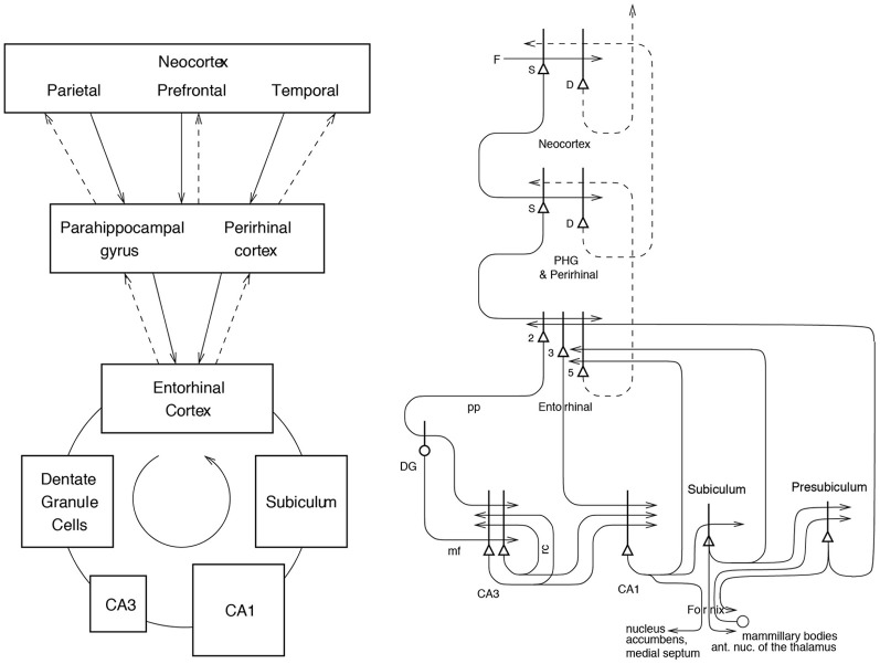
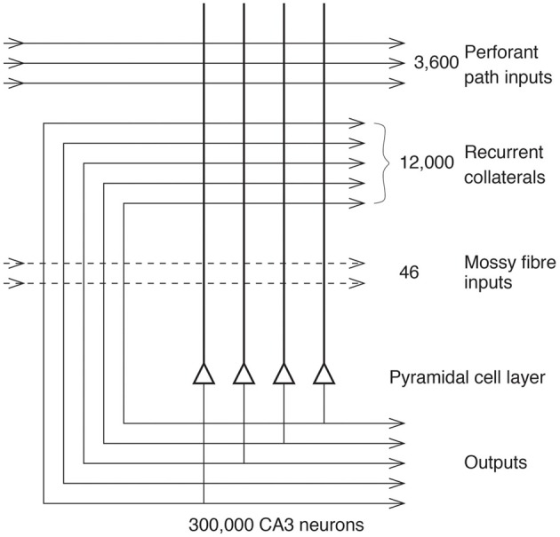
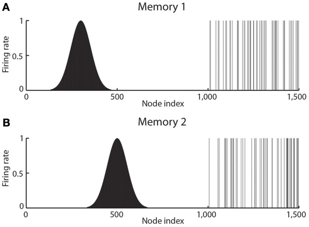
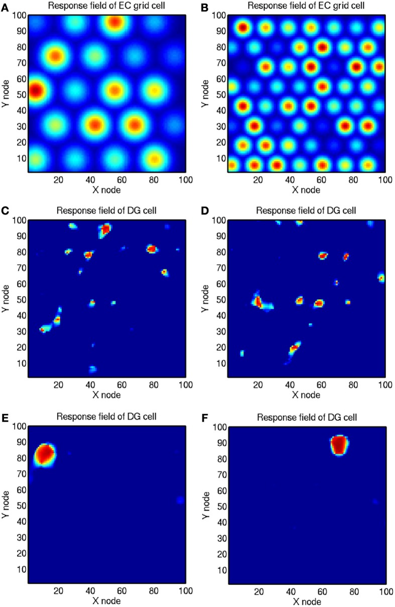
The mechanisms for pattern completion and pattern separation are described in the context of a theory of hippocampal function in which the hippocampal CA3 system operates as a single attractor or autoassociation network to enable rapid, one-trial, associations between any spatial location (place in rodents, or spatial view in primates) and an object or reward, and to provide for completion of the whole memory during recall from any part. The factors important in the pattern completion in CA3 together with a large number of independent memories stored in CA3 include a sparse distributed representation which is enhanced by the graded firing rates of CA3 neurons, representations that are independent due to the randomizing effect of the mossy fibers, heterosynaptic long-term depression as well as long-term potentiation in the recurrent collateral synapses, and diluted connectivity to minimize the number of multiple synapses between any pair of CA3 neurons which otherwise distort the basins of attraction. Recall of information from CA3 is implemented by the entorhinal cortex perforant path synapses to CA3 cells, which in acting as a pattern associator allow some pattern generalization. Pattern separation is performed in the dentate granule cells using competitive learning to convert grid-like entorhinal cortex firing to place-like fields. Pattern separation in CA3, which is important for completion of any one of the stored patterns from a fragment, is provided for by the randomizing effect of the mossy fiber synapses to which neurogenesis may contribute, by the large number of dentate granule cells each with a sparse representation, and by the sparse independent representations in CA3. Recall to the neocortex is achieved by a reverse hierarchical series of pattern association networks implemented by the hippocampo-cortical backprojections, each one of which performs some pattern generalization, to retrieve a complete pattern of cortical firing in higher-order cortical areas.
Overview
The claims and statistics of this paper haven’t been checked yet.
Run the check — free, no account needed. It reads the open-access text and checks the sources it cites.
Integrity signals
Registry
Cites
Statistics
Figures
Verification
Claims the paper makes
The claims and statistics of this paper haven’t been checked yet.
Run the check — free, no account needed. It reads the open-access text and checks the sources it cites.
Claims other papers make about this one
Each is checked against this paper’s own text: does it say what the citing paper says it does?
The source backs part of the claim, not all of it — or backs it for a different population, condition or comparison.
In the citing paper
“The failure in the use of a strategy based on local cues after the massive modification of the global shape of the surrounding environment could be indicative of the triggering of a global remapping that lead the subjects to perceive the substantially altered experimental setup as a novel environment”
From Spatial Cognition in Teleost Fish: Strategies and Mechanisms (2021) · cites this paper as [80]
Experimental — accuracy not yet measuredThe cited source says what the claim attributes to it.
In the citing paper
“This feature seems to depend on a pattern completion mechanism that is able to reinstate complete memories after partial cueing, based on the operation of hippocampal autoassociative neural networks”
From Spatial Cognition in Teleost Fish: Strategies and Mechanisms (2021) · cites this paper as [80]
The cited source says what the claim attributes to it.
In the citing paper
“It has been proposed [79,80,190,206,207] that similar spatial behavior capabilities likely depend, in mammals, on two hypothetical hippocampal mechanisms: first, the projections from the dentate gyrus to the CA3 area of the hippocampus seem to operate as a sparse coding device that orthogonalizes and disambiguates similar inputs patterns, performing pattern separation during initial encoding and”
From Spatial Cognition in Teleost Fish: Strategies and Mechanisms (2021) · cites this paper as [80]
The cited source says what the claim attributes to it.
In the citing paper
“second, the extensive recurrent-collateral network of the CA3 neurons operates as an autoassociative network that allows the storage of arbitrary associations between stimuli, and that functions as a pattern completion mechanism that reinstates the complete memories in response to incomplete input patterns.”
From Spatial Cognition in Teleost Fish: Strategies and Mechanisms (2021) · cites this paper as [80]
Self-check
Percentages, sums and reported differences re-computed from the paper’s own numbers.
The self-check needs the paper’s full text.
Citations
Cites
An Introduction to the Bootstrap
Efron, B., & Tibshirani, R. J. (1994) · doi:10.1201/9780429246593 · Cited by 40,500
Neural networks and physical systems with emergent collective computational abilities.
Hopfield, J. J. (1982) · Proceedings of the National Academy of Sciences · doi:10.1073/pnas.79.8.2554 · Cited by 19,270
The magical number seven, plus or minus two: Some limits on our capacity for processing information.
Miller, G. A. (1956) · Psychological Review · doi:10.1037/h0043158 · Cited by 17,812
The magical number seven, plus or minus two: Some limits on our capacity for processing information.
Miller, G. A. (1994) · Psychological Review · doi:10.1037/0033-295x.101.2.343 · Cited by 17,559
Self-Organization and Associative Memory
Kohonen, T. (1989) · Springer Series in Information Sciences · doi:10.1007/978-3-642-88163-3 · Cited by 8,740
(1978) · UA Campus Repository (The University of Arizona) · Cited by 8,010
Introduction to the theory of neural computation
Hertz, J., Krogh, A., & Palmer, R. G. (1994) · Neural Networks · doi:10.1016/0893-6080(94)90014-0 · Cited by 6,410
(2023) · doi:10.1126/science · Cited by 6,063
McClelland, J. L., McNaughton, B. L., & O'Reilly, R. C. (1995) · Psychological Review · doi:10.1037/0033-295x.102.3.419 · Cited by 5,256
The hippocampus as a cognitive map
Passingham, R. E. (1979) · Neuroscience · doi:10.1016/0306-4522(79)90015-0 · Cited by 5,128
Research works printed but not linked: 7
- Amaral D. G. Price J. L. Pitkanen A. Carmichael S. T. (1992). Anatomical organization of the primate amygdaloid complex, in The Amygdala, ed Aggleton J. P. (New York, NY: Wiley-Liss), 1– 66
- Morris R. G. M. (1989). Does synaptic plasticity play a role in information storage in the vertebrate brain? in Parallel Distributed Processing: Implications for Psychology and Neurobiology, ed Morris R. G. M. (Oxford: Oxford University Press), 248– 285
- Rolls E. T. (1987). Information representation, processing and storage in the brain: analysis at the single neuron level, in The Neural and Molecular Bases of Learning, eds Changeux J.-P. Konishi M. (Chichester: Wiley), 503– 540
- Rolls E. T. (1989a). Functions of neuronal networks in the hippocampus and cerebral cortex in memory, in Models of Brain Function, ed Cotterill R. M. J. (Cambridge: Cambridge University Press), 15– 33
- Schultz S. Panzeri S. Rolls E. T. Treves A. (2000). Chap. 14. Quantitive model analysis of a Schaffer collateral model, in Information Theory and the Brain, ed Hancock P. Foldiak P. Baddeley R. (Cambridge: Cambridge University Press), 257– 272
- Storm-Mathiesen J. Zimmer J. Ottersen O. P. (eds.). (1990). Understanding the Brain Through the Hippocampus. Oxford: Elsevier
- Witter M. P. Naber P. A. Van Haeften T. Machielsen W. C. Rombouts S. A. Barkhof F. (2000a). Cortico-hippocampal communication by way of parallel parahippocampal-subicular pathways. Hippocampus 10, 398– 410 10.1002/1098-1063(2000)10:4<398::AID-HIPO6>3.3.CO;2-B 10985279 · DOI
Cited by
Sorted by citations
Cognitive impairment in schizophrenia: aetiology, pathophysiology, and treatment
McCutcheon, R. A., Keefe, R. S. E., & McGuire, P. K. (2023) · Molecular Psychiatry · doi:10.1038/s41380-023-01949-9 · Cited by 861
Domain generality versus modality specificity: the paradox of statistical learning
Frost, R., Armstrong, B. C., Siegelman, N., & Christiansen, M. H. (2015) · Trends in Cognitive Sciences · doi:10.1016/j.tics.2014.12.010 · Cited by 601
Limbic systems for emotion and for memory, but no single limbic system
Rolls, E. T. (2013) · Cortex · doi:10.1016/j.cortex.2013.12.005 · Cited by 456
Self-citationHow Can Hearing Loss Cause Dementia?
Griffiths, T. D., Lad, M., Kumar, S., Holmes, E., McMurray, B., Maguire, E. A., Billig, A. J., & Sedley, W. (2020) · Neuron · doi:10.1016/j.neuron.2020.08.003 · Cited by 389
Structural imaging of hippocampal subfields in healthy aging and Alzheimer’s disease
de Florès, R., Joie, R. L., & Chételat, G. (2015) · Neuroscience · doi:10.1016/j.neuroscience.2015.08.033 · Cited by 387
A computational theory of hippocampal function, and tests of the theory: New developments
Kesner, R. P., & Rolls, E. T. (2015) · Neuroscience & Biobehavioral Reviews · doi:10.1016/j.neubiorev.2014.11.009 · Cited by 347
Self-citationHow the hippocampus preserves order: the role of prediction and context
Davachi, L., & DuBrow, S. (2015) · Trends in Cognitive Sciences · doi:10.1016/j.tics.2014.12.004 · Cited by 328
Illuminating dendritic function with computational models
Poirazi, P., & Papoutsi, A. (2020) · Nature reviews. Neuroscience · doi:10.1038/s41583-020-0301-7 · Cited by 313
Synaptic mechanisms of pattern completion in the hippocampal CA3 network
Guzman, S. J., Schlögl, A., Frotscher, M., & Jónás, P. (2016) · Science · doi:10.1126/science.aaf1836 · Cited by 288
Hippocampal neurogenesis: Learning to remember
Lazarov, O., & Hollands, C. (2016) · Progress in Neurobiology · doi:10.1016/j.pneurobio.2015.12.006 · Cited by 297
Spatial Representations of Granule Cells and Mossy Cells of the Dentate Gyrus
GoodSmith, D., Chen, X., Wang, C., Kim, S. H., Song, H., Burgalossi, A., Christian, K. M., & Knierim, J. J. (2017) · Neuron · doi:10.1016/j.neuron.2016.12.026 · Cited by 267
Transcending time in the brain: How event memories are constructed from experience
Clewett, D., DuBrow, S., & Davachi, L. (2019) · Hippocampus · doi:10.1002/hipo.23074 · Cited by 252
Prediction and memory: A predictive coding account
Barron, H. C., Auksztulewicz, R., & Friston, K. (2020) · Progress in Neurobiology · doi:10.1016/j.pneurobio.2020.101821 · Cited by 220
Neuronal pattern separation in the olfactory bulb improves odor discrimination learning
Gschwend, O., Abraham, N. M., Lagier, S., Begnaud, F., Rodríguez, I., & Carleton, A. (2015) · Nature Neuroscience · doi:10.1038/nn.4089 · Cited by 198
The Neurobiology of Fear Generalization
Asok, A., Kandel, E. R., & Rayman, J. B. (2019) · Frontiers in Behavioral Neuroscience · doi:10.3389/fnbeh.2018.00329 · Cited by 201
Jiménez-Baladó, J., & Eich, T. S. (2021) · Seminars in Cell and Developmental Biology · doi:10.1016/j.semcdb.2021.01.005 · Cited by 194
The storage and recall of memories in the hippocampo-cortical system
Rolls, E. T. (2017) · Cell and Tissue Research · doi:10.1007/s00441-017-2744-3 · Cited by 195
Self-citationWang, Y.-S., Shen, C., & Jiang, J. (2019) · Pharmacological Research · doi:10.1016/j.phrs.2019.104520 · Cited by 189
Cerebral cortex : principles of operation
Rolls, E. T. (2016) · doi:10.82444/warw.33712699 · Cited by 187
Self-citationThe fate of memory: Reconsolidation and the case of Prediction Error
Fernández, R. S., Boccia, M. M., & Pedreira, M. E. (2016) · Neuroscience & Biobehavioral Reviews · doi:10.1016/j.neubiorev.2016.06.004 · Cited by 189
Hippocampal Maturation Drives Memory from Generalization to Specificity
Keresztes, A., Ngo, C. T., Lindenberger, U., Werkle‐Bergner, M., & Newcombe, N. S. (2018) · Trends in Cognitive Sciences · doi:10.1016/j.tics.2018.05.004 · Cited by 170
Spatial representations in the primate hippocampus, and their functions in memory and navigation
Rolls, E. T., & Wirth, S. (2018) · Progress in Neurobiology · doi:10.1016/j.pneurobio.2018.09.004 · Cited by 173
Self-citationSun, Q., Sotayo, A. O., Cazzulino, A. S., Snyder, A. M., Denny, C. A., & Siegelbaum, S. A. (2017) · Neuron · doi:10.1016/j.neuron.2017.07.012 · Cited by 159
Suicidal behaviour and memory: A systematic review and meta-analysis
Richard-Devantoy, S., Berlim, M. T., & Jollant, F. (2015) · The World Journal of Biological Psychiatry · doi:10.3109/15622975.2014.925584 · Cited by 153
Griffiths, B., Parish, G., Roux, F., Michelmann, S., van der Plas, M., Kolibius, L. D., Chelvarajah, R., Rollings, D. T., Sawlani, V., Hamer, H. M., et al. (2019) · Proceedings of the National Academy of Sciences · doi:10.1073/pnas.1914180116 · Cited by 166
Dynamic learning and memory, synaptic plasticity and neurogenesis: an update
Stuchlı́k, A. (2014) · Frontiers in Behavioral Neuroscience · doi:10.3389/fnbeh.2014.00106 · Cited by 172
The effects of physical exercise and cognitive training on memory and neurotrophic factors.
Heisz, J. J., Clark, I. B., Bonin, K., Paolucci, E. M., Michalski, B., Becker, S., & Fahnestock, M. (2017) · Journal of Cognitive Neuroscience · doi:10.1162/jocn_a_01164 · Cited by 144
Interaction between Neurogenesis and Hippocampal Memory System: New VistasPreprint
Abrous, D. N., & Wojtowicz, J. M. (2015) · Cold Spring Harbor Perspectives in Biology · doi:10.1101/cshperspect.a018952 · Cited by 128
Toward an integration of deep learning and neuroscience
Marblestone, A. H., Wayne, G., & Kording, K. P. (2016) · Frontiers in Computational Neuroscience · doi:10.3389/fncom.2016.00094 · Cited by 119
Laminar activity in the hippocampus and entorhinal cortex related to novelty and episodic encoding
Maaß, A., Schütze, H., Speck, O., Yonelinas, A. P., Tempelmann, C., Heinze, H., Berron, D., Cárdenas‐Blanco, A., Brodersen, K. H., Stephan, K. E., et al. (2014) · Nature Communications · doi:10.1038/ncomms6547 · Cited by 110
Loss of pattern separation performance in schizophrenia suggests dentate gyrus dysfunction
Das, T., Ivleva, E. I., Wagner, A. D., Stark, C. E. L., & Tamminga, C. A. (2014) · Schizophrenia Research · doi:10.1016/j.schres.2014.05.006 · Cited by 110
Huang, C., Rolls, E. T., Hsu, C. H., Feng, J., & Lin, C. (2021) · Cerebral Cortex · doi:10.1093/cercor/bhab113 · Cited by 112
Self-citationA novel pyramidal cell type promotes sharp-wave synchronization in the hippocampus
Hunt, D. L., Linaro, D., Si, B., Romani, S., & Spruston, N. (2018) · Nature Neuroscience · doi:10.1038/s41593-018-0172-7 · Cited by 109
Organizing memories for generalization in complementary learning systems
Sun, W., Advani, M., Spruston, N., Saxe, A., & Fitzgerald, J. E. (2023) · Nature Neuroscience · doi:10.1038/s41593-023-01382-9 · Cited by 103
Neocortical synaptic engrams for remote contextual memories
Lee, J., Kim, W. B., Park, E. H., & Cho, J.-H. (2022) · Nature Neuroscience · doi:10.1038/s41593-022-01223-1 · Cited by 116
Reagh, Z. M., & Ranganath, C. (2023) · Nature Communications · doi:10.1038/s41467-023-36805-5 · Cited by 99
Selective alterations of neurons and circuits related to early memory loss in Alzheimer’s disease
Llorens-Martı́n, M., Blázquez‐Llorca, L., Benavides‐Piccione, R., Rábano, A., Hernández, F., Ávila, J., & DeFelipe, J. (2014) · Frontiers in Neuroanatomy · doi:10.3389/fnana.2014.00038 · Cited by 94
Kowalski, J., Gan, J., Jonas, P., & Pernía‐Andrade, A. J. (2016) · Hippocampus · doi:10.1002/hipo.22550 · Cited by 96
Cross-species neuroscience: closing the explanatory gap
Barron, H. C., Mars, R. B., Dupret, D., Lerch, J. P., & Sampaio‐Baptista, C. (2020) · Philosophical Transactions of the Royal Society B Biological Sciences · doi:10.1098/rstb.2019.0633 · Cited by 89
Andres‐Alonso, M., Ammar, M. R., Butnaru, I., Gomes, G. M., Sanhueza, G. A., Raman, R., Yuanxiang, P., Borgmeyer, M., Lopez‐Rojas, J., Raza, S. A., et al. (2019) · Nature Communications · doi:10.1038/s41467-019-13224-z · Cited by 84
Monday, H. R., Kharod, S. C., Yoon, Y. J., Singer, R. H., & Castillo, P. E. (2021) · Neuron · doi:10.1016/j.neuron.2022.05.024 · Cited by 85
Parallel processing streams in the hippocampus
Lee, H., GoodSmith, D., & Knierim, J. J. (2020) · Current Opinion in Neurobiology · doi:10.1016/j.conb.2020.03.004 · Cited by 93
Billig, A. J., Lad, M., Sedley, W., & Griffiths, T. D. (2022) · Progress in Neurobiology · doi:10.1016/j.pneurobio.2022.102326 · Cited by 92
Rolls, E. T. (2022) · Hippocampus · doi:10.1002/hipo.23467 · Cited by 87
Self-citationKCC2 Regulates Neuronal Excitability and Hippocampal Activity via Interaction with Task-3 Channels
Goutierre, M., Awabdh, S. A., Donneger, F., François, E., Gómez-Domínguez, D., Irinopoulou, T., de la Prida, L. M., & Poncer, J. C. (2019) · Cell Reports · doi:10.1016/j.celrep.2019.06.001 · Cited by 84
Daugherty, A. M., Flinn, R., & Ofen, N. (2017) · NeuroImage · doi:10.1016/j.neuroimage.2017.03.047 · Cited by 77
Running improves pattern separation during novel object recognition
Bolz, L., Heigele, S., & Bischofberger, J. (2015) · Brain Plasticity · doi:10.3233/bpl-150010 · Cited by 80
Long, N. M., Lee, H., & Kuhl, B. A. (2016) · Journal of Neuroscience · doi:10.1523/jneurosci.1850-16.2016 · Cited by 74
Electrophysiological signatures of memory reactivation in humans
Schreiner, T., & Staudigl, T. (2020) · Philosophical Transactions of the Royal Society B Biological Sciences · doi:10.1098/rstb.2019.0293 · Cited by 87
Mack, M. L., & Preston, A. R. (2015) · NeuroImage · doi:10.1016/j.neuroimage.2015.12.015 · Cited by 86
Dillon, S., Tsivos, D., Knight, M. J., McCann, B., Pennington, C. M., Shiel, A. I., Conway, M. E., Newson, M., Kauppinen, R. A., & Coulthard, E. J. (2017) · Scientific Reports · doi:10.1038/s41598-017-13853-8 · Cited by 80
Tests of pattern separation and pattern completion in humans—A systematic review
Liu, K. Y., Gould, R. L., Coulson, M., Ward, E. V., & Howard, R. J. (2015) · Hippocampus · doi:10.1002/hipo.22561 · Cited by 77
Bolding, K. A., Nagappan, S., Han, B.-X., Wang, F., & Franks, K. M. (2020) · eLife · doi:10.7554/elife.53125 · Cited by 88
O’Callaghan, C., Walpola, I. C., & Shine, J. M. (2020) · Philosophical Transactions of the Royal Society B Biological Sciences · doi:10.1098/rstb.2019.0699 · Cited by 77
Building up and wearing down episodic memory: mnemonic discrimination and relational binding
Ngo, C. T., Lin, Y., Newcombe, N. S., & Olson, I. R. (2019) · Journal of Experimental Psychology General · doi:10.1037/xge0000583 · Cited by 65
Female-specific synaptic dysfunction and cognitive impairment in a mouse model of PCDH19 disorder
Hoshina, N., Johnson‐Venkatesh, E., Hoshina, M., & Umemori, H. (2021) · Science · doi:10.1126/science.aaz3893 · Cited by 68
An historical perspective on Endel Tulving's episodic-semantic distinction
Renoult, L., & Rugg, M. D. (2020) · Neuropsychologia · doi:10.1016/j.neuropsychologia.2020.107366 · Cited by 87
Wesierska, M., Dziendzikowska, K., Gromadzka-Ostrowska, J., Dudek, J., Polkowska–Motrenko, H., Audinot, J., Gutleb, A. C., Lankoff, A., & Kruszewski, M. K. (2018) · Toxicology Letters · doi:10.1016/j.toxlet.2018.03.019 · Cited by 65
Canada, K. L., Ngo, C. T., Newcombe, N. S., Geng, F., & Riggins, T. (2018) · Cerebral Cortex · doi:10.1093/cercor/bhy211 · Cited by 74
Function of local circuits in the hippocampal dentate gyrus-CA3 system
Senzai, Y. (2018) · Neuroscience Research · doi:10.1016/j.neures.2018.11.003 · Cited by 71
Eye movements support behavioral pattern completion
Wynn, J. S., Ryan, J. D., & Buchsbaum, B. R. (2020) · Proceedings of the National Academy of Sciences · doi:10.1073/pnas.1917586117 · Cited by 60
Extra-hippocampal contributions to pattern separation
Amer, T., & Davachi, L. (2023) · eLife · doi:10.7554/elife.82250 · Cited by 84
Augusto‐Oliveira, M., Arrifano, G. D. P., Malva, J. O., & Lopez, M. E. C. (2019) · Cells · doi:10.3390/cells8020125 · Cited by 65
Prefrontal feature representations drive memory recall
Yadav, N., Noble, C., Niemeyer, J. E., Terceros, A., Victor, J., Liston, C., & Rajasethupathy, P. (2022) · Nature · doi:10.1038/s41586-022-04936-2 · Cited by 61
Elliott, S. B., Harvey‐Girard, E., Giassi, A. C. C., & Maler, L. (2017) · Journal of Comparative Neurology · doi:10.1002/cne.24060 · Cited by 61
Spatial Cognition in Teleost Fish: Strategies and Mechanisms
Rodríguez, F., Quintero, B., Amores, L., Madrid, D., Salas-Peña, C., & Salas, C. (2021) · Animals · doi:10.3390/ani11082271 · Cited by 59
“It has been proposed [79,80,190,206,207] that similar spatial behavior capabilities likely depend, in mammals, on two hypothetical hippocampal mechanisms: first, the projections from the dentate gyrus to the CA3 area of the hippocampus seem to operate as a sparse coding device that orthogonalizes and disambiguates sim…”
BackgroundCross-Situational Learning Is Supported by Propose-but-Verify Hypothesis Testing
Berens, S. C., Horst, J. S., & Bird, C. M. (2018) · Current Biology · doi:10.1016/j.cub.2018.02.042 · Cited by 60
Sex Differences in Remote Contextual Fear Generalization in Mice
Asok, A., Hijazi, J., Harvey, L., Kosmidis, S., Kandel, E. R., & Rayman, J. B. (2019) · Frontiers in Behavioral Neuroscience · doi:10.3389/fnbeh.2019.00056 · Cited by 61
Ekman, M., Kusch, S., & de Lange, F. P. (2023) · eLife · doi:10.7554/elife.78904 · Cited by 63
Unsupervised Hebbian learning experimentally realized with analogue memristive crossbar arrays
Hansen, M., Zahari, F., Kohlstedt, H., & Ziegler, M. (2018) · Scientific Reports · doi:10.1038/s41598-018-27033-9 · Cited by 57
Stark, S. M., Frithsen, A., & Stark, C. E. L. (2020) · Hippocampus · doi:10.1002/hipo.23259 · Cited by 52
Lifanov, J., Domingo, J. L., & Wimber, M. (2021) · Nature Communications · doi:10.1038/s41467-021-23288-5 · Cited by 69
Seeking motivation and reward: Roles of dopamine, hippocampus, and supramammillo-septal pathway
Kesner, A. J., Calva, C. B., & Ikemoto, S. (2022) · Progress in Neurobiology · doi:10.1016/j.pneurobio.2022.102252 · Cited by 61
Tuoc, T., Dere, E., Radyushkin, K., Pham, L., Nguyen, H., Tonchev, A. B., Sun, G., Ronnenberg, A., Shi, Y., Staiger, J. F., et al. (2016) · Molecular Neurobiology · doi:10.1007/s12035-016-9948-5 · Cited by 53
Music-selective neural populations arise without musical training
Boebinger, D., Norman-Haignere, S., McDermott, J. H., & Kanwisher, N. (2021) · Journal of Neurophysiology · doi:10.1152/jn.00588.2020 · Cited by 53
Triple-Memory Networks: A Brain-Inspired Method for Continual Learning
Wang, L., Lei, B., Li, Q., Su, H., Zhu, J., & Zhong, Y. (2021) · IEEE Transactions on Neural Networks and Learning Systems · doi:10.1109/tnnls.2021.3111019 · Cited by 57
Davidson, P. S. R., Vidjen, P. S., Trincao-Batra, S., & Collin, C. A. (2018) · The Journals of Gerontology Series B · doi:10.1093/geronb/gby130 · Cited by 51
Cortical reactivations predict future sensory responses
Nguyen, N. D., Lutas, A., Amsalem, O., Fernando, J., Ahn, A. Y.-E., Hakim, R., Vergara, J., McMahon, J., Dimidschstein, J., Sabatini, B. L., et al. (2023) · Nature · doi:10.1038/s41586-023-06810-1 · Cited by 53
A theory of hippocampal function: New developments
Rolls, E. T., & Treves, A. (2024) · Progress in Neurobiology · doi:10.1016/j.pneurobio.2024.102636 · Cited by 54
Self-citationExtensive cortical functional connectivity of the human hippocampal memory system
Ma, Q., Rolls, E. T., Huang, C., Cheng, W., & Feng, J. (2021) · Cortex · doi:10.1016/j.cortex.2021.11.014 · Cited by 51
Self-citationScene-selective coding by single neurons in the human parahippocampal cortex
Mormann, F., Kornblith, S., Cerf, M., Ison, M. J., Kraskov, A., Tran, M., Knieling, S., Quiroga, R. Q., Koch, C., & Fried, I. (2017) · Proceedings of the National Academy of Sciences · doi:10.1073/pnas.1608159113 · Cited by 54
Mechanisms Underlying the Antidepressant Response of Acupuncture via PKA/CREB Signaling Pathway
Jiang, H., Zhang, X., Wang, Y., Zhang, H., Li, J., Yang, X., Zhao, B., Zhang, C., Yu, M., Xu, M., et al. (2017) · Neural Plasticity · doi:10.1155/2017/4135164 · Cited by 51
The Hippocampal Horizon: Constructing and Segmenting Experience for Episodic Memory
Ross, T. W., & Easton, A. (2021) · Neuroscience & Biobehavioral Reviews · doi:10.1016/j.neubiorev.2021.11.038 · Cited by 50
Effect of anxiety on behavioural pattern separation in humans
Balderston, N. L., Mathur, A., Adu‐Brimpong, J., Hale, E. A., Ernst, M., & Grillon, C. (2015) · Cognition & Emotion · doi:10.1080/02699931.2015.1096235 · Cited by 47
Maurer, A. P., Johnson, S. A., Hernandez, A. R., Reasor, J. E., Cossio, D. M., Fertal, K. E., Mizell, J. M., Lubke, K. N., Clark, B. J., & Burke, S. N. (2017) · Frontiers in Systems Neuroscience · doi:10.3389/fnsys.2017.00049 · Cited by 49
Nash, M. I., Hodges, C. B., Muncy, N. M., & Kirwan, C. B. (2021) · Hippocampus · doi:10.1002/hipo.23299 · Cited by 49
cAMP-Dependent Synaptic Plasticity at the Hippocampal Mossy Fiber Terminal
Shahoha, M., Cohen, R., Ben-Simon, Y., & Ashery, U. (2022) · Frontiers in Synaptic Neuroscience · doi:10.3389/fnsyn.2022.861215 · Cited by 46
Abrous, D. N., Koehl, M., & Lemoine, M. (2021) · Molecular Psychiatry · doi:10.1038/s41380-021-01172-4 · Cited by 51
Adult‐born neurons promote cognitive flexibility by improving memory precision and indexing
Berdugo‐Vega, G., Lee, C., Garthe, A., Kempermann, G., & Calegari, F. (2021) · Hippocampus · doi:10.1002/hipo.23373 · Cited by 45
Finnie, P. S. B., Komorowski, R. W., & Bear, M. F. (2021) · Current Biology · doi:10.1016/j.cub.2021.06.079 · Cited by 44
Neurons including hippocampal spatial view cells, and navigation in primates including humans
Rolls, E. T. (2021) · Hippocampus · doi:10.1002/hipo.23324 · Cited by 45
Self-citationOffline ensemble co-reactivation links memories across days
Zaki, Y., Pennington, Z. T., Morales-Rodriguez, D., Bacon, M. E., Ko, B., Francisco, T. R., LaBanca, A. R., Sompolpong, P., Dong, Z., Lamsifer, S. I., et al. (2024) · Nature · doi:10.1038/s41586-024-08168-4 · Cited by 55
Modulation of Learning and Memory: A Shared Framework for Interference and Generalization
Herszage, J., & Censor, N. (2018) · Neuroscience · doi:10.1016/j.neuroscience.2018.08.006 · Cited by 41
The Entorhinal Cortex and Adult Neurogenesis in Major Depression
Kim, I. B., & Park, S. (2021) · International Journal of Molecular Sciences · doi:10.3390/ijms222111725 · Cited by 48
Sherrill, S. P., Timme, N. M., Beggs, J. M., & Newman, E. L. (2021) · PLoS Computational Biology · doi:10.1371/journal.pcbi.1009196 · Cited by 41
Cryptic laminar and columnar organization in the dorsolateral pallium of a weakly electric fish
Trinh, A., Harvey‐Girard, E., Teixeira, F., & Maler, L. (2016) · Journal of Comparative Neurology · doi:10.1002/cne.23874 · Cited by 44
Scharfman, H. E., & Myers, C. E. (2015) · Neurobiology of Learning and Memory · doi:10.1016/j.nlm.2015.09.005 · Cited by 39
Rolls, E. T., & Deco, G. (2014) · Brain Research · doi:10.1016/j.brainres.2014.09.021 · Cited by 40
Self-citationStructure and function of the hippocampal CA3 module
Sammons, R. P., Vezir, M., Velasquez, L. M., Cano, G., Orlando, M., Sievers, M., Grasso, E., Metodieva, V. D., Kempter, R., Schmidt, H., et al. (2024) · Proceedings of the National Academy of Sciences · doi:10.1073/pnas.2312281120 · Cited by 46
Effect of Threat on Right dlPFC Activity during Behavioral Pattern Separation
Balderston, N. L., Hsiung, A., Ernst, M., & Grillon, C. (2017) · Journal of Neuroscience · doi:10.1523/jneurosci.0717-17.2017 · Cited by 35
Metadata
- Authors
- Rolls, E. T.
- Affiliations
- University of Warwick (GB)
- Corresponding author
- Rolls, Edmund T.
- ORCID
- Rolls, Edmund T.
- Venue
- Frontiers in Systems Neuroscience
- Publisher
- Frontiers Media SA
- Published
- 2013
- Volume
- 7
- Pages
- 74-74
- ISSN
- 1662-5137 (ISSN-L)
- PMC
- PMC3812781
- OpenAlex
- W2016454630
- Cited by
- 545
- References
- 191
- Citations by year
- 2026: 55 · 2025: 66 · 2024: 49 · 2023: 42 · 2022: 42 · 2021: 77 · 2020: 42 · 2019: 30 · 2018: 34 · 2017: 32 · 2016: 33 · 2015: 29 · 2014: 12 · 2013: 2
- FWCI
- 2.15
- Type
- journal article
- Language
- en
- Open access
- gold
- doi.org
- Licence
- cc-by
- Locations
- Frontiers in Systems Neuroscience (publishedVersion, cc-by)
- Europe PMC (PubMed Central) (publishedVersion, cc-by)
- PubMed Central (publishedVersion, cc-by)
- PubMed (publishedVersion)
- Warwick Research Archive Portal (University of Warwick) (acceptedVersion)
- DOAJ (DOAJ: Directory of Open Access Journals) (submittedVersion, cc-by-sa)
- Funders
- Medical Research Council
- Topics
- Life Sciences › Neuroscience › Cognitive Neuroscience › Memory and Neural Mechanisms
- Life Sciences › Neuroscience › Cellular and Molecular Neuroscience › Neuroscience and Neuropharmacology Research
- Life Sciences › Neuroscience › Cognitive Neuroscience › Neural dynamics and brain function
- Keywords
- pattern completion, pattern separation, hippocampus, CA3, attractor networks, autoassociation, sparse distributed representations, mossy fibers, long-term potentiation, dentate gyrus granule cells, entorhinal cortex, perforant path, place cells, grid cells, pattern generalization, neurogenesis, memory retrieval
About these checks
Registry, retraction, Cites and statistics results are computed, not judged, with one exception: a printed reference that no index lists is read once by a model before it counts as a citation error. Claims are extracted by a model and kept only when their quote appears verbatim in the text; each cited source is then checked, with a model’s help, for whether it says what the claim relies on. Those verdicts can be wrong — read the claim’s quote and the cited source before relying on one.
Statistics and arithmetic are recomputed, not judged.
Confidence bands are shown only for fields calibrated on our annotated test set. None is calibrated yet, so no band is shown.
What counts as a claim ›How we measure confidence ›How we check this ›
Are you an author, or do you see a problem with this page? Request a review
