

The DOI matches its Crossref record; 3 of the 3 references it cites were found in a registry or an index.
Regulation of Huntingtin Gene Expression by miRNA-137, -214, -148a, and Their Respective isomiRs
Abstract
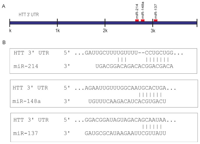
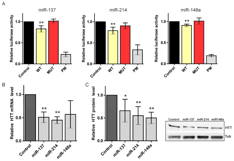
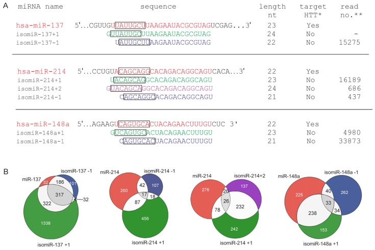
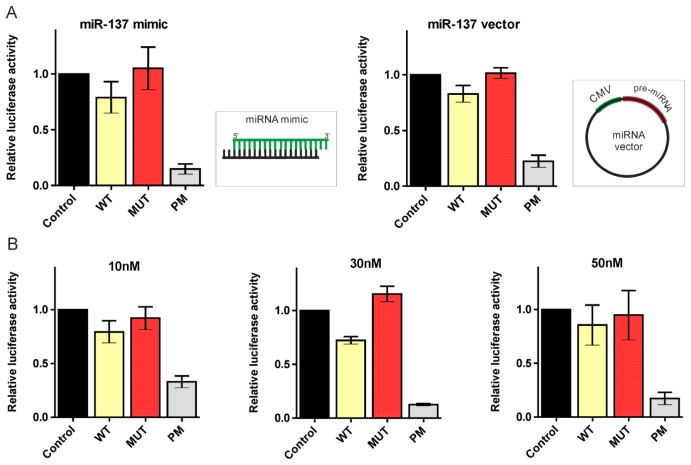
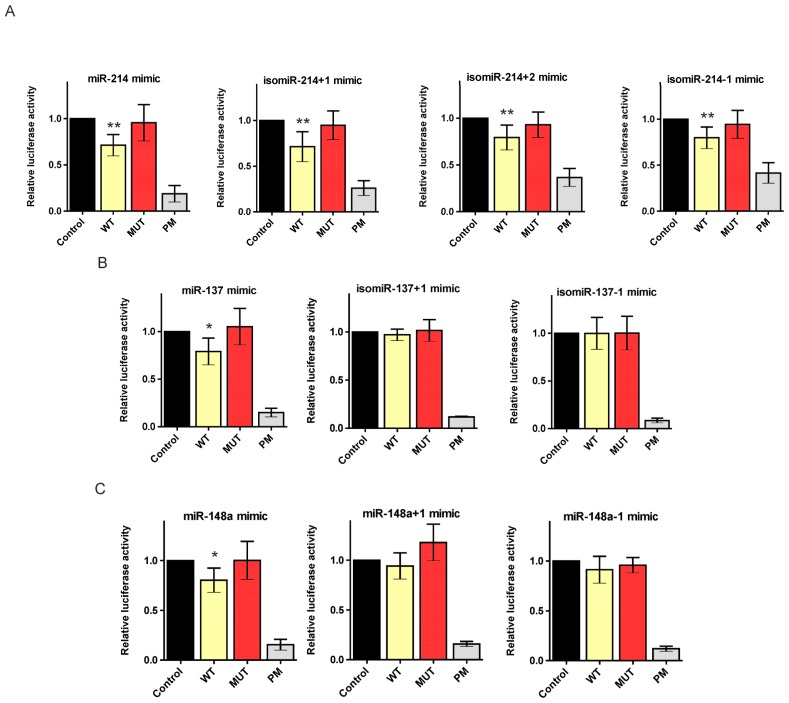
With the advent of deep sequencing technology, a variety of miRNA length and sequence variants, termed isomiRNAs (isomiRs), have been discovered. However, the functional roles of these commonly detected isomiRs remain unknown. In this paper, we demonstrated that miRNAs regulate the expression of the HTT gene, whose mutation leads to Huntington’s disease (HD), a hereditary degenerative disorder. Specifically, we validated the interactions of canonical miRNAs, miR-137, miR-214, and miR-148a, with the HTT 3'UTR using a luciferase assay. Moreover, we applied synthetic miRNA mimics to examine whether a slight shifting of miRNA seed regions might alter the regulation of the HTT transcript. We also examined miR-137, miR-214, and miR-148a isomiRs and showed the activity of these isoforms on reporter constructs bearing appropriate sequences from the HTT 3'UTR. Hence, we demonstrated that certain 5'-end variants of miRNAs might be functional for the regulation of the same targets as canonical miRNAs.
Overview
The claims and statistics of this paper haven’t been checked yet.
Run the check — free, no account needed. It reads the open-access text and checks the sources it cites.
Integrity signals
Registry
Cites
Statistics
Figures
Verification
Claims the paper makes
The claims and statistics of this paper haven’t been checked yet.
Run the check — free, no account needed. It reads the open-access text and checks the sources it cites.
Claims other papers make about this one
Each is checked against this paper’s own text: does it say what the citing paper says it does?
In the citing paper
“miRNAs that target the HTT 3′UTR and reduce HTT transcript levels in vitro, miR-148a-5p, miR-150-5p and miR-214-5p”
From miR-10b-5p expression in Huntington’s disease brain relates to age of onset and the extent of striatal involvement (2015) · cites this paper as [38]
Self-check
Percentages, sums and reported differences re-computed from the paper’s own numbers.
The self-check needs the paper’s full text.
Citations
Cites
A Mammalian microRNA Expression Atlas Based on Small RNA Library Sequencing
Landgraf, P., Rusu, M., Sheridan, R., Sewer, A., Iovino, N., Aravin, A., Pfeffer, S., Rice, A., Kamphorst, A. O., Landthaler, M., et al. (2007) · Cell · doi:10.1016/j.cell.2007.04.040 · Cited by 3,626
The role of site accessibility in microRNA target recognition
Kertesz, M., Iovino, N., Unnerstall, U., Gaul, U., & Segal, E. (2007) · Nature Genetics · doi:10.1038/ng2135 · Cited by 2,460
MicroRNAs and their isomiRs function cooperatively to target common biological pathways
Cloonan, N., Wani, S., Xu, Q., Gu, J., Lea, K., Heater, S., Barbacioru, C., Steptoe, A. L., Martin, H. C., Nourbakhsh, E., et al. (2011) · Genome Biology · doi:10.1186/gb-2011-12-12-r126 · Cited by 345
Research works printed but not linked: 60
- Chekulaeva, 2009, Mechanisms of miRNA-mediated post-transcriptional regulation in animal cells, Curr. Opin. Cell Biol, 21, 452, 10.1016/j.ceb.2009.04.009 · DOI · Paper page
- Bartel, 2009, MicroRNAs: Target recognition and regulatory functions, Cell, 136, 215, 10.1016/j.cell.2009.01.002 · DOI · Paper page
- Kim, 2009, Biogenesis of small RNAs in animals, Nat. Rev. Mol. Cell Biol, 10, 126, 10.1038/nrm2632 · DOI · Paper page
- Krol, 2010, The widespread regulation of microRNA biogenesis, function and decay, Nat. Rev. Genet, 11, 597, 10.1038/nrg2843 · DOI · Paper page
- Koscianska, 2011, The role of the precursor structure in the biogenesis of microRNA, Cell. Mol. Life Sci, 68, 2859, 10.1007/s00018-011-0726-2 · DOI
- Winter, 2009, Many roads to maturity: microRNA biogenesis pathways and their regulation, Nat. Cell Biol, 11, 228, 10.1038/ncb0309-228 · DOI
- Saini, 2008, miRBase: Tools for microRNA genomics, Nucleic Acids Res, 36, D154
- Morin, 2008, Application of massively parallel sequencing to microRNA profiling and discovery in human embryonic stem cells, Genome Res, 18, 610, 10.1101/gr.7179508 · DOI · Paper page
- Krol, 2011, Structural basis of microRNA length variety, Nucleic Acids Res, 39, 257, 10.1093/nar/gkq727 · DOI
- Seitz, 2008, Argonaute loading improves the 5′ precision of both MicroRNAs and their miRNA* strands in flies, Curr. Biol, 18, 147, 10.1016/j.cub.2007.12.049 · DOI · Paper page
- Wu, 2009, Alternative processing of primary microRNA transcripts by Drosha generates 5′ end variation of mature microRNA, PLoS One, 4, e7566, 10.1371/journal.pone.0007566 · DOI
- Frank, 2010, Structural basis for 5′-nucleotide base-specific recognition of guide RNA by human AGO2, Nature, 465, 818, 10.1038/nature09039 · DOI · Paper page
- Ruby, 2006, Large-scale sequencing reveals 21U-RNAs and additional microRNAs and endogenous siRNAs in C. elegans, Cell, 127, 1193, 10.1016/j.cell.2006.10.040 · DOI
- Wu, 2007, miRNA profiling of naive, effector and memory CD8 T cells, PLoS One, 2, e1020, 10.1371/journal.pone.0001020 · DOI
- Huse, 2007, Accuracy and quality of massively parallel DNA pyrosequencing, Genome Biol, 8, R143, 10.1186/gb-2007-8-7-r143 · DOI
- Tian, 2010, Sequencing bias: Comparison of different protocols of microRNA library construction, BMC Biotechnol, 10, 64, 10.1186/1472-6750-10-64 · DOI
- Lee, 2012, TRBP alters human precursor microRNA processing in vitro, RNA, 18, 2012, 10.1261/rna.035501.112 · DOI · Paper page
- Liu, 2011, The exoribonuclease Nibbler controls 3′ end processing of microRNAs in Drosophila, Curr. Biol, 21, 1888, 10.1016/j.cub.2011.10.006 · DOI
- Friedman, 2009, Most mammalian mRNAs are conserved targets of microRNAs, Genome Res, 19, 92, 10.1101/gr.082701.108 · DOI · Paper page
- Ross, 2011, Huntington’s disease: From molecular pathogenesis to clinical treatment, Lancet Neurol, 10, 83, 10.1016/S1474-4422(10)70245-3 · DOI
- Fiszer, 2013, RNA toxicity in polyglutamine disorders: Concepts, models, and progress of research, J. Mol. Med, 91, 683, 10.1007/s00109-013-1016-2 · DOI
- Marti, 2010, A myriad of miRNA variants in control and Huntington’s disease brain regions detected by massively parallel sequencing, Nucleic Acids Res, 38, 7219, 10.1093/nar/gkq575 · DOI · Paper page
- Chiang, 2010, Mammalian microRNAs: Experimental evaluation of novel and previously annotated genes, Genes Dev, 24, 992, 10.1101/gad.1884710 · DOI · Paper page
- Kawahara, 2008, Frequency and fate of microRNA editing in human brain, Nucleic Acids Res, 36, 5270, 10.1093/nar/gkn479 · DOI · Paper page
- Taft, 2010, Dynamic isomiR regulation in Drosophila development, RNA, 16, 1881, 10.1261/rna.2379610 · DOI · Paper page
- Khvorova, 2003, Functional siRNAs and miRNAs exhibit strand bias, Cell, 115, 209, 10.1016/S0092-8674(03)00801-8 · DOI · Paper page
- Schwarz, 2003, Asymmetry in the assembly of the RNAi enzyme complex, Cell, 115, 199, 10.1016/S0092-8674(03)00759-1 · DOI · Paper page
- Oguri, 2008, Characterization of endogenous human Argonautes and their miRNA partners in RNA silencing, Proc. Natl. Acad. Sci. USA, 105, 7964, 10.1073/pnas.0800334105 · DOI
- Humphreys, 2012, Complexity of murine cardiomyocyte miRNA biogenesis, sequence variant expression and function, PLoS One, 7, e30933, 10.1371/journal.pone.0030933 · DOI
- Lee, 2010, Complexity of the microRNA repertoire revealed by next-generation sequencing, RNA, 16, 2170, 10.1261/rna.2225110 · DOI
- Neilsen, 2012, IsomiRs—The overlooked repertoire in the dynamic microRNAome, Trends Genet, 28, 544, 10.1016/j.tig.2012.07.005 · DOI · Paper page
- Faghihi, 2010, Evidence for natural antisense transcript-mediated inhibition of microRNA function, Genome Biol., 11, R56, 10.1186/gb-2010-11-5-r56 · DOI · Paper page
- Llorens, 2013, A highly expressed miR-101 isomiR is a functional silencing small RNA, BMC Genomics, 14, 104, 10.1186/1471-2164-14-104 · DOI
- Chan, 2013, Concordant and discordant regulation of target genes by miR-31 and its isoforms, PLoS One, 8, e58169, 10.1371/journal.pone.0058169 · DOI
- Sinha, 2011, Micro RNA-214,-150,-146a and-125b target Huntingtin gene, RNA Biol, 8, 1005, 10.4161/rna.8.6.16035 · DOI · Paper page
- Witkos, 2011, Practical aspects of microRNA target prediction, Curr. Mol. Med, 11, 93, 10.2174/156652411794859250 · DOI
- Dweep, 2011, miRWalk—Database: Prediction of possible miRNA binding sites by “walking” the genes of three genomes, J. Biomed. Inform, 44, 839, 10.1016/j.jbi.2011.05.002 · DOI
- Hsu, 2011, miRTarBase: A database curates experimentally validated microRNA-target interactions, Nucleic Acids Res, 39, D163, 10.1093/nar/gkq1107 · DOI
- Sinha, 2010, Altered microRNAs in STHdh(Q111)/Hdh(Q111) cells: miR-146a targets TBP, Biochem. Biophys. Res. Commun, 396, 742, 10.1016/j.bbrc.2010.05.007 · DOI
- Soldati, 2013, Dysregulation of REST-regulated coding and non-coding RNAs in a cellular model of Huntington’s disease, J. Neurochem, 124, 418, 10.1111/jnc.12090 · DOI
- Kiriakidou, 2004, A combined computational-experimental approach predicts human microRNA targets, Genes Dev, 18, 1165, 10.1101/gad.1184704 · DOI
- John, 2004, Human microRNA targets, PLoS Biol, 2, e363, 10.1371/journal.pbio.0020363 · DOI · Paper page
- Krek, 2005, Combinatorial microRNA target predictions, Nat. Genet, 37, 495, 10.1038/ng1536 · DOI · Paper page
- Lewis, 2005, Conserved seed pairing, often flanked by adenosines, indicates that thousands of human genes are microRNA targets, Cell, 120, 15, 10.1016/j.cell.2004.12.035 · DOI · Paper page
- Salmena, 2011, A ceRNA hypothesis: The Rosetta Stone of a hidden RNA language?, Cell, 146, 353, 10.1016/j.cell.2011.07.014 · DOI · Paper page
- Bicchi, 2013, MicroRNAs and molecular mechanisms of neurodegeneration, Genes, 4, 244, 10.3390/genes4020244 · DOI
- Costa, 2012, Non-coding RNA and pseudogenes in neurodegenerative diseases: “The (un)Usual Suspects”, Front. Genet, 3, 231, 10.3389/fgene.2012.00231 · DOI
- Koscianska, 2007, Prediction and preliminary validation of oncogene regulation by miRNAs, BMC Mol. Biol, 8, 79, 10.1186/1471-2199-8-79 · DOI
- Baek, 2008, The impact of microRNAs on protein output, Nature, 455, 64, 10.1038/nature07242 · DOI · Paper page
- Guo, 2010, Mammalian microRNAs predominantly act to decrease target mRNA levels, Nature, 466, 835, 10.1038/nature09267 · DOI · Paper page
- Cheng, 2013, YM500: A small RNA sequencing (smRNA-seq) database for microRNA research, Nucleic Acids Res, 41, D285, 10.1093/nar/gks1238 · DOI
- Robins, 2005, Human microRNAs target a functionally distinct population of genes with AT-rich 3′ UTRs, Proc. Natl. Acad. Sci. USA, 102, 15557, 10.1073/pnas.0507443102 · DOI
- Grimson, 2007, MicroRNA targeting specificity in mammals: Determinants beyond seed pairing, Mol. Cell, 27, 91, 10.1016/j.molcel.2007.06.017 · DOI · Paper page
- Goswami, 2010, MicroRNA-340-mediated degradation of microphthalmia-associated transcription factor mRNA is inhibited by the coding region determinant-binding protein, J. Biol. Chem, 285, 20532, 10.1074/jbc.M110.109298 · DOI
- Brennecke, 2005, Principles of microRNA-target recognition, PLoS Biol, 3, e85, 10.1371/journal.pbio.0030085 · DOI
- Ebhardt, 2009, Meta-analysis of small RNA-sequencing errors reveals ubiquitous post-transcriptional RNA modifications, Nucleic Acids Res, 37, 2461, 10.1093/nar/gkp093 · DOI
- Felice, 2009, The 5′ terminal uracil of let-7a is critical for the recruitment of mRNA to Argonaute2, Biochem. J, 422, 329, 10.1042/BJ20090534 · DOI
- Mi, 2008, Sorting of small RNAs into Arabidopsis argonaute complexes is directed by the 5′ terminal nucleotide, Cell, 133, 116, 10.1016/j.cell.2008.02.034 · DOI
- Koscianska, 2011, High-resolution northern blot for a reliable analysis of microRNAs and their precursors, Scientific World Journal, 11, 102, 10.1100/tsw.2011.11 · DOI
- Koscianska, 2011, Northern blotting analysis of microRNAs, their precursors and RNA interference triggers, BMC Mol. Biol, 12, 14, 10.1186/1471-2199-12-14 · DOI
Cited by
Sorted by citations
MiR-137: an important player in neural development and neoplastic transformation
Mahmoudi, E., & Cairns, M. J. (2017) · Molecular Psychiatry · doi:10.1038/mp.2016.150 · Cited by 198
Hoss, A. G., Labadorf, A., Latourelle, J. C., Kartha, V. K., Hadzi, T. C., Gusella, J. F., MacDonald, M. E., Chen, J.-F., Akbarian, S., Weng, Z., et al. (2015) · BMC Medical Genomics · doi:10.1186/s12920-015-0083-3 · Cited by 151
Metadata
- Authors
- Kozlowska, E., Krzyzosiak, W., & Koscianska, E.
- Affiliations
- Department of Molecular Biomedicine, Institute of Bioorganic Chemistry, Polish Academy of Sciences, Noskowskiego 12/14 Str., 61-704 Poznan, Poland
- ORCID
- Koscianska, Edyta
- Venue
- International Journal of Molecular Sciences
- Publisher
- MDPI AG
- Published
- 2013-08-19
- Volume
- 14
- Issue
- 8
- Pages
- 16999-17016
- ISSN
- 1422-0067
- PMC
- PMC3759948
- Cited by
- 45
- References
- 3
- Language
- en
About these checks
Registry, retraction, Cites and statistics results are computed, not judged, with one exception: a printed reference that no index lists is read once by a model before it counts as a citation error. Claims are extracted by a model and kept only when their quote appears verbatim in the text; each cited source is then checked, with a model’s help, for whether it says what the claim relies on. Those verdicts can be wrong — read the claim’s quote and the cited source before relying on one.
Statistics and arithmetic are recomputed, not judged.
Confidence bands are shown only for fields calibrated on our annotated test set. None is calibrated yet, so no band is shown.
What counts as a claim ›How we measure confidence ›How we check this ›
Are you an author, or do you see a problem with this page? Request a review
