

Asbestos and Iron
Open accessThe DOI matches its Crossref record; 152 of the 152 references it cites were found in a registry or an index; 48 of the 159 sources its claims cite were checked for support.
Abstract
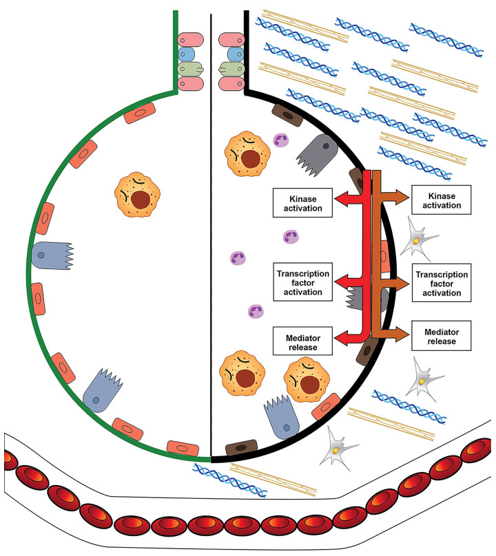
Theories of disease pathogenesis following asbestos exposure have focused on the participation of iron. After exposure, an open network of negatively charged functional groups on the fiber surface complexes host metals with a preference for iron. Competition for iron between the host and the asbestos results in a functional metal deficiency. The homeostasis of iron in the host is modified by the cell response, including increased import to correct the loss of the metal to the fiber surface. The biological effects of asbestos develop in response to and are associated with the disruption of iron homeostasis. Cell iron deficiency in the host following fiber exposure activates kinases and transcription factors, which are associated with the release of mediators coordinating both inflammatory and fibrotic responses. Relative to serpentine chrysotile, the clearance of amphiboles is incomplete, resulting in translocation to the mesothelial surface of the pleura. Since the biological effect of asbestos is dependent on retention of the fiber, the sequestration of iron by the surface, and functional iron deficiency in the cell, the greater clearance (i.e., decreased persistence) of chrysotile results in its diminished impact. An inability to clear asbestos from the lower respiratory tract initiates a host process of iron biomineralization (i.e., asbestos body formation). Host cells attempt to mobilize the metal sequestered by the fiber surface by producing superoxide at the phagosome membrane. The subsequent ferrous cation is oxidized and undergoes hydrolysis, creating poorly crystalline iron oxyhydroxide (i.e., ferrihydrite) included in the coat of the asbestos body.
Overview
CoChat Summary
TL;DR
This review argues that asbestos-related disease pathogenesis depends on disruption of host iron homeostasis after surface complexation of iron by the fiber, rather than on structural iron or oxidant chemistry alone. It proposes that amphiboles are more biopersistent and therefore have greater biological effects than chrysotile, and that asbestos body formation reflects a host iron biomineralization response.
Key findings
- Iron content in standard asbestos reference samples was greater in amosite (28%) and crocidolite (27%) than in chrysotile (0–2%), but the paper concludes current evidence does not support structural iron in the crystal lattice as the main driver of biological effect.
- Asbestos surfaces have negatively charged silanol groups that complex Fe3+ with high affinity; crystalline silicates show critical stability constants up to 1 × 10^17, while transferrin binds iron with a Ksc of approximately 10^20 M−1 at pH 7.4, and the labile iron pool is less than 1–5 µM.
- Functional iron deficiency after asbestos exposure increases DMT1 and ferritin expression, activates MAP kinases and transcription factors, and increases inflammatory mediators such as IL-6 and IL-8; iron pretreatment diminishes these responses.
- Asbestos bodies form on biopersistent fibers: uncoated fibers account for 95–97% of total inhaled asbestos, fibers less than 20 µm rarely become coated while virtually all fibers 80 µm or greater are coated, chrysotile asbestos bodies are usually less than 1–3% (mean 0.14%), and the coat can contain iron up to 10% w/w and calcium up to 1% w/w, with ferrihydrite as the major iron oxyhydroxide and more crystalline forms absent or below 5%.
Methods
- Narrative review and conceptual synthesis of literature on asbestos and iron; no primary experimental design is reported.
- Evidence cited includes animal models, human lung tissue and autopsy studies, in vitro macrophage and respiratory cell exposures, mineralogical/spectroscopic analyses, and epidemiologic/pathology reports.
- Main analysis integrates asbestos crystal lattice chemistry, surface complexation, cell iron homeostasis, inflammatory/fibrotic signaling, and ferruginous body formation.
Limitations
- The paper states no formal limitations section and describes no systematic search or meta-analytic method.
- The text notes that the precise composition of the asbestos body coat is not known, that there are no data supporting the fiber as the source of coat iron, and that dissolution has not been delineated during asbestos body formation.
- Evident from the text: the mechanistic hypothesis is largely inferential and synthesized from heterogeneous cited studies rather than tested by new primary data.
Model-written summary. Check it against the paper before relying on it.
Integrity signals
Registry
Cites
Statistics
Figures



Verification
Claims the paper makes
37 Valid · 6 Not valid · 42 Not checked · 58 Own results and interpretations
At least one cited source doesn’t support this claim or points to a different paper, and none confirmed it — see the verdicts below.
In the paper
“Iron in the coat of asbestos bodies does not appear to directly participate in the generation of reactive oxygen species [100].”
§ Asbestos and Iron > 5. The Formation of Ferruginous Bodies and Iron
What the sources say
- Disputedcites [100] Bardelli F. Brun F. De Panfilis S. Cloetens P. Capella S. Belluso E. Bellis D. Di Napoli A. Cedola A. Chemo-physical properties of asbestos bodies in human lung tissues studied at the nano-scale by non-invasive, label free X-ray imaging and spectroscopic techniques Toxicol. Lett. 2021 348 18 27 10.1016/j.toxlet.2021.05.002 34023437 sourceExperimental — accuracy not yet measured
The Introduction (page not provided) says that experiments found asbestos bodies induce free-radical generation and that iron in their coating is catalytically active. That is contrary to the claim that coating iron does not appear to participate directly in reactive-oxygen-species generation. (LLM, conf=0.85)
Checked by a reasoning model — may be wrong.
hedged- Asserted: passes — The authors put it forward as true.
- Checkable: passes — It can be checked against evidence.
- Load-bearing: unclear — It is unclear whether the paper’s conclusions depend on it.
- Based: passes — The paper gives a reason why it holds.
- Disputed
The sources with a verdict support this claim; the rest weren’t checked yet, have no full text we could read, or their verdicts aren’t rated yet.
In the paper
“There is support for metals’ involvement in the biological effect of asbestos, with iron chelation therapy and phlebotomy demonstrating possible preventive effects against asbestos-induced carcinogenesis in animal models [5,6].”
§ Asbestos and Iron > 1. Introduction
What the sources say
- Not checked — no full text of the sourcecites [5] Nagai H. Okazaki Y. Chew S.H. Misawa N. Yasui H. Toyokuni S. Deferasirox induces mesenchymal-epithelial transition in crocidolite-induced mesothelial carcinogenesis in rats Cancer Prev. Res. 2013 6 1222 1230 10.1158/1940-6207.CAPR-13-0244 24027214 source
- Supportedcites [6] Ohara Y. Chew S.H. Shibata T. Okazaki Y. Yamashita K. Toyokuni S. Phlebotomy as a preventive measure for crocidolite-induced mesothelioma in male rats Cancer Sci. 2018 109 330 339 10.1111/cas.13460 29193587 PMC5797813 source
The Introduction reports preventive effects of iron chelators against asbestos-induced mesothelioma in rats. The Discussion reports that phlebotomy reduced tumor burden and improved survival in male rats, while cautiously describing it as a possible preventive measure. (LLM, conf=0.93)
hedgedstates a possible cause- Asserted: passes — The authors put it forward as true.
- Checkable: passes — It can be checked against evidence.
- Load-bearing: passes — The paper’s conclusions depend on it.
- Based: passes — The paper gives a reason why it holds.
The cited sources support only part of this claim — read each source’s verdict below.
In the paper
“An inability to clear asbestos from the lower respiratory tract initiates a host process of iron biomineralization (a process through which a living organism produces a mineral including an oxide) [97,98].”
§ Asbestos and Iron > 5. The Formation of Ferruginous Bodies and Iron
What the sources say
- Partly supportedcites [97] Pascolo L. Gianoncelli A. Kaulich B. Rizzardi C. Schneider M. Bottin C. Polentarutti M. Kiskinova M. Longoni A. Synchrotron soft X-ray imaging and fluorescence microscopy reveal novel features of asbestos body morphology and composition in human lung tissues Part. Fibre Toxicol. 2011 8 7 10.1186/1743-8977-8-7 21299853 PMC3041679 sourceExperimental — accuracy not yet measured
The Abstract Background describes asbestos bodies in lung tissue as products of biomineralization that deposits endogenous iron around inhaled fibres. The Background also describes their formation as a host protective mechanism and mentions fibre retention in the distal lung. It does not establish that an inability to clear asbestos initiates that process; the formation mechanism is described as not well defined. Page numbers are unavailable in our copy. (LLM, conf=0.82)
- Supportedcites [98] Bardelli F. Veronesi G. Capella S. Bellis D. Charlet L. Cedola A. Belluso E. New insights on the biomineralisation process developing in human lungs around inhaled asbestos fibres Sci. Rep. 2017 7 44862 10.1038/srep44862 28332562 PMC5362951 source
The Abstract states that asbestos in the lungs induces biomineralisation, forming an iron-rich coating around the fibres. The Discussion describes the fibres’ high persistence in tissue and iron deposition during their prolonged residence in the lungs, supporting the claim’s description of a process following failure to clear them. (LLM, conf=0.89)
plainstates a cause- Asserted: passes — The authors put it forward as true.
- Checkable: passes — It can be checked against evidence.
- Load-bearing: passes — The paper’s conclusions depend on it.
- Based: passes — The paper gives a reason why it holds.
- Partly supported
The cited sources support only part of this claim — read each source’s verdict below.
In the paper
“Following human inhalation, the greatest deposition of particles/fibers is in the 16th to 19th generations of airways, which includes the respiratory bronchioles [85,86,87,88].”
§ Asbestos and Iron > 4. Asbestos, Cell Iron Homeostasis, and Their Biological Effects
What the sources say
- Partly supportedcites [85] Lippmann M. Yeates D.B. Albert R.E. Deposition, retention, and clearance of inhaled particles Br. J. Ind. Med. 1980 37 337 362 10.1136/oem.37.4.337 7004477 PMC1008751 sourceExperimental — accuracy not yet measured
The Deposition Mechanisms section (page unavailable) says inhaled particles can deposit in bronchioles and alveolar spaces, supporting deposition in small airways generally. It does not identify the 16th–19th airway generations, establish that they include respiratory bronchioles, or say that they receive the greatest deposition of particles or fibers. (LLM, conf=0.78)
- Not checked — no full text of the sourcecites [86] Hiller F.C. Deposition of sidestream cigarette smoke in the human respiratory tract Prev. Med. 1984 13 602 607 10.1016/S0091-7435(84)80010-9 6536937 source
- Not checked — no full text of the sourcecites [87] Choi J.I. Kim C.S. Mathematical analysis of particle deposition in human lungs: An improved single path transport model Inhal. Toxicol. 2007 19 925 939 10.1080/08958370701513014 17849277 source
- Could not verifycites [88] Kim S.Y. Sim S. Choi H.G. Active and passive smoking impacts on asthma with quantitative and temporal relations: A Korean Community Health Survey Sci. Rep. 2018 8 8614 10.1038/s41598-018-26895-3 29872096 PMC5988685 sourceExperimental — accuracy not yet measured
The Discussion passages address tobacco smoke, airway inflammation, and asthma, but not where inhaled particles or fibers deposit. Searches for deposition in the 16th–19th airway generations and for respiratory bronchioles found no passage addressing the claim; page numbers are unavailable in our copy. (LLM, conf=0.74)
plainstates a cause- Asserted: passes — The authors put it forward as true.
- Checkable: passes — It can be checked against evidence.
- Load-bearing: unclear — It is unclear whether the paper’s conclusions depend on it.
- Based: passes — The paper gives a reason why it holds.
- Partly supported
The cited sources support only part of this claim — read each source’s verdict below.
In the paper
“Particle/fiber deposition can often be greatest in the lower lung fields [89].”
§ Asbestos and Iron > 4. Asbestos, Cell Iron Homeostasis, and Their Biological Effects
What the sources say
- Partly supportedcites [89] Lambert A.R. O’Shaughnessy P. Tawhai M.H. Hoffman E.A. Lin C.L. Regional deposition of particles in an image-based airway model: Large-eddy simulation and left-right lung ventilation asymmetry Aerosol. Sci. Technol. 2011 45 11 25 10.1080/02786826.2010.517578 21307962 PMC3034252 sourceExperimental — accuracy not yet measured
The Discussion states that particle deposition was greatest in the lower lung lobes and reports a similar finding in a prior study. This supports the claim for particles, but our copy does not establish the same pattern for fibers. (LLM, conf=0.83)
plain- Asserted: passes — The authors put it forward as true.
- Checkable: passes — It can be checked against evidence.
- Load-bearing: unclear — It is unclear whether the paper’s conclusions depend on it.
- Based: passes — The paper gives a reason why it holds.
- Partly supported
91 more claims · 22 with their cited sources checked
Create a free account to see all 143 claims and to verify the 5 references still open.
- It may state two separate claims as one. 41
- Probably not a substantial claim. 7
Claims other papers make about this one
Each is checked against this paper’s own text: does it say what the citing paper says it does?
The cited source says what the claim attributes to it.
In the citing paper
“Increasing the bioavailability of iron, however, can overcome this process [84].”
From Environmental Contaminants, Iron Deficiency, and Iron‐Deficiency Anemia: A Review of the Literature (2025) · cites this paper as [84]
Citations
Cites
Oxyl radicals, redox-sensitive signalling cascades and antioxidants
Genestra, M. (2007) · Cellular Signalling · doi:10.1016/j.cellsig.2007.04.009 · Cited by 4,930
Production of Reactive Oxygen Species by Mitochondria
Chen, Q., Vazquez, E. J., Moghaddas, S., Hoppel, C. L., & Lesnefsky, E. J. (2003) · Journal of Biological Chemistry · doi:10.1074/jbc.m304854200 · Cited by 1,616 · cited 1×
“The electron transport chain in the mitochondria can be a major source of superoxide production [46,47].”
BackgroundMesoscale Iron Enrichment Experiments 1993-2005: Synthesis and Future Directions
Boyd, P. W., Jickells, T., Law, C. S., Blain, S., Boyle, E. A., Buesseler, K. O., Coale, K. H., Cullen, J. J., de Baar, H. J. W., Follows, M., et al. (2007) · Science · doi:10.1126/science.1131669 · Cited by 1,542 · cited 1×
“Iron is essential for almost every organism, and its lack of availability can restrict life in environments ranging in size from the ecosystem of the Pacific Ocean to a bacterium [33,34].”
BackgroundKakhlon, O., & Cabantchik, Z. I. (2002) · Free Radical Biology and Medicine · doi:10.1016/s0891-5849(02)01006-7 · Cited by 839 · cited 1×
“Intracellular iron levels available for complexation by the fiber surface can approach the concentration of the labile iron pool (less than 1–5 μM) [39,40].”
BackgroundMitogen-activated protein kinase cascades and regulation of gene expression
Su, B., & Karin, M. (1996) · Current Opinion in Immunology · doi:10.1016/s0952-7915(96)80131-2 · Cited by 747
Mortality from Lung Cancer in Asbestos Workers
Doll, R. (1955) · Occupational and Environmental Medicine · doi:10.1136/oem.12.2.81 · Cited by 717 · cited 1×
“In 1955, epidemiologic studies initially established an association between lung cancer and asbestos exposure [2,3].”
BackgroundHansel, C. M., Benner, S. G., Neiss, J., Dohnalkova, A., Kukkadapu, R. K., & Fendorf, S. (2003) · Geochimica et Cosmochimica Acta · doi:10.1016/s0016-7037(03)00276-x · Cited by 634 · cited 1×
“Subsequently, ferrihydrite in the coat can further react through hydrolysis– precipitation processes to produce goethite (Fe(OH)O), lepidocrocite (γ-FeO(OH)), magnetite, and hematite (Fe2O3) [113].”
BackgroundCompeting Fe (II)-induced mineralization pathways of ferrihydrite
Hansel, C. M., Benner, S. G., & Fendorf, S. (2005) · Environmental Science & Technology · doi:10.1021/es050666z · Cited by 619
Heyder, J. (2004) · Proceedings of the American Thoracic Society · doi:10.1513/pats.200409-046ta · Cited by 601
Deposition, retention, and clearance of inhaled particles.
Lippmann, M., Yeates, D. B., & Albert, R. E. (1980) · Occupational and Environmental Medicine · doi:10.1136/oem.37.4.337 · Cited by 557
Fels, A. O., & Cohn, Z. A. (1986) · Journal of Applied Physiology · doi:10.1152/jappl.1986.60.2.353 · Cited by 506
Recent progress in understanding the mechanism of heavy metals retention by iron (oxyhydr)oxides
Shi, M., Min, X., Ke, Y., Lin, Z., Yang, Z., Wang, S., Peng, N., Yan, X., Luo, S., Wu, J., et al. (2021) · Science of The Total Environment · doi:10.1016/j.scitotenv.2020.141930 · Cited by 430 · cited 1×
“The retention mechanisms of metal cations and oxyanions on various iron oxyhydroxides can include (1) surface complexation, (2) surface precipitation, and (3) structural incorporation [119].”
BackgroundXiao, G. G., Wang, M., Li, N., Loo, J. A., & Nel, A. E. (2003) · Journal of Biological Chemistry · doi:10.1074/jbc.m306423200 · Cited by 428
Boland, D. D., Collins, R. N., Miller, C. J., Glover, C. J., & Waite, T. D. (2014) · Environmental Science & Technology · doi:10.1021/es4043275 · Cited by 406 · cited 1×
“Due to its strong reducing capacity, Fe2+ further reacts with and provokes the conversion of ferrihydrite to secondary, more crystalline forms [111,112].”
BackgroundThe transformation of ferrihydrite into goethite or hematite, revisited
Cudennec, Y., & Lecerf, A. (2006) · Journal of Solid State Chemistry · doi:10.1016/j.jssc.2005.11.030 · Cited by 388 · cited 1×
“residence time, all of which are primarily a function of Fe2+ concentration; temperature and pH can also contribute [114,115].”
BackgroundGeneration of superoxide by the mitochondrial Complex I
Grivennikova, V. G., & Vinogradov, A. D. (2006) · Biochimica et Biophysica Acta (BBA) - Bioenergetics · doi:10.1016/j.bbabio.2006.03.013 · Cited by 379 · cited 1×
“The electron transport chain in the mitochondria can be a major source of superoxide production [46,47].”
BackgroundBreuer, W., Epsztejn, S., & Cabantchik, Z. I. (1995) · Journal of Biological Chemistry · doi:10.1074/jbc.270.41.24209 · Cited by 326
Labile iron in cells and body fluids: Physiology, pathology, and pharmacology
Cabantchik, Z. I. (2014) · Frontiers in Pharmacology · doi:10.3389/fphar.2014.00045 · Cited by 306 · cited 1×
“Intracellular iron levels available for complexation by the fiber surface can approach the concentration of the labile iron pool (less than 1–5 μM) [39,40].”
BackgroundIron-dependent regulation of the divalent metal ion transporter
Gunshin, H., Allerson, C. R., Polycarpou-Schwarz, M., Rofts, A., Rogers, J. T., Kishi, F., Hentze, M. W., Rouault, T. A., Andrews, N. C., & Hediger, M. A. (2001) · FEBS Letters · doi:10.1016/s0014-5793(01)03189-1 · Cited by 305 · cited 2×
“The expression of divalent metal transporter 1 (DMT1) is increased after asbestos exposure, reflecting the diminished iron concentrations available in the cells [44,45].”
BackgroundChua, F., Gauldie, J., & Laurent, G. J. (2005) · American Journal of Respiratory Cell and Molecular Biology · doi:10.1165/rcmb.2005-0062tr · Cited by 305 · cited 1×
“Pulmonary fibrosis is also included in the host response to reverse the loss of essential metal after its sequestration by asbestos (Figures 3 and 4) [64].”
BackgroundSurface Hydroxyl Site Densities on Metal Oxides as a Measure for the Ion-Exchange Capacity
Tamura, H., Tanaka, A., Mita, K.-Y., & Furuichi, R. (1999) · Journal of Colloid and Interface Science · doi:10.1006/jcis.1998.5877 · Cited by 252 · cited 1×
“In oxides, this cation-exchange capacity can directly correlate with the surface hydroxyl site density [25].”
BackgroundLi, N., Kim, S., Wang, M., Froines, J., Sioutas, C., & Nel, A. (2002) · Inhalation Toxicology · doi:10.1080/089583701753678571 · Cited by 249
Katzenstein, A.-L. A., Mukhopadhyay, S., Zanardi, C., & Dexter, E. (2010) · Human Pathology · doi:10.1016/j.humpath.2009.09.003 · Cited by 234
Deng, X., Rui, W., Zhang, F., & Ding, W. (2013) · Cell Biology and Toxicology · doi:10.1007/s10565-013-9242-5 · Cited by 227
Tripathi, R. D., Tripathi, P., Dwivedi, S., Kumar, A., Mishra, A., Chauhan, P. S., Norton, G. J., & Nautiyal, C. S. (2014) · Metallomics · doi:10.1039/c4mt00111g · Cited by 218 · cited 2×
“Such iron plaques include crystalline iron oxyhydroxides and demonstrate positive staining with Perls’ Prussian blue [142,143].”
BackgroundFIBROSIS OF THE LUNGS DUE TO THE INHALATION OF ASBESTOS DUST
Cooke, W. E. (1924) · BMJ · doi:10.1136/bmj.2.3317.147 · Cited by 214
Pathology of Asbestos-Associated Diseases
(2014) · doi:10.1007/978-3-642-41193-9 · Cited by 199
Self-citationCallens, C., Coulon, S., Naudin, J., Radford-Weiss, I., Boissel, N., Raffoux, E., Wang, P. H. M., Agarwal, S., Tamouza, H., Paubelle, E., et al. (2010) · Journal of Experimental Medicine · doi:10.1084/jem.20091488 · Cited by 197 · cited 1×
“Through a comparable pathway, cellular superoxide generation can increase after exposure to iron chelators [48,49].”
BackgroundRoot Iron Plaque on Wetland Plants as a Dynamic Pool of Nutrients and Contaminants
Khan, N., Seshadri, B., Bolan, N., Saint, C. P., Kirkham, M. B., Chowdhury, S., Yamaguchi, N., Lee, D. Y., Li, G., Kunhikrishnan, A., et al. (2016) · Advances in Agronomy · doi:10.1016/bs.agron.2016.04.002 · Cited by 194 · cited 1×
“In another related pathway of iron biomineralization, iron plaque formation in plants is dependent on the availability of Fe2+, which reacts with oxygen to generate Fe3+ oxyhydroxides and deposits it on root surfaces [140,141].”
BackgroundIron chemistry at the service of life
Sánchez, M., Sabio, L., Gálvez, N., Capdevila, M., & Dominguez‐Vera, J. M. (2017) · IUBMB Life · doi:10.1002/iub.1602 · Cited by 190 · cited 1×
“Its unique coordination chemistry led to its evolutionary selection for numerous essential cell functions [35].”
BackgroundDiffuse Pleural Mesothelioma and Asbestos Exposure in the North Western Cape Province
Wagner, J. C., Sleggs, C. A., & Marchand, P. (1960) · Occupational and Environmental Medicine · doi:10.1136/oem.17.4.260 · Cited by 182
Vargas, G., Cypriano, J., Correa, T., Leão, P., Bazylinski, D. A., & Abreu, F. (2018) · Molecules · doi:10.3390/molecules23102438 · Cited by 180 · cited 1×
“Finally, in a pathway of iron biomineralization approximating asbestos body formation, several bacteria incorporate iron from their environment to synthesize intracellular nanoparticles of magnetite or greigite (Fe3S4) in organelles called magnetosomes [137,138].”
BackgroundBoulanger, G., Andujar, P., Pairon, J.-C., Billon-Galland, M.-A., Dion, C., Dumortier, P., Brochard, P., Sobaszek, A., Bartsch, P., Paris, C., et al. (2014) · Environmental Health · doi:10.1186/1476-069x-13-59 · Cited by 176 · cited 1×
“This is efficient when the mean diameter of the particles/fibers is smaller than the mean diameter of the macrophage (10 microns) but, when the size exceeds this, there can be incomplete phagocytosis and clearance [93].”
BackgroundLambert, A. R., O'shaughnessy, P. T., Tawhai, M. H., Hoffman, E. A., & Lin, C.-L. (2011) · Aerosol Science and Technology · doi:10.1080/02786826.2010.517578 · Cited by 175 · cited 1×
“Particle/fiber deposition can often be greatest in the lower lung fields [89].”
BackgroundExtracellular Iron Biomineralization by Photoautotrophic Iron-Oxidizing Bacteria
Miot, J., Benzerara, K., Obst, M., Kappler, A., Hegler, F., Schädler, S., Bouchez, C., Guyot, F., & Morin, G. (2009) · Applied and Environmental Microbiology · doi:10.1128/aem.00490-09 · Cited by 172 · cited 1×
“, lipopolysaccharide) fibers with a high negative charge as a template for biomineralization [110].”
BackgroundHealth risk of chrysotile revisited
Bernstein, D., Dunnigan, J., Hesterberg, T., Brown, R., Velasco, J. A. L., Barrera, R., Hoskins, J., & Gibbs, A. (2013) · Critical Reviews in Toxicology · doi:10.3109/10408444.2012.756454 · Cited by 172 · cited 1×
“After exposure to the lower respiratory tract, the crystal lattice of amphiboles does not change and, as a result of its insolubility at any pH value, is biopersistent [19,96].”
BackgroundAsbestos and other ferruginous bodies: their formation and clinical significance.
(1981) · PubMed Central · Cited by 155
Asbestos minerals in modern technology
Speil, S., & Leineweber, J. P. (1969) · Environmental Research · doi:10.1016/0013-9351(69)90036-x · Cited by 134 · cited 1×
“As a result of the lack of acid-soluble surface groups, the amphiboles have negligible solubility at any pH that might be encountered in a living system [18] (Figure 1A).”
BackgroundRole of surface complexed iron in oxidant generation and lung inflammation induced by silicates
Ghio, A. J., Kennedy, T. P., Whorton, A. R., Crumbliss, A. L., Hatch, G. E., & Hoidal, J. R. (1992) · American Journal of Physiology-Lung Cellular and Molecular Physiology · doi:10.1152/ajplung.1992.263.5.l511 · Cited by 132 · cited 2×
“Iron homeostasis in the host must be odified and the cell response will include the upregulation of import to correct the functional deficiency followi g loss f the metal to t e fiber surface [42,43].”
BackgroundSelf-citationCase, B. W., Abraham, J. L., Meeker, G., Pooley, F. D., & Pinkerton, K. E. (2011) · Journal of Toxicology and Environmental Health, Part B · doi:10.1080/10937404.2011.556045 · Cited by 123 · cited 1×
“The single serpentine fiber, chrysotile, is composed of sheets of silicon-centered tetrahedra joined to sheets of octahedral coordinated magnesium (brucite or Mg(OH)2) in a flexible structure [10].”
BackgroundTahir, S. S., & Rauf, N. (2004) · Journal of Environmental Management · doi:10.1016/j.jenvman.2004.06.009 · Cited by 106
(1992) · American Mineralogist · Cited by 105
Involvement of Superoxide Radical in Extracellular Ferric Reduction by Iron-Deficient Bean Roots
Cakmak, I., van de Wetering, D. A. M., Marschner, H., & Bienfait, H. F. (1987) · Plant Physiology · doi:10.1104/pp.85.1.310 · Cited by 102 · cited 1×
“Such ferrireduction is an essential, and frequently limiting, reaction in iron acquisition, transport, and translocation [50,51].”
BackgroundMathematical analysis of particle deposition in human lungs: An improved single path transport model
Choi, J.-I., & Kim, C. S. (2007) · Inhalation Toxicology · doi:10.1080/08958370701513014 · Cited by 101
Amor, M., Mathon, F. P., Monteil, C. L., Busigny, V., & Lefevre, C. T. (2020) · Environmental Microbiology · doi:10.1111/1462-2920.15098 · Cited by 98 · cited 1×
“Finally, in a pathway of iron biomineralization approximating asbestos body formation, several bacteria incorporate iron from their environment to synthesize intracellular nanoparticles of magnetite or greigite (Fe3S4) in organelles called magnetosomes [137,138].”
BackgroundBernstein, D. M., Chevalier, J., & Smith, P. (2005) · Inhalation Toxicology · doi:10.1080/08958370591002012 · Cited by 97
Xiao, W., Jones, A. M., Li, X., Collins, R. N., & Waite, T. D. (2018) · Environmental Science & Technology · doi:10.1021/acs.est.7b05098 · Cited by 96 · cited 1×
“Due to its strong reducing capacity, Fe2+ further reacts with and provokes the conversion of ferrihydrite to secondary, more crystalline forms [111,112].”
BackgroundGardi, C., Arezzini, B., Fortino, V., & Comporti, M. (2002) · Biochemical Pharmacology · doi:10.1016/s0006-2952(02)01257-1 · Cited by 94
Ito, F., Kato, K., Yanatori, I., Murohara, T., & Toyokuni, S. (2021) · Redox Biology · doi:10.1016/j.redox.2021.102174 · Cited by 88 · cited 1×
“Postulates regarding the pathogenesis of asbestosrelated disease include oxidative stress mediated by the in vivo production of hydroxyl radicals by iron and, more recently, accompanied by ferroptosis [7,8].”
BackgroundPascolo, L., Gianoncelli, A., Schneider, G., Salomé, M., Schneider, M., Calligaro, C., Kiskinova, M., Melato, M., & Rizzardi, C. (2013) · Scientific Reports · doi:10.1038/srep01123 · Cited by 88 · cited 2×
“residence time, all of which are primarily a function of Fe2+ concentration; temperature and pH can also contribute [114,115].”
BackgroundCell signaling and transcription factor activation by asbestos in lung injury and disease
Shukla, A., Ramos-Nino, M., & Mossman, B. (2003) · The International Journal of Biochemistry & Cell Biology · doi:10.1016/s1357-2725(02)00315-1 · Cited by 87
Cheng, S.-E., Lee, I.-T., Lin, C.-C., Kou, Y. R., & Yang, C.-M. (2010) · Free Radical Biology and Medicine · doi:10.1016/j.freeradbiomed.2010.02.026 · Cited by 86
The biopersistence of brazilian chrysotile asbestos following inhalation
Bernstein, D. M., Rogers, R., & Smith, P. (2004) · Inhalation Toxicology · doi:10.1080/08958370490490176 · Cited by 84
Asbestos Bodies, Their Formation, Composition and Character
Pooley, F. D. (1972) · Environmental Research · doi:10.1016/0013-9351(72)90039-4 · Cited by 73
Direct identification of reaction sites on ferrihydrite
Boily, J., & Song, X. (2020) · Communications Chemistry · doi:10.1038/s42004-020-0325-y · Cited by 72 · cited 1×
“Hydroxyl groups on ferrihydrite control its chemical reactivity [120].”
BackgroundGawda, A., Majka, G., Nowak, B., Śróttek, M., Walczewska, M., & Marcinkiewicz, J. (2018) · Inflammation Research · doi:10.1007/s00011-018-1165-4 · Cited by 71
Brown, J. S., Zeman, K. L., & Bennett, W. D. (2001) · Journal of Aerosol Medicine · doi:10.1089/08942680152744659 · Cited by 71
Li, H., Zheng, X., Tao, L., Yang, Y., Gao, L., & Xiong, J. (2019) · Rice · doi:10.1186/s12284-019-0291-0 · Cited by 69 · cited 1×
“Such iron plaques include crystalline iron oxyhydroxides and demonstrate positive staining with Perls’ Prussian blue [142,143].”
BackgroundChan, J. K., Charrier, J. G., Kodani, S. D., Vogel, C. F., Kado, S. Y., Anderson, D. S., Anastasio, C., & Van Winkle, L. S. (2013) · Particle and Fibre Toxicology · doi:10.1186/1743-8977-10-34 · Cited by 69
Histochemistry of Aortic Elastin in Patients With Nonspecific Abdominal Aortic Aneurysmal Disease
Tilson, M. D. (1988) · Archives of Surgery · doi:10.1001/archsurg.1988.01400280113023 · Cited by 68
Deferoxamine induces prolonged cardiac preconditioning via accumulation of oxygen radicals
Dendorfer, A., Heidbreder, M., Hellwig-Bürgel, T., Jöhren, O., Qadri, F., & Dominiak, P. (2005) · Free Radical Biology and Medicine · doi:10.1016/j.freeradbiomed.2004.10.015 · Cited by 68 · cited 1×
“Through a comparable pathway, cellular superoxide generation can increase after exposure to iron chelators [48,49].”
BackgroundBernstein, D. M., Rogers, R., & Smith, P. (2005) · Inhalation Toxicology · doi:10.1080/08958370590885663 · Cited by 67
A note on the composition and properties of ferritin iron cores
Treffry, A., Harrison, P. M., Cleton, M. I., de Bruijn, W. C., & Mann, S. (1987) · Journal of Inorganic Biochemistry · doi:10.1016/0162-0134(87)85001-8 · Cited by 66
Iron Homeostasis in the Lung Following Asbestos Exposure
Ghio, A. J., Stonehuerner, J., Richards, J., & Devlin, R. B. (2008) · Antioxidants & Redox Signaling · doi:10.1089/ars.2007.1909 · Cited by 65 · cited 1×
“An alternative hypothesis was submitted stating that asbestos-related disease reflected the disruption of host iron homeostasis following fiber exposure [9].”
BackgroundInfluentialSelf-citationSynthetic vitreous fibers: A review toxicology, epidemiology and regulations
Bernstein, D. M. (2007) · Critical Reviews in Toxicology · doi:10.1080/10408440701524592 · Cited by 65
Asbestos health hazard: A spectroscopic study of synthetic geoinspired Fe-doped chrysotile
Foresti, E., Fornero, E., Lesci, I. G., Rinaudo, C., Zuccheri, T., & Roveri, N. (2009) · Journal of Hazardous Materials · doi:10.1016/j.jhazmat.2009.01.103 · Cited by 60 · cited 1×
“Chrysotile fibrils are curved either concentrically or spirally into tubular structures [11].”
BackgroundKnörr-Wittmann, C., Hengstermann, A., Gebel, S., Alam, J., & Müller, T. (2005) · Free Radical Biology and Medicine · doi:10.1016/j.freeradbiomed.2005.07.003 · Cited by 60
Enhanced deposition of low-molecular-weight hyaluronan in lungs of cigarette smoke-exposed mice
Bracke, K. R., Dentener, M. A., Papakonstantinou, E., Vernooy, J. H. J., Demoor, T., Pauwels, N. S., Cleutjens, J., van Suylen, R. J., Joos, G. F., Brusselle, G. G., et al. (2010) · American Journal of Respiratory Cell and Molecular Biology · doi:10.1165/rcmb.2008-0424oc · Cited by 59
Huang, C.-Y., Wu, C.-H., Yang, J.-I., Li, Y.-H., & Kuo, J.-M. (2015) · Journal of Food and Drug Analysis · doi:10.1016/j.jfda.2014.06.009 · Cited by 58
Aqueous and solid complexes of iron(III) with hyaluronic acid
Mercê, A. L. R., Marques Carrera, L. C., Santos Romanholi, L. K., & Lobo Recio, M. Á. (2002) · Journal of Inorganic Biochemistry · doi:10.1016/s0162-0134(01)00422-6 · Cited by 58
The health risk of chrysotile asbestos
Bernstein, D. M. (2014) · Current Opinion in Pulmonary Medicine · doi:10.1097/mcp.0000000000000064 · Cited by 57
Mobilization of iron from ferritin: New steps and details
La, A., Nguyen, T., Tran, K., Sauble, E., Tu, D., Gonzalez, A., Kidane, T. Z., Soriano, C., Morgan, J., Doan, M., et al. (2018) · Metallomics · doi:10.1039/c7mt00284j · Cited by 56 · cited 1×
“However, the process of asbestos body formation occurs in the lysosome, which is normally the site of the degradation of both ferritin and hemosiderin to iron [129].”
BackgroundCatalytic role of copper and iron ions in the depolymerization of hyaluronic acid by ascorbic acid
Matsumura, G., & Pigman, W. (1965) · Archives of Biochemistry and Biophysics · doi:10.1016/0003-9861(65)90446-7 · Cited by 56 · cited 1×
“, collagen, elastin, and GAG) will be degraded [76,83,84].”
BackgroundHypothesis: Is lung disease after silicate inhalation caused by oxidant generation?
Ghio, A. J., Kennedy, T. P., Schapira, R. M., Crumbliss, A. L., & Hoidal, J. R. (1990) · The Lancet · doi:10.1016/0140-6736(90)92421-d · Cited by 56 · cited 1×
“Postulates regarding the pathogenesis of asbestosrelated disease include oxidative stress mediated by the in vivo production of hydroxyl radicals by iron and, more recently, accompanied by ferroptosis [7,8].”
BackgroundSelf-citationThe Battle for Iron between Humans and Microbes
Carver, P. L. (2018) · Current Medicinal Chemistry · doi:10.2174/0929867324666170720110049 · Cited by 54 · cited 1×
“Iron is essential for almost every organism, and its lack of availability can restrict life in environments ranging in size from the ecosystem of the Pacific Ocean to a bacterium [33,34].”
BackgroundDeposition of sidestream cigarette smoke in the human respiratory tract
Charles Hiller, F. (1984) · Preventive Medicine · doi:10.1016/s0091-7435(84)80010-9 · Cited by 53
Gualtieri, A. F., Lusvardi, G., Zoboli, A., Di Giuseppe, D., & Lassinantti Gualtieri, M. (2019) · Environmental Research · doi:10.1016/j.envres.2019.01.011 · Cited by 52 · cited 3×
“In contrast, macrophages dissociate the crystalline structure of chrysotile, leaving an unstable and amorphous silicate [20].”
BackgroundIdo, Y., Muto, N., Inada, A., Kohroki, J., Mano, M., Odani, T., Itoh, N., Yamamoto, K., & Tanaka, K. (1999) · Cell Proliferation · doi:10.1046/j.1365-2184.1999.3210063.x · Cited by 52
Induction of Ferritin Synthesis in Human Lung Epithelial Cells Treated with Crocidolite Asbestos
Fang, R., & Aust, A. E. (1997) · Archives of Biochemistry and Biophysics · doi:10.1006/abbi.1997.9892 · Cited by 52
Pollastri, S., D’Acapito, F., Trapananti, A., Colantoni, I., Andreozzi, G. B., & Gualtieri, A. F. (2015) · Journal of Hazardous Materials · doi:10.1016/j.jhazmat.2015.05.010 · Cited by 51 · cited 2×
“The involvement of structural iron in oxidative stress and biological effects by asbestos would subsequently be dependent on fiber destruction [16].”
BackgroundA Comparison of Some Aspects of the Aqueous Coordination Chemistry of Aluminum(III) and Iron(III)
Crumbliss, A. L., & Garrison, J. M. (1988) · Comments on Inorganic Chemistry · doi:10.1080/02603598808048670 · Cited by 50 · cited 1×
“As a result of its electropositivity, Fe3+ has a high affinity for oxygen-donor ligands [26].”
BackgroundDumortier, P., Çoplü, L., Broucke, I., Emri, S., Selcuk, T., de Maertelaer, V, De Vuyst, P., & Baris, I. (2001) · Occupational and Environmental Medicine · doi:10.1136/oem.58.4.261 · Cited by 49 · cited 1×
“This includes cigarette smoke particles; sheet silicates such as talc, mica, and kaolinite; diatomaceous earth; metal oxides; metals (with cores of titanium, iron, chromium, and aluminum); coal dust; woodstove dust; zeolites including erionite; silicon carbide; fiberglass; cotton dust; synthetic textile fibers; and hu…”
BackgroundPascolo, L., Gianoncelli, A., Kaulich, B., Rizzardi, C., Schneider, M., Bottin, C., Polentarutti, M., Kiskinova, M., Longoni, A., & Melato, M. (2011) · Particle and Fibre Toxicology · doi:10.1186/1743-8977-8-7 · Cited by 47 · cited 1×
“An inability to clear asbestos from the lower respiratory tract initiates a host process of iron biomineralization (a process through which a living organism produces a mineral including an oxide) [97,98].”
BackgroundThe biopersistence of Canadian chrysotile asbestos following inhalation
Bernstein, D. M., Rogers, R., & Smith, P. (2003) · Inhalation Toxicology · doi:10.1080/08958370390241713 · Cited by 46
Borghi, E. B., Occhiuzzi, M., Foresti, E., Lesci, I. G., & Roveri, N. (2009) · Physical Chemistry Chemical Physics · doi:10.1039/b915182f · Cited by 45 · cited 1×
“This is comparable to aluminum, which can also be a common foreign element in fibers [13].”
BackgroundMetal catalysis in the depolymerization of hyaluronic acid by autoxidants
Harris, M. J., Herp, A., & Pigma, W. (1972) · Journal of the American Chemical Society · doi:10.1021/ja00776a047 · Cited by 44 · cited 1×
“, collagen, elastin, and GAG) will be degraded [76,83,84].”
BackgroundKim, S. Y., Sim, S., & Choi, H. G. (2018) · Scientific Reports · doi:10.1038/s41598-018-26895-3 · Cited by 43
Ryan, A. J., Larson-Casey, J. L., He, C., Murthy, S., & Carter, A. B. (2014) · Journal of Biological Chemistry · doi:10.1074/jbc.m114.579870 · Cited by 43 · cited 1×
“This latter pathway can account for the rapidity of changes in the metal concentration [124].”
BackgroundWood Smoke Particle Sequesters Cell Iron to Impact a Biological Effect
Ghio, A. J., Soukup, J. M., Dailey, L. A., Tong, H., Kesic, M. J., Budinger, G. R. S., & Mutlu, G. M. (2015) · Chemical Research in Toxicology · doi:10.1021/acs.chemrestox.5b00270 · Cited by 41 · cited 1×
“Comparable to MAP kinases, increased iron availability decreases the activation of transcription factors following exposure to particles/fibers [53,54,63].”
BackgroundSelf-citationFluctuations of intracellular iron modulate elastin production
Bunda, S., Kaviani, N., & Hinek, A. (2005) · Journal of Biological Chemistry · doi:10.1074/jbc.m409897200 · Cited by 40
Characterization of Tubular Chrysotile by Thermoporometry, Nitrogen Sorption, Drifts, and Tem
Titulaer, M. K., van Miltenburg, J. C., Jansen, J. B. H., & Geus, J. W. (1993) · Clays and Clay Minerals · doi:10.1346/ccmn.1993.0410410 · Cited by 40
Ghio, A. J., Soukup, J. M., Dailey, L. A., & Madden, M. C. (2020) · Free Radical Biology and Medicine · doi:10.1016/j.freeradbiomed.2020.02.007 · Cited by 39 · cited 1×
“Similar to asbestos, these other particles/fibers function to complex iron [131].”
BackgroundSelf-citationBernstein, D. M., Rogers, R. A., Sepulveda, R., Donaldson, K., Schuler, D., Gaering, S., Kunzendorf, P., Chevalier, J., & Holm, S. E. (2011) · Inhalation Toxicology · doi:10.3109/08958378.2011.575413 · Cited by 39
Nagai, H., Okazaki, Y., Chew, S. H., Misawa, N., Yasui, H., & Toyokuni, S. (2013) · Cancer Prevention Research · doi:10.1158/1940-6207.capr-13-0244 · Cited by 39 · cited 1×
“There is support for metals’ involvement in the biological effect of asbestos, with iron chelation therapy and phlebotomy demonstrating possible preventive effects against asbestos-induced carcinogenesis in animal models [5,6].”
BackgroundReaction of montmorillonite with iron (III)
Herrera, R., & Peech, M. (1970) · Soil Science Society of America Journal · doi:10.2136/sssaj1970.03615995003400050021x · Cited by 38 · cited 1×
“Dose-dependent complexation of inorganic iron has been demonstrated for crystalline silicates with critical stability constants (Ksc) up to 1 × 10¹⁷ [27,28].”
BackgroundGeorgiou, N. A., Van Der Bruggen, T., Oudshoorn, M., Hider, R. C., Marx, J. J. M., & Van Asbeck, B. S. (2002) · European Journal of Clinical Investigation · doi:10.1046/j.1365-2362.2002.0320s1091.x · Cited by 38
Sequestration of mitochondrial iron by silica particle initiates a biological effect
Ghio, A. J., Tong, H., Soukup, J. M., Dailey, L. A., Cheng, W.-Y., Samet, J. M., Kesic, M. J., Bromberg, P. A., Turi, J. L., Upadhyay, D., et al. (2013) · American Journal of Physiology-Lung Cellular and Molecular Physiology · doi:10.1152/ajplung.00099.2013 · Cited by 37 · cited 1×
“Comparable to MAP kinases, increased iron availability decreases the activation of transcription factors following exposure to particles/fibers [53,54,63].”
BackgroundSelf-citationFlieger, J., Kawka, J., Płaziński, W., Panek, R., & Madej, J. (2020) · Materials · doi:10.3390/ma13225271 · Cited by 37
Bernstein, D. M., Rogers, R. A., Sepulveda, R., Kunzendorf, P., Bellmann, B., Ernst, H., Creutzenberg, O., & Phillips, J. I. (2015) · Toxicology and Applied Pharmacology · doi:10.1016/j.taap.2014.12.012 · Cited by 36
Collagen fiber immobilized Fe(III): A novel catalyst for photo-assisted degradation of dyes
Tang, R., Liao, X.-P., Liu, X., & Shi, B. (2005) · Chemical Communications · doi:10.1039/b512184a · Cited by 36
Gandolfi, N. B., Gualtieri, A. F., Pollastri, S., Tibaldi, E., & Belpoggi, F. (2016) · Journal of Hazardous Materials · doi:10.1016/j.jhazmat.2015.11.050 · Cited by 34 · cited 5×
“Most inhaled asbestos fibers do not have a ferruginous coat, with uncoated fibers accounting for 95–97% of the total [19].”
BackgroundInfluentialDuenas-Ramirez, P., Bertagnolli, C., Müller, R., Sartori, K., Boos, A., Elhabiri, M., Bégin-Colin, S., & Mertz, D. (2020) · Journal of Colloid and Interface Science · doi:10.1016/j.jcis.2020.06.013 · Cited by 34
DNA strand breaks following in vitro exposure to asbestos increase with surface-complexed [Fe3+]
Ghio, A. J., Kennedy, T. P., Stonehuerner, J. G., Crumbliss, A. L., & Hoidal, J. R. (1994) · Archives of Biochemistry and Biophysics · doi:10.1006/abbi.1994.1202 · Cited by 33 · cited 2×
“Iron homeostasis in the host must be odified and the cell response will include the upregulation of import to correct the functional deficiency followi g loss f the metal to t e fiber surface [42,43].”
BackgroundSelf-citationDevelopment of asbestos bodies on amosite, chrysotile and crocidolite fibres in guinea-pig lungs
Botham, S. K., & Holt, P. F. (1971) · The Journal of Pathology · doi:10.1002/path.1711050303 · Cited by 32 · cited 1×
“, anthophyllite, tremolite, and chrysotile) [103,104].”
Characterization of Fe(III) Adsorption onto Zeolite and Bentonite
Bakalár, T., Kaňuchová, M., Girová, A., Pavolová, H., Hromada, R., & Hajduová, Z. (2020) · International Journal of Environmental Research and Public Health · doi:10.3390/ijerph17165718 · Cited by 32
(1986) · PubMed · Cited by 31
Pascolo, L., Borelli, V., Canzonieri, V., Gianoncelli, A., Birarda, G., Bedolla, D. E., Salomé, M., Vaccari, L., Calligaro, C., Cotte, M., et al. (2015) · Scientific Reports · doi:10.1038/srep12129 · Cited by 30
Perls’ Prussian Blue Stains of Lung Tissue, Bronchoalveolar Lavage, and Sputum
Ghio, A. J., & Roggli, V. L. (2021) · Journal of Environmental Pathology, Toxicology and Oncology · doi:10.1615/jenvironpatholtoxicoloncol.2020036292 · Cited by 29
Self-citationHOLMES, A., MORGAN, A., & SANDALLS, F. J. (1971) · American Industrial Hygiene Association Journal · doi:10.1080/0002889718506461 · Cited by 28 · cited 1×
“The determination of iron in standard reference samples of asbestos revealed that their concentrations in amosite and crocidolite (28% and 27%, respectively) were greater than those in chrysotile (0–2%) [12].”
BackgroundPhlebotomy as a preventive measure for crocidolite-induced mesothelioma in male rats
Ohara, Y., Chew, S., Shibata, T., Okazaki, Y., Yamashita, K., & Toyokuni, S. (2018) · Cancer Science · doi:10.1111/cas.13460 · Cited by 28
Dlamini, C. L., De Kock, L.-A., Kefeni, K. K., Mamba, B. B., & Msagati, T. A. M. (2019) · Journal of Environmental Health Science and Engineering · doi:10.1007/s40201-019-00388-5 · Cited by 28
NAKAMURA, E., MAKISHIMA, A., HAGINO, K., & OKABE, K. (2009) · Proceedings of the Japan Academy, Series B · doi:10.2183/pjab.85.229 · Cited by 27 · cited 1×
“Furthermore, gadolinium and radium can be demonstrated in asbestos bodies following their use in radiographic imaging [123].”
BackgroundIkeda, H., Wu, G. Y., & Wu, C. H. (1992) · Hepatology · doi:10.1002/hep.1840150218 · Cited by 27
Andrew J. Ghio Ann LeFurgey, V. (1997) · Journal of Toxicology and Environmental Health · doi:10.1080/009841097160537 · Cited by 27 · cited 1×
“There is diminished generation of oxidative stress and reduced toxicity in coated fibers relative to uncoated fibers [20,132].”
BackgroundBardelli, F., Veronesi, G., Capella, S., Bellis, D., Charlet, L., Cedola, A., & Belluso, E. (2017) · Scientific Reports · doi:10.1038/srep44862 · Cited by 26 · cited 7×
“An inability to clear asbestos from the lower respiratory tract initiates a host process of iron biomineralization (a process through which a living organism produces a mineral including an oxide) [97,98].”
BackgroundInfluentialBorelli, V., Brochetta, C., Melato, M., Rizzardi, C., Polentarutti, M., Busatto, C., Vita, F., Abbate, R., Gotter, R., & Zabucchi, G. (2007) · Journal of Toxicology and Environmental Health, Part A · doi:10.1080/15287390701380906 · Cited by 26
Oxidative stress activates anion exchange protein 2 and AP-1 in airway epithelial cells
Turi, J. L., Jaspers, I., Dailey, L. A., Madden, M. C., Brighton, L. E., Carter, J. D., Nozik-Grayck, E., Piantadosi, C. A., & Ghio, A. J. (2002) · American Journal of Physiology-Lung Cellular and Molecular Physiology · doi:10.1152/ajplung.00398.2001 · Cited by 26 · cited 1×
“Such ferrireduction is an essential, and frequently limiting, reaction in iron acquisition, transport, and translocation [50,51].”
BackgroundSelf-citationEffect of UV irradiation on the physico-chemical properties of iron crosslinked collagen
Fathima, N. N., Rao, J. R., & Nair, B. U. (2011) · Journal of Photochemistry and Photobiology B Biology · doi:10.1016/j.jphotobiol.2011.09.003 · Cited by 26 · cited 1×
“, tanning, which most commonly is achieved with chromium, but iron can be employed) [71,72].”
BackgroundGuo, C., Weber, R. J. M., Buckley, A., Mazzolini, J., Robertson, S., Delgado-Saborit, J. M., Rappoport, J. Z., Warren, J., Hodgson, A., Sanderson, P., et al. (2021) · International Journal of Molecular Sciences · doi:10.3390/ijms22020556 · Cited by 25
Leaching of Constituents of Chrysotile Asbestos in vivo
HOLMES, A., & Morgan, A. E. (1967) · Nature · doi:10.1038/215441b0 · Cited by 25 · cited 1×
“However, after inducing radioactivity in chrysotile using neutron irradiation, there was very little translocation of any metal from the asbestos in an animal model, indicating that metal mobilization with in vivo dissolution was either an extremely slow process or did not occur [24].”
BackgroundThe formation of asbestos bodies by mouse peritoneal macrophages. An in vitro study.
(1990) · PubMed Central · Cited by 25
Chen, F., Sun, S., Kuhn, D. C., Gaydos, L. J., Shi, X., Lu, Y., & Demers, L. M. (1997) · American Journal of Physiology-Lung Cellular and Molecular Physiology · doi:10.1152/ajplung.1997.272.4.l779 · Cited by 24
Singha, K. T., Sebastian, A., & Prasad, M. N. V. (2019) · International Journal of Phytoremediation · doi:10.1080/15226514.2018.1474442 · Cited by 23 · cited 1×
“In another related pathway of iron biomineralization, iron plaque formation in plants is dependent on the availability of Fe2+, which reacts with oxygen to generate Fe3+ oxyhydroxides and deposits it on root surfaces [140,141].”
BackgroundHuman inhalation exposure to iron oxide particles
Lewinski, N., Graczyk, H., & Riediker, M. (2013) · BioNanoMaterials · doi:10.1515/bnm-2013-0007 · Cited by 22
Industrial Aspects of Bronchiogenic Neoplasms
Breslow, L. (1955) · Diseases of the Chest · doi:10.1378/chest.28.4.421 · Cited by 22 · cited 1×
“In 1955, epidemiologic studies initially established an association between lung cancer and asbestos exposure [2,3].”
BackgroundFeIII-Binding Collagen Mimetics
Kinberger, G. A., Taulane, J. P., & Goodman, M. (2006) · Inorganic Chemistry · doi:10.1021/ic0520059 · Cited by 21
The degradation of hyaluronic acid by ferrous ions
Kennedy, J. F., & Cho Tun, H. (1972) · Carbohydrate Research · doi:10.1016/s0008-6215(00)85724-9 · Cited by 21 · cited 1×
“, collagen, elastin, and GAG) will be degraded [76,83,84].”
BackgroundPascolo, L., Zabucchi, G., Gianoncelli, A., Kourousias, G., Trevisan, E., Pascotto, E., Casarsa, C., Ryan, C., Lucattelli, M., Lungarella, G., et al. (2016) · Toxicology Letters · doi:10.1016/j.toxlet.2015.11.016 · Cited by 20 · cited 1×
“The amount of calcium and iron deposited on the fiber can range up to 1% and 10% (w/w), respectively, the content of the two metals can be directly proportional to each other, and they can co-localize in the coat [101,122].”
BackgroundWerckmann, J., Cypriano, J., Lefèvre, C. T., Dembelé, K., Ersen, O., Bazylinski, D. A., Lins, U., & Farina, M. (2017) · Scientific Reports · doi:10.1038/s41598-017-08994-9 · Cited by 20 · cited 1×
“This biomineralization also occurs within intracellular membranous vesicles that originate from invaginations of the cytoplasmic membrane [139].”
BackgroundIron diminishes the in vitro biological effect of vanadium
Ghio, A. J., Stonehuerner, J., Soukup, J. M., Dailey, L. A., Kesic, M. J., & Cohen, M. D. (2015) · Journal of Inorganic Biochemistry · doi:10.1016/j.jinorgbio.2015.03.008 · Cited by 19 · cited 1×
“Comparable to MAP kinases, increased iron availability decreases the activation of transcription factors following exposure to particles/fibers [53,54,63].”
BackgroundSelf-citationEvidences on As(III) and As(V) interaction with iron(III) oxides: Hematite and goethite
Ajith, N., Satpati, A. K., Debnath, A. K., & Swain, K. K. (2021) · Journal of Environmental Science and Health, Part A · doi:10.1080/10934529.2021.1959173 · Cited by 19
Staining of elastic tissue by Verhoeff's iron hematoxylin.
Pieraggi, M. T., Nejjar, I., Julian, M., & Bouissou, H. (1986) · PubMed · Cited by 18
Iron Accumulation and Expression of Iron-Related Proteins Following Murine Exposure to Crocidolite
Ghio, A., Tan, R. J., Ghio, K., Fattman, C. L., & Oury, T. D. (2009) · Journal of Environmental Pathology, Toxicology and Oncology · doi:10.1615/jenvironpatholtoxicoloncol.v28.i2.70 · Cited by 18
Self-citationSannes, P. L. (1986) · Histochemistry · doi:10.1007/bf00493420 · Cited by 17
David, S. R., Ihiawakrim, D., Regis, R., & Geoffroy, V. A. (2020) · Journal of Hazardous Materials · doi:10.1016/j.jhazmat.2020.122532 · Cited by 16
Tanji, T., Yada, K., & Akatsuka, Y. (1984) · Clays and Clay Minerals · doi:10.1346/ccmn.1984.0320512 · Cited by 15
Sorption and precipitation of iron of kaolinite. I. Factors involved in sorption equilibria
Fordham, A. (1969) · Australian Journal of Soil Research · doi:10.1071/sr9690185 · Cited by 15 · cited 1×
“Dose-dependent complexation of inorganic iron has been demonstrated for crystalline silicates with critical stability constants (Ksc) up to 1 × 10¹⁷ [27,28].”
BackgroundUsing in vitro iron deposition on asbestos to model asbestos bodies formed in human lung
Shen, Z., Bosbach, D., Hochella, Bish, D. L., Williams, Dodson, R. F., & Aust, A. E. (2000) · Chemical Research in Toxicology · doi:10.1021/tx000025b · Cited by 15 · cited 1×
“There is an association between the iron-containing coat and the SiO2 layer of the asbestos since the latter provides the template for the initial complexation of the metal, and this is followed by its reduction, displacement, and precipitation as ferrihydrite [107,109].”
BackgroundNanoscale transformations of amphiboles within human alveolar epithelial cells
Vigliaturo, R., Jamnik, M., Dražić, G., Podobnik, M., Žnidarič, M. T., Ventura, G. D., Redhammer, G. J., Žnidaršič, N., Caserman, S., & Gieré, R. (2022) · Scientific Reports · doi:10.1038/s41598-022-05802-x · Cited by 14 · cited 2×
“There is an association between the iron-containing coat and the SiO2 layer of the asbestos since the latter provides the template for the initial complexation of the metal, and this is followed by its reduction, displacement, and precipitation as ferrihydrite [107,109].”
BackgroundIron and Iron-Related Proteins in Asbestosis
Ghio, A. J., Pavlisko, E. N., & Roggli, V. L. (2015) · Journal of Environmental Pathology, Toxicology and Oncology · doi:10.1615/jenvironpatholtoxicoloncol.2015013397 · Cited by 13 · cited 7×
“The expression of divalent metal transporter 1 (DMT1) is increased after asbestos exposure, r flecting the diminished iron conce trations available in the cells [44,45].”
BackgroundInfluentialSelf-citationOxalate deposition on asbestos bodies
Ghio, A. J., Roggli, V. L., Richards, J. H., Crissman, K. M., Stonehuerner, J. D., & Piantadosi, C. A. (2003) · Human Pathology · doi:10.1016/s0046-8177(03)00252-1 · Cited by 12
Self-citationThe biological effect of asbestos exposure is dependent on changes in iron homeostasis
Ghio, A. J., Soukup, J. M., Dailey, L. A., Richards, J. H., & Tong, H. (2016) · Inhalation Toxicology · doi:10.1080/08958378.2016.1257665 · Cited by 12 · cited 5×
“Mitochondrial iron concentrations decrease after the exposure of respiratory cells to asbestos as the surface sequesters this host metal [41].”
BackgroundInfluentialSelf-citationParker, C. W., Senko, J. M., Auler, A. S., Sasowsky, I. D., Schulz, F., Woyke, T., & Barton, H. A. (2022) · Scientific Reports · doi:10.1038/s41598-022-21365-3 · Cited by 12
Higher iron bioavailability of a human-like collagen iron complex
Zhu, C., Yang, F., Fan, D., Wang, Y., & Yu, Y. (2017) · Journal of Biomaterials Applications · doi:10.1177/0885328217708638 · Cited by 11
Askri, D., Ouni, S., Galai, S., Chovelon, B., Arnaud, J., Lehmann, S. G., Sakly, M., Sève, M., & Amara, S. (2018) · Journal of Trace Elements in Medicine and Biology · doi:10.1016/j.jtemb.2018.06.006 · Cited by 11
Kanagy, J. R., & Kronstadt, R. A. (1943) · Journal of research of the National Bureau of Standards · doi:10.6028/jres.031.017 · Cited by 8 · cited 1×
“, tanning, which most commonly is achieved with chromium, but iron can be employed) [71,72].”
BackgroundWarnock, M. L., & Churg, A. M. (1980) · Chest · doi:10.1378/chest.77.2.129 · Cited by 8 · cited 1×
“Within a short period of time following asbestos exposure, this iron accumulates in the cytoplasm of the macrophages in close proximity to the fibers [108].”
BackgroundBardelli, F., Brun, F., De Panfilis, S., Cloetens, P., Capella, S., Belluso, E., Bellis, D., Di Napoli, A., & Cedola, A. (2021) · Toxicology Letters · doi:10.1016/j.toxlet.2021.05.002 · Cited by 6 · cited 6×
“With the exception of silicon, all elements appear to co-localize to the coat on the asbestos body [100].”
BackgroundInfluentialStudies on the structure of collagen V. The site of binding of trivalent iron on collagen
Rosmus, J., Vančíková, O., Marc, J., & Deyl, Z. (1967) · Experientia · doi:10.1007/bf02136206 · Cited by 6
Koksal, D., Karcioglu, O., Babaoglu, E., Sarınc Ulasli, S., & Onder, S. (2020) · Archives of Environmental & Occupational Health · doi:10.1080/19338244.2018.1544113 · Cited by 6
Enhanced c-fos expression after intracellular iron deprivation.
Fukuchi, K., Tomoyasu, S., Watanabe, K., Watanabe, H., Takagi, Y., Tsuruoka, N., & Gomi, K. (1993) · PubMed · Cited by 6
In situ preparation of colloidal iron by microwave irradiation for transmission electron microscopy.
(2006) · PubMed · doi:10.18926/amo/30753 · Cited by 2
Cited by
Exploring microbial diversity and interactions for asbestos modifying properties
Wong, L. C., Rodenburg, U., Leite, R. R., Korthals, G. W., Pover, J., Koerten, H. K., Kuramae, E. E., & Bodelier, P. L. E. (2024) · The Science of The Total Environment · doi:10.1016/j.scitotenv.2024.175577 · Cited by 7
Environmental Contaminants, Iron Deficiency, and Iron‐Deficiency Anemia: A Review of the Literature
Lichtler, R., & Cowley, M. (2025) · Scientifica · doi:10.1155/sci5/5007983 · Cited by 6
“Increasing the bioavailability of iron, however, can overcome this process [84].”
Zahiu, T., Mihu, C. M., Boşca, A. B., Mărginean, M. V., Mocan, L. P., Lupean, R.-A., Suflețel, R. T., Mihu, C., & Melincovici, C. S. (2025) · Diagnostics · doi:10.3390/diagnostics15111323 · Cited by 6
“As-bestos exposure lowers intracellular iron levels by binding the metal with fiber surfaces, disrupting essential cellular functions and causing cell death or apoptosis [23,24].”
Cellular defense mechanisms against asbestos fibers
Barlow, C. A., & Mossman, B. T. (2025) · Frontiers in Public Health · doi:10.3389/fpubh.2025.1566473 · Cited by 6
“Since amosite and crocidolite asbestos are high iron-containing and the most potent in the induction of human mesotheliomas, most studies have focused on their mechanisms of oxidant stress and defense (40, 48, 49).”
InfluentialGualtieri, A. F., Ferrari, É., Rigamonti, L., Ruozi, B., Mirata, S., Almonti, V., Passalacqua, M., Vernazza, S., Valerio, S. D., Tossetta, G., et al. (2024) · Current Research in Toxicology · doi:10.1016/j.crtox.2024.100202 · Cited by 3
Metadata
- Authors
- Ghio, A. J., Stewart, M., Sangani, R. G., Pavlisko, E. N., & Roggli, V. L.
- Affiliations
- Environmental Protection Agency (US)
- Johns Hopkins University (US)
- West Virginia University (US)
- Duke Medical Center (US)
- Corresponding author
- Ghio, Andrew J.
- Venue
- International Journal of Molecular Sciences
- Publisher
- MDPI AG
- Published
- 2023-08-03
- Volume
- 24
- Issue
- 15
- Pages
- 12390
- ISSN
- 1422-0067 (ISSN-L)
- PMC
- PMC10419076
- OpenAlex
- W4385507347
- Semantic Scholar
- 3adf5b7d7da4ae390667d7b3a13b9a9b7649da01
- Cited by
- 15
- References
- 152
- Citations by year
- 2026: 5 · 2025: 6 · 2024: 4
- FWCI
- 0.69
- Type
- journal article
- Language
- en
- Open access
- gold
- doi.org
- Licence
- Locations
- International Journal of Molecular Sciences (publishedVersion, cc-by)
- PubMed Central (publishedVersion, cc-by)
- PubMed (publishedVersion)
- Topics
- Health Sciences › Medicine › Pulmonary and Respiratory Medicine › Occupational and environmental lung diseases
- Health Sciences › Medicine › Hematology › Iron Metabolism and Disorders
- Health Sciences › Medicine › Pulmonary and Respiratory Medicine › Interstitial Lung Diseases and Idiopathic Pulmonary Fibrosis
- Keywords
- asbestos, iron, iron homeostasis, chrysotile, amphibole fibers, serpentine asbestos, metal sequestration, iron deficiency, pleural mesothelium, ferrihydrite, iron biomineralization, oxidative stress, inflammation, fibrosis, pathogenesis
- MeSH terms
- Animals, Asbestos, Asbestosis, Homeostasis, Humans, Iron, Asbestos, Serpentine
About these checks
Registry, retraction, Cites and statistics results are computed, not judged, with one exception: a printed reference that no index lists is read once by a model before it counts as a citation error. Claims are extracted by a model and kept only when their quote appears verbatim in the text; each cited source is then checked, with a model’s help, for whether it says what the claim relies on. Those verdicts can be wrong — read the claim’s quote and the cited source before relying on one.
The claim verdicts this report counts are experimental: their accuracy is not yet measured.
A language model drew the claims from the paper on 2026-10-10, under version 3 of the claim definition (model deepseek-v4.1-flash, prompt extract-v3).
A second model labelled each claim on 2026-10-10 (model jev-1.13-20260917, definition revision gold-guideline-v0.2).
Cited sources are checked with a language model’s help (gpt-6-sol).
Statistics and arithmetic are recomputed, not judged.
Confidence bands are shown only for fields calibrated on our annotated test set. None is calibrated yet, so no band is shown.
What counts as a claim ›How we measure confidence ›How we check this ›
Are you an author, or do you see a problem with this page? Request a review
Data sources
Bibliographic data: Semantic Scholar, OpenAlex, Crossref.
Contains information from the Semantic Scholar Academic Graph Datasets, made available under the ODC Attribution License (ODC-BY 1.0).
Semantic Scholar’s data is described in: Kinney, R., et al. (2023). The Semantic Scholar Open Data Platform. arXiv:2301.10140.
