

The DOI matches its Crossref record; 10 of the 10 references it cites were found in a registry or an index.
FoxO3 Modulates Circadian Rhythms in Neural Stem Cells
Abstract
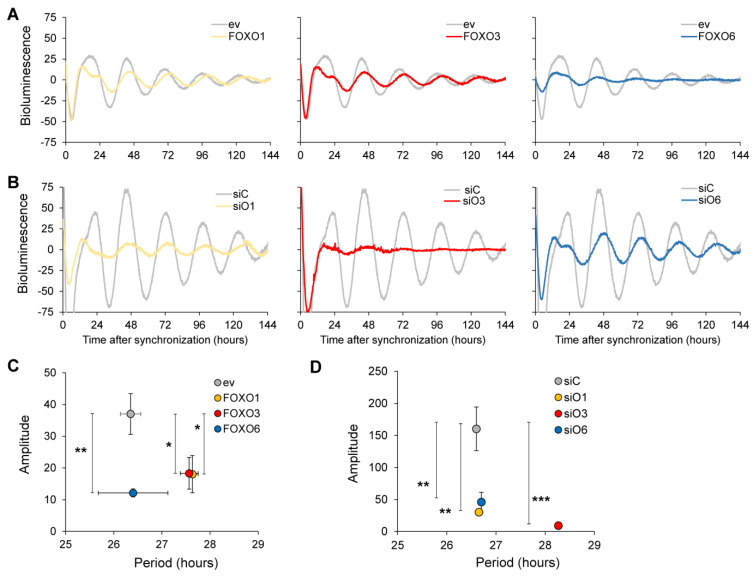
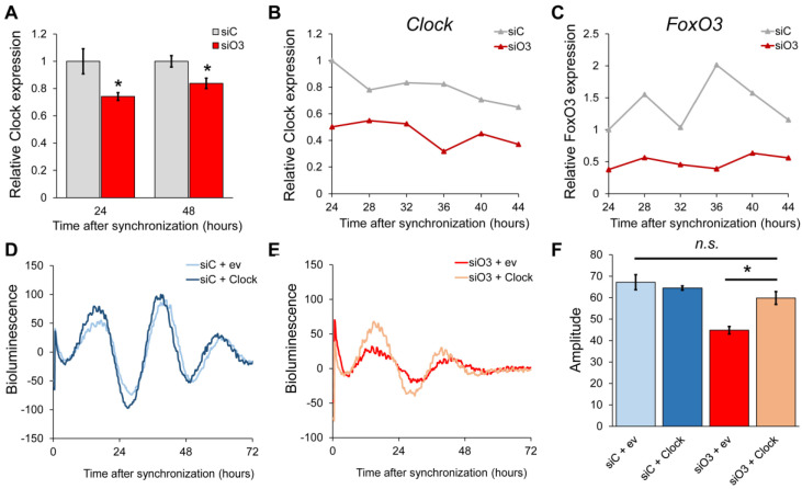
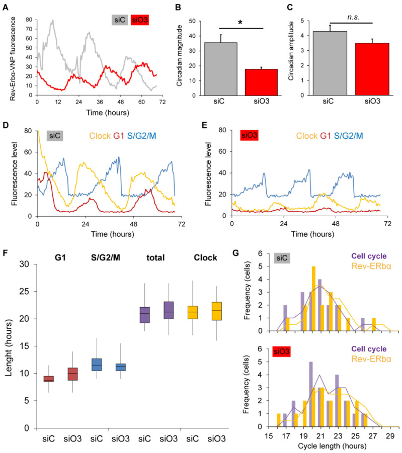
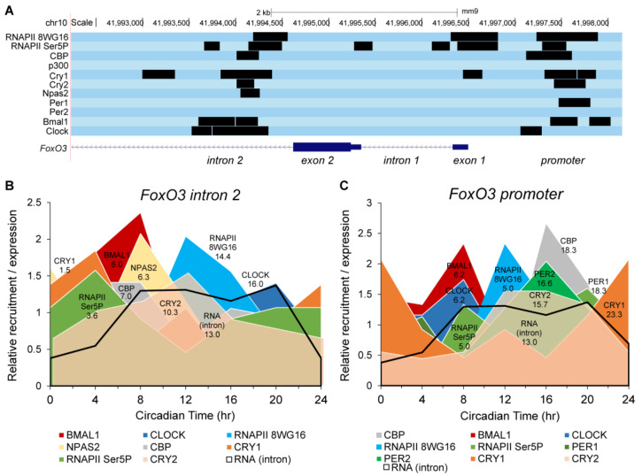
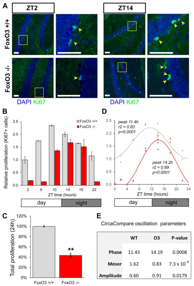
Both FoxO transcription factors and the circadian clock act on the interface of metabolism and cell cycle regulation and are important regulators of cellular stress and stem cell homeostasis. Importantly, FoxO3 preserves the adult neural stem cell population by regulating cell cycle and cellular metabolism and has been shown to regulate circadian rhythms in the liver. However, whether FoxO3 is a regulator of circadian rhythms in neural stem cells remains unknown. Here, we show that loss of FoxO3 disrupts circadian rhythmicity in cultures of neural stem cells, an effect that is mediated via regulation of Clock transcriptional levels. Using Rev-Erbα-VNP as a reporter, we then demonstrate that loss of FoxO3 does not disrupt circadian rhythmicity at the single cell level. A meta-analysis of published data revealed dynamic co-occupancy of multiple circadian clock components within FoxO3 regulatory regions, indicating that FoxO3 is a Clock-controlled gene. Finally, we examined proliferation in the hippocampus of FoxO3-deficient mice and found that loss of FoxO3 delayed the circadian phase of hippocampal proliferation, indicating that FoxO3 regulates correct timing of NSC proliferation. Taken together, our data suggest that FoxO3 is an integral part of circadian regulation of neural stem cell homeostasis.
Overview
The claims and statistics of this paper haven’t been checked yet.
Run the check — free, no account needed. It reads the open-access text and checks the sources it cites.
Integrity signals
Registry
Cites
Statistics
Figures
Verification
Claims the paper makes
The claims and statistics of this paper haven’t been checked yet.
Run the check — free, no account needed. It reads the open-access text and checks the sources it cites.
Claims other papers make about this one
Each is checked against this paper’s own text: does it say what the citing paper says it does?
The source backs part of the claim, not all of it — or backs it for a different population, condition or comparison.
In the citing paper
“PTEN could activate FOXO activity and inhibit the insulin/IGF pathway, suppressing neurogenesis [28–31].”
From M2 microglia-derived small extracellular vesicles modulate NSC fate after ischemic stroke via miR-25-3p/miR-93-5p-TGFBR/PTEN/FOXO3 axis (2025) · cites this paper as [31]
Experimental — accuracy not yet measured
Self-check
Percentages, sums and reported differences re-computed from the paper’s own numbers.
The self-check needs the paper’s full text.
Citations
Cites
Transcriptional architecture and chromatin landscape of the core circadian clock in mammals
Koike, N., Yoo, S.-H., Huang, H.-C., Kumar, V., Lee, C., Kim, T.-K., & Takahashi, J. S. (2012) · Science · doi:10.1126/science.1226339 · Cited by 1,464
Molecular regulation of stem cell quiescence
Cheung, T. H., & Rando, T. A. (2013) · Nature Reviews Molecular Cell Biology · doi:10.1038/nrm3591 · Cited by 1,148 · cited 1×
“DNA damage during life and allows for long-lived populations [11,12].”
BackgroundNPAS2: an analog of clock operative in the mammalian forebrain
Reick, M., Garcia, J. A., Dudley, C., & McKnight, S. L. (2001) · Science · doi:10.1126/science.1060699 · Cited by 572
Metabolism and cancer: the circadian clock connection
Sahar, S., & Sassone-Corsi, P. (2009) · Nature Reviews Cancer · doi:10.1038/nrc2747 · Cited by 551
cAMP-dependent signaling as a core component of the mammalian circadian pacemaker
O'Neill, J. S., Maywood, E. S., Chesham, J. E., Takahashi, J. S., & Hastings, M. H. (2008) · Science · doi:10.1126/science.1152506 · Cited by 418
The circadian molecular clock creates epidermal stem cell heterogeneity
Janich, P., Pascual, G., Merlos-Suárez, A., Batlle, E., Ripperger, J., Albrecht, U., Cheng, H.-Y. M., Obrietan, K., Di Croce, L., & Benitah, S. A. (2011) · Nature · doi:10.1038/nature10649 · Cited by 326
Adult Neurogenesis Is Sustained by Symmetric Self-Renewal and Differentiation
Obernier, K., Cebrian-Silla, A., Thomson, M., Parraguez, J. I., Anderson, R., Guinto, C., Rodas Rodriguez, J., Garcia-Verdugo, J.-M., & Alvarez-Buylla, A. (2018) · Cell Stem Cell · doi:10.1016/j.stem.2018.01.003 · Cited by 250
Characterization of the direct targets of FOXO transcription factors throughout evolution
Webb, A. E., Kundaje, A., & Brunet, A. (2016) · Aging Cell · doi:10.1111/acel.12479 · Cited by 182
FOXO3 shares common targets with ASCL1 genome-wide and inhibits ASCL1-dependent neurogenesis
Webb, A. E., Pollina, E. A., Vierbuchen, T., Urbán, N., Ucar, D., Leeman, D. S., Martynoga, B., Sewak, M., Rando, T. A., Guillemot, F., et al. (2013) · Cell Reports · doi:10.1016/j.celrep.2013.06.035 · Cited by 164
Schäffner, I., Minakaki, G., Khan, M. A., Balta, E.-A., Schlötzer-Schrehardt, U., Schwarz, T. J., Beckervordersandforth, R., Winner, B., Webb, A. E., DePinho, R. A., et al. (2018) · Neuron · doi:10.1016/j.neuron.2018.08.017 · Cited by 145
Research works printed but not linked: 50
- Dibner, 2010, The mammalian circadian timing system: Organization and coordination of central and peripheral clocks, Annu. Rev. Physiol., 72, 517, 10.1146/annurev-physiol-021909-135821 · DOI · Paper page
- Zhang, 2014, A circadian gene expression atlas in mammals: Implications for biology and medicine, Proc. Natl. Acad. Sci. USA, 111, 16219, 10.1073/pnas.1408886111 · DOI · Paper page
- Buhr, 2013, Molecular components of the mammalian circadian clock, Handb. Exp. Pharmacol., 217, 3, 10.1007/978-3-642-25950-0_1 · DOI · Paper page
- Takahashi, 2016, Transcriptional architecture of the mammalian circadian clock, Nat. Rev. Genet., 18, 164, 10.1038/nrg.2016.150 · DOI
- Brown, 2014, Circadian clock-mediated control of stem cell division and differentiation: Beyond night and day, Development, 141, 3105, 10.1242/dev.104851 · DOI
- Janich, 2013, Human Epidermal Stem Cell Function Is Regulated by Circadian Oscillations, Cell Stem Cell, 13, 745, 10.1016/j.stem.2013.09.004 · DOI
- Draijer, 2019, The circadian clock in adult neural stem cell maintenance, Prog. Neurobiol., 173, 41, 10.1016/j.pneurobio.2018.05.007 · DOI
- Taupin, 2002, Adult neurogenesis and neural stem cells of the central nervous system in mammals, J. Neurosci. Res., 69, 745, 10.1002/jnr.10378 · DOI
- Merkle, 2006, Neural stem cells in mammalian development, Curr. Opin. Cell Biol., 18, 704, 10.1016/j.ceb.2006.09.008 · DOI
- Basak, 2009, Stem cells of the adult mammalian brain and their niche, Cell. Mol. Life Sci., 66, 1057, 10.1007/s00018-008-8544-x · DOI
- Blomfield, 2019, Quiescence of Adult Mammalian Neural Stem Cells: A Highly Regulated Rest, Neuron, 104, 834, 10.1016/j.neuron.2019.09.026 · DOI
- Berg, 2016, Return to quiescence of mouse neural stem cells by degradation of a proactivation protein, Science, 353, 292, 10.1126/science.aaf4802 · DOI
- Encinas, 2012, Neural stem cell deforestation as the main force driving the age-related decline in adult hippocampal neurogenesis, Behav. Brain Res., 227, 433, 10.1016/j.bbr.2011.10.010 · DOI
- Ro, 2013, FoxOs in neural stem cell fate decision, Arch. Biochem. Biophys., 534, 55, 10.1016/j.abb.2012.07.017 · DOI
- Borgs, L., Beukelaers, P., Vandenbosch, R., Nguyen, L., Moonen, G., Maquet, P., Albrecht, U., Belachew, S., and Malgrange, B. (2009). Period 2 regulates neural stem/progenitor cell proliferation in the adult hippocampus. BMC Neurosci., 10. · DOI
- Yuen, 2013, The Circadian Molecular Clock Regulates Adult Hippocampal Neurogenesis by Controlling the Timing of Cell-Cycle Entry and Exit, Cell Rep., 5, 961, 10.1016/j.celrep.2013.10.037 · DOI
- Schnell, A., Chappuis, S., Schmutz, I., Brai, E., Ripperger, J.A., Schaad, O., Welzl, H., Descombes, P., Alberi, L., and Albrecht, U. (2014). The Nuclear Receptor REV-ERBα Regulates Fabp7 and Modulates Adult Hippocampal Neurogenesis. PLoS ONE, 9. · DOI
- Renault, 2009, FoxO3 Regulates Neural Stem Cell Homeostasis, Cell Stem Cell, 5, 527, 10.1016/j.stem.2009.09.014 · DOI · Paper page
- Paik, 2009, FoxOs Cooperatively Regulate Diverse Pathways Governing Neural Stem Cell Homeostasis, Cell Stem Cell, 5, 540, 10.1016/j.stem.2009.09.013 · DOI · Paper page
- Greer, 2005, FOXO transcription factors at the interface between longevity and tumor suppression, Oncogene, 24, 7410, 10.1038/sj.onc.1209086 · DOI
- Gross, 2008, The role of FoxO in the regulation of metabolism, Oncogene, 27, 2320, 10.1038/onc.2008.25 · DOI
- Rafalski, 2011, Energy metabolism in adult neural stem cell fate, Prog. Neurobiol., 93, 182, 10.1016/j.pneurobio.2010.10.007 · DOI
- Brunet, 1999, Akt Promotes Cell Survival by Phosphorylating and Inhibiting a Forkhead Transcription Factor, Cell, 96, 857, 10.1016/S0092-8674(00)80595-4 · DOI · Paper page
- Hoekman, 2004, The ins and outs of FoxO shuttling: Mechanisms of FoxO translocation and transcriptional regulation, Biochem. J., 380, 297, 10.1042/bj20040167 · DOI
- Chaves, 2014, Insulin-FOXO3 Signaling Modulates Circadian Rhythms via Regulation of Clock Transcription, Curr. Biol., 24, 1248, 10.1016/j.cub.2014.04.018 · DOI
- Laywell, 2000, Identification of a multipotent astrocytic stem cell in the immature and adult mouse brain, Proc. Natl. Acad. Sci. USA, 97, 13883, 10.1073/pnas.250471697 · DOI
- Fitzsimons, 2013, Knockdown of the glucocorticoid receptor alters functional integration of newborn neurons in the adult hippocampus and impairs fear-motivated behavior, Mol. Psychiatry, 18, 993, 10.1038/mp.2012.123 · DOI
- Hoekman, 2006, Spatial and temporal expression of FoxO transcription factors in the developing and adult murine brain, Gene Expr. Patterns, 6, 134, 10.1016/j.modgep.2005.07.003 · DOI
- Nagoshi, 2004, Circadian gene expression in individual fibroblasts: Cell-autonomous and self-sustained oscillators pass time to daughter cells, Cell, 119, 693, 10.1016/j.cell.2004.11.015 · DOI
- Feillet, 2014, Phase locking and multiple oscillating attractors for the coupled mammalian clock and cell cycle, Proc. Natl. Acad. Sci. USA, 111, 9828, 10.1073/pnas.1320474111 · DOI
- Gaucher, 2018, Molecular Cogs: Interplay between Circadian Clock and Cell Cycle, Trends Cell Biol., 28, 368, 10.1016/j.tcb.2018.01.006 · DOI
- Farshadi, 2020, Molecular Links between the Circadian Clock and the Cell Cycle, J. Mol. Biol., 432, 3515, 10.1016/j.jmb.2020.04.003 · DOI
- Vollmers, 2012, Circadian oscillations of protein-coding and regulatory RNAs in a highly dynamic mammalian liver epigenome, Cell Metab., 16, 833, 10.1016/j.cmet.2012.11.004 · DOI
- Masri, 2013, The circadian clock and cell cycle: Interconnected biological circuits, Curr. Opin. Cell Biol., 25, 730, 10.1016/j.ceb.2013.07.013 · DOI
- Hosaka, 2004, Disruption of forkhead transcription factor (FOXO) family members in mice reveals their functional diversification, Proc. Natl. Acad. Sci. USA, 101, 2975, 10.1073/pnas.0400093101 · DOI
- Taupin, 2007, BrdU immunohistochemistry for studying adult neurogenesis: Paradigms, pitfalls, limitations, and validation, Brain Res. Rev., 53, 198, 10.1016/j.brainresrev.2006.08.002 · DOI · Paper page
- Parsons, 2022, CircaCompare: A method to estimate and statistically support differences in mesor, amplitude and phase, between circadian rhythms, Bioinformatics, 36, 1208, 10.1093/bioinformatics/btz730 · DOI
- Lützner, N., Kalbacher, H., Krones-Herzig, A., and Rösl, F. (2012). FOXO3 Is a Glucocorticoid Receptor Target and Regulates LKB1 and Its Own Expression Based on Cellular AMP Levels via a Positive Autoregulatory Loop. PLoS ONE, 7. · DOI
- Herzig, 2018, AMPK: Guardian of metabolism and mitochondrial homeostasis, Nat. Rev. Mol. Cell Biol., 19, 121, 10.1038/nrm.2017.95 · DOI
- Dickmeis, T., Lahiri, K., Nica, G., Vallone, D., Santoriello, C., Neumann, C.J., Hammerschmidt, M., and Foulkes, N.S. (2007). Glucocorticoids Play a Key Role in Circadian Cell Cycle Rhythms. PLoS Biol., 5. · DOI
- Schouten, 2020, Circadian glucocorticoid oscillations preserve a population of adult hippocampal neural stem cells in the aging brain, Mol. Psychiatry, 25, 1382, 10.1038/s41380-019-0440-2 · DOI
- Chen, 2007, A Conserved DNA damage response pathway responsible for coupling the cell division cycle to the circadian and metabolic cycles, Cell Cycle, 6, 2906, 10.4161/cc.6.23.5041 · DOI
- Janich, 2014, Circadian control of tissue homeostasis and adult stem cells, Curr. Opin. Cell Biol., 31, 8, 10.1016/j.ceb.2014.06.010 · DOI
- Bass, 2010, Circadian Integration of Metabolism and Energetics, Science, 330, 1349, 10.1126/science.1195027 · DOI · Paper page
- Altmann, C., Keller, S., and Schmidt, M.H.H. (2019). The Role of SVZ Stem Cells in Glioblastoma. Cancers, 11. · DOI
- Salih, 2008, FoxO transcription factors in the maintenance of cellular homeostasis during aging, Curr. Opin. Cell Biol., 20, 126, 10.1016/j.ceb.2008.02.005 · DOI · Paper page
- Sato, 2017, Circadian Reprogramming in the Liver Identifies Metabolic Pathways of Aging, Cell, 170, 664, 10.1016/j.cell.2017.07.042 · DOI
- Farajnia, 2012, Evidence for neuronal desynchrony in the aged suprachiasmatic nucleus clock, J. Neurosci., 32, 5891, 10.1523/JNEUROSCI.0469-12.2012 · DOI · Paper page
- Benitah, 2020, Circadian Regulation of Adult Stem Cell Homeostasis and Aging, Cell Stem Cell, 26, 817, 10.1016/j.stem.2020.05.002 · DOI
- Farshadi, 2019, The positive circadian regulators CLOCK and BMAL1 control G2/M cell cycle transition through Cyclin B1, Cell Cycle, 18, 16, 10.1080/15384101.2018.1558638 · DOI
Cited by
Sorted by citations
Zhang, Q., Yi, Y., Chen, T., Ai, Y., Chen, Z., Liu, G., Tang, Z., Chen, J., Xu, T., Chen, X., et al. (2025) · Journal of Nanobiotechnology · doi:10.1186/s12951-025-03390-2 · Cited by 27
Metadata
- Authors
- Draijer, S., Timmerman, R., Pannekeet, J., van Harten, A., Farshadi, E. A., Kemmer, J., van Gilst, D., Chaves, I., & Hoekman, M. F. M.
- Affiliations
- Swammerdam Institute of Life Sciences, University of Amsterdam, 1018 WB Amsterdam, The Netherlands
- Department of Molecular Genetics, Erasmus University Medical Center, 3000 CA Rotterdam, The Netherlands
- ORCID
- Chaves, Inês
- Venue
- International Journal of Molecular Sciences
- Publisher
- MDPI AG
- Published
- 2023-09-04
- Volume
- 24
- Issue
- 17
- Pages
- 13662
- ISSN
- 1422-0067
- PMC
- PMC10563086
- Cited by
- 7
- References
- 10
- Language
- en
About these checks
Registry, retraction, Cites and statistics results are computed, not judged, with one exception: a printed reference that no index lists is read once by a model before it counts as a citation error. Claims are extracted by a model and kept only when their quote appears verbatim in the text; each cited source is then checked, with a model’s help, for whether it says what the claim relies on. Those verdicts can be wrong — read the claim’s quote and the cited source before relying on one.
Statistics and arithmetic are recomputed, not judged.
Confidence bands are shown only for fields calibrated on our annotated test set. None is calibrated yet, so no band is shown.
What counts as a claim ›How we measure confidence ›How we check this ›
Are you an author, or do you see a problem with this page? Request a review
