

The DOI matches its Crossref record; 11 of the 11 references it cites were found in a registry or an index.
Small Extracellular Vesicles’ miRNAs: Biomarkers and Therapeutics for Neurodegenerative Diseases
Abstract
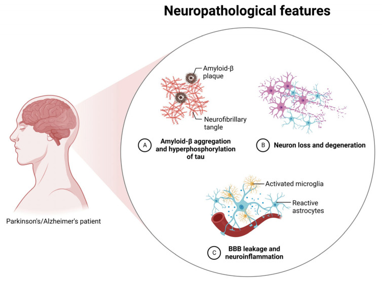
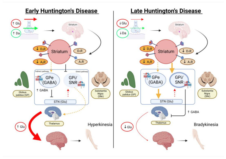
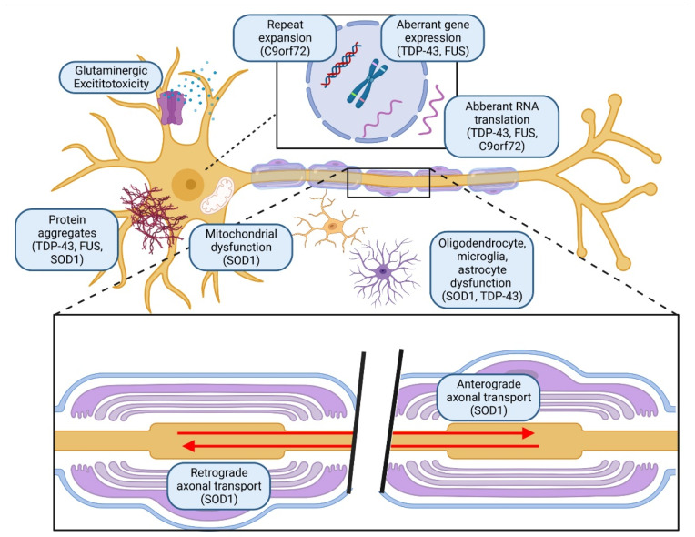
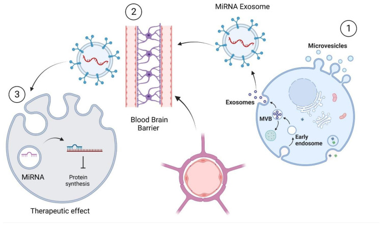
Neurodegenerative diseases are critical in the healthcare system as patients suffer from progressive diseases despite currently available drug management. Indeed, the growing ageing population will burden the country’s healthcare system and the caretakers. Thus, there is a need for new management that could stop or reverse the progression of neurodegenerative diseases. Stem cells possess a remarkable regenerative potential that has long been investigated to resolve these issues. Some breakthroughs have been achieved thus far to replace the damaged brain cells; however, the procedure’s invasiveness has prompted scientists to investigate using stem-cell small extracellular vesicles (sEVs) as a non-invasive cell-free therapy to address the limitations of cell therapy. With the advancement of technology to understand the molecular changes of neurodegenerative diseases, efforts have been made to enrich stem cells’ sEVs with miRNAs to increase the therapeutic efficacy of the sEVs. In this article, the pathophysiology of various neurodegenerative diseases is highlighted. The role of miRNAs from sEVs as biomarkers and treatments is also discussed. Lastly, the applications and delivery of stem cells and their miRNA-enriched sEVs for treating neurodegenerative diseases are emphasised and reviewed.
Overview
The claims and statistics of this paper haven’t been checked yet.
Run the check — free, no account needed. It reads the open-access text and checks the sources it cites.
Integrity signals
Registry
Cites
Statistics
Figures
Verification
Claims the paper makes
The claims and statistics of this paper haven’t been checked yet.
Run the check — free, no account needed. It reads the open-access text and checks the sources it cites.
Claims other papers make about this one
Each is checked against this paper’s own text: does it say what the citing paper says it does?
The cited source says what the claim attributes to it.
In the citing paper
“Since miRNAs are released in EVs by cells, they may also reflect their pathological state [56].”
From miR-92a-3p and miR-320a are Upregulated in Plasma Neuron-Derived Extracellular Vesicles of Patients with Frontotemporal Dementia (2025) · cites this paper as [56]
Self-check
Percentages, sums and reported differences re-computed from the paper’s own numbers.
The self-check needs the paper’s full text.
Citations
Cites
1 retracted
Dementia prevention, intervention, and care: 2020 report of the Lancet Commission
Livingston, G., Huntley, J., Sommerlad, A., Ames, D., Ballard, C., Banerjee, S., Brayne, C., Burns, A., Cohen-Mansfield, J., Cooper, C., et al. (2020) · The Lancet · doi:10.1016/s0140-6736(20)30367-6 · Cited by 10,430
RetractedOverview of MicroRNA Biogenesis, Mechanisms of Actions, and Circulation
O'Brien, J., Hayder, H., Zayed, Y., & Peng, C. (2018) · Frontiers in Endocrinology · doi:10.3389/fendo.2018.00402 · Cited by 5,287
Wiklander, O. P. B., Nordin, J. Z., O'Loughlin, A., Gustafsson, Y., Corso, G., Mäger, I., Vader, P., Lee, Y., Sork, H., Seow, Y., et al. (2015) · Journal of Extracellular Vesicles · doi:10.3402/jev.v4.26316 · Cited by 1,730
Fiandaca, M. S., Kapogiannis, D., Mapstone, M., Boxer, A., Eitan, E., Schwartz, J. B., Abner, E. L., Petersen, R. C., Federoff, H. J., Miller, B. L., et al. (2015) · Alzheimer's & Dementia · doi:10.1016/j.jalz.2014.06.008 · Cited by 959
α-Synuclein binds to TOM20 and inhibits mitochondrial protein import in Parkinson’s disease
Di Maio, R., Barrett, P. J., Hoffman, E. K., Barrett, C. W., Zharikov, A., Borah, A., Hu, X., McCoy, J., Chu, C. T., Burton, E. A., et al. (2016) · Science Translational Medicine · doi:10.1126/scitranslmed.aaf3634 · Cited by 619
Banks, W. A., Sharma, P., Bullock, K. M., Hansen, K. M., Ludwig, N., & Whiteside, T. L. (2020) · International Journal of Molecular Sciences · doi:10.3390/ijms21124407 · Cited by 561
Elucidation of Exosome Migration Across the Blood–Brain Barrier Model In Vitro
Chen, C. C., Liu, L., Ma, F., Wong, C. W., Guo, X. E., Chacko, J. V., Farhoodi, H. P., Zhang, S. X., Zimak, J., Ségaliny, A., et al. (2016) · Cellular and Molecular Bioengineering · doi:10.1007/s12195-016-0458-3 · Cited by 537
Perets, N., Betzer, O., Shapira, R., Brenstein, S., Angel, A., Sadan, T., Ashery, U., Popovtzer, R., & Offen, D. (2019) · Nano Letters · doi:10.1021/acs.nanolett.8b04148 · Cited by 382
Shi, M., Chu, F., Zhu, F., & Zhu, J. (2022) · Frontiers in Aging Neuroscience · doi:10.3389/fnagi.2022.870517 · Cited by 275
de Godoy, M. A., Saraiva, L. M., de Carvalho, L. R. P., Vasconcelos-dos-Santos, A., Beiral, H. J. V., Ramos, A. B., Silva, L. R. D. P., Leal, R. B., Monteiro, V. H. S., Braga, C. V., et al. (2018) · Journal of Biological Chemistry · doi:10.1074/jbc.m117.807180 · Cited by 214
Momin, M. Y., Gaddam, R. R., Kravitz, M., Gupta, A., & Vikram, A. (2021) · Cells · doi:10.3390/cells10113097 · Cited by 107
Research works printed but not linked: 143
- Rekatsina, 2020, Pathophysiology and Therapeutic Perspectives of Oxidative Stress and Neurodegenerative Diseases: A Narrative Review, Adv. Ther., 37, 113, 10.1007/s12325-019-01148-5 · DOI
- Fayazi, 2021, Stem Cell-Derived Exosomes: A New Strategy of Neurodegenerative Disease Treatment, Mol. Neurobiol., 58, 3494, 10.1007/s12035-021-02324-x · DOI
- Parambi, 2022, Gene Therapy Approach with an Emphasis on Growth Factors: Theoretical and Clinical Outcomes in Neurodegenerative Diseases, Mol. Neurobiol., 59, 191, 10.1007/s12035-021-02555-y · DOI
- Muddapu, 2020, Neurodegenerative Diseases–Is Metabolic Deficiency the Root Cause?, Front. Neurosci., 14, 213, 10.3389/fnins.2020.00213 · DOI
- Whalen, K. (2014). Pharmacology, Lippincott Williams & Wilkins.
- Ogbodo, 2022, Alzheimer’s Disease: Pathogenesis and Therapeutic Interventions, Curr. Aging Sci., 15, 2, 10.2174/1874609814666210302085232 · DOI
- Kim, 2021, Efficacy and tolerability of aripiprazole versus D2 antagonists in the early course of schizophrenia: A systematic review and meta-analysis, Npj Schizophr., 7, 29, 10.1038/s41537-021-00158-z · DOI
- Ferrini, 2021, Persistency of Mesenchymal Stromal/Stem Cells in Lungs, Front. Cell Dev. Biol., 9, 1825, 10.3389/fcell.2021.709225 · DOI
- Wang, 2015, Excess Integrins Cause Lung Entrapment of Mesenchymal Stem Cells, Stem Cells, 33, 3315, 10.1002/stem.2087 · DOI
- Witwer, 2018, Minimal information for studies of extracellular vesicles 2018 (MISEV2018): A position statement of the International Society for Extracellular Vesicles and update of the MISEV2014 guidelines, J. Extracell. Vesicles, 7, 1535750, 10.1080/20013078.2018.1535750 · DOI · Paper page
- Abdelaal, N.E., and Abdelhai, M.F. (2021). Role of Exosomes in Biological Communication Systems, Springer.
- Beatriz, 2021, Exosomes: Innocent Bystanders or Critical Culprits in Neurodegenerative Diseases, Front. Cell Dev. Biol., 9, 635104, 10.3389/fcell.2021.635104 · DOI
- Takahashi, 2023, Extracellular vesicle-based therapeutics: Extracellular vesicles as therapeutic targets and agents, Pharmacol. Ther., 242, 108352, 10.1016/j.pharmthera.2023.108352 · DOI
- Sarvar, 2016, Mesenchymal stem cell-derived exosomes: New opportunity in cell-free therapy, Adv. Pharm. Bull., 6, 293, 10.15171/apb.2016.041 · DOI
- Yang, 2019, MiR-124 Enriched Exosomes Promoted the M2 Polarization of Microglia and Enhanced Hippocampus Neurogenesis After Traumatic Brain Injury by Inhibiting TLR4 Pathway, Neurochem. Res., 44, 811, 10.1007/s11064-018-02714-z · DOI · Paper page
- Salarpour, 2022, The application of exosomes and Exosome-nanoparticle in treating brain disorders, J. Mol. Liq., 350, 118549, 10.1016/j.molliq.2022.118549 · DOI
- Alzheimer, 1906, Uber einen eigenartigen schweren Erkrankungsprozess der Hirninde, Neurol. Cent., 25, 1134
- Inga, Z. (2015). Alzheimer’s Disease, IntechOpen. Chapter 10.
- Chow, 2014, CDK5 activator protein p25 preferentially binds and activates GSK3β, Proc. Natl. Acad. Sci. USA, 111, E4887, 10.1073/pnas.1402627111 · DOI
- Bourdenx, 2017, Protein aggregation and neurodegeneration in prototypical neurodegenerative diseases: Examples of amyloidopathies, tauopathies and synucleinopathies, Prog. Neurobiol., 155, 171, 10.1016/j.pneurobio.2015.07.003 · DOI
- Singh, A., Kukreti, R., Saso, L., and Kukreti, S. (2019). Oxidative Stress: A Key Modulator in Neurodegenerative Diseases. Molecules, 24. · DOI
- Neniskyte, 2017, Errant gardeners: Glial-cell-dependent synaptic pruning and neurodevelopmental disorders, Nat. Rev. Neurosci., 18, 658, 10.1038/nrn.2017.110 · DOI
- Actor, J.K., and Smith, K.C. (2019). Translational Inflammation, Academic Press. · DOI
- Zuroff, 2017, Clearance of cerebral Aβ in Alzheimer’s disease: Reassessing the role of microglia and monocytes, Cell. Mol. Life Sci., 74, 2167, 10.1007/s00018-017-2463-7 · DOI
- Wang, 2015, TREM2 lipid sensing sustains the microglial response in an Alzheimer’s disease model, Cell, 160, 1061, 10.1016/j.cell.2015.01.049 · DOI · Paper page
- Khan, 2019, Awareness and current knowledge of Parkinson’s disease: A neurodegenerative disorder, Int. J. Neurosci., 129, 55, 10.1080/00207454.2018.1486837 · DOI
- Parashos, S.A., and Wichmann, R. (2020). Navigating Life with Parkinson’s Disease, Oxford University Press. [2nd ed.].
- Bellucci, 2020, Nuclear Factor-κB Dysregulation and α-Synuclein Pathology: Critical Interplay in the Pathogenesis of Parkinson’s Disease, Front. Aging Neurosci., 12, 68, 10.3389/fnagi.2020.00068 · DOI
- Ma, 2018, C-terminal truncation exacerbates the aggregation and cytotoxicity of α-Synuclein: A vicious cycle in Parkinson’s disease, Biochim. Biophys. Acta (BBA) Mol. Basis Dis., 1864, 3714, 10.1016/j.bbadis.2018.10.003 · DOI
- Sato, 2011, Authentically phosphorylated α-synuclein at Ser129 accelerates neurodegeneration in a rat model of familial Parkinson’s disease, J. Neurosci., 31, 16884, 10.1523/JNEUROSCI.3967-11.2011 · DOI
- Park, 2011, Elevated levels of α-synuclein oligomer in the cerebrospinal fluid of drug-naïve patients with Parkinson’s disease, J. Clin. Neurol., 7, 215, 10.3988/jcn.2011.7.4.215 · DOI
- Jones, R. (2010). The roles of PINK1 and Parkin in Parkinson’s disease. PLoS Biol., 8. · DOI
- Behl, 2022, Exploring the Role of Ubiquitin–Proteasome System in Parkinson’s Disease, Mol. Neurobiol., 59, 4257, 10.1007/s12035-022-02851-1 · DOI
- Liu, 2022, Deubiquitinating enzymes (DUBs): Decipher underlying basis of neurodegenerative diseases, Mol. Psychiatry, 27, 259, 10.1038/s41380-021-01233-8 · DOI
- Zhang, 2022, MANF Inhibits α-Synuclein Accumulation through Activation of Autophagic Pathways, Oxid. Med. Cell. Longev., 2022, 7925686
- Wilkaniec, 2021, Exogenous Alpha-Synuclein Evoked Parkin Downregulation Promotes Mitochondrial Dysfunction in Neuronal Cells. Implications for Parkinson’s Disease Pathology, Front. Aging Neurosci., 13, 591475, 10.3389/fnagi.2021.591475 · DOI
- Stoker, T.B., and Greenland, J.C. (2018). Parkinson’s Disease: Pathogenesis and Clinical Aspects, Codon Publications. · DOI
- Stoker, 2022, Huntington’s disease: Diagnosis and management, Pract. Neurol., 22, 32, 10.1136/practneurol-2021-003074 · DOI
- Licitra, 2017, Huntington’s Disease: Mechanisms of Pathogenesis and Therapeutic Strategies, Cold Spring Harb. Perspect. Med., 7, a024240, 10.1101/cshperspect.a024240 · DOI
- Kostrzewa, R.M. (2021). Handbook of Neurotoxicity, Springer International Publishing. · DOI
- Carmo, 2018, Mitochondrial Dysfunction in Huntington’s Disease, Adv. Exp. Med. Biol., 1049, 59, 10.1007/978-3-319-71779-1_3 · DOI
- Paul, 2014, Cystathionine γ-lyase deficiency mediates neurodegeneration in Huntington’s disease, Nature, 509, 96, 10.1038/nature13136 · DOI
- Catanesi, M., d’Angelo, M., Tupone, M.G., Benedetti, E., Giordano, A., Castelli, V., and Cimini, A. (2020). MicroRNAs Dysregulation and Mitochondrial Dysfunction in Neurodegenerative Diseases. Int. J. Mol. Sci., 21. · DOI
- Buren, C. (2017). Global Neuroprotection of Huntingtin in Culture and Alterations of Cortico-Striatal Connections in Huntington’s Disease Culture and Mouse Models. [Ph.D. Thesis, University of British Columbia].
- Raymond, 2011, Pathophysiology of Huntington’s disease: Time-dependent alterations in synaptic and receptor function, Neuroscience, 198, 252, 10.1016/j.neuroscience.2011.08.052 · DOI
- Watanabe, 2022, Impaired neuromuscular transmission in facial muscles of amyotrophic lateral sclerosis: A single-fiber electromyography study, Neurol. Clin. Neurosci., 10, 78, 10.1111/ncn3.12580 · DOI
- Satarker, S., Bojja, S.L., Gurram, P.C., Mudgal, J., Arora, D., and Nampoothiri, M. (2022). Astrocytic Glutamatergic Transmission and Its Implications in Neurodegenerative Disorders. Cells, 11. · DOI
- Didonna, 2019, The role of neurofilament aggregation in neurodegeneration: Lessons from rare inherited neurological disorders, Mol. Neurodegener., 14, 19, 10.1186/s13024-019-0318-4 · DOI
- Van den Bos, M.A.J., Geevasinga, N., Higashihara, M., Menon, P., and Vucic, S. (2019). Pathophysiology and Diagnosis of ALS: Insights from Advances in Neurophysiological Techniques. Int. J. Mol. Sci., 20. · DOI
- He, 2021, Diagnostic and Therapeutic Potential of Exosomal MicroRNAs for Neurodegenerative Diseases, Neural Plast., 2021, 8884642, 10.1155/2021/8884642 · DOI
- Matsumoto, 2017, Transmission of α-synuclein-containing erythrocyte-derived extracellular vesicles across the blood-brain barrier via adsorptive mediated transcytosis: Another mechanism for initiation and progression of Parkinson’s disease?, Acta Neuropathol. Commun., 5, 71, 10.1186/s40478-017-0470-4 · DOI · Paper page
- Manna, I., De Benedittis, S., Quattrone, A., Maisano, D., Iaccino, E., and Quattrone, A. (2020). Exosomal miRNAs as Potential Diagnostic Biomarkers in Alzheimer’s Disease. Pharmaceuticals, 13. · DOI
- Mathew, B., Mansuri, M.S., Williams, K.R., and Nairn, A.C. (2021). Exosomes as Emerging Biomarker Tools in Neurodegenerative and Neuropsychiatric Disorders-A Proteomics Perspective. Brain Sci., 11. · DOI
- Cheng, 2014, Exosomes provide a protective and enriched source of miRNA for biomarker profiling compared to intracellular and cell-free blood, J. Extracell. Vesicles, 3, 23743, 10.3402/jev.v3.23743 · DOI · Paper page
- Fenoglio, 2019, Exosome Determinants of Physiological Aging and Age-Related Neurodegenerative Diseases, Front. Aging Neurosci., 11, 232, 10.3389/fnagi.2019.00232 · DOI · Paper page
- Kumar, 2016, Are circulating microRNAs peripheral biomarkers for Alzheimer’s disease?, Biochim. Biophys. Acta, 1862, 1617, 10.1016/j.bbadis.2016.06.001 · DOI
- Thomi, 2019, Exosomes derived from umbilical cord mesenchymal stem cells reduce microglia-mediated neuroinflammation in perinatal brain injury, Stem Cell Res. Ther., 10, 105, 10.1186/s13287-019-1207-z · DOI
- Foo, J.B., Looi, Q.H., How, C.W., Lee, S.H., Al-Masawa, M.E., Chong, P.P., and Law, J.X. (2021). Mesenchymal Stem Cell-Derived Exosomes and MicroRNAs in Cartilage Regeneration: Biogenesis, Efficacy, miRNA Enrichment and Delivery. Pharmaceuticals, 14. · DOI
- Markoutsa, 2022, Extracellular vesicles derived from inflammatory-educated stem cells reverse brain inflammation-implication of miRNAs, Mol. Ther., 30, 816, 10.1016/j.ymthe.2021.08.008 · DOI
- Chen, 2021, Exosomal Proteins and miRNAs as Mediators of Amyotrophic Lateral Sclerosis, Front. Cell Dev. Biol., 9, 718803, 10.3389/fcell.2021.718803 · DOI
- Yang, 2017, Exosome Mediated Delivery of miR-124 Promotes Neurogenesis after Ischemia, Mol. Ther. Nucleic Acids, 7, 278, 10.1016/j.omtn.2017.04.010 · DOI · Paper page
- Cheng, 2009, MiR-124 Regulates Adult Neurogenesis in the Subventricular Zone Stem Cell Niche, Nat. Neurosci., 12, 399, 10.1038/nn.2294 · DOI · Paper page
- Yu, 2011, MicroRNA miR-133b is essential for functional recovery after spinal cord injury in adult zebrafish, Eur. J. Neurosci., 33, 1587, 10.1111/j.1460-9568.2011.07643.x · DOI
- Xin, 2017, MicroRNA cluster miR-17–92 Cluster in Exosomes Enhance Neuroplasticity and Functional Recovery After Stroke in Rats, Stroke, 48, 747, 10.1161/STROKEAHA.116.015204 · DOI
- Zuo, 2019, MicroRNA-132 attenuates cerebral injury by protecting blood-brain-barrier in MCAO mice, Exp. Neurol., 316, 12, 10.1016/j.expneurol.2019.03.017 · DOI
- Ma, 2014, MiR-181a Regulates Blood-Tumor Barrier Permeability by Targeting Krüppel-Like Factor 6, J. Cereb. Blood Flow Metab., 34, 1826, 10.1038/jcbfm.2014.152 · DOI
- Yu, 2019, Exosomes derived from microRNA-199a-overexpressing mesenchymal stem cells inhibit glioma progression by down-regulating AGAP2, Aging, 11, 5300, 10.18632/aging.102092 · DOI
- Li, 2019, MiR-199a Inhibits Tumor Growth and Attenuates Chemoresistance by Targeting K-RAS via AKT and ERK Signalings, Front. Oncol., 9, 1071, 10.3389/fonc.2019.01071 · DOI
- Wang, 2021, Challenges in the development and establishment of exosome-based drug delivery systems, J. Control. Release, 329, 894, 10.1016/j.jconrel.2020.10.020 · DOI
- Wang, 2014, The role of miR-146a in dorsal root ganglia neurons of experimental diabetic peripheral neuropathy, Neuroscience, 259, 155, 10.1016/j.neuroscience.2013.11.057 · DOI
- Wang, 2020, The Role of Exosomal microRNAs and Oxidative Stress in Neurodegenerative Diseases, Oxid. Med. Cell. Longev., 2020, 3232869, 10.1155/2020/3232869 · DOI
- Yu, 2021, The role of exosomal microRNAs in central nervous system diseases, Mol. Cell. Biochem., 476, 2111, 10.1007/s11010-021-04053-0 · DOI
- Bolukbasi, 2012, miR-1289 and “Zipcode”-like Sequence Enrich mRNAs in Microvesicles, Mol. Ther. Nucleic Acids, 1, e10, 10.1038/mtna.2011.2 · DOI
- Hooper, C., Sainz-Fuertes, R., Lynham, S., Hye, A., Killick, R., Warley, A., Bolondi, C., Pocock, J., and Lovestone, S. (2012). Wnt3a induces exosome secretion from primary cultured rat microglia. BMC Neurosci., 13. · DOI
- Trindade, 2021, Diagnostic and therapeutic potential of exosomes in Alzheimer’s disease, J. Neurochem., 156, 162, 10.1111/jnc.15112 · DOI
- Cui, 2017, Exosomes derived from hypoxia-preconditioned mesenchymal stromal cells ameliorate cognitive decline by rescuing synaptic dysfunction and regulating inflammatory responses in APP/PS1 mice, FASEB J., 32, 654, 10.1096/fj.201700600R · DOI · Paper page
- Cai, 2018, Exosomes: A novel therapeutic target for Alzheimer’s disease?, Neural Regen. Res., 13, 930, 10.4103/1673-5374.232490 · DOI
- Katsuda, 2013, Human adipose tissue-derived mesenchymal stem cells secrete functional neprilysin-bound exosomes, Sci. Rep., 3, 1197, 10.1038/srep01197 · DOI
- Shen, 2018, MiR-125b-5p is involved in oxygen and glucose deprivation injury in PC-12 cells via CBS/H(2)S pathway, Nitric Oxide, 78, 11, 10.1016/j.niox.2018.05.004 · DOI
- Sharples, 2008, Inhibition of gamma-secretase causes increased secretion of amyloid precursor protein C-terminal fragments in association with exosomes, FASEB J., 22, 1469, 10.1096/fj.07-9357com · DOI
- Yuyama, 2014, Decreased amyloid-β pathologies by intracerebral loading of glycosphingolipid-enriched exosomes in Alzheimer model mice, J. Biol. Chem., 289, 24488, 10.1074/jbc.M114.577213 · DOI
- Falker, 2016, Exosomal cellular prion protein drives fibrillization of amyloid beta and counteracts amyloid beta-mediated neurotoxicity, J. Neurochem., 137, 88, 10.1111/jnc.13514 · DOI
- Xin, 2012, Exosome-mediated transfer of miR-133b from multipotent mesenchymal stromal cells to neural cells contributes to neurite outgrowth, Stem Cells, 30, 1556, 10.1002/stem.1129 · DOI
- Ba, 2015, Schisandrin B shows neuroprotective effect in 6-OHDA-induced Parkinson’s disease via inhibiting the negative modulation of miR-34a on Nrf2 pathway, Biomed. Pharm., 75, 165, 10.1016/j.biopha.2015.07.034 · DOI
- Narbute, 2019, Intranasal Administration of Extracellular Vesicles Derived from Human Teeth Stem Cells Improves Motor Symptoms and Normalizes Tyrosine Hydroxylase Expression in the Substantia Nigra and Striatum of the 6-Hydroxydopamine-Treated Rats, Stem Cells Transl. Med., 8, 490, 10.1002/sctm.18-0162 · DOI · Paper page
- Mao, 2015, Secreted miR-34a in astrocytic shedding vesicles enhanced the vulnerability of dopaminergic neurons to neurotoxins by targeting Bcl-2, Protein Cell, 6, 529, 10.1007/s13238-015-0168-y · DOI · Paper page
- Chen, 2019, Activation of the miR-34a-Mediated SIRT1/mTOR Signaling Pathway by Urolithin A Attenuates D-Galactose-Induced Brain Aging in Mice, Neurotherapeutics, 16, 1269, 10.1007/s13311-019-00753-0 · DOI
- Jiang, 2019, Serum secreted miR-137-containing exosomes affects oxidative stress of neurons by regulating OXR1 in Parkinson’s disease, Brain Res., 1722, 146331, 10.1016/j.brainres.2019.146331 · DOI
- Oh, 2017, The Cleavage Effect of Mesenchymal Stem Cell and Its Derived Matrix Metalloproteinase-2 on Extracellular α-Synuclein Aggregates in Parkinsonian Models, Stem Cells Transl. Med., 6, 949, 10.5966/sctm.2016-0111 · DOI
- Lee, 2021, Exosomes as a potential messenger unit during heterochronic parabiosis for amelioration of Huntington’s disease, Neurobiol. Dis., 155, 105374, 10.1016/j.nbd.2021.105374 · DOI
- Lee, 2016, Exosomes from adipose-derived stem cells ameliorate phenotype of Huntington’s disease in vitro model, Eur. J. Neurosci., 44, 2114, 10.1111/ejn.13275 · DOI · Paper page
- Jeon, 2016, Human-to-mouse prion-like propagation of mutant huntingtin protein, Acta Neuropathol., 132, 577, 10.1007/s00401-016-1582-9 · DOI
- Das, 2013, MicroRNA-124 targets CCNA2 and regulates cell cycle in STHdh(Q111)/Hdh(Q111) cells, Biochem. Biophys. Res. Commun., 437, 217, 10.1016/j.bbrc.2013.06.041 · DOI
- Lee, 2017, Exosome-Based Delivery of miR-124 in a Huntington’s Disease Model, J. Mov. Disord., 10, 45, 10.14802/jmd.16054 · DOI · Paper page
- Ananbeh, H., Vodicka, P., and Kupcova Skalnikova, H. (2021). Emerging Roles of Exosomes in Huntington’s Disease. Int. J. Mol. Sci., 22. · DOI
- Rizzuti, 2018, MicroRNA expression analysis identifies a subset of downregulated miRNAs in ALS motor neuron progenitors, Sci. Rep., 8, 10105, 10.1038/s41598-018-28366-1 · DOI
- Pusic, 2014, IFNγ-stimulated dendritic cell exosomes as a potential therapeutic for remyelination, J. Neuroimmunol., 266, 12, 10.1016/j.jneuroim.2013.10.014 · DOI
- Derkus, 2017, A new approach in stem cell research—Exosomes: Their mechanism of action via cellular pathways, Cell Biol. Int., 41, 466, 10.1002/cbin.10742 · DOI
- Lugli, G., Cohen, A.M., Bennett, D.A., Shah, R.C., Fields, C.J., Hernandez, A.G., and Smalheiser, N.R. (2015). Plasma Exosomal miRNAs in Persons with and without Alzheimer Disease: Altered Expression and Prospects for Biomarkers. PLoS ONE, 10. · DOI · Paper page
- Barbagallo, 2020, Specific Signatures of Serum miRNAs as Potential Biomarkers to Discriminate Clinically Similar Neurodegenerative and Vascular-Related Diseases, Cell. Mol. Neurobiol., 40, 531, 10.1007/s10571-019-00751-y · DOI
- Yang, 2018, The Serum Exosome Derived MicroRNA−135a, −193b, and −384 Were Potential Alzheimer’s Disease Biomarkers, Biomed. Environ. Sci., 31, 87
- Liu, 2014, MicroRNA-193b is a regulator of amyloid precursor protein in the blood and cerebrospinal fluid derived exosomal microRNA-193b is a biomarker of Alzheimer’s disease, Mol. Med. Rep., 10, 2395, 10.3892/mmr.2014.2484 · DOI · Paper page
- Riancho, 2017, MicroRNA Profile in Patients with Alzheimer’s Disease: Analysis of miR-9–5p and miR-598 in Raw and Exosome Enriched Cerebrospinal Fluid Samples, J. Alzheimer’s Dis., 57, 483, 10.3233/JAD-161179 · DOI · Paper page
- Yao, 2018, Circulating exosomal miRNAs as diagnostic biomarkers in Parkinson’s disease, Eur. Rev. Med. Pharmacol. Sci., 22, 5278
- Gui, 2015, Altered microRNA profiles in cerebrospinal fluid exosome in Parkinson disease and Alzheimer disease, Oncotarget, 6, 37043, 10.18632/oncotarget.6158 · DOI · Paper page
- Xu, 2018, Comparison of the extraction and determination of serum exosome and miRNA in serum and the detection of miR-27a-3p in serum exosome of ALS patients, Intractable Rare Dis. Res., 7, 13, 10.5582/irdr.2017.01091 · DOI
- Freischmidt, 2013, Systemic dysregulation of TDP-43 binding microRNAs in amyotrophic lateral sclerosis, Acta Neuropathol. Commun., 1, 42, 10.1186/2051-5960-1-42 · DOI
- Toh, 2018, MSC exosome works through a protein-based mechanism of action, Biochem. Soc. Trans., 46, 843, 10.1042/BST20180079 · DOI
- Albanese, M., Chen, Y.A., Hüls, C., Gärtner, K., Tagawa, T., Mejias-Perez, E., Keppler, O.T., Göbel, C., Zeidler, R., and Shein, M. (2021). MicroRNAs are minor constituents of extracellular vesicles that are rarely delivered to target cells. PLoS Genet., 17. · DOI
- Chevillet, 2014, Quantitative and stoichiometric analysis of the microRNA content of exosomes, Proc. Natl. Acad. Sci. USA, 111, 14888, 10.1073/pnas.1408301111 · DOI
- Song, 2017, Exosomal miR-146a Contributes to the Enhanced Therapeutic Efficacy of Interleukin-1β-Primed Mesenchymal Stem Cells Against Sepsis, Stem Cells, 35, 1208, 10.1002/stem.2564 · DOI
- Tejeda-Mora, H., Leon, L.G., Demmers, J., Baan, C.C., Reinders, M.E.J., Bleck, B., Lombardo, E., Merino, A., and Hoogduijn, M.J. (2021). Proteomic Analysis of Mesenchymal Stromal Cell-Derived Extracellular Vesicles and Reconstructed Membrane Particles. Int. J. Mol. Sci., 22. · DOI
- Akbari, 2020, Free and hydrogel encapsulated exosome-based therapies in regenerative medicine, Life Sci., 249, 117447, 10.1016/j.lfs.2020.117447 · DOI
- Cui, 2019, RVG-modified exosomes derived from mesenchymal stem cells rescue memory deficits by regulating inflammatory responses in a mouse model of Alzheimer’s disease, Immun. Ageing, 16, 10, 10.1186/s12979-019-0150-2 · DOI
- Haney, 2015, Exosomes as drug delivery vehicles for Parkinson’s disease therapy, J. Control. Release, 207, 18, 10.1016/j.jconrel.2015.03.033 · DOI · Paper page
- Liu, 2017, Integration of stem cell-derived exosomes with in situ hydrogel glue as a promising tissue patch for articular cartilage regeneration, Nanoscale, 9, 4430, 10.1039/C7NR00352H · DOI
- Tejedor, 2017, The Effect of Stereotactic Injections on Demyelination and Remyelination: A Study in the Cuprizone Model, J. Mol. Neurosci., 61, 479, 10.1007/s12031-017-0888-y · DOI
- Neffendorf, 2015, Stereotactic radiotherapy for wet age-related macular degeneration: Current perspectives, Clin. Ophthalmol., 9, 1829
- Kim, 2015, Stereotactic brain injection of human umbilical cord blood mesenchymal stem cells in patients with Alzheimer’s disease dementia: A phase 1 clinical trial, Alzheimers Dement., 1, 95, 10.1016/j.trci.2015.06.007 · DOI
- Bors, 2018, Evaluation of intranasal delivery route of drug administration for brain targeting, Brain Res. Bull., 143, 155, 10.1016/j.brainresbull.2018.10.009 · DOI
- Matsumoto, 2017, Accelerated growth of B16BL6 tumor in mice through efficient uptake of their own exosomes by B16BL6 cells, Cancer Sci., 108, 1803, 10.1111/cas.13310 · DOI
- Kodali, M., Castro, O.W., Kim, D.K., Thomas, A., Shuai, B., Attaluri, S., Upadhya, R., Gitai, D., Madhu, L.N., and Prockop, D.J. (2019). Intranasally Administered Human MSC-Derived Extracellular Vesicles Pervasively Incorporate into Neurons and Microglia in both Intact and Status Epilepticus Injured Forebrain. Int. J. Mol. Sci., 21. · DOI
- Wu, 2008, From nose to brain: Understanding transport capacity and transport rate of drugs, Expert Opin. Drug Deliv., 5, 1159, 10.1517/17425247.5.10.1159 · DOI
- Wei, 2021, Mesenchymal Stem Cell–Derived Exosomes: A Promising Biological Tool in Nanomedicine, Front. Pharmacol., 11, 590470, 10.3389/fphar.2020.590470 · DOI
- Shi, 2020, Exosomes from oral tissue stem cells: Biological effects and applications, Cell Biosci., 10, 108, 10.1186/s13578-020-00471-7 · DOI
- Munir, J., Yoon, J.K., and Ryu, S. (2020). Therapeutic miRNA-Enriched Extracellular Vesicles: Current Approaches and Future Prospects. Cells, 9. · DOI
- Luarte, 2016, Potential Therapies by Stem Cell-Derived Exosomes in CNS Diseases: Focusing on the Neurogenic Niche, Stem Cells Int., 2016, 5736059, 10.1155/2016/5736059 · DOI
- Jeske, 2020, Human Pluripotent Stem Cell-Derived Extracellular Vesicles: Characteristics and Applications, Tissue Eng. Part B Rev., 26, 129, 10.1089/ten.teb.2019.0252 · DOI
- Araldi, R.P., D’Amelio, F., Vigerelli, H., de Melo, T.C., and Kerkis, I. (2020). Stem Cell-Derived Exosomes as Therapeutic Approach for Neurodegenerative Disorders: From Biology to Biotechnology. Cells, 9. · DOI
- Joo, H.S., Suh, J.H., Lee, H.J., Bang, E.S., and Lee, J.M. (2020). Current Knowledge and Future Perspectives on Mesenchymal Stem Cell-Derived Exosomes as a New Therapeutic Agent. Int. J. Mol. Sci., 21. · DOI
- Rezaie, 2018, Exosomes and their Application in Biomedical Field: Difficulties and Advantages, Mol. Neurobiol., 55, 3372, 10.1007/s12035-017-0582-7 · DOI
- Vinaiphat, 2019, Clinical implications of extracellular vesicles in neurodegenerative diseases, Expert Rev. Mol. Diagn., 19, 813, 10.1080/14737159.2019.1657407 · DOI · Paper page
- 2016, Exosomes as a potential novel therapeutic tools against neurodegenerative diseases, Pharmacol. Res., 113, 816, 10.1016/j.phrs.2016.02.002 · DOI
- Kalavrizioti, 2006, RNA-mediated therapeutics: From gene inactivation to clinical application, Curr. Top. Med. Chem., 6, 1737, 10.2174/156802606778194253 · DOI
- Qiu, L., Tan, E.K., and Zeng, L. (2015). microRNA: Medical Evidence: From Molecular Biology to Clinical Practice, Springer.
- Li, 2014, Therapeutic targeting of microRNAs: Current status and future challenges, Nat. Rev. Drug Discov., 13, 622, 10.1038/nrd4359 · DOI
- Choo, 2018, M1 macrophage-derived nanovesicles potentiate the anticancer efficacy of immune checkpoint inhibitors, ACS Nano, 12, 8977, 10.1021/acsnano.8b02446 · DOI
- Pulliam, 2020, Using neuronal extracellular vesicles and machine learning to predict cognitive deficits in HIV, J. Neurovirol., 26, 880, 10.1007/s13365-020-00877-6 · DOI
- Yee, 2015, Stem Cell Therapy for Neurodegenerative Diseases, Hanyang Med. Rev., 35, 229, 10.7599/hmr.2015.35.4.229 · DOI
- Smyth, 2015, Biodistribution and delivery efficiency of unmodified tumor-derived exosomes, J. Control. Release, 199, 145, 10.1016/j.jconrel.2014.12.013 · DOI
- Takakura, 2020, Therapeutic application of small extracellular vesicles (sEVs): Pharmaceutical and pharmacokinetic challenges, Biol. Pharm. Bull., 43, 576, 10.1248/bpb.b19-00831 · DOI
- Maciotta, 2013, The involvement of microRNAs in neurodegenerative diseases, Front. Cell. Neurosci., 7, 265, 10.3389/fncel.2013.00265 · DOI
- Junn, 2012, MicroRNAs in neurodegenerative diseases and their therapeutic potential, Pharmacol. Ther., 133, 142, 10.1016/j.pharmthera.2011.10.002 · DOI · Paper page
Cited by
Sorted by citations
Singh, R., Rai, S., Bharti, P. S., Zehra, S., Gorai, P. K., Modi, G. P., Rani, N., Dev, K., Inampudi, K. K., Y., V. V., et al. (2024) · BMC Medicine · doi:10.1186/s12916-024-03475-z · Cited by 28
Manzini, V., Cappelletti, P., Orefice, N. S., Brentari, I., Rigby, M. J., Lo Giudice, M., Feligioni, M., Rivabene, R., Crestini, A., Manfredi, F., et al. (2025) · Molecular Neurobiology · doi:10.1007/s12035-024-04386-z · Cited by 16
Metadata
- Authors
- Lim, W. Q., Michelle Luk, K. H., Lee, K. Y., Nurul, N., Loh, S. J., Yeow, Z. X., Wong, Q. X., Daniel Looi, Q. H., Chong, P. P., How, C. W., Hamzah, S., & Foo, J. B.
- Affiliations
- School of Pharmacy, Faculty of Health and Medical Sciences, Taylor’s University, Subang Jaya 47500, Selangor, Malaysia
- My CytoHealth Sdn. Bhd., Lab 6, DMC Level 2, Hive 5, Taman Teknologi MRANTI, Bukit Jalil, Kuala Lumpur 57000, Malaysia
- National Orthopaedic Centre of Excellence for Research and Learning (NOCERAL), Department of Orthopaedic Surgery, Faculty of Medicine, Universiti Malaya, Kuala Lumpur 50603, Malaysia
- School of Pharmacy, Monash University Malaysia, Bandar Sunway, Subang Jaya 47500, Selangor, Malaysia
- Venue
- Pharmaceutics
- Publisher
- MDPI AG
- Published
- 2023-04-11
- Volume
- 15
- Issue
- 4
- Pages
- 1216
- ISSN
- 1999-4923
- PMC
- PMC10143523
- Cited by
- 25
- References
- 11
- Language
- en
- Funders
- My CytoHealth Sdn. Bhd
About these checks
Registry, retraction, Cites and statistics results are computed, not judged, with one exception: a printed reference that no index lists is read once by a model before it counts as a citation error. Claims are extracted by a model and kept only when their quote appears verbatim in the text; each cited source is then checked, with a model’s help, for whether it says what the claim relies on. Those verdicts can be wrong — read the claim’s quote and the cited source before relying on one.
Statistics and arithmetic are recomputed, not judged.
Confidence bands are shown only for fields calibrated on our annotated test set. None is calibrated yet, so no band is shown.
What counts as a claim ›How we measure confidence ›How we check this ›
Are you an author, or do you see a problem with this page? Request a review
