

The DOI matches its Crossref record; 1 of the 1 references it cites were found in a registry or an index.
Interleukin 3- receptor targeted exosomes inhibit in vitro and in vivo Chronic Myelogenous Leukemia cell growth
Overview
The claims and statistics of this paper haven’t been checked yet.
Run the check — free, no account needed. It reads the open-access text and checks the sources it cites.
Integrity signals
Registry
Cites
Statistics
Figures
Verification
Claims the paper makes
The claims and statistics of this paper haven’t been checked yet.
Run the check — free, no account needed. It reads the open-access text and checks the sources it cites.
Claims other papers make about this one
Each is checked against this paper’s own text: does it say what the citing paper says it does?
The cited source says what the claim attributes to it.
In the citing paper
“These findings suggest that IL3-targeted exosomes are a promising strategy for overcoming pharmacological resistance in CML [166].”
From Emerging role of exosomes in cancer therapy: progress and challenges (2025) · cites this paper as [166]
In the citing paper
“These modifications can enhance the ability of EVs to selectively bind to specific cell types or tissues, thereby reducing off-target delivery and increasing the therapeutic potential of the encapsulated cargo,215,216 including in cancer217,218 and CNS pathologies.219,220”
From MiR-128-3p – a gray eminence of the human central nervous system (2024) · cites this paper as [217]
In the citing paper
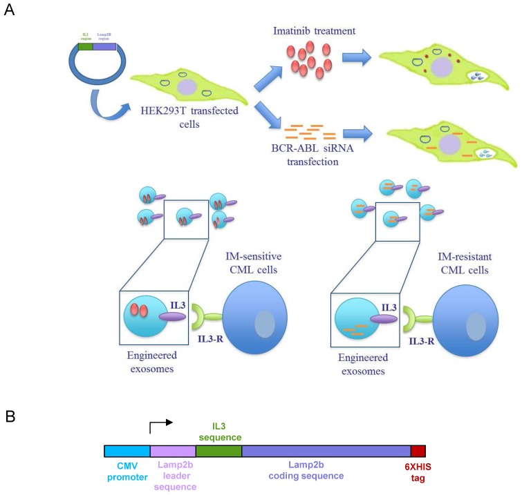
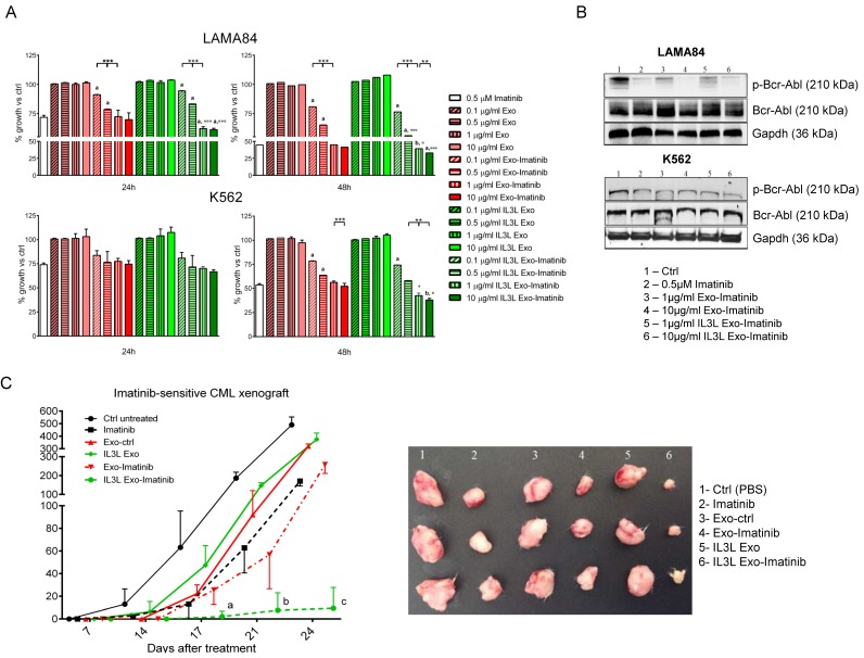
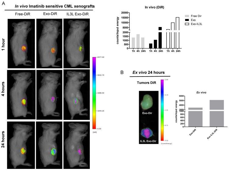
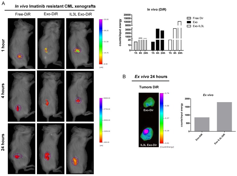
“They demonstrate that engineered exosomes containing IL3-Lamp2B, loaded with Imatinib, can selectively target tumor cells in vivo, resulting in a reduction in tumor size.”
From Emerging role of exosomes in cancer therapy: progress and challenges (2025) · cites this paper as [166]
In the citing paper
“The interleukin-3 receptor (IL3-R) is known to be excessively expressed in CML and acute myeloid leukemia (AML) blasts, while it shows minimal or no expression in normal hematopoietic stem cells, indicating that IL3-R could be a potential receptor site for an exosome-based drug delivery system to suppress BCR-ABL.”
From Emerging role of exosomes in cancer therapy: progress and challenges (2025) · cites this paper as [166]
In the citing paper
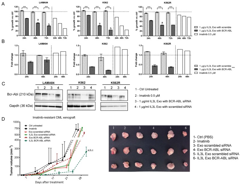
“Moreover, these modified exosomes effectively transport effective BCR-ABL siRNA to Imatinib-resistant CML cells.”
From Emerging role of exosomes in cancer therapy: progress and challenges (2025) · cites this paper as [166]
In the citing paper
“| Chronic Myelogenous Leukemia | Modified exosomes containing IL3-Lamp2B, loaded with Imatinib | Reduced tumor size | [166] |”
From Emerging role of exosomes in cancer therapy: progress and challenges (2025) · cites this paper as [166]
Self-check
Percentages, sums and reported differences re-computed from the paper’s own numbers.
The self-check needs the paper’s full text.
Citations
Cites
Johnsen, K. B., Gudbergsson, J. M., Skov, M. N., Pilgaard, L., Moos, T., & Duroux, M. (2014) · Biochimica et Biophysica Acta (BBA) - Reviews on Cancer · doi:10.1016/j.bbcan.2014.04.005 · Cited by 704
Research works printed but not linked: 41
- [1]Faderl S Kantarjian HM Talpaz M Chronic myelogenous leukemia: update on biology and treatment Oncology (Williston Park) 1999 13 169 80 discussion 81, 84 10079468
- [2]Smith DL Burthem J Whetton AD Molecular pathogenesis of chronic myeloid leukaemia Expert Rev Mol Med 2003 5 1 27 14987402
- [3]Druker BJ Lydon NB Lessons learned from the development of an abl tyrosine kinase inhibitor for chronic myelogenous leukemia J Clin Invest 2000 105 3 7 10619854 PMC382593
- [4]Jabbour E Kantarjian H Cortes J Use of second- and third-generation tyrosine kinase inhibitors in the treatment of chronic myeloid leukemia: an evolving treatment paradigm Clin Lymphoma Myeloma Leuk 2015 15 323 34 25971713 PMC5141582
- [5]Mughal TI Schrieber A Principal long-term adverse effects of imatinib in patients with chronic myeloid leukemia in chronic phase Biologics 2010 4 315 23 21209726 PMC3010822
- [6]Farmer S Horvath-Puho E Vestergaard H Hermann AP Frederiksen H Chronic myeloproliferative neoplasms and risk of osteoporotic fractures; a nationwide population-based cohort study Br J Haematol 2013 163 603 10 24111669
- [7]Mahon FX Deininger MW Schultheis B Chabrol J Reiffers J Goldman JM Selection and characterization of BCR-ABL positive cell lines with differential sensitivity to the tyrosine kinase inhibitor STI571: diverse mechanisms of resistance Blood 2000 96 1070 9 10910924
- [8]Pinilla-Ibarz J Sweet K Emole J Fradley M Long-term BCR-ABL1 Tyrosine Kinase Inhibitor Therapy in Chronic Myeloid Leukemia Anticancer Res 2015 35 6355 64 26637844
- [9]Koldehoff M Zakrzewski JL Beelen DW Elmaagacli AH Additive antileukemia effects by GFI1B- and BCR-ABL-specific siRNA in advanced phase chronic myeloid leukemic cells Cancer Gene Ther 2013 20 421 7 23788109
- [10]Zhao RC McIvor RS Griffin JD Verfaillie CM Gene therapy for chronic myelogenous leukemia (CML): a retroviral vector that renders hematopoietic progenitors methotrexate-resistant and CML progenitors functionally normal and nontumorigenic in vivo Blood 1997 90 4687 98 9389683
- [11]Burnett JC Rossi JJ RNA-based therapeutics: current progress and future prospects Chem Biol 2012 19 60 71 22284355 PMC3269031
- [12]Yingchoncharoen P Kalinowski DS Richardson DR Lipid-Based Drug Delivery Systems in Cancer Therapy: What Is Available and What Is Yet to Come Pharmacol Rev 2016 68 701 87 27363439 PMC4931871
- [13]Raimondo S Corrado C Raimondi L De Leo G Alessandro R Role of Extracellular Vesicles in Hematological Malignancies Biomed Res Int 2015 2015 821613 26583135 PMC4637071
- [14]Corrado C Raimondo S Chiesi A Ciccia F De Leo G Alessandro R Exosomes as intercellular signaling organelles involved in health and disease: basic science and clinical applications Int J Mol Sci 2013 14 5338 66 23466882 PMC3634447
- [15]Raimondo S Naselli F Fontana S Monteleone F Lo Dico A Saieva L Citrus limon-derived nanovesicles inhibit cancer cell proliferation and suppress CML xenograft growth by inducing TRAIL-mediated cell death Oncotarget 2015 6 19514 27 26098775 PMC4637302
- [16]Conigliaro A Costa V Lo Dico A Saieva L Buccheri S Dieli F CD90+ liver cancer cells modulate endothelial cell phenotype through the release of exosomes containing H19 lncRNA Mol Cancer 2015 14 155 26272696 PMC4536801
- [17]Simons M Raposo G Exosomes-vesicular carriers for intercellular communication Curr Opin Cell Biol 2009 21 575 81 19442504
- [18]El Andaloussi S Lakhal S Mager I Wood MJ Exosomes for targeted siRNA delivery across biological barriers Adv Drug Deliv Rev 2013 65 391 7 22921840
- [20]Hu G Drescher KM Chen XM Exosomal miRNAs: Biological Properties and Therapeutic Potential Front Genet 2012 3 56 22529849 PMC3330238
- [21]Ohno S Takanashi M Sudo K Ueda S Ishikawa A Matsuyama N Systemically injected exosomes targeted to EGFR deliver antitumor microRNA to breast cancer cells Mol Ther 2013 21 185 91 PMC3538304 23032975
- [22]Haney MJ Klyachko NL Zhao Y Gupta R Plotnikova EG He Z Exosomes as drug delivery vehicles for Parkinson's disease therapy J Control Release 2015 207 18 30 25836593 PMC4430381
- [23]Saari H Lazaro-Ibanez E Viitala T Vuorimaa-Laukkanen E Siljander P Yliperttula M Microvesicle- and exosome-mediated drug delivery enhances the cytotoxicity of Paclitaxel in autologous prostate cancer cells J Control Release 2015 26390807
- [24]Alvarez-Erviti L Seow Y Yin H Betts C Lakhal S Wood MJ Delivery of siRNA to the mouse brain by systemic injection of targeted exosomes Nat Biotechnol 2011 29 341 5 21423189
- [25]Nievergall E Ramshaw HS Yong AS Biondo M Busfield SJ Vairo G Monoclonal antibody targeting of IL-3 receptor alpha with CSL362 effectively depletes CML progenitor and stem cells Blood 2014 123 1218 28 24363400
- [26]Testa U Riccioni R Militi S Coccia E Stellacci E Samoggia P Elevated expression of IL-3Ralpha in acute myelogenous leukemia is associated with enhanced blast proliferation, increased cellularity, and poor prognosis Blood 2002 100 2980 8 12351411
- [27]Testa U Pelosi E Frankel A CD 123 is a membrane biomarker and a therapeutic target in hematologic malignancies Biomark Res 2014 2 4 24513123 PMC3928610
- [28]Li J Chen X Yi J Liu Y Li D Wang J Identification and Characterization of 293T Cell-Derived Exosomes by Profiling the Protein, mRNA and MicroRNA Components PLoS One 2016 11 e0163043 27649079 PMC5029934
- [29]Bumcrot D Manoharan M Koteliansky V Sah DW RNAi therapeutics: a potential new class of pharmaceutical drugs Nat Chem Biol 2006 2 711 9 17108989 PMC7097247
- [30]Mendonca LS Moreira JN de Lima MC Simoes S Co-encapsulation of anti-BCR-ABL siRNA and imatinib mesylate in transferrin receptor-targeted sterically stabilized liposomes for chronic myeloid leukemia treatment Biotechnol Bioeng 2010 107 884 93 20632368
- [31]Landry B Valencia-Serna J Gul-Uludag H Jiang X Janowska-Wieczorek A Brandwein J Progress in RNAi-mediated Molecular Therapy of Acute and Chronic Myeloid Leukemia Mol Ther Nucleic Acids 2015 4 e240 25965550
- [32]Koldehoff M Steckel NK Beelen DW Elmaagacli AH Therapeutic application of small interfering RNA directed against bcr-abl transcripts to a patient with imatinib-resistant chronic myeloid leukaemia Clin Exp Med 2007 7 47 55 17609876
- [33]Greco KA Franzen CA Foreman KE Flanigan RC Kuo PC Gupta GN PLK-1 Silencing in Bladder Cancer by siRNA Delivered With Exosomes Urology 2016 91 241 e1-7 26876462
- [34]Cooper JM Wiklander PB Nordin JZ Al-Shawi R Wood MJ Vithlani M Systemic exosomal siRNA delivery reduced alpha-synuclein aggregates in brains of transgenic mice Mov Disord 2014 29 1476 85 25112864 PMC4204174
- [35]Tian Y Li S Song J Ji T Zhu M Anderson GJ A doxorubicin delivery platform using engineered natural membrane vesicle exosomes for targeted tumor therapy Biomaterials 2014 35 2383 90 24345736
- [36]Kim MS Haney MJ Zhao Y Mahajan V Deygen I Klyachko NL Development of exosome-encapsulated paclitaxel to overcome MDR in cancer cells Nanomedicine 2016 12 655 64 26586551 PMC4809755
- [37]Sun D Zhuang X Xiang X Liu Y Zhang S Liu C A novel nanoparticle drug delivery system: the anti-inflammatory activity of curcumin is enhanced when encapsulated in exosomes Mol Ther 2010 18 1606 14 20571541 PMC2956928
- [38]Frolova O Benito J Brooks C Wang RY Korchin B Rowinsky EK SL-401 and SL-501, targeted therapeutics directed at the interleukin-3 receptor, inhibit the growth of leukaemic cells and stem cells in advanced phase chronic myeloid leukaemia Br J Haematol 2014 166 862 74 24942980 PMC4146738
- [39]Simhadri VR Reiners KS Hansen HP Topolar D Simhadri VL Nohroudi K Dendritic cells release HLA-B-associated transcript-3 positive exosomes to regulate natural killer function PLoS One 2008 3 e3377 18852879 PMC2566590
- [40]El-Andaloussi S Lee Y Lakhal-Littleton S Li J Seow Y Gardiner C Exosome-mediated delivery of siRNA in vitro and in vivo Nat Protoc 2012 7 2112 26 23154783
- [41]Raimondo S Saieva L Corrado C Fontana S Flugy A Rizzo A Chronic myeloid leukemia-derived exosomes promote tumor growth through an autocrine mechanism Cell Commun Signal 2015 13 8 25644060 PMC4320527
- [42]Bende G Kollipara S Sekar V Saha R UV-spectrophotometric determination of imatinib mesylate and its application in solubility studies Pharmazie 2008 63 641 5 18819515
Cited by
Sorted by citations
Engineering exosomes for targeted drug delivery
Liang, Y., Duan, L., Lu, J., & Xia, J. (2021) · Theranostics · doi:10.7150/thno.52570 · Cited by 1,547
Exosome engineering: Current progress in cargo loading and targeted delivery
Fu, S., Wang, Y., Xia, X., & Zheng, J. C. (2020) · NanoImpact · doi:10.1016/j.impact.2020.100261 · Cited by 444
Engineered Extracellular Vesicles for Cancer Therapy
Zhang, X., Zhang, H., Gu, J., Zhang, J., Shi, H., Qian, H., Wang, D., Xu, W., Pan, J., & Santos, H. A. (2021) · Advanced Materials · doi:10.1002/adma.202005709 · Cited by 428
Extracellular vesicles: A bright star of nanomedicine
Wu, P., Zhang, B., Ocansey, D. K. W., Xu, W., & Qian, H. (2021) · Biomaterials · doi:10.1016/j.biomaterials.2020.120467 · Cited by 368
Recent advances in exosome-mediated nucleic acid delivery for cancer therapy
Zhang, Y., Liu, Q., Zhang, X., Huang, H., Tang, S., Chai, Y., Xu, Z., Li, M., Chen, X., Liu, J., et al. (2022) · Journal of Nanobiotechnology · doi:10.1186/s12951-022-01472-z · Cited by 306
Metadata
- Authors
- Bellavia, D., Raimondo, S., Calabrese, G., Forte, S., Cristaldi, M., Patinella, A., Memeo, L., Manno, M., Raccosta, S., Diana, P., Cirrincione, G., Giavaresi, G., Monteleone, F., Fontana, S., De Leo, G., & Alessandro, R.
- Venue
- Theranostics
- Publisher
- Ivyspring International Publisher
- Published
- 2017
- Volume
- 7
- Issue
- 5
- Pages
- 1333-1345
- ISSN
- 1838-7640
- PMC
- PMC5399597
- Cited by
- 368
- References
- 1
- Language
- en
About these checks
Registry, retraction, Cites and statistics results are computed, not judged, with one exception: a printed reference that no index lists is read once by a model before it counts as a citation error. Claims are extracted by a model and kept only when their quote appears verbatim in the text; each cited source is then checked, with a model’s help, for whether it says what the claim relies on. Those verdicts can be wrong — read the claim’s quote and the cited source before relying on one.
Statistics and arithmetic are recomputed, not judged.
Confidence bands are shown only for fields calibrated on our annotated test set. None is calibrated yet, so no band is shown.
What counts as a claim ›How we measure confidence ›How we check this ›
Are you an author, or do you see a problem with this page? Request a review
