

The DOI matches its Crossref record; 8 of the 8 references it cites were found in a registry or an index.
ANGPTL4 mediates shuttling of lipid fuel to brown adipose tissue during sustained cold exposure
Abstract
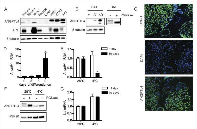
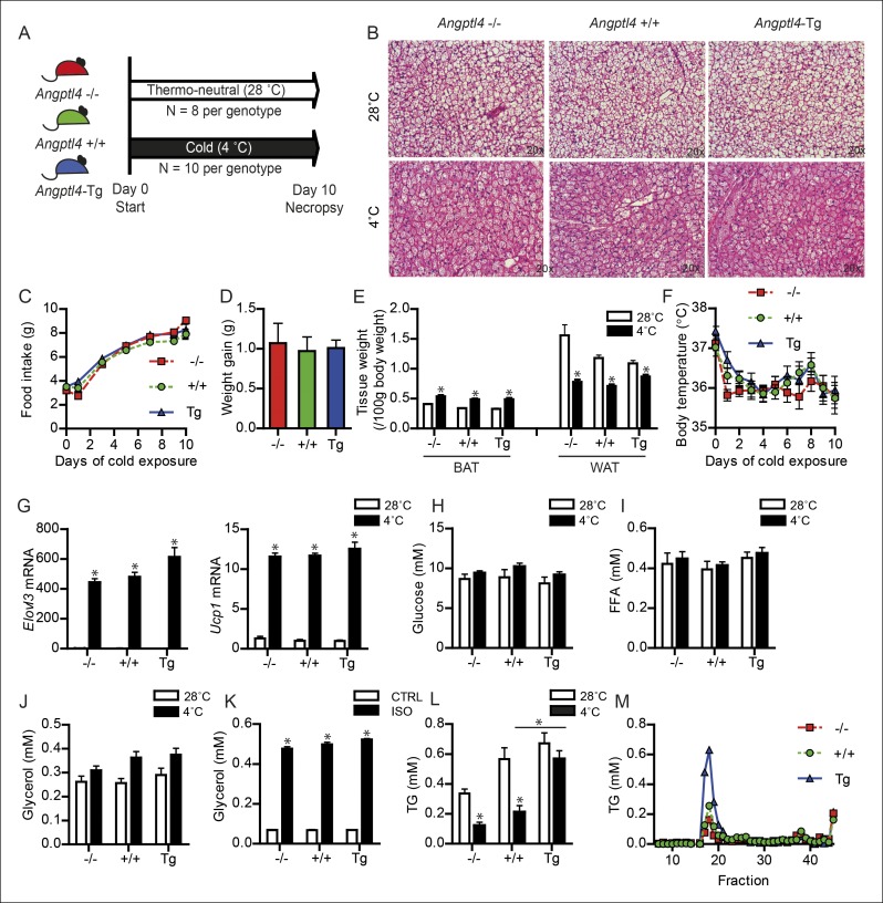
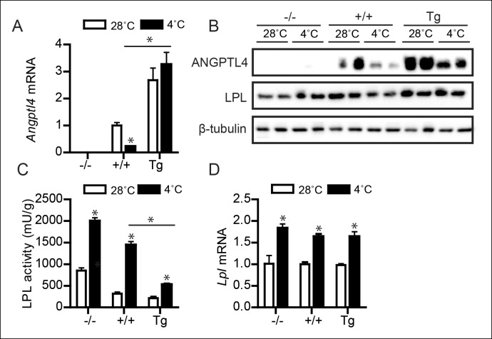
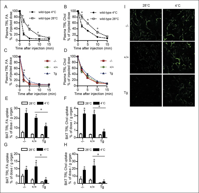
Brown adipose tissue (BAT) activation via cold exposure is increasingly scrutinized as a potential approach to ameliorate cardio-metabolic risk. Transition to cold temperatures requires changes in the partitioning of energy substrates, re-routing fatty acids to BAT to fuel non-shivering thermogenesis. However, the mechanisms behind the redistribution of energy substrates to BAT remain largely unknown. Angiopoietin-like 4 (ANGPTL4), a protein that inhibits lipoprotein lipase (LPL) activity, is highly expressed in BAT. Here, we demonstrate that ANGPTL4 is part of a shuttling mechanism that directs fatty acids derived from circulating triglyceride-rich lipoproteins to BAT during cold. Specifically, we show that cold markedly down-regulates ANGPTL4 in BAT, likely via activation of AMPK, enhancing LPL activity and uptake of plasma triglyceride-derived fatty acids. In contrast, cold up-regulates ANGPTL4 in WAT, abolishing a cold-induced increase in LPL activity. Together, our data indicate that ANGPTL4 is an important regulator of plasma lipid partitioning during sustained cold.
Overview
The claims and statistics of this paper haven’t been checked yet.
Run the check — free, no account needed. It reads the open-access text and checks the sources it cites.
Integrity signals
Registry
Cites
Statistics
Figures
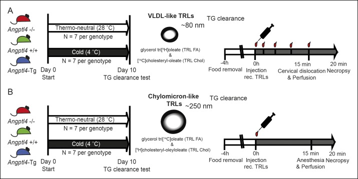
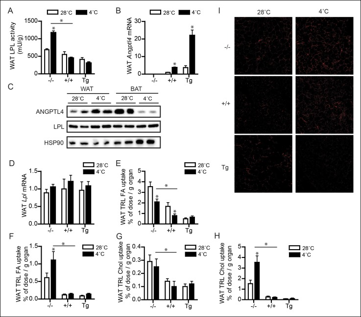
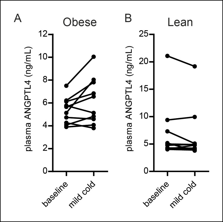
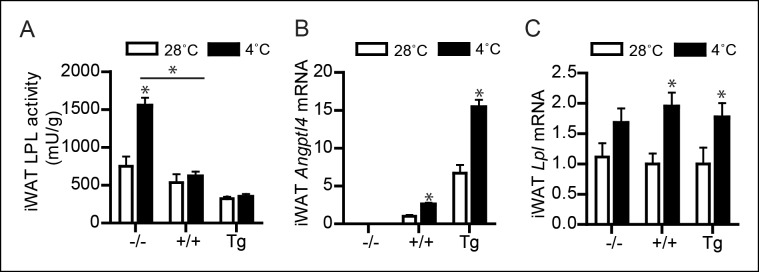
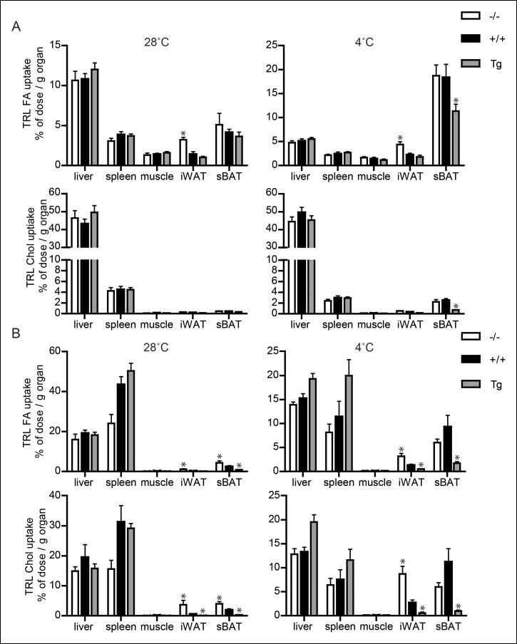
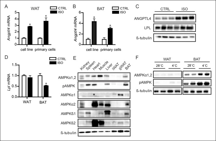
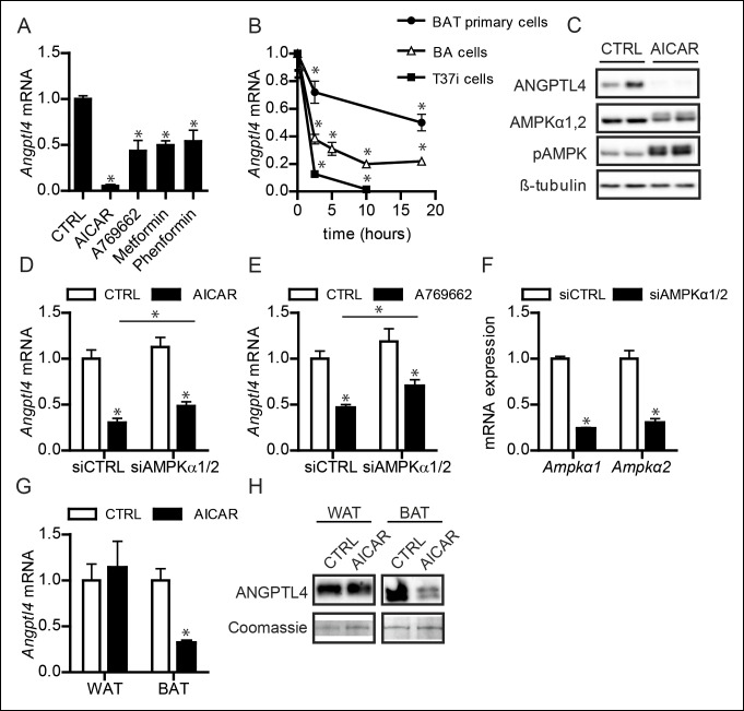
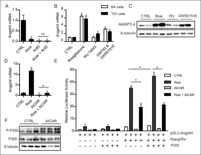
Verification
Claims the paper makes
The claims and statistics of this paper haven’t been checked yet.
Run the check — free, no account needed. It reads the open-access text and checks the sources it cites.
Claims other papers make about this one
Each is checked against this paper’s own text: does it say what the citing paper says it does?
The cited source doesn’t support the claim as written.
In the citing paper
“It is important to note that ANGPTL proteins are likely not the primary driver of the alterations in lipid partitioning that occur during thermogenesis.”
From Regulation of lipoprotein metabolism by ANGPTL3, ANGPTL4, and ANGPTL8 (2021) · cites this paper as [71]
Checked by a reasoning model — may be wrong.
Experimental — accuracy not yet measuredThe source backs part of the claim, not all of it — or backs it for a different population, condition or comparison.
In the citing paper
“Consistent with a role for ANGPTL4 in thermogenic triglyceride partitioning, either whole body or BAT-specific deletion of ANGPTL4 resulted in increased LPL activity and triglyceride uptake in BAT (33, 71).”
From Regulation of lipoprotein metabolism by ANGPTL3, ANGPTL4, and ANGPTL8 (2021) · cites this paper as [71]
Experimental — accuracy not yet measuredThe source backs part of the claim, not all of it — or backs it for a different population, condition or comparison.
In the citing paper
“Angptl4 gene expression decreases in brown adipose tissue in the cold (33, 70, 71).”
From Regulation of lipoprotein metabolism by ANGPTL3, ANGPTL4, and ANGPTL8 (2021) · cites this paper as [71]
Experimental — accuracy not yet measuredThe cited source says what the claim attributes to it.
In the citing paper
“It is important to note, however, that modulation of ANGPTL4 expression is not essential for cold-induced thermogenesis or even cold-induced changes in triglyceride partitioning.”
From Regulation of lipoprotein metabolism by ANGPTL3, ANGPTL4, and ANGPTL8 (2021) · cites this paper as [71]
The cited source says what the claim attributes to it.
In the citing paper
“Dijk et al. (71) found that greater uptake of TGs into BAT was induced by cold both in ANGPTL4-deficient mice and in mice overexpressing ANGPTL4.”
From Regulation of lipoprotein metabolism by ANGPTL3, ANGPTL4, and ANGPTL8 (2021) · cites this paper as [71]
In the citing paper
“With cold exposure, large amounts of triglyceride are funneled to brown adipose tissue in an LPL-dependent manner (68, 69, 71).”
From Regulation of lipoprotein metabolism by ANGPTL3, ANGPTL4, and ANGPTL8 (2021) · cites this paper as [71]
In the citing paper
“With prolonged cold exposure, expression of ANGPTL4 decreases in BAT and increases in WAT (33, 70, 71).”
From Regulation of lipoprotein metabolism by ANGPTL3, ANGPTL4, and ANGPTL8 (2021) · cites this paper as [71]
In the citing paper
“The resulting decrease in ANGPTL4 protein leads to increased LPL activity in BAT, increasing uptake of triglycerides for thermogenic oxidation (71).”
From Regulation of lipoprotein metabolism by ANGPTL3, ANGPTL4, and ANGPTL8 (2021) · cites this paper as [71]
In the citing paper
“Dijk et al. (71) also observed that neither whole body knockout of ANGPTL4 nor ANGPTL4 overexpression had a significant effect on body temperature after prolonged cold exposure.”
From Regulation of lipoprotein metabolism by ANGPTL3, ANGPTL4, and ANGPTL8 (2021) · cites this paper as [71]
In the citing paper
“Cold exposure did not increase LPL activity in white adipose tissue, nor was ANGPTL4 expression decreased (71), indicating that these changes were specific to brown adipose tissue.”
From Regulation of lipoprotein metabolism by ANGPTL3, ANGPTL4, and ANGPTL8 (2021) · cites this paper as [71]
Self-check
Percentages, sums and reported differences re-computed from the paper’s own numbers.
The self-check needs the paper’s full text.
Citations
Cites
Brown Adipose Tissue: Function and Physiological Significance
CANNON, B., & NEDERGAARD, J. (2004) · Physiological Reviews · doi:10.1152/physrev.00015.2003 · Cited by 6,433
Cold-Activated Brown Adipose Tissue in Healthy Men
van Marken Lichtenbelt, W. D., Vanhommerig, J. W., Smulders, N. M., Drossaerts, J. M. A. F. L., Kemerink, G. J., Bouvy, N. D., Schrauwen, P., & Teule, G. J. J. (2009) · New England Journal of Medicine · doi:10.1056/nejmoa0808718 · Cited by 3,452
Functional Brown Adipose Tissue in Healthy Adults
Virtanen, K. A., Lidell, M. E., Orava, J., Heglind, M., Westergren, R., Niemi, T., Taittonen, M., Laine, J., Savisto, N.-J., Enerbäck, S., et al. (2009) · New England Journal of Medicine · doi:10.1056/nejmoa0808949 · Cited by 3,058
Dyslipidemia in obesity: Mechanisms and potential targets
Klop, B., Elte, J., & Cabezas, M. (2013) · Nutrients · doi:10.3390/nu5041218 · Cited by 1,665
Brown adipose tissue activity controls triglyceride clearance
Bartelt, A., Bruns, O. T., Reimer, R., Hohenberg, H., Ittrich, H., Peldschus, K., Kaul, M. G., Tromsdorf, U. I., Weller, H., Waurisch, C., et al. (2011) · Nature Medicine · doi:10.1038/nm.2297 · Cited by 1,608
Self-citationKöster, A., Chao, Y. B., Mosior, M., Ford, A., Gonzalez-DeWhitt, P. A., Hale, J. E., Li, D., Qiu, Y., Fraser, C. C., Yang, D. D., et al. (2005) · Endocrinology · doi:10.1210/en.2005-0476 · Cited by 427
AMPK inhibition in health and disease
Viollet, B., Horman, S., Leclerc, J., Lantier, L., Foretz, M., Billaud, M., Giri, S., & Andreelli, F. (2010) · Critical Reviews in Biochemistry and Molecular Biology · doi:10.3109/10409238.2010.488215 · Cited by 383
AMPK: Lessons from transgenic and knockout animals
Viollet, B. (2009) · Frontiers in bioscience · doi:10.2741/3229 · Cited by 314
Research works printed but not linked: 53
- Alex, 2013, Short-chain fatty acids stimulate angiopoietin-like 4 synthesis in human colon adenocarcinoma cells by activating peroxisome proliferator-activated receptor, Molecular and Cellular Biology, 33, 1303, 10.1128/MCB.00858-12 · DOI
- Alex, 2014, ANGPTL4 is produced by entero-endocrine cells in the human intestinal tract, Histochemistry and Cell Biology, 141, 383, 10.1007/s00418-013-1157-y · DOI · Paper page
- An, 2005, beta-Agonist stimulation produces changes in cardiac AMPK and coronary lumen LPL only during increased workload, AJP: Endocrinology and Metabolism, 288, 10.1152/ajpendo.00588.2004 · DOI
- Bartelt, 2013, Effects of adipocyte lipoprotein lipase on de novo lipogenesis and white adipose tissue browning, Biochimica Et Biophysica Acta (BBA) - Molecular and Cell Biology of Lipids, 1831, 934, 10.1016/j.bbalip.2012.11.011 · DOI
- Bauwens, 2011, Cold tolerance, cold-induced hyperphagia, and nonshivering thermogenesis are normal in α1-AMPK-/- mice, AJP: Regulatory, Integrative and Comparative Physiology, 301, R473, 10.1152/ajpregu.00444.2010 · DOI
- Berbée, 2015, Brown fat activation reduces hypercholesterolaemia and protects from atherosclerosis development, Nature Communications, 6:, 10.1038/ncomms7356 · DOI
- Bergeron, 2001, Effect of 5-aminoimidazole-4-carboxamide-1-beta-D-ribofuranoside infusion on in vivo glucose and lipid metabolism in lean and obese zucker rats, Diabetes, 50, 1076, 10.2337/diabetes.50.5.1076 · DOI
- Bertin, 1985, Effects of cold acclimation on the activity of lipoprotein lipase in adipose tissues of genetically obese zucker rats, Comparative Biochemistry and Physiology Part B: Comparative Biochemistry, 81, 797, 10.1016/0305-0491(85)90408-0 · DOI
- Bruns, 2009, Real-time magnetic resonance imaging and quantification of lipoprotein metabolism in vivo using nanocrystals, Nature Nanotechnology, 4, 193, 10.1038/nnano.2008.405 · DOI
- Buhl, 2002, Long-term AICAR administration reduces metabolic disturbances and lowers blood pressure in rats displaying features of the insulin resistance syndrome, Diabetes, 51, 2199, 10.2337/diabetes.51.7.2199 · DOI
- Carneheim, 1988, Cold-induced beta-adrenergic recruitment of lipoprotein lipase in brown fat is due to increased transcription, The American Journal of Physiology, 254
- Catoire, 2014, Fatty acid-inducible ANGPTL4 governs lipid metabolic response to exercise, Proceedings of the National Academy of Sciences of the United States of America, 111, 10.1073/pnas.1400889111 · DOI · Paper page
- Cheang, 2014, Metformin protects endothelial function in diet-induced obese mice by inhibition of endoplasmic reticulum stress through 5' adenosine monophosphate-activated protein kinase-peroxisome proliferator-activated receptor pathway, Arteriosclerosis, Thrombosis, and Vascular Biology, 34, 830, 10.1161/ATVBAHA.113.301938 · DOI
- Dijk, 2014, Regulation of lipoprotein lipase by Angptl4, Trends in Endocrinology & Metabolism, 25, 146, 10.1016/j.tem.2013.12.005 · DOI
- Garcia-Arcos, 2013, Adipose-specific lipoprotein lipase deficiency more profoundly affects brown than white fat biology, Journal of Biological Chemistry, 288, 14046, 10.1074/jbc.M113.469270 · DOI · Paper page
- Ge, 2004, Oligomerization state-dependent hyperlipidemic effect of angiopoietin-like protein 4, The Journal of Lipid Research, 45, 2071, 10.1194/jlr.M400138-JLR200 · DOI · Paper page
- Geerling, 2014, Metformin lowers plasma triglycerides by promoting VLDL-triglyceride clearance by brown adipose tissue in mice, Diabetes, 63, 880, 10.2337/db13-0194 · DOI
- Gelman, 1999, P300 interacts with the n- and c-terminal part of PPAR 2 in a ligand-independent and -dependent manner, respectively, Journal of Biological Chemistry, 274, 7681, 10.1074/jbc.274.12.7681 · DOI
- Georgiadi, 2010, Induction of cardiac Angptl4 by dietary fatty acids is mediated by peroxisome proliferator-activated receptor beta /delta and protects against fatty acid-induced oxidative stress, Circulation Research, 106, 1712, 10.1161/CIRCRESAHA.110.217380 · DOI · Paper page
- Giralt, 1990, Lipoprotein lipase mRNA expression in brown adipose tissue: translational and/or posttranslational events are involved in the modulation of enzyme activity, Biochimica Et Biophysica Acta (BBA) - Gene Structure and Expression, 1048, 270, 10.1016/0167-4781(90)90066-B · DOI
- Jong, 2001, Apolipoprotein c-III deficiency accelerates triglyceride hydrolysis by lipoprotein lipase in wild-type and apoE knockout mice, Journal of Lipid Research, 42, 1578, 10.1016/S0022-2275(20)32211-2 · DOI
- Kaddatz, 2010, Transcriptional profiling identifies functional interactions of TGFβ and PPARβ/δ signaling, Journal of Biological Chemistry, 285, 29469, 10.1074/jbc.M110.142018 · DOI
- Kersten, 2000, Characterization of the fasting-induced adipose factor FIAF, a novel peroxisome proliferator-activated receptor target gene, Journal of Biological Chemistry, 275, 28488, 10.1074/jbc.M004029200 · DOI · Paper page
- Kersten, 2009, Caloric restriction and exercise increase plasma ANGPTL4 levels in humans via elevated free fatty acids, Arteriosclerosis, Thrombosis, and Vascular Biology, 29, 969, 10.1161/ATVBAHA.108.182147 · DOI · Paper page
- Kersten, 2014, Physiological regulation of lipoprotein lipase, Biochimica Et Biophysica Acta (BBA) - Molecular and Cell Biology of Lipids, 1841, 919, 10.1016/j.bbalip.2014.03.013 · DOI
- Khedoe, 2014, Brown adipose tissue takes up plasma triglycerides mostly after lipolysis, Journal of Lipid Research, 569, 51
- Kim, 2000, Hepatic expression, synthesis and secretion of a novel fibrinogen/angiopoietin-related protein that prevents endothelial-cell apoptosis, Biochemical Journal, 346, 603, 10.1042/bj3460603 · DOI
- Klingenspor, 1989, Short photoperiod and cold activate brown fat lipoprotein lipase in the djungarian hamster, The American Journal of Physiology, 257, R1123
- Klingenspor, 1996, Multiple regulatory steps are involved in the control of lipoprotein lipase activity in brown adipose tissue, Journal of Lipid Research, 37, 1685, 10.1016/S0022-2275(20)39111-2 · DOI
- Kroupa, 2012, Linking nutritional regulation of Angptl4, Gpihbp1, and Lmf1 to lipoprotein lipase activity in rodent adipose tissue, BMC Physiology, 12, 13, 10.1159/000327774 · DOI
- Leff, 2003, AMP-activated protein kinase regulates gene expression by direct phosphorylation of nuclear proteins, Biochemical Society Transactions, 31, 224, 10.1042/bst0310224 · DOI
- Lichtenstein, 2010, Angptl4 protects against severe proinflammatory effects of saturated fat by inhibiting fatty acid uptake into mesenteric lymph node macrophages, Cell Metabolism, 12, 580, 10.1016/j.cmet.2010.11.002 · DOI · Paper page
- Lim, 2012, AMP-activated protein kinase inhibits TGF-β-induced fibrogenic responses of hepatic stellate cells by targeting transcriptional coactivator p300, Journal of Cellular Physiology, 227, 1081, 10.1002/jcp.22824 · DOI
- Mandard, 2004, The direct peroxisome proliferator-activated receptor target fasting-induced adipose factor (FIAF/PGAR/ANGPTL4) is present in blood plasma as a truncated protein that is increased by fenofibrate treatment, Journal of Biological Chemistry, 279, 34411, 10.1074/jbc.M403058200 · DOI · Paper page
- Mandard, 2006, The fasting-induced adipose Factor/Angiopoietin-like protein 4 is physically associated with lipoproteins and governs plasma lipid levels and adiposity, Journal of Biological Chemistry, 281, 934, 10.1074/jbc.M506519200 · DOI · Paper page
- Mitchell, 1992, Regulation of expression of the lipoprotein lipase gene in brown adipose tissue, The American Journal of Physiology, 263
- Mulligan, 2007, Upregulation of AMPK during cold exposure occurs via distinct mechanisms in brown and white adipose tissue of the mouse, The Journal of Physiology, 580, 677, 10.1113/jphysiol.2007.128652 · DOI
- Namgaladze, 2013, AICAR inhibits PPAR during monocyte differentiation to attenuate inflammatory responses to atherogenic lipids, Cardiovascular Research, 98, 479, 10.1093/cvr/cvt073 · DOI
- Ohira, 2009, Metformin promotes induction of lipoprotein lipase in skeletal muscle through activation of adenosine monophosphate-activated protein kinase, Metabolism, 58, 1408, 10.1016/j.metabol.2009.04.024 · DOI
- Rensen, 1995, Selective liver targeting of antivirals by recombinant chylomicrons—a new therapeutic approach to hepatitis b, Nature Medicine, 1, 221, 10.1038/nm0395-221 · DOI
- Ruge, 2004, Nutritional regulation of lipoprotein lipase in mice, The International Journal of Biochemistry & Cell Biology, 36, 320, 10.1016/S1357-2725(03)00256-5 · DOI
- Sozio, 2011, Activated AMPK inhibits PPAR- and PPAR- transcriptional activity in hepatoma cells, AJP: Gastrointestinal and Liver Physiology, 301, G739, 10.1152/ajpgi.00432.2010 · DOI
- Uldry, 2006, Complementary action of the PGC-1 coactivators in mitochondrial biogenesis and brown fat differentiation, Cell Metabolism, 3, 333, 10.1016/j.cmet.2006.04.002 · DOI
- Vijgen, 2013, Increased oxygen consumption in human adipose tissue from the 'brown adipose tissue' region, The Journal of Clinical Endocrinology & Metabolism, 98, 10.1210/jc.2013-1348 · DOI
- Vo, 2001, CREB-binding protein and p300 in transcriptional regulation, Journal of Biological Chemistry, 276, 13505, 10.1074/jbc.R000025200 · DOI
- Wang, 2011, Brown adipose tissue in humans is activated by elevated plasma catecholamines levels and is inversely related to central obesity, PLoS ONE, 6, 10.1371/journal.pone.0021006.t002 · DOI
- Wijers, 2010, Cold-induced adaptive thermogenesis in lean and obese, Obesity, 18, 1092, 10.1038/oby.2010.74 · DOI
- Wu, 2012, Beige adipocytes are a distinct type of thermogenic fat cell in mouse and human, Cell, 150, 366, 10.1016/j.cell.2012.05.016 · DOI
- Yang, 2001, Regulation of transcription by AMP-activated protein kinase: PHOSPHORYLATION of p300 BLOCKS ITS INTERACTION with NUCLEAR RECEPTORS, Journal of Biological Chemistry, 276, 38341, 10.1074/jbc.C100316200 · DOI
- Yang, 2008, Suppression of the Raf/MEK/ERK signaling cascade and inhibition of angiogenesis by the carboxyl terminus of angiopoietin-like protein 4, Arteriosclerosis, Thrombosis, and Vascular Biology, 28, 835, 10.1161/ATVBAHA.107.157776 · DOI
- Yoon, 2000, Peroxisome proliferator-activated receptor gamma target gene encoding a novel angiopoietin-related protein associated with adipose differentiation, Molecular and Cellular Biology, 20, 5343, 10.1128/MCB.20.14.5343-5349.2000 · DOI · Paper page
- Young, 2013, Biochemistry and pathophysiology of intravascular and intracellular lipolysis, Genes & Development, 27, 459, 10.1101/gad.209296.112 · DOI
- Zennaro, 1998, Hibernoma development in transgenic mice identifies brown adipose tissue as a novel target of aldosterone action, Journal of Clinical Investigation, 101, 1254, 10.1172/JCI1915 · DOI
Cited by
Sorted by citations
Energetic Trade-Offs and Hypometabolic States Promote Disease Tolerance
Ganeshan, K., Nikkanen, J., Man, K., Leong, Y. A., Sogawa, Y., Maschek, J. A., Van Ry, T., Chagwedera, D. N., Cox, J. E., & Chawla, A. (2019) · Cell · doi:10.1016/j.cell.2019.01.050 · Cited by 286
Hepatokines and metabolism: Deciphering communication from the liver
Jensen-Cody, S. O., & Potthoff, M. J. (2021) · Molecular Metabolism · doi:10.1016/j.molmet.2020.101138 · Cited by 252
Angiopoietin-like 3 in lipoprotein metabolism
Kersten, S. (2017) · Nature Reviews Endocrinology · doi:10.1038/nrendo.2017.119 · Cited by 220
Self-citationRegulation of lipoprotein metabolism by ANGPTL3, ANGPTL4, and ANGPTL8
Sylvers-Davie, K. L., & Davies, B. S. J. (2021) · American Journal of Physiology-Endocrinology and Metabolism · doi:10.1152/ajpendo.00195.2021 · Cited by 154
‘Browning’ the cardiac and peri-vascular adipose tissues to modulate cardiovascular risk
Aldiss, P., Davies, G., Woods, R., Budge, H., Sacks, H. S., & Symonds, M. E. (2017) · International Journal of Cardiology · doi:10.1016/j.ijcard.2016.11.074 · Cited by 148
Janssen, A. W. F., Katiraei, S., Bartosinska, B., Eberhard, D., Willems van Dijk, K., & Kersten, S. (2018) · Diabetologia · doi:10.1007/s00125-018-4583-5 · Cited by 96
Self-citationDijk, W., Ruppert, P. M. M., Oost, L. J., & Kersten, S. (2018) · Journal of Biological Chemistry · doi:10.1074/jbc.ra118.002426 · Cited by 72
Self-citationIngerslev, B., Hansen, J. S., Hoffmann, C., Clemmesen, J. O., Secher, N. H., Scheler, M., Hrabĕ de Angelis, M., Häring, H. U., Pedersen, B. K., Weigert, C., et al. (2017) · Molecular Metabolism · doi:10.1016/j.molmet.2017.06.018 · Cited by 61
“The results are in accordance with data obtained in white and brown adipocytes, showing that b-adrenergic stimulation increases cAMP and ANGPTL4 mRNA and protein abundance [34].”
BackgroundResult
Metadata
- Authors
- Dijk, W., Heine, M., Vergnes, L., Boon, M. R., Schaart, G., Hesselink, M. K., Reue, K., van Marken Lichtenbelt, W. D., Olivecrona, G., Rensen, P. C., Heeren, J., & Kersten, S.
- Affiliations
- Nutrition, Metabolism and Genomics group, Division of Human Nutrition, Wageningen University, Wageningen, The Netherlands
- Department of Biochemistry and Molecular Cell Biology, University Medical Center Hamburg-Eppendorf, Hamburg, Germany
- Department of Human Genetics, David Geffen School of Medicine, University of California, Los Angeles, Los Angeles, United States
- Department of Medicine, Division of Endocrinology, Leiden University Medical Center, Leiden, The Netherlands
- Department of Human Movement Sciences, Maastricht University, Maastricht, The Netherlands
- Department of Human Biology, Maastricht University Medical Centre, Maastricht, The Netherlands
- Department of Medical Biosciences/Physiological Chemistry, Umeå University, Umeå, Sweden
- Venue
- eLife
- Publisher
- eLife Sciences Publications, Ltd
- Published
- 2015-10-17
- Volume
- 4
- ISSN
- 2050-084X
- PMC
- PMC4709329
- Cited by
- 114
- References
- 8
- Language
- en
- Licence
- Funders
- Fondation Leducq (12CVD04)
- Forskningsrådet om Hälsa, Arbetsliv och Välfärd (12203)
- Hartstichting (2009T038)
About these checks
Registry, retraction, Cites and statistics results are computed, not judged, with one exception: a printed reference that no index lists is read once by a model before it counts as a citation error. Claims are extracted by a model and kept only when their quote appears verbatim in the text; each cited source is then checked, with a model’s help, for whether it says what the claim relies on. Those verdicts can be wrong — read the claim’s quote and the cited source before relying on one.
Statistics and arithmetic are recomputed, not judged.
Confidence bands are shown only for fields calibrated on our annotated test set. None is calibrated yet, so no band is shown.
What counts as a claim ›How we measure confidence ›How we check this ›
Are you an author, or do you see a problem with this page? Request a review
