

The DOI matches its Crossref record; 79 of the 80 references it cites were found in a registry or an index.
Large-scale determination of previously unsolved protein structures using evolutionary information
Open accessAbstract
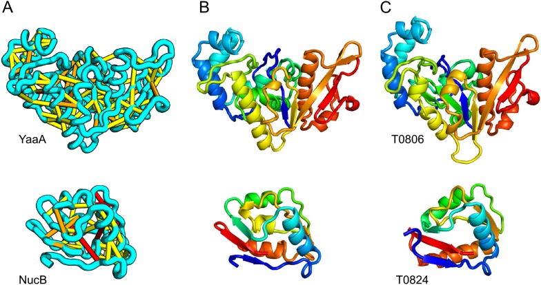
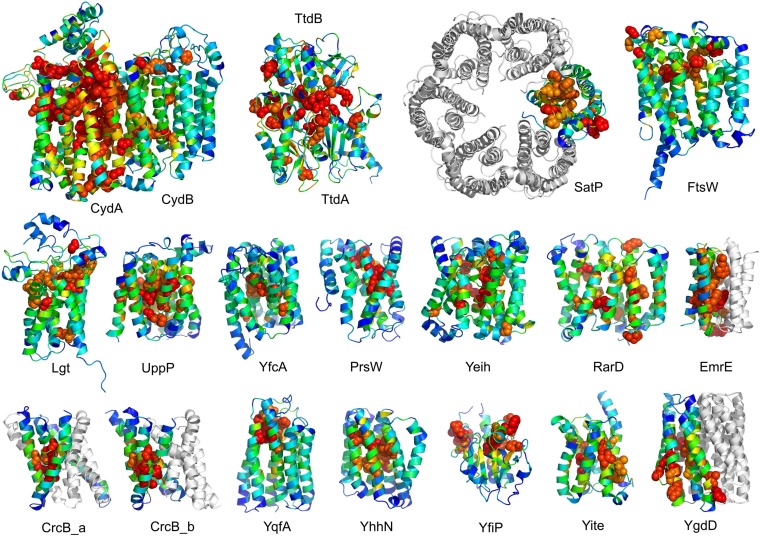
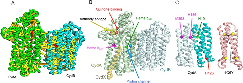
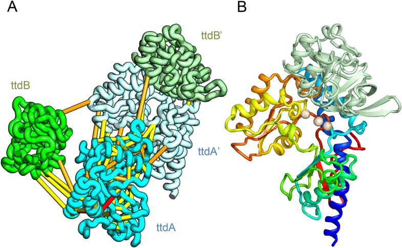
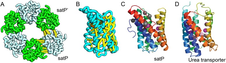
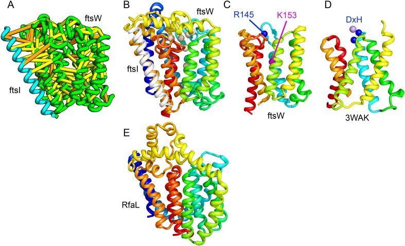
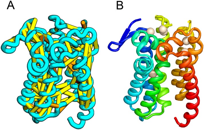
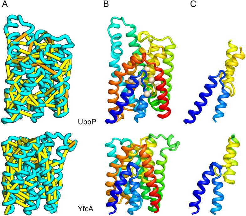
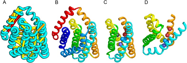
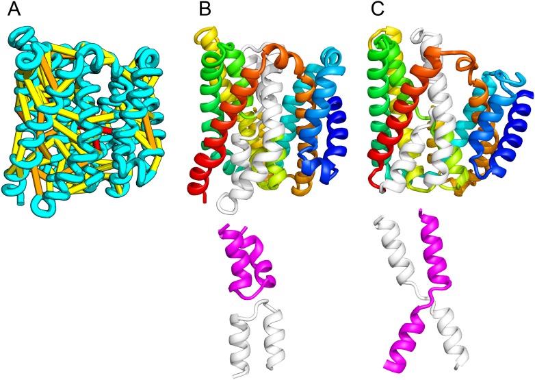
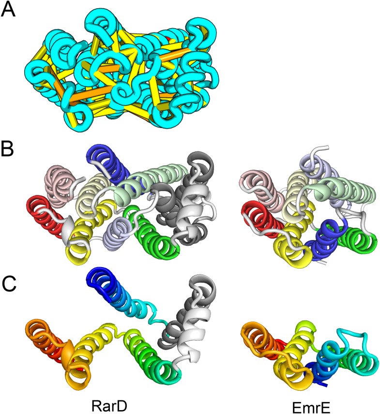
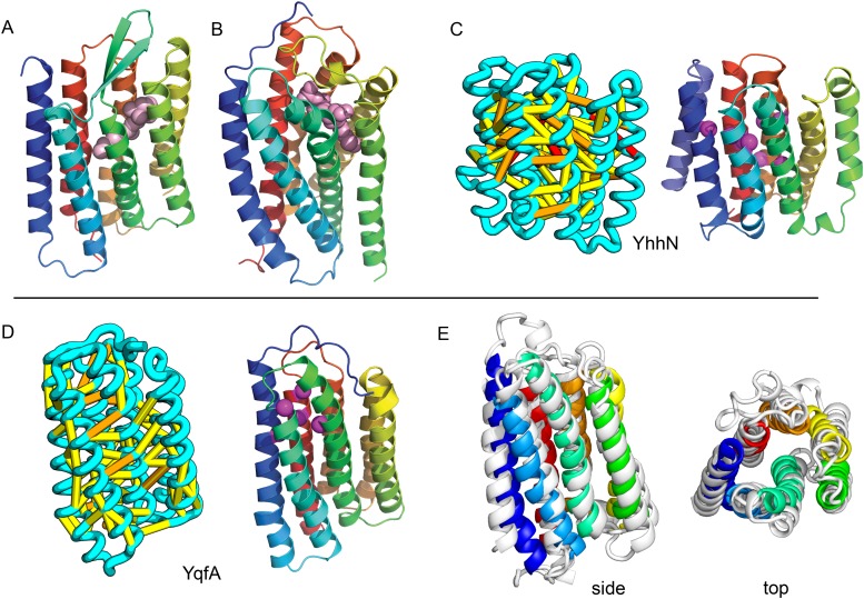
The prediction of the structures of proteins without detectable sequence similarity to any protein of known structure remains an outstanding scientific challenge. Here we report significant progress in this area. We first describe de novo blind structure predictions of unprecendented accuracy we made for two proteins in large families in the recent CASP11 blind test of protein structure prediction methods by incorporating residue-residue co-evolution information in the Rosetta structure prediction program. We then describe the use of this method to generate structure models for 58 of the 121 large protein families in prokaryotes for which three-dimensional structures are not available. These models, which are posted online for public access, provide structural information for the over 400,000 proteins belonging to the 58 families and suggest hypotheses about mechanism for the subset for which the function is known, and hypotheses about function for the remainder.
Overview
The claims and statistics of this paper haven’t been checked yet.
Run the check — free, no account needed. It reads the open-access text and checks the sources it cites.
Integrity signals
Registry
Cites
Statistics
Figures
Verification
Claims the paper makes
The claims and statistics of this paper haven’t been checked yet.
Run the check — free, no account needed. It reads the open-access text and checks the sources it cites.
Claims other papers make about this one
Each is checked against this paper’s own text: does it say what the citing paper says it does?
In the citing paper
“Traditional computational prediction pipelines reliant on inter-residue distance restraints calculated from deep multiple sequence alignments (MSAs) have historically struggled to accurately predict the structures of these proteins and their movements.”
From Sampling alternative conformational states of transporters and receptors with AlphaFold2 (2022) · cites this paper as Ovchinnikov et al., 2015
Self-check
Percentages, sums and reported differences re-computed from the paper’s own numbers.
The self-check needs the paper’s full text.
Citations
Cites
TM-align: a protein structure alignment algorithm based on the TM-score
Zhang, Y. (2005) · Nucleic Acids Research · doi:10.1093/nar/gki524 · Cited by 3,774
Structure and function of the global ocean microbiome
Sunagawa, S., Coelho, L. P., Chaffron, S., Kultima, J. R., Labadie, K., Salazar, G., Djahanschiri, B., Zeller, G., Mende, D. R., Alberti, A., et al. (2015) · Science · doi:10.1126/science.1261359 · Cited by 2,978
Protein homology detection by HMM–HMM comparison
Söding, J. (2004) · Bioinformatics · doi:10.1093/bioinformatics/bti125 · Cited by 2,459
Scoring function for automated assessment of protein structure template quality
Zhang, Y., & Skolnick, J. (2004) · Proteins: Structure, Function, and Bioinformatics · doi:10.1002/prot.20264 · Cited by 2,392
HHblits: lightning-fast iterative protein sequence searching by HMM-HMM alignment
Remmert, M., Biegert, A., Hauser, A., & Söding, J. (2012) · Nature Methods · doi:10.1038/nmeth.1818 · Cited by 2,288
Protein Structure Prediction Using Rosetta
Rohl, C. A., Strauss, C. E. M., Misura, K. M. S., & Baker, D. A. (2004) · Methods in enzymology on CD-ROM/Methods in enzymology · doi:10.1016/s0076-6879(04)83004-0 · Cited by 1,754
Activities at the Universal Protein Resource (UniProt)
The UniProt Consortium (2013) · Nucleic Acids Research · doi:10.1093/nar/gkt1140 · Cited by 1,736
PISCES: a protein sequence culling server
Wang, G., & Dunbrack, R. L. (2003) · Bioinformatics · doi:10.1093/bioinformatics/btg224 · Cited by 1,664
UniRef: comprehensive and non-redundant UniProt reference clusters
Suzek, B. E., Huang, H., McGarvey, P., Mazumder, R., & Wu, C. H. (2007) · Bioinformatics · doi:10.1093/bioinformatics/btm098 · Cited by 1,613
Expanded microbial genome coverage and improved protein family annotation in the COG database
Galperin, M. Y., Makarova, K. S., Wolf, Y. I., & Koonin, E. V. (2014) · Nucleic Acids Research · doi:10.1093/nar/gku1223 · Cited by 1,547
Research works printed but not linked: 1
- Hayat, 2014, Accurate prediction of transmembrane β-barrel proteins from sequences, 10.1101/006577 · DOI
Cited by
Sorted by citations
Highly accurate protein structure prediction for the human proteome
Tunyasuvunakool, K., Adler, J., Wu, Z., Green, T., Zielinski, M., Žídek, A., Bridgland, A., Cowie, A., Meyer, C., Laydon, A., et al. (2021) · Nature · doi:10.1038/s41586-021-03828-1 · Cited by 3,025
The coming of age of de novo protein design
Huang, P.-S., Boyken, S. E., & Baker, D. (2016) · Nature · doi:10.1038/nature19946 · Cited by 1,478
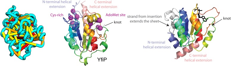
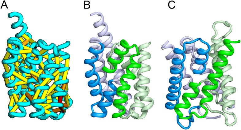
“…are selected for experimental characterization. d , Blind, de novo structure prediction (left) for the critical assessment of protein structure prediction (CASP)11 target T0806, which has no sequence similarity to any protein of known structure, using coevolution-derived contact constraints 27 .”
BackgroundSelf-citationUnified rational protein engineering with sequence-based deep representation learning
Alley, E. C., Khimulya, G., Biswas, S., AlQuraishi, M., & Church, G. M. (2019) · Nature Methods · doi:10.1038/s41592-019-0598-1 · Cited by 1,131
Advances in protein structure prediction and design
Kuhlman, B., & Bradley, P. (2019) · Nature Reviews Molecular Cell Biology · doi:10.1038/s41580-019-0163-x · Cited by 806
Mutation effects predicted from sequence co-variation
Hopf, T. A., Ingraham, J. B., Poelwijk, F. J., Schärfe, C. P. I., Springer, M., Sander, C., & Marks, D. S. (2017) · Nature Biotechnology · doi:10.1038/nbt.3769 · Cited by 775
Deep generative models of genetic variation capture the effects of mutations
Riesselman, A. J., Ingraham, J. B., & Marks, D. S. (2018) · Nature Methods · doi:10.1038/s41592-018-0138-4 · Cited by 601
Epistasis in protein evolution
Starr, T. N., & Thornton, J. W. (2016) · Protein Science · doi:10.1002/pro.2897 · Cited by 598
Sampling alternative conformational states of transporters and receptors with AlphaFold2
del Alamo, D., Sala, D., Mchaourab, H. S., & Meiler, J. (2022) · eLife · doi:10.7554/elife.75751 · Cited by 511
“The resulting models are unnaturally compact and frequently distorted, preventing critical questions about ligand and/or drug binding modes from being addressed ( Ovchinnikov et al., 2015 ; Nicoludis and Gaudet, 2018 ).”
BackgroundMolecular Mechanisms of Two-Component Signal Transduction
Zschiedrich, C. P., Keidel, V., & Szurmant, H. (2016) · Journal of Molecular Biology · doi:10.1016/j.jmb.2016.08.003 · Cited by 540
Protein structure determination using metagenome sequence data
Ovchinnikov, S., Park, H., Varghese, N., Huang, P.-S., Pavlopoulos, G. A., Kim, D. E., Kamisetty, H., Kyrpides, N. C., & Baker, D. (2017) · Science · doi:10.1126/science.aah4043 · Cited by 520
Self-citationTaguchi, A., Welsh, M. A., Marmont, L. S., Lee, W., Sjodt, M., Kruse, A. C., Kahne, D. E., Bernhardt, T. G., & Walker, S. (2019) · Nature Microbiology · doi:10.1038/s41564-018-0345-x · Cited by 335
The EVcouplings Python framework for coevolutionary sequence analysis
Hopf, T. A., Green, A. G., Schubert, B., Mersmann, S., Schärfe, C., Ingraham, J., Tóth-Petróczy, Á., Brock, K. P., Riesselman, A. J., Palmedo, P., et al. (2018) · Bioinformatics · doi:10.1093/bioinformatics/bty862 · Cited by 286
Michaeli, K., Kantor-Uriel, N., Naaman, R., & Waldeck, D. H. (2016) · Chemical Society Reviews · doi:10.1039/c6cs00369a · Cited by 291
Crystal structure of the human sterol transporter ABCG5/ABCG8
Lee, J., Kinch, L. N., Borek, D., Wang, J., Wang, J., Urbatsch, I. L., Xie, X., Grishin, N. V., Cohen, J. C., Otwinowski, Z., et al. (2016) · Nature · doi:10.1038/nature17666 · Cited by 281
Protein interaction networks revealed by proteome coevolution
Cong, Q., Anishchenko, I., Ovchinnikov, S., & Baker, D. (2019) · Science · doi:10.1126/science.aaw6718 · Cited by 241
Self-citationStructural basis of ER-associated protein degradation mediated by the Hrd1 ubiquitin ligase complex
Wu, X., Siggel, M., Ovchinnikov, S., Mi, W., Svetlov, V. V., Nudler, E., Liao, M., Hummer, G., & Rapoport, T. A. (2020) · Science · doi:10.1126/science.aaz2449 · Cited by 236
Architectures of Lipid Transport Systems for the Bacterial Outer Membrane
Ekiert, D. C., Bhabha, G., Isom, G. L., Greenan, G. A., Ovchinnikov, S., Henderson, I. R., Cox, J. S., & Vale, R. D. (2017) · Cell · doi:10.1016/j.cell.2017.03.019 · Cited by 233
Chirality-induced spin polarization places symmetry constraints on biomolecular interactions
Kumar, A., Capua, E., Kesharwani, M., Martin, J. M. L., Sitbon, E., Waldeck, D., & Naaman, R. (2017) · Proceedings of the National Academy of Sciences of the United States of America · doi:10.1073/pnas.1611467114 · Cited by 210
Protocols for Molecular Modeling with Rosetta3 and RosettaScripts
Bender, B. J., Cisneros, A., Duran, A. M., Finn, J. A., Fu, D., Lokits, A. D., Mueller, B. K., Sangha, A. K., Sauer, M. F., Sevy, A. M., et al. (2016) · Biochemistry · doi:10.1021/acs.biochem.6b00444 · Cited by 202
Cryo-EM structure of the protein-conducting ERAD channel Hrd1 in complex with Hrd3
Schoebel, S., Mi, W., Stein, A., Ovchinnikov, S., Pavlovicz, R. E., DiMaio, F., Baker, D. A., Chambers, M. G., Su, H., Li, D., et al. (2017) · Nature · doi:10.1038/nature23314 · Cited by 205
Structures of the stator complex that drives rotation of the bacterial flagellum
Deme, J. C., Johnson, S., Vickery, O. N., Aron, A., Monkhouse, H., Griffiths, T., James, R. H., Berks, B. C., Coulton, J. W., Stansfeld, P. J., et al. (2020) · Nature Microbiology · doi:10.1038/s41564-020-0788-8 · Cited by 196
Accelerated cryo-EM-guided determination of three-dimensional RNA-only structures
Kappel, K., Zhang, K., Su, Z., Watkins, A. M., Kladwang, W., Li, S., Pintilie, G. D., Topkar, V. V., Rangan, R., Zheludev, I. N., et al. (2020) · Nature Methods · doi:10.1038/s41592-020-0878-9 · Cited by 186
Emami, K., Guyet, A., Kawai, Y., Devi, J., Wu, L. J., Allenby, N., Daniel, R. A., & Errington, J. (2017) · Nature Microbiology · doi:10.1038/nmicrobiol.2016.253 · Cited by 187
Origins of coevolution between residues distant in protein 3D structures
Anishchenko, I., Ovchinnikov, S., Kamisetty, H., & Baker, D. (2017) · Proceedings of the National Academy of Sciences · doi:10.1073/pnas.1702664114 · Cited by 185
Self-citationStructure of the core of the type III secretion system export apparatus
Kuhlen, L., Abrusci, P., Johnson, S., Gault, J., Deme, J. C., Caesar, J. J. E., Dietsche, T., Mebrhatu, M. T., Ganief, T. A., Maček, B., et al. (2018) · Nature Structural & Molecular Biology · doi:10.1038/s41594-018-0086-9 · Cited by 175
Structure of a bd oxidase indicates similar mechanisms for membrane-integrated oxygen reductases
Safarian, S., Rajendran, C., Müller, H., Preu, J., Langer, J. D., Ovchinnikov, S., Hirose, T., Kusumoto, T., Sakamoto, J., & Michel, H. (2016) · Science · doi:10.1126/science.aaf2477 · Cited by 163
Inferring protein 3D structure from deep mutation scans
Rollins, N. J., Brock, K. P., Poelwijk, F. J., Stiffler, M. A., Gauthier, N. P., Sander, C., & Marks, D. S. (2019) · Nature Genetics · doi:10.1038/s41588-019-0432-9 · Cited by 166
Wen, B., Zeng, W., Liao, Y., Shi, Z., Savage, S. R., Jiang, W., & Zhang, B. (2020) · PROTEOMICS · doi:10.1002/pmic.201900335 · Cited by 160
Determining protein structures using deep mutagenesis
Schmiedel, J. M., & Lehner, B. (2019) · Nature Genetics · doi:10.1038/s41588-019-0431-x · Cited by 145
Potts Hamiltonian models of protein co-variation, free energy landscapes, and evolutionary fitness
Levy, R. M., Haldane, A., & Flynn, W. F. (2016) · Current Opinion in Structural Biology · doi:10.1016/j.sbi.2016.11.004 · Cited by 134
Structure of the peptidoglycan polymerase RodA resolved by evolutionary coupling analysis
Sjodt, M., Brock, K. P., Dobihal, G. S., Rohs, P. D. A., Green, A. G., Hopf, T. A., Meeske, A. J., Srisuknimit, V., Kahne, D. E., Walker, S., et al. (2018) · Nature · doi:10.1038/nature25985 · Cited by 135
“This is sufficient to map the binding site between bPBPs and RodA to TM8 and TM9 (Figure 4a, b; Extended Data Figure 8), corresponding to the proposed interaction site between the divisome SEDS protein FtsW and its corresponding bPBP, FtsI 10,21,22 .”
Harnessing Conformational Plasticity to Generate Designer Enzymes
Crean, R., Gardner, J. M., & Kamerlin, S. C. L. (2020) · Journal of the American Chemical Society · doi:10.1021/jacs.0c04924 · Cited by 129
Active site rearrangement and structural divergence in prokaryotic respiratory oxidases
Safarian, S., Hahn, A. S., Mills, D. J., Radloff, M., Eisinger, M., Nikolaev, A., Meier‐Credo, J., Melin, F., Miyoshi, H., Gennis, R. B., et al. (2019) · Science · doi:10.1126/science.aay0967 · Cited by 127
Yang, X., McQuillen, R. J., Lyu, Z., Phillips‐Mason, P. J., Cruz, A. D. L., McCausland, J. W., Liang, H., DeMeester, K. E., Santiago, C. C., Grimes, C. L., et al. (2021) · Nature Microbiology · doi:10.1038/s41564-020-00853-0 · Cited by 115
Green, A. G., Elhabashy, H., Brock, K. P., Maddamsetti, R., Kohlbacher, O., & Marks, D. S. (2021) · Nature Communications · doi:10.1038/s41467-021-21636-z · Cited by 114
Leclercq, S., Derouaux, A., Olatunji, S., Fraipont, C., Egan, A. J. F., Vollmer, W., Breukink, E. J., & Terrak, M. (2017) · Scientific Reports · doi:10.1038/srep43306 · Cited by 113
“A model of the interaction between the TMs of FtsW and PBP3 was proposed using a co-evolutionary approach 34 .”
Ovchinnikov, S., Kim, D. E., Wang, R. Y., Liu, Y., DiMaio, F., & Baker, D. A. (2015) · Proteins Structure Function and Bioinformatics · doi:10.1002/prot.24974 · Cited by 111
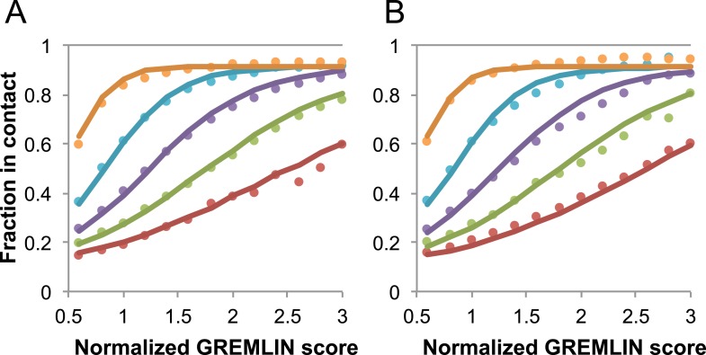
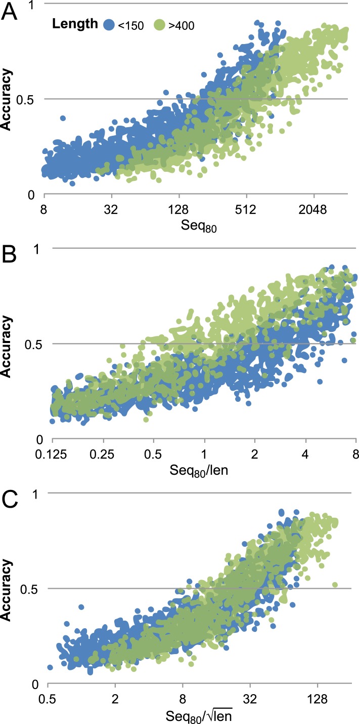
“35 This study also showed that the number of non-redundant (at 80% identity cutoff) sequences over the square-root of length is a better predictor of accuracy than sequences per length when there are 5 L sequences.”
BackgroundMolecular structure of the intact bacterial flagellar basal body
Johnson, S., Furlong, E., Deme, J. C., Nord, A. L., Caesar, J. J. E., Chevance, F. F. V., Berry, R. M., Hughes, K. T., & Lea, S. M. (2021) · Nature Microbiology · doi:10.1038/s41564-021-00895-y · Cited by 109
“Co-evolutionary contacts were determined by the Gremlin web-server 50 .”
Uguzzoni, G., Lovis, S. J., Oteri, F., Schug, A. H., Szurmant, H., & Weigt, M. (2017) · Proceedings of the National Academy of Sciences · doi:10.1073/pnas.1615068114 · Cited by 108
“Such information has proven useful in the prediction of tertiary protein structures (10-13), conformational transitions (14-16), and similarly RNA structures (17, 18).”
BackgroundDeciphering protein evolution and fitness landscapes with latent space models
Ding, X., Zou, Z., & Brooks III, C. L. (2019) · Nature Communications · doi:10.1038/s41467-019-13633-0 · Cited by 106
Protein structure prediction using Rosetta in CASP12
Ovchinnikov, S., Park, H., Kim, D. E., DiMaio, F., & Baker, D. A. (2017) · Proteins Structure Function and Bioinformatics · doi:10.1002/prot.25390 · Cited by 109
“converted to probabilities based on the number of effective sequences and length.(5,13) This predicted contact map is aligned to the contact maps of known structures in the PDB for fold recognition.”
BackgroundBiogenesis and functions of aminocarboxypropyluridine in tRNA
Takakura, M., Ishiguro, K., Akichika, S., Miyauchi, K., & Suzuki, T. (2019) · Nature Communications · doi:10.1038/s41467-019-13525-3 · Cited by 94
Abriata, L. A., Tamò, G. E., Monastyrskyy, B., Kryshtafovych, A., & Peraro, M. D. (2017) · Proteins Structure Function and Bioinformatics · doi:10.1002/prot.25423 · Cited by 88
Protein contact prediction using metagenome sequence data and residual neural networks
Wu, Q., Peng, Z., Anishchenko, I. V., Cong, Q., Baker, D. A., & Yang, J. (2019) · Bioinformatics · doi:10.1093/bioinformatics/btz477 · Cited by 87
Structure of bacterial phospholipid transporter MlaFEDB with substrate bound
Coudray, N., Isom, G. L., MacRae, M. R., Saiduddin, M. N., Bhabha, G., & Ekiert, D. C. (2020) · eLife · doi:10.7554/elife.62518 · Cited by 74
Computational approaches to investigating allostery
Schueler‐Furman, O., & Wodak, S. J. (2016) · Current Opinion in Structural Biology · doi:10.1016/j.sbi.2016.06.017 · Cited by 72
DESTINI: A deep-learning approach to contact-driven protein structure prediction
Gao, M., Zhou, H., & Skolnick, J. (2019) · Scientific Reports · doi:10.1038/s41598-019-40314-1 · Cited by 61
“Overall, protein contact information is the most important component in modern structural modelling packages 3,5,10 .”
BackgroundPark, Y., LaCourse, K. D., Cambillau, C., DiMaio, F., Mougous, J. D., & Veesler, D. (2018) · Nature Communications · doi:10.1038/s41467-018-07796-5 · Cited by 60
Ghachi, M. E., Howe, N., Huang, C., Oliéric, V., Warshamanage, R., Touzé, T., Weichert, D., Stansfeld, P. J., Wang, M., Kerff, F., et al. (2018) · Nature Communications · doi:10.1038/s41467-018-03477-5 · Cited by 61
Structural and Evolutionary Insights Point to Allosteric Regulation of TRP Ion Channels
Hilton, J. K., Kim, M., & Horn, W. D. V. (2019) · Accounts of Chemical Research · doi:10.1021/acs.accounts.9b00075 · Cited by 56
AlphaFold Models of Small Proteins Rival the Accuracy of Solution NMR Structures
Tejero, R., Huang, Y. J., Ramelot, T. A., & Montelione, G. T. (2022) · Frontiers in Molecular Biosciences · doi:10.3389/fmolb.2022.877000 · Cited by 56
“…et al., 2017; Huang et al., 2019), contact prediction based on sequence covariance (Marks et al., 2011; Morcos et al., 2011; Marks et al., 2012; Ovchinnikov et al., 2015; Ovchinnikov et al., 2016; Buchan and Jones, 2018), the massive and still growing databases of genomic sequence data, and…”
BackgroundWorkman, S. D., Worrall, L. J., & Strynadka, N. C. J. (2018) · Nature Communications · doi:10.1038/s41467-018-03547-8 · Cited by 55
The evolution of gene regulatory networks controlling Arabidopsis thaliana L. trichome development
Doroshkov, A. V., Konstantinov, D. K., Afonnikov, D. A., & Владимирович, Г. К. (2019) · BMC Plant Biology · doi:10.1186/s12870-019-1640-2 · Cited by 54
Inferring interaction partners from protein sequences using mutual information
Bitbol, A. (2018) · PLoS Computational Biology · doi:10.1371/journal.pcbi.1006401 · Cited by 53
Huang, Y. J., Zhang, N., Bersch, B., Fidelis, K., Inouye, M., Ishida, Y., Kryshtafovych, A., Kobayashi, N., Kuroda, Y., Liu, G., et al. (2021) · Proteins: Structure, Function, and Bioinformatics · doi:10.1002/prot.26246 · Cited by 49
Cooper, C. J., Zheng, K., Rush, K. W., Johs, A., Sanders, B., Pavlopoulos, G. A., Kyrpides, N. C., Podar, M., Ovchinnikov, S., Ragsdale, S. W., et al. (2020) · Communications Biology · doi:10.1038/s42003-020-1047-5 · Cited by 59
Enzyme structures of the bacterial peptidoglycan and wall teichoic acid biogenesis pathways
Caveney, N. A., Li, F. K. K., & Strynadka, N. C. J. (2018) · Current Opinion in Structural Biology · doi:10.1016/j.sbi.2018.05.002 · Cited by 53
Enhanced unbiased sampling of protein dynamics using evolutionary coupling information
Shamsi, Z., Moffett, A. S., & Shukla, D. (2017) · Scientific Reports · doi:10.1038/s41598-017-12874-7 · Cited by 52
Federating Structural Models and Data: Outcomes from A Workshop on Archiving Integrative Structures
Berman, H. M., Adams, P. D., Bonvin, A. M. J. J., Burley, S. K., Carragher, B. O., Chiu, W., DiMaio, F., Ferrin, T. E., Gabanyi, M. J., Goddard, T. D., et al. (2019) · Structure · doi:10.1016/j.str.2019.11.002 · Cited by 51
Sun, P., Li, J., Zhang, X., Guan, Z., Xiao, Q., Zhao, C., Song, M., Zhou, Y., Mou, L., Ke, M., et al. (2018) · Journal of Biological Chemistry · doi:10.1074/jbc.ra118.003876 · Cited by 47
Li, Y., Gong, H., Zhan, R., Ouyang, S., Park, K., Lutkenhaus, J., & Du, S. (2021) · PLoS Genetics · doi:10.1371/journal.pgen.1009366 · Cited by 46
A Slippery Scaffold: Synthesis and Recycling of the Bacterial Cell Wall Carrier Lipid
Workman, S. D., & Strynadka, N. C. J. (2020) · Journal of Molecular Biology · doi:10.1016/j.jmb.2020.03.025 · Cited by 45
Chan, Y. H., Venev, S. V., Zeldovich, K. B., & Matthews, C. R. (2017) · Nature Communications · doi:10.1038/ncomms14614 · Cited by 49
Alexandre, C. M., Urton, J. R., Jean-Baptiste, K., Huddleston, J., Dorrity, M. W., Cuperus, J. T., Sullivan, A. M., Bemm, F., Jolić, D., Arsovski, A. A., et al. (2017) · Molecular Biology and Evolution · doi:10.1093/molbev/msx326 · Cited by 44
“…where we can computationally predict important domains within proteins; indeed, largescale cross-species analysis of coevolution allows accurate predictions of protein structures, a prerequisite for fully understanding gene function (Ovchinnikov et al. 2015; Tang et al. 2015; Stein et al. 2015).”
BackgroundProtein sequence‐to‐structure learning: Is this the end(‐to‐end revolution)?
Laine, E., Eismann, S., Elofsson, A., & Grudinin, S. (2021) · Proteins: Structure, Function, and Bioinformatics · doi:10.1002/prot.26235 · Cited by 44
“These advances have significantly increased the protein structure modelling coverage of genomes [8, 9, 10], and also of bacterial interactomes [11, 12, 13].”
BackgroundApplications of contact predictions to structural biology
Šimkovic, F., Ovchinnikov, S., Baker, D. A., & Rigden, D. J. (2017) · IUCrJ · doi:10.1107/s2052252517005115 · Cited by 42
Protein structural motifs in prediction and design
Mackenzie, C. O., & Grigoryan, G. (2017) · Current Opinion in Structural Biology · doi:10.1016/j.sbi.2017.03.012 · Cited by 40
Large-scale structure prediction by improved contact predictions and model quality assessment
Michel, M., Hurtado, D. M., Uziela, K., & Elofsson, A. (2017) · Bioinformatics · doi:10.1093/bioinformatics/btx239 · Cited by 38
Structure and mechanism of a tripartite ATP-independent periplasmic TRAP transporter
Davies, J. S., Currie, M. J., North, R. A., Scalise, M., Wright, J. D., Copping, J. M., Remus, D. M., Gulati, A., Morado, D. R., Jamieson, S. A., et al. (2023) · Nature Communications · doi:10.1038/s41467-023-36590-1 · Cited by 34
“The predicted bindingmode is similar to themode first predicted by Ovchinnikov et al.(36), as well as that suggested for the HiSiaPQMsystem.”
ResultDubey, S., Ager-Wick, E., Kumar, J., Karunasagar, I., Karunasagar, I., Peng, B., Evensen, Ø., Sørum, H. R., & Munang’andu, H. M. (2022) · Frontiers in Microbiology · doi:10.3389/fmicb.2022.1008870 · Cited by 41
A lipophilicity-based energy function for membrane-protein modelling and design
Weinstein, J. J., Elazar, A., & Fleishman, S. J. (2019) · PLoS Computational Biology · doi:10.1371/journal.pcbi.1007318 · Cited by 35
Mi3-GPU: MCMC-based inverse Ising inference on GPUs for protein covariation analysis
Haldane, A., & Levy, R. M. (2020) · Computer Physics Communications · doi:10.1016/j.cpc.2020.107312 · Cited by 38
Condon, S. G. F., Mahbuba, D., Armstrong, C. R., Vázquez, G. D., Craven, S. J., LaPointe, L. M., Khadria, A. S., Chadda, R., Crooks, J. A., Rangarajan, N., et al. (2017) · Journal of Biological Chemistry · doi:10.1074/jbc.ra117.000426 · Cited by 35
“Additionally, sigmoidal distance restraints were placed between the C atoms of each pair of top co-evolving positions (83), using the following form,”
MethodSahoo, A., Khare, S., Devanarayanan, S., Jain, P. C., & Varadarajan, R. (2015) · eLife · doi:10.7554/elife.09532 · Cited by 36
“…et al., 2004 ), chemical shift data ( Shen et al., 2008 ) from NMR experiments, co-varying residues identified from statistical analysis of genomic data ( Hopf et al., 2012 ; Jones et al., 2012 ; Ovchinnikov et al., 2014 ; Ovchinnikov et al., 2015 ; Sulkowska, et al., 2012 ) to determine structure.”
BackgroundBig-data approaches to protein structure prediction
Söding, J. (2017) · Science · doi:10.1126/science.aal4512 · Cited by 36
Paulsen, V. S., Mardirossian, M., Blencke, H., Benincasa, M., Runti, G., Nepa, M., Haug, T., Stensvåg, K., & Scocchi, M. (2016) · Microbiology · doi:10.1099/mic.0.000249 · Cited by 34
Feng, S., Zhang, W.-X., Yang, J., Yang, Y., & Shen, H. (2019) · Journal of Molecular Biology · doi:10.1016/j.jmb.2019.12.007 · Cited by 35
Blind assessment of monomeric AlphaFold2 protein structure models with experimental NMR data
Li, E. H., Spaman, L. E., Tejero, R., Huang, Y. J., Ramelot, T. A., Fraga, K. J., Prestegard, J. H., Kennedy, M. A., & Montelione, G. T. (2023) · Journal of Magnetic Resonance · doi:10.1016/j.jmr.2023.107481 · Cited by 34
Modeling of protein conformational changes with Rosetta guided by limited experimental data
Sala, D., del Alamo, D., Mchaourab, H. S., & Meiler, J. (2022) · Structure · doi:10.1016/j.str.2022.04.013 · Cited by 30
Lytvynenko, I., Brill, S., Oswald, C., & Pos, K. M. (2015) · Journal of Molecular Biology · doi:10.1016/j.jmb.2015.12.006 · Cited by 31
Synthetic protein alignments by CCMgen quantify noise in residue-residue contact prediction
Vorberg, S., Seemayer, S., & Söding, J. (2018) · PLoS Computational Biology · doi:10.1371/journal.pcbi.1006526 · Cited by 36
Barth, P., & Senes, A. (2016) · Nature Structural & Molecular Biology · doi:10.1038/nsmb.3231 · Cited by 30
Ji, S., Oruç, T., Mead, L., Rehman, M. F. U., Thomas, C. M., Butterworth, S., & Winn, P. J. (2019) · PLoS ONE · doi:10.1371/journal.pone.0205214 · Cited by 30
“It has been shown that there is a correlation between the Nf value and the structure prediction accuracy [26].”
BackgroundCampaña, M. B., Irudayanathan, F. J., Davis, T. R., McGovern-Gooch, K. R., Loftus, R., Ashkar, M., Escoffery, N., Navarro, M., Sieburg, M. A., Nangia, S., et al. (2019) · Journal of Biological Chemistry · doi:10.1074/jbc.ac119.009749 · Cited by 30
Structural model of human PORCN illuminates disease-associated variants and drug-binding sites
Yu, J., Liao, P.-J., Xu, W., Jones, J. R., Everman, D. B., Flanagan‐Steet, H. R., Keller, T. H., & Virshup, D. M. (2021) · Journal of Cell Science · doi:10.1242/jcs.259383 · Cited by 29
Comparing co-evolution methods and their application to template-free protein structure prediction
de Oliveira, S. H. P., Shi, J., & Deane, C. M. (2016) · Bioinformatics · doi:10.1093/bioinformatics/btw618 · Cited by 28
A large-scale comparative assessment of methods for residue–residue contact prediction
Wuyun, Q., Zheng, W. H., Peng, Z., & Yang, J. (2016) · Briefings in Bioinformatics · doi:10.1093/bib/bbw106 · Cited by 28
Cao, L., Yu, B., Kong, D., Cong, Q., Tao, Y., Chen, Z., Hu, Z., Chang, H., Zhong, J., Baker, D. A., et al. (2019) · PLoS Pathogens · doi:10.1371/journal.ppat.1007759 · Cited by 31
Tools and Approaches for Dissecting Protein Bacteriocin Import in Gram-Negative Bacteria
Atanasković, I., & Kleanthous, C. (2019) · Frontiers in Microbiology · doi:10.3389/fmicb.2019.00646 · Cited by 28
Co-evolution techniques are reshaping the way we do structural bioinformatics
de Oliveira, S. H. P., & Deane, C. M. (2017) · F1000Research · doi:10.12688/f1000research.11543.1 · Cited by 28
Protein structure prediction: making AWSEM AWSEM‐ER by adding evolutionary restraints
Sirovetz, B. J., Schafer, N. P., & Wolynes, P. G. (2017) · Proteins Structure Function and Bioinformatics · doi:10.1002/prot.25367 · Cited by 27
“15,16 Being tied to function, ultimately, coevolutionary data contain information that goes beyond an individual protein ’ s structure.”
BackgroundChemical tools to characterize peptidoglycan synthases
Taguchi, A., Kahne, D. E., & Walker, S. (2019) · Current Opinion in Chemical Biology · doi:10.1016/j.cbpa.2019.07.009 · Cited by 25
Observation selection bias in contact prediction and its implications for structural bioinformatics
Orlando, G., Raimondi, D., & Vranken, W. (2016) · Scientific Reports · doi:10.1038/srep36679 · Cited by 25
SARS-CoV2 billion-compound docking
Rogers, D. O. W., Agarwal, R., Vermaas, J. V., Smith, M. D., Tatikonda, R. R., Cooper, C. J., Sedova, A., Boehm, S., Baker, M. B., Gläser, J., et al. (2023) · Scientific Data · doi:10.1038/s41597-023-01984-9 · Cited by 25
Goojani, H. G., Konings, J., Hakvoort, H. W. J., Hong, S., Gennis, R. B., Sakamoto, J., Lill, H., & Bald, D. (2020) · Biochimica et Biophysica Acta (BBA) - Bioenergetics · doi:10.1016/j.bbabio.2020.148175 · Cited by 24
Structurally Mapping Endogenous Heme in the CcmCDE Membrane Complex for Cytochrome c Biogenesis
Sutherland, M. C., Jarodsky, J. M., Ovchinnikov, S., Baker, D., & Kranz, R. G. (2018) · Journal of Molecular Biology · doi:10.1016/j.jmb.2018.01.022 · Cited by 23
“[46,47], facilitate discussion of the mechanisms underlying holoCcmE formation (Step 1 in Fig.”
BackgroundSelf-citationRoles of the DedD Protein in Escherichia coli Cell Constriction
Liu, B., Hale, C. A., Persons, L., Phillips‐Mason, P. J., & de Boer, P. A. J. (2019) · Journal of Bacteriology · doi:10.1128/jb.00698-18 · Cited by 24
Targeting Lipoprotein Biogenesis: Considerations towards Antimicrobials
Arnaout, T. E., & Soulimane, T. (2019) · Trends in Biochemical Sciences · doi:10.1016/j.tibs.2019.03.007 · Cited by 23
Using molecular simulation to explore the nanoscale dynamics of the plant kinome
Moffett, A. S., & Shukla, D. (2018) · Biochemical Journal · doi:10.1042/bcj20170299 · Cited by 23
Pancsa, R., Zsolyomi, F., & Tompa, P. (2018) · International Journal of Molecular Sciences · doi:10.3390/ijms19113315 · Cited by 22
“This is well supported by the fact that recent large-scale residue co-variation analyses have all focused on bacterial protein complexes [9,14,15].”
Background
Metadata
- Authors
- Ovchinnikov, S., Kinch, L., Park, H., Liao, Y., Pei, J., Kim, D. E., Kamisetty, H., Grishin, N. V., & Baker, D.
- Affiliations
- University of Washington (US)
- Howard Hughes Medical Institute (US)
- The University of Texas Southwestern Medical Center (US)
- Meta (United States) (US)
- Corresponding author
- Baker, David
- ORCID
- Venue
- eLife
- Publisher
- eLife Sciences Publications, Ltd
- Published
- 2015-09-03
- Volume
- 4
- Pages
- e09248-e09248
- ISSN
- 2050-084X (ISSN-L)
- PMC
- PMC4602095
- OpenAlex
- W1895528264
- Cited by
- 222
- References
- 80
- Citations by year
- 2026: 3 · 2025: 4 · 2024: 2 · 2023: 12 · 2022: 17 · 2021: 20 · 2020: 25 · 2019: 42 · 2018: 30 · 2017: 39 · 2016: 24 · 2015: 4
- FWCI
- 14.18
- Type
- journal article
- Language
- en
- Open access
- gold
- doi.org
- Licence
- Locations
- eLife (publishedVersion, cc-by)
- Europe PMC (PubMed Central) (publishedVersion, cc-by)
- PubMed Central (publishedVersion, cc-by)
- PubMed (publishedVersion)
- DOAJ (DOAJ: Directory of Open Access Journals) (submittedVersion, cc-by-sa)
- Funders
- National Institutes of Health (NIH) (R01GM092802, GM094575)
- Welch Foundation (Robert A. Welch Foundation) (I-1505)
- Topics
- Life Sciences › Biochemistry, Genetics and Molecular Biology › Molecular Biology › Protein Structure and Dynamics
- Life Sciences › Biochemistry, Genetics and Molecular Biology › Structural Biology › Enzyme Structure and Function
- Life Sciences › Biochemistry, Genetics and Molecular Biology › Molecular Biology › RNA and protein synthesis mechanisms
- Keywords
- protein structure prediction, de novo structure prediction, Rosetta, prokaryotic proteins, structural model, protein function prediction, evolutionary information
- MeSH terms
- Bacterial Proteins, Models, Molecular, Protein Conformation, Evolution, Molecular, Computational Biology
About these checks
Registry, retraction, Cites and statistics results are computed, not judged, with one exception: a printed reference that no index lists is read once by a model before it counts as a citation error. Claims are extracted by a model and kept only when their quote appears verbatim in the text; each cited source is then checked, with a model’s help, for whether it says what the claim relies on. Those verdicts can be wrong — read the claim’s quote and the cited source before relying on one.
Statistics and arithmetic are recomputed, not judged.
Confidence bands are shown only for fields calibrated on our annotated test set. None is calibrated yet, so no band is shown.
What counts as a claim ›How we measure confidence ›How we check this ›
Are you an author, or do you see a problem with this page? Request a review
