

The DOI matches its Crossref record; 18 of the 18 references it cites were found in a registry or an index.
Transformative Impact of Nanocarrier‐Mediated Drug Delivery: Overcoming Biological Barriers and Expanding Therapeutic Horizons
Abstract
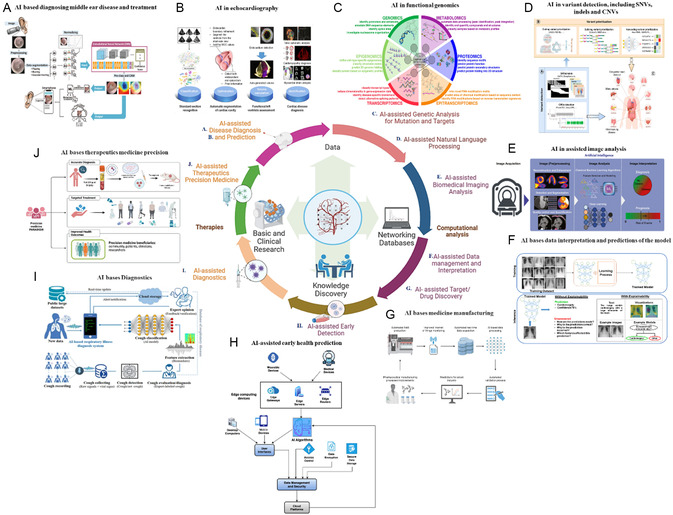
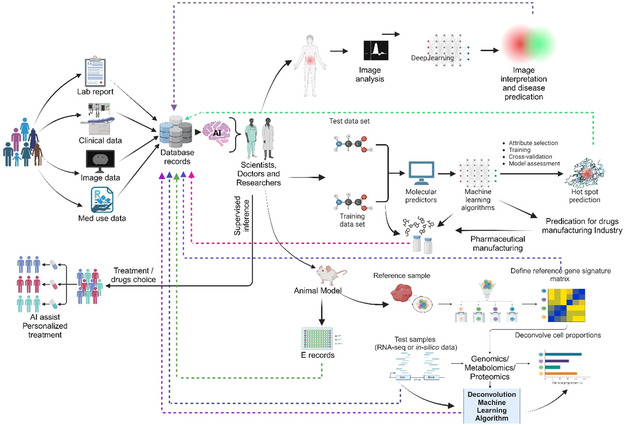
Advancing therapeutic progress is centered on developing drug delivery systems (DDS) that control therapeutic molecule release, ensuring precise targeting and optimal concentrations. Targeted DDS enhances treatment efficacy and minimizes off‐target effects, but struggles with drug degradation. Over the last three decades, nanopharmaceuticals have evolved from laboratory concepts into clinical products, highlighting the profound impact of nanotechnology in medicine. Despite advancements, the effective delivery of therapeutics remains challenging because of biological barriers. Nanocarriers offer a solution with a small size, high surface‐to‐volume ratios, and customizable properties. These systems address physiological and biological challenges, such as shear stress, protein adsorption, and quick clearance. They allow targeted delivery to specific tissues, improve treatment outcomes, and reduce adverse effects. Nanocarriers exhibit controlled release, decreased degradation, and enhanced efficacy. Their size facilitates cell membrane penetration and intracellular delivery. Surface modifications increase affinity for specific cell types, allowing precise treatment delivery. This study also elucidates the potential integration of artificial intelligence with nanoscience to innovate future nanocarrier systems.
Overview
The claims and statistics of this paper haven’t been checked yet.
Run the check — free, no account needed. It reads the open-access text and checks the sources it cites.
Integrity signals
Registry
Cites
Statistics
Figures
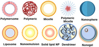
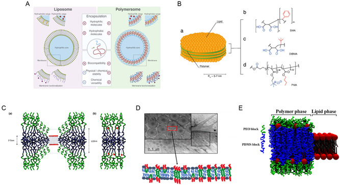
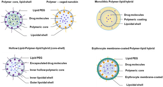
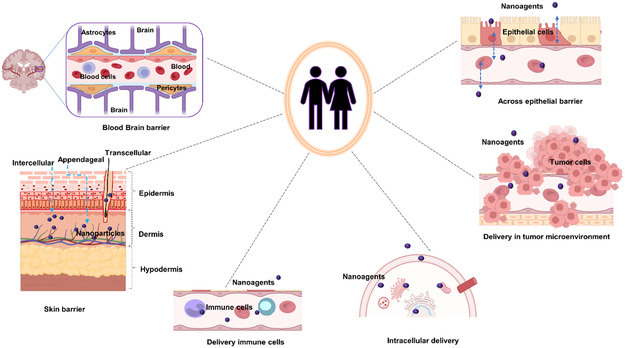
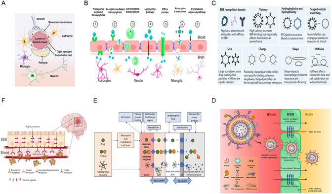
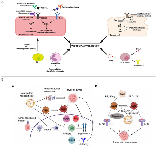
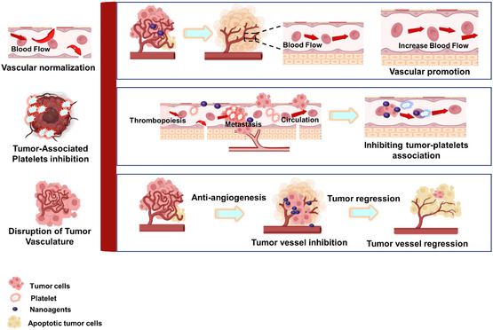
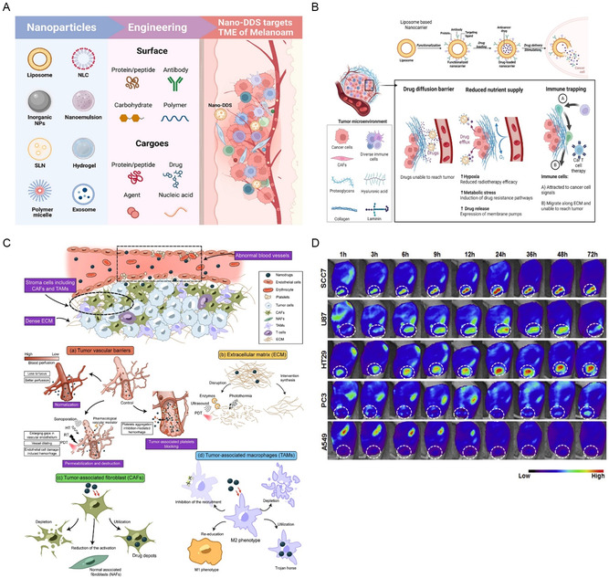
Verification
Claims the paper makes
The claims and statistics of this paper haven’t been checked yet.
Run the check — free, no account needed. It reads the open-access text and checks the sources it cites.
Claims other papers make about this one
Each is checked against this paper’s own text: does it say what the citing paper says it does?
In the citing paper
“Ligand-functionalized platforms add active binding, trans-barrier transport, and facilitated uptake to improve selectivity and distribution [79] (Fig. 5).”
From Overcoming tumor microenvironment barriers: transformable and bioinspired nanomedicine strategies for deep tumor penetration (2026) · cites this paper as [79]
Self-check
Percentages, sums and reported differences re-computed from the paper’s own numbers.
The self-check needs the paper’s full text.
Citations
Cites
Principles of nanoparticle design for overcoming biological barriers to drug delivery
Blanco, E., Shen, H., & Ferrari, M. (2015) · Nature Biotechnology · doi:10.1038/nbt.3330 · Cited by 7,002
Doxil® — The first FDA-approved nano-drug: Lessons learned
Barenholz, Y. (. (2012) · Journal of Controlled Release · doi:10.1016/j.jconrel.2012.03.020 · Cited by 4,792
Micellar Nanocarriers: Pharmaceutical Perspectives
Torchilin, V. P. (2006) · Pharmaceutical Research · doi:10.1007/s11095-006-9132-0 · Cited by 1,910
The blood–brain barrier: Structure, regulation and drug delivery
Wu, D., Chen, Q., Chen, X., Han, F., Chen, Z., & Wang, Y. (2023) · Signal Transduction and Targeted Therapy · doi:10.1038/s41392-023-01481-w · Cited by 1,906
High performance plasma amyloid-β biomarkers for Alzheimer's disease
Nakamura, A., Kaneko, N., Villemagne, V. L., Kato, T., Doecke, J., Doré, V., Fowler, C., Li, Q.-X., Martins, R., Rowe, C., et al. (2018) · Nature · doi:10.1038/nature25456 · Cited by 1,681
The evolution of commercial drug delivery technologies
Vargason, A. M., Anselmo, A. C., & Mitragotri, S. (2021) · Nature Biomedical Engineering · doi:10.1038/s41551-021-00698-w · Cited by 1,361
The big picture on nanomedicine: the state of investigational and approved nanomedicine products
Etheridge, M. L., Campbell, S. A., Erdman, A. G., Haynes, C. L., Wolf, S. M., & McCullough, J. (2013) · Nanomedicine: Nanotechnology, Biology and Medicine · doi:10.1016/j.nano.2012.05.013 · Cited by 1,088
Polymeric micelles: nanocarriers for cancer-targeted drug delivery
Zhang, Y., Huang, Y., & Li, S. (2014) · AAPS PharmSciTech · doi:10.1208/s12249-014-0113-z · Cited by 367
Yokoyama, M. (1998) · Journal of Controlled Release · doi:10.1016/s0168-3659(97)00115-6 · Cited by 308
Nazir, S., Dickson, D. M., & Akram, M. U. (2023) · Computers in Biology and Medicine · doi:10.1016/j.compbiomed.2023.106668 · Cited by 283
Research works printed but not linked: 262
- 10.1038/s43588-021-00060-9 · DOI
- 10.1038/s41540-022-00247-4 · DOI
- 10.1016/j.addr.2019.07.010 · DOI
- 10.2147/IJN.S236927 · DOI
- 10.2147/IJN.S406415 · DOI
- 10.1088/1748-605X/abf7d5 · DOI
- 10.2147/IJN.S146315 · DOI
- 10.1021/acs.chemrev.5b00589 · DOI
- 10.1016/j.heliyon.2023.e17488 · DOI · Paper page
- 10.3390/nano14070557 · DOI
- 10.1038/s41565-019-0567-y · DOI
- 10.2174/092986712800784676 · DOI
- 10.3390/pharmaceutics16020188 · DOI
- 10.1016/j.jddst.2021.102426 · DOI · Paper page
- 10.3390/molecules25092193 · DOI
- 10.1016/B978-0-323-89925-3.00013-7 · DOI
- 10.1186/s12951-018-0392-8 · DOI
- 10.2174/1567201818666210609161301 · DOI
- Hafner A., 2014, Int. J. Nanomed., 9, 1005
- 10.1038/s41577-019-0269-6 · DOI
- 10.1016/j.ijbiomac.2022.07.118 · DOI
- 10.1021/acsomega.2c05976 · DOI · Paper page
- 10.1007/s40089-017-0208-0 · DOI
- 10.1039/9781782622345-00001 · DOI
- Sabine Gressler C. H., 2024, Nanocarrier. Part I: Overview and Categorization of Nanocarriers
- 10.2217/nnm-2018-0120 · DOI
- 10.1089/jamp.2023.29089.sb · DOI
- 10.1016/j.ijpharm.2020.119098 · DOI
- 10.1002/adma.202002047 · DOI
- 10.2174/1574891X113089990038 · DOI
- 10.3390/pharmaceutics15020413 · DOI
- 10.1016/j.ijbiomac.2020.11.052 · DOI
- 10.3390/nano14080672 · DOI
- 10.1080/17425247.2016.1244187 · DOI
- 10.1016/j.ijbiomac.2020.10.104 · DOI
- 10.1007/978-981-99-3307-5_7 · DOI
- 10.3390/molecules25163731 · DOI · Paper page
- 10.1016/j.jare.2021.11.008 · DOI
- 10.1016/j.cocis.2017.12.004 · DOI
- 10.3389/fphar.2021.683935 · DOI
- 10.1016/j.ejpb.2011.07.005 · DOI
- 10.1016/j.ijpharm.2019.118796 · DOI
- 10.3390/pharmaceutics12090837 · DOI
- 10.1016/j.neuron.2018.07.039 · DOI · Paper page
- 10.2217/nnm.14.156 · DOI
- 10.1080/10717544.2018.1461955 · DOI
- 10.1016/j.ijbiomac.2020.11.207 · DOI
- 10.1016/j.jconrel.2017.05.019 · DOI · Paper page
- 10.1016/j.jconrel.2018.09.001 · DOI
- 10.1038/s41578-020-00269-6 · DOI · Paper page
- Shazwani S. S., 2024, ACS Omega, 9, 17379
- 10.1016/j.ijpharm.2005.04.029 · DOI
- 10.3109/02652049509010292 · DOI
- Melis Ç., 2014, Application of Nanotechnology in Drug Delivery
- 10.3390/pharmaceutics14102195 · DOI
- 10.1111/cns.12580 · DOI
- 10.1016/j.cis.2015.08.011 · DOI
- 10.1016/S0928-0987(99)00071-8 · DOI
- Scheffel U., 1972, J. Nucl. Med., 13, 498
- 10.1016/j.pneurobio.2014.05.002 · DOI · Paper page
- 10.4103/0250-474X.57282 · DOI
- 10.1080/17425255.2019.1621289 · DOI
- 10.1016/j.drudis.2019.01.004 · DOI
- 10.1016/j.ejphar.2014.11.014 · DOI
- 10.1155/2021/8844030 · DOI
- 10.3390/molecules23040938 · DOI
- 10.3389/fphar.2023.1159131 · DOI
- 10.1016/S1359-6446(04)03276-3 · DOI
- 10.1016/S1367-5931(02)00347-2 · DOI
- 10.1021/acsami.2c13129 · DOI
- 10.1016/j.drudis.2022.01.001 · DOI
- 10.1016/B978-0-323-91924-1.00010-1 · DOI
- 10.1186/s13036-022-00298-5 · DOI
- 10.1016/j.addr.2023.115005 · DOI
- 10.3390/polym10060595 · DOI
- 10.1073/pnas.1003103107 · DOI
- 10.1039/D1TB02144C · DOI
- 10.1021/acschemneuro.7b00301 · DOI
- 10.1016/j.colsurfb.2021.111764 · DOI
- 10.3390/polym14122510 · DOI
- 10.2174/1381612826666200116150426 · DOI
- 10.1021/nn800275r · DOI
- 10.1016/j.mattod.2013.09.002 · DOI
- 10.1021/nn3023602 · DOI
- 10.1039/c2sm07188f · DOI
- 10.1529/biophysj.104.048223 · DOI
- 10.1021/acsapm.2c01272 · DOI
- 10.1016/j.bpj.2021.12.005 · DOI
- 10.1016/j.biomaterials.2009.01.005 · DOI
- 10.3390/pharmaceutics13081291 · DOI
- 10.1016/j.biomaterials.2008.12.013 · DOI
- 10.1021/ja9017336 · DOI
- 10.1016/S0169-409X(99)00035-6 · DOI
- 10.1074/jbc.272.4.2382 · DOI
- 10.1002/anie.201004867 · DOI
- 10.1016/j.mimet.2019.03.017 · DOI
- 10.1039/b709566j · DOI
- 10.1016/j.bios.2003.10.017 · DOI
- 10.1073/pnas.1106634108 · DOI
- 10.1039/c2cs35103j · DOI
- 10.1007/s11095-011-0568-5 · DOI
- 10.1002/anie.201101554 · DOI
- 10.1080/17425247.2019.1614911 · DOI
- 10.3390/polym15092196 · DOI
- 10.1007/s12272-013-0272-6 · DOI
- 10.1038/s41573-020-0090-8 · DOI · Paper page
- 10.3390/ijms222111654 · DOI
- 10.1111/j.1471-4159.2008.05492.x · DOI · Paper page
- 10.1016/S0962-8924(02)02282-1 · DOI
- 10.1016/j.chemphyslip.2015.03.003 · DOI
- 10.1016/j.jconrel.2016.01.046 · DOI
- 10.1186/s12951-016-0177-x · DOI
- 10.1016/j.jconrel.2009.03.002 · DOI · Paper page
- 10.1023/A:1006948027674 · DOI
- 10.1016/j.colsurfb.2016.04.041 · DOI
- 10.1016/j.jtice.2013.09.024 · DOI
- 10.1016/j.lfs.2005.05.085 · DOI
- 10.1016/j.ijpharm.2011.05.062 · DOI
- 10.1016/j.jconrel.2008.10.016 · DOI
- 10.4161/derm.1.4.9501 · DOI
- 10.1039/D2NA00530A · DOI
- 10.1166/jnn.2015.9185 · DOI
- 10.1016/j.jconrel.2004.06.015 · DOI
- 10.1016/j.ijpharm.2010.09.037 · DOI
- 10.3390/ijms22158319 · DOI
- 10.1080/17425247.2020.1727883 · DOI
- Uner M., 2006, Pharmazie, 61, 375
- 10.1016/B978-0-323-89925-3.00008-3 · DOI
- Sakhrani N. M., 2013, Drug Des. Dev. Ther., 2013, 585
- 10.1016/j.ijpharm.2015.10.053 · DOI
- 10.1016/j.biomaterials.2016.04.026 · DOI
- 10.1016/j.drudis.2017.08.011 · DOI
- 10.1016/j.nano.2014.02.014 · DOI
- 10.1016/j.jconrel.2014.09.031 · DOI
- 10.1016/S0168-3659(99)00228-X · DOI
- 10.1002/adma.201902791 · DOI
- 10.7150/thno.57828 · DOI
- 10.3390/pharmaceutics15061610 · DOI
- 10.3390/pharmaceutics11120632 · DOI
- 10.1016/j.biopha.2018.09.097 · DOI
- 10.1016/j.addr.2024.115239 · DOI
- 10.3390/biom13121723 · DOI
- 10.1016/j.eurpolymj.2020.110058 · DOI
- 10.1007/s10895‐023‐03562‐z · DOI
- 10.1021/acsami.6b02721 · DOI
- 10.1016/j.bioadv.2022.212725 · DOI
- 10.1038/s41427-021-00303-1 · DOI
- 10.3390/biology13010001 · DOI
- 10.1016/j.trac.2022.116714 · DOI
- 10.1021/acsnano.8b06924 · DOI
- 10.1016/j.polymer.2015.12.040 · DOI
- 10.1002/app.42650 · DOI
- 10.1073/pnas.1210096109 · DOI
- 10.1016/j.drudis.2022.103469 · DOI
- 10.1016/j.biomaterials.2022.121656 · DOI
- 10.1038/nnano.2016.160 · DOI
- 10.1016/j.ijpharm.2022.122570 · DOI
- 10.1371/journal.pone.0179545 · DOI
- 10.1038/onc.2017.119 · DOI
- 10.1186/s11658-018-0088-y · DOI
- Tei K., 2002, Cancer Res., 62, 6289
- 10.1096/fj.99-1049com · DOI
- 10.1096/fj.05-4493com · DOI · Paper page
- 10.3389/fonc.2018.00367 · DOI
- 10.1080/10717544.2020.1809559 · DOI
- 10.3389/fimmu.2018.00455 · DOI
- 10.1016/j.addr.2021.02.019 · DOI
- 10.1186/s12935-023-02927-5 · DOI
- Lupo G., 2016, Front. Pharmacol., 7, 519
- 10.1021/acsnano.6b03110 · DOI
- 10.1016/j.ijpharm.2013.05.018 · DOI
- 10.1016/j.nantod.2017.06.010 · DOI
- 10.1002/smll.201903323 · DOI
- 10.1002/anie.201506262 · DOI
- 10.1007/s11894-010-0131-2 · DOI
- 10.1016/S0006-3495(01)75844-4 · DOI
- 10.1080/10717544.2018.1501119 · DOI
- 10.1080/10717544.2022.2058646 · DOI
- 10.1080/10717544.2017.1279238 · DOI · Paper page
- 10.1016/j.biomaterials.2007.02.021 · DOI
- 10.1016/j.ijmm.2007.05.004 · DOI
- 10.1016/j.addr.2008.11.002 · DOI
- 10.1016/j.ejpb.2014.12.024 · DOI
- 10.1016/j.ijpharm.2014.05.047 · DOI
- 10.1016/j.ejpb.2015.01.022 · DOI
- 10.1007/s11051-012-1353-z · DOI
- 10.1021/mp500439c · DOI
- 10.1016/j.biomaterials.2013.04.053 · DOI
- 10.1016/j.biomaterials.2008.09.050 · DOI · Paper page
- 10.1016/j.biomaterials.2013.04.028 · DOI
- 10.1093/toxsci/kfm217 · DOI
- 10.1034/j.1600-0854.2001.020301.x · DOI
- 10.1002/btm2.10143 · DOI · Paper page
- 10.37349/ebmx.2024.00009 · DOI
- 10.1007/978-1-61779-052-2_21 · DOI
- Pillai G., 2014, Nanomedicines for Cancer Therapy : An Update of FDA Approved and those Under Various Stages of Development
- 10.3390/pharmaceutics14010106 · DOI
- 10.1093/rb/rbw011 · DOI
- 10.1089/apc.1995.9.306 · DOI
- FDA approves KS drug, Food and Drug Administration, Florida Department of Health and Rehabilitation Services, 1996, AIDS Alert, 11, 11
200 of 262 shown
Cited by
Sorted by citations
Sheng, J., Yuan, W., Zhang, M., Chen, Z., Qi, Y., Zhao, Y., & Zhang, S. (2026) · Journal of Nanobiotechnology · doi:10.1186/s12951-026-04028-7 · Cited by 35
Self-citation
Metadata
- Authors
- Kim, M., Shin, M., Zhao, Y., Ghosh, M., & Son, Y.
- Venue
- Small Science
- Publisher
- Wiley
- Published
- 2024-09-17
- Volume
- 4
- Issue
- 11
- ISSN
- 2688-4046
- PMC
- PMC11935121
- Cited by
- 97
- References
- 18
- Language
- en
- Funders
- National Research Foundation of Korea (2020R1A2C2004128, RS‐2023‐00208776, 2019R1A6A1A10072987, 2022H1D3A2A02053110, 2023RIS‐009, 2020R1A6C101A188)
About these checks
Registry, retraction, Cites and statistics results are computed, not judged, with one exception: a printed reference that no index lists is read once by a model before it counts as a citation error. Claims are extracted by a model and kept only when their quote appears verbatim in the text; each cited source is then checked, with a model’s help, for whether it says what the claim relies on. Those verdicts can be wrong — read the claim’s quote and the cited source before relying on one.
Statistics and arithmetic are recomputed, not judged.
Confidence bands are shown only for fields calibrated on our annotated test set. None is calibrated yet, so no band is shown.
What counts as a claim ›How we measure confidence ›How we check this ›
Are you an author, or do you see a problem with this page? Request a review
