

The DOI matches its Crossref record; 45 of the 46 references it cites were found in a registry or an index.
A European multicentre PET study of fibrillar amyloid in Alzheimer’s disease
Open accessAbstract
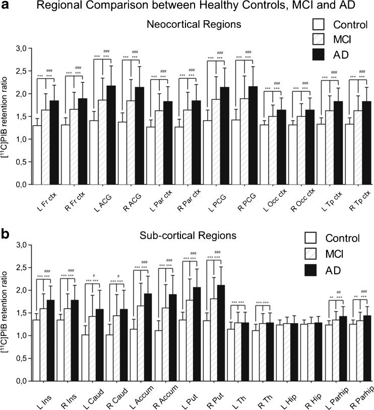
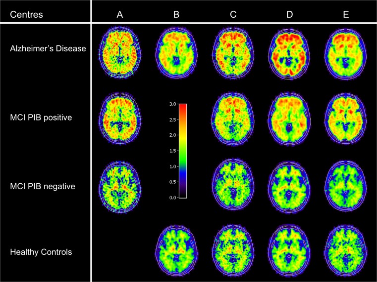
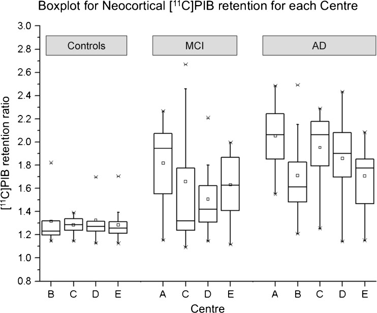
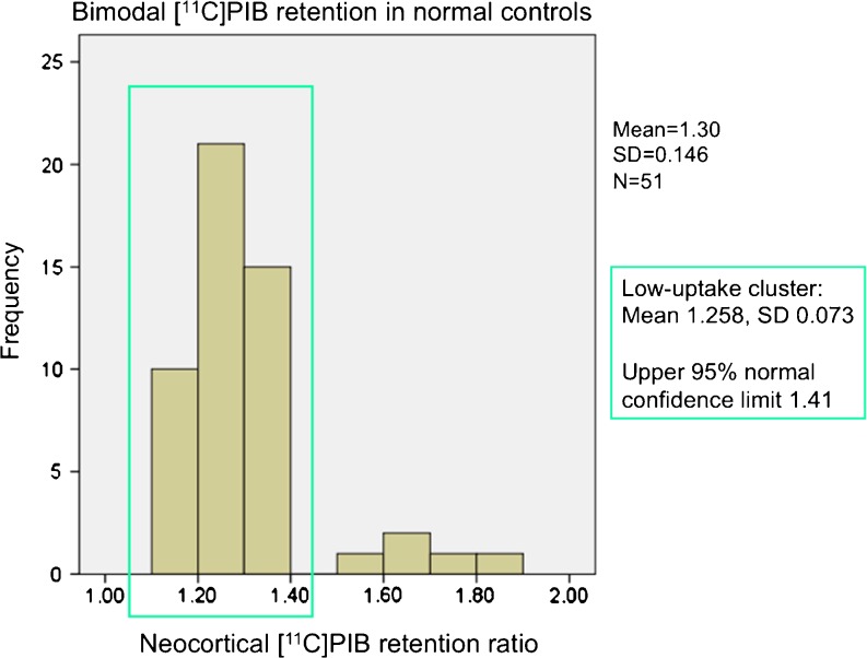
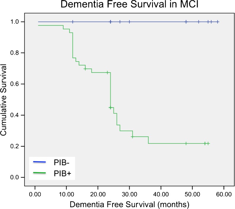
PURPOSE: Amyloid PET tracers have been developed for in vivo detection of brain fibrillar amyloid deposition in Alzheimer's disease (AD). To serve as an early biomarker in AD the amyloid PET tracers need to be analysed in multicentre clinical studies. METHODS: In this study 238 [(11)C]Pittsburgh compound-B (PIB) datasets from five different European centres were pooled. Of these 238 datasets, 18 were excluded, leaving [(11)C]PIB datasets from 97 patients with clinically diagnosed AD (mean age 69 ± 8 years), 72 patients with mild cognitive impairment (MCI; mean age 67.5 ± 8 years) and 51 healthy controls (mean age 67.4 ± 6 years) available for analysis. Of the MCI patients, 64 were longitudinally followed for 28 ± 15 months. Most participants (175 out of 220) were also tested for apolipoprotein E (ApoE) genotype. RESULTS: [(11)C]PIB retention in the neocortical and subcortical brain regions was significantly higher in AD patients than in age-matched controls. Intermediate [(11)C]PIB retention was observed in MCI patients, with a bimodal distribution (64 % MCI PIB-positive and 36 % MCI PIB-negative), which was significantly different the pattern in both the AD patients and controls. Higher [(11)C]PIB retention was observed in MCI ApoE ε4 carriers compared to non-ApoE ε4 carriers (p < 0.005). Of the MCI PIB-positive patients, 67 % had converted to AD at follow-up while none of the MCI PIB-negative patients converted. CONCLUSION: This study demonstrated the robustness of [(11)C]PIB PET as a marker of neocortical fibrillar amyloid deposition in brain when assessed in a multicentre setting. MCI PIB-positive patients showed more severe memory impairment than MCI PIB-negative patients and progressed to AD at an estimated rate of 25 % per year. None of the MCI PIB-negative patients converted to AD, and thus PIB negativity had a 100 % negative predictive value for progression to AD. This supports the notion that PIB-positive scans in MCI patients are an indicator of prodromal AD.
Overview
The claims and statistics of this paper haven’t been checked yet.
Run the check — free, no account needed. It reads the open-access text and checks the sources it cites.
Integrity signals
Registry
Cites
Statistics
Figures
Verification
Claims the paper makes
The claims and statistics of this paper haven’t been checked yet.
Run the check — free, no account needed. It reads the open-access text and checks the sources it cites.
Claims other papers make about this one
Each is checked against this paper’s own text: does it say what the citing paper says it does?
The source backs part of the claim, not all of it — or backs it for a different population, condition or comparison.
In the citing paper
“Numerous studies have shown that CU individuals with abnormal amyloid biomarkers have more rapid progression of atrophy, hypometabolism, and clinical/cognitive decline than individuals without biomarker evidence of Aβ deposition [13,33,80,93–99]”
From NIA‐AA Research Framework: Toward a biological definition of Alzheimer's disease (2018) · cites this paper as [94]
Experimental — accuracy not yet measured
Self-check
Percentages, sums and reported differences re-computed from the paper’s own numbers.
The self-check needs the paper’s full text.
Citations
Cites
McKhann, G., Drachman, D., Folstein, M., Katzman, R., Price, D., & Stadlan, E. M. (1984) · Neurology · doi:10.1212/wnl.34.7.939 · Cited by 27,915
McKhann, G. M., Knopman, D. S., Chertkow, H., Hyman, B. T., Jack, C. R., Kawas, C. H., Klunk, W. E., Koroshetz, W. J., Manly, J. J., Mayeux, R., et al. (2011) · Alzheimer's & Dementia · doi:10.1016/j.jalz.2011.03.005 · Cited by 18,657
Mild cognitive impairment as a diagnostic entity
Petersen, R. C. (2004) · Journal of Internal Medicine · doi:10.1111/j.1365-2796.2004.01388.x · Cited by 7,864
Ashburner, J., & Friston, K. J. (2005) · NeuroImage · doi:10.1016/j.neuroimage.2005.02.018 · Cited by 7,507
Diagnostic and Statistical Manual of Mental Disorders ( DSM ‐ IV ‐ TR )
Segal, D. L. (2010) · Wiley eBooks · doi:10.1002/9780470479216.corpsy0271 · Cited by 4,687
Imaging brain amyloid in Alzheimer's disease with Pittsburgh Compound‐B
Klunk, W. E., Engler, H., Nordberg, A., Wang, Y., Blomqvist, G., Holt, D. P., Bergström, M., Savitcheva, I., Huang, G., Estrada, S., et al. (2004) · Annals of Neurology · doi:10.1002/ana.20009 · Cited by 4,496
Self-citationResearch criteria for the diagnosis of Alzheimer's disease: revising the NINCDS–ADRDA criteria
Dubois, B., Feldman, H. H., Jacova, C., DeKosky, S. T., Barberger-Gateau, P., Cummings, J., Delacourte, A., Galasko, D., Gauthier, S., Jicha, G., et al. (2007) · The Lancet Neurology · doi:10.1016/s1474-4422(07)70178-3 · Cited by 4,411
The Alzheimer’s disease neuroimaging initiative (ADNI): MRI methods
Jack, C. R., Bernstein, M. A., Fox, N. C., Thompson, P., Alexander, G., Harvey, D., Borowski, B., Britson, P. J., L. Whitwell, J., Ward, C., et al. (2008) · Journal of Magnetic Resonance Imaging · doi:10.1002/jmri.21049 · Cited by 4,347
Revising the definition of Alzheimer's disease: a new lexicon
Dubois, B., Feldman, H. H., Jacova, C., Cummings, J. L., DeKosky, S. T., Barberger-Gateau, P., Delacourte, A., Frisoni, G., Fox, N. C., Galasko, D., et al. (2010) · The Lancet Neurology · doi:10.1016/s1474-4422(10)70223-4 · Cited by 1,869
Frequency of Stages of Alzheimer-Related Lesions in Different Age Categories
Braak, H., & Braak, E. (1997) · Neurobiology of Aging · doi:10.1016/s0197-4580(97)00056-0 · Cited by 1,656
Research works printed but not linked: 1
- Collignon A, Maes F, Delaere D, Vandermeulen D, Suetens P, Marchal G. Automated multi-modality image registration based on information theory. Proceedings of the International Conference on Information Processing in Medical Imaging. Dordrecht: Kluwer Academic; 1995.
Cited by
Sorted by citations
NIA‐AA Research Framework: Toward a biological definition of Alzheimer's disease
Jack, C. R., Bennett, D. A., Blennow, K., Carrillo, M. C., Dunn, B., Haeberlein, S. B., Holtzman, D. M., Jagust, W., Jessen, F., Karlawish, J., et al. (2018) · Alzheimer's & Dementia · doi:10.1016/j.jalz.2018.02.018 · Cited by 10,537
Masters, C. L., Bateman, R., Blennow, K., Rowe, C. C., Sperling, R. A., & Cummings, J. L. (2015) · Nature Reviews Disease Primers · doi:10.1038/nrdp.2015.56 · Cited by 1,853
Biomarker Modeling of Alzheimer’s Disease
Jack, C. R., & Holtzman, D. M. (2013) · Neuron · doi:10.1016/j.neuron.2013.12.003 · Cited by 1,002
Johnson, K. A., Minoshima, S., Bohnen, N. I., Donohoe, K. J., Foster, N. L., Herscovitch, P., Karlawish, J. H., Rowe, C. C., Carrillo, M. C., Hartley, D. M., et al. (2013) · Alzheimer's & Dementia · doi:10.1016/j.jalz.2013.01.002 · Cited by 603
Villeneuve, S., Rabinovici, G. D., Cohn-Sheehy, B. I., Madison, C., Ayakta, N., Ghosh, P. M., La Joie, R., Arthur-Bentil, S. K., Vogel, J. W., Marks, S. M., et al. (2015) · Brain · doi:10.1093/brain/awv112 · Cited by 408
Johnson, K. A., Minoshima, S., Bohnen, N. I., Donohoe, K. J., Foster, N. L., Herscovitch, P., Karlawish, J. H., Rowe, C. C., Carrillo, M. C., Hartley, D. M., et al. (2013) · Journal of Nuclear Medicine · doi:10.2967/jnumed.113.120618 · Cited by 329
Rodriguez‐Vieitez, E., Saint‐Aubert, L., Carter, S. F., Almkvist, O., Farid, K., Schöll, M., Chiotis, K., Þórðardóttir, S., Graff, C., Wall, A. E., et al. (2016) · Brain · doi:10.1093/brain/awv404 · Cited by 307
Self-citationMultimodal imaging in Alzheimer's disease: validity and usefulness for early detection
Teipel, S. J., Drzezga, A. E., Grothe, M. J., Barthel, H., Chételat, G., Schuff, N. W., Skudlarski, P., Cavedo, E., Frisoni, G. B., Hoffmann, W. U., et al. (2015) · The Lancet Neurology · doi:10.1016/s1474-4422(15)00093-9 · Cited by 285
Self-citationHuijbers, W., Mormino, E. C., Schultz, A. P., Wigman, S. E., Ward, A. M., Larvie, M., Amariglio, R. E., Marshall, G. A., Rentz, D. M., Johnson, K. A., et al. (2015) · Brain · doi:10.1093/brain/awv007 · Cited by 254
Sharma, R. A., Varga, A. W., Bubu, O. M., Pirraglia, E., Kam, K., Parekh, A., Wohlleber, M. E., Miller, M. D., Andrade, A. G., Lewis, C., et al. (2017) · American Journal of Respiratory and Critical Care Medicine · doi:10.1164/rccm.201704-0704oc · Cited by 259
Roberts, R. O., Aakre, J. A., Kremers, W. K., Vassilaki, M., Knopman, D. S., Mielke, M. M., Alhurani, R., Geda, Y. E., Machulda, M. M., Coloma, P., et al. (2018) · JAMA Neurology · doi:10.1001/jamaneurol.2018.0629 · Cited by 223
Rowe, C. C., Bourgeat, P., Ellis, K. A., Brown, B., Lim, Y. Y., Mulligan, R., Jones, G., Maruff, P., Woodward, M., Price, R., et al. (2013) · Annals of Neurology · doi:10.1002/ana.24040 · Cited by 222
Jack, C. R., Wiste, H. J., Botha, H., Weigand, S. D., Therneau, T. M., Knopman, D. S., Graff-Radford, J., Jones, D. T., Ferman, T. J., Boeve, B. F., et al. (2019) · Brain · doi:10.1093/brain/awz268 · Cited by 200
Unbiased Approach to Counteract Upward Drift in Cerebrospinal Fluid Amyloid-β 1–42 Analysis Results
Tijms, B. M., Willemse, E. A. J., Zwan, M. D., Mulder, S. D., Visser, P. J., van Berckel, B. N. M., van der Flier, W. M., Scheltens, P., & Teunissen, C. E. (2017) · Clinical Chemistry · doi:10.1373/clinchem.2017.281055 · Cited by 209
Smailagic, N., Vacante, M., Hyde, C., Martin, S., Ukoumunne, O. C., & Sachpekidis, C. (2015) · Cochrane Database of Systematic Reviews · doi:10.1002/14651858.cd010632.pub2 · Cited by 235
Amyloid-first and neurodegeneration-first profiles characterize incident amyloid PET positivity
Jack, C. R., Wiste, H. J., Weigand, S. D., Knopman, D. S., Lowe, V. J., Vemuri, P., Mielke, M. M., Jones, D. T., Senjem, M. L., Gunter, J. L., et al. (2013) · Neurology · doi:10.1212/01.wnl.0000435556.21319.e4 · Cited by 203
Florbetapir F 18 amyloid PET and 36-month cognitive decline:a prospective multicenter study
AV45-A11 Study Group, Doraiswamy, P. M., Sperling, R. A., Johnson, K., Reiman, E. M., Wong, T. Z., Sabbagh, M. N., Sadowsky, C. H., Fleisher, A. S., Carpenter, A., et al. (2014) · Molecular Psychiatry · doi:10.1038/mp.2014.9 · Cited by 195
Korolev, I. O., Symonds, L. L., Bozoki, A., & Initiative, A. D. N. (2016) · PLoS ONE · doi:10.1371/journal.pone.0138866 · Cited by 203
Blennow, K., Shaw, L. M. J., Stomrud, E., Mattsson, N., Toledo, J. B., Buck, K., Wahl, S., Eichenlaub, U., Lifke, V., Simon, M., et al. (2019) · Scientific Reports · doi:10.1038/s41598-019-54204-z · Cited by 194
Rowe, C. C., Pejoska, S., Mulligan, R. S., Jones, G. R., Chan, J. G., Svensson, S., Cselényi, Z., Masters, C. L., & Villemagne, V. L. (2013) · Journal of Nuclear Medicine · doi:10.2967/jnumed.112.114785 · Cited by 195
Chandra, A., Valkimadi, P., Pagano, G., Cousins, O., Dervenoulas, G., Politis, M., & Initiative, A. D. N. (2019) · Human Brain Mapping · doi:10.1002/hbm.24782 · Cited by 197
Cheng, Y.-W., Chen, T.-F., & Chiu, M.-J. (2017) · Neuropsychiatric Disease and Treatment · doi:10.2147/ndt.s123428 · Cited by 150
Schöll, M., Carter, S. F., Westman, E., Rodriguez‐Vieitez, E., Almkvist, O., Þórðardóttir, S., Wall, A. E., Graff, C., Långström, B., & Nordberg, A. K. (2015) · Scientific Reports · doi:10.1038/srep16404 · Cited by 153
Self-citationHampel, H. J., Lista, S., Teipel, S. J., Garaci, F., Nisticò, R., Blennow, K., Zetterberg, H., Bertram, L., Duyckaerts, C. J., Bakardjian, H., et al. (2013) · Biochemical Pharmacology · doi:10.1016/j.bcp.2013.11.009 · Cited by 148
Self-citationAlzheimer’s disease profiled by fluid and imaging markers: tau PET best predicts cognitive decline
Bucci, M., Chiotis, K., Nordberg, A., & Initiative, F. T. A. D. N. (2021) · Molecular Psychiatry · doi:10.1038/s41380-021-01263-2 · Cited by 140
Self-citationAmyloid tracers detect multiple binding sites in Alzheimer's disease brain tissue
Ni, R., Gillberg, P., Bergfors, A., Marutle, A., & Nordberg, A. K. (2013) · Brain · doi:10.1093/brain/awt142 · Cited by 135
Self-citationPérez‐Grijalba, V., Romero, J., Pesini, P., Sarasa, L., Monleón, I., José, I. S., Arbizu, J., Martínez‐Lage, P., Munuera, J., Ruiz, A., et al. (2019) · The Journal of Prevention of Alzheimer s Disease · doi:10.14283/jpad.2018.41 · Cited by 148
Leuzy, A., Chiotis, K., Hasselbalch, S. G., Rinne, J. O., de Mendonça, A., Otto, M., Lleó, A., Castelo-Branco, M. S., Santana, I. J., Johansson, J. J., et al. (2016) · Brain · doi:10.1093/brain/aww160 · Cited by 135
Self-citationImaging in-vivo tau pathology in Alzheimer’s disease with THK5317 PET in a multimodal paradigm
Chiotis, K., Saint‐Aubert, L., Savitcheva, I., Jelić, V., Andersen, P., Jonasson, M., Eriksson, J., Lubberink, M., Almkvist, O., Wall, A. E., et al. (2016) · European Journal of Nuclear Medicine and Molecular Imaging · doi:10.1007/s00259-016-3363-z · Cited by 131
Self-citationMitsis, E. M., Riggio, S., Kostakoğlu, L., Dickstein, D. L., Macháč, J., Delman, B. N., Goldstein, M., Jennings, D. L., D’Antonio, E., Martin, J. B., et al. (2014) · Translational Psychiatry · doi:10.1038/tp.2014.91 · Cited by 124
Patterns of microglial cell activation in frontotemporal lobar degeneration
Lant, S. B., Robinson, A. C., Thompson, J. C., Rollinson, S., Pickering‐Brown, S., Snowden, J. S., Davidson, Y. S., Gerhard, A., & Mann, D. M. A. (2014) · Neuropathology and Applied Neurobiology · doi:10.1111/nan.12092 · Cited by 102
Pilebro, B., Arvidsson, S., Lindqvist, P., Sundström, T., Westermark, P., Antoni, G., Suhr, O. B., & Sørensen, J. (2016) · Journal of Nuclear Cardiology · doi:10.1007/s12350-016-0638-5 · Cited by 112
Ewers, M., Brendel, M., Rizk‐Jackson, A., Rominger, A., Bartenstein, P., Schuff, N. W., & Weiner, M. W. (2013) · NeuroImage Clinical · doi:10.1016/j.nicl.2013.10.018 · Cited by 116
Aβ imaging with 18F-florbetaben in prodromal Alzheimer's disease: a prospective outcome study
Ong, K. T., Villemagne, V. L., Bahar‐Fuchs, A., Lamb, F., Langdon, N., Catafau, A. M., Stephens, A. W., Seibyl, J. P., Dinkelborg, L. M., Reininger, C. B., et al. (2014) · Journal of Neurology Neurosurgery & Psychiatry · doi:10.1136/jnnp-2014-308094 · Cited by 100
Amyloid PET in clinical practice: Its place in the multidimensional space of Alzheimer's disease
Vandenberghe, R., Adamczuk, K., Dupont, P., Laere, K. V., & Chételat, G. (2013) · NeuroImage: Clinical · doi:10.1016/j.nicl.2013.03.014 · Cited by 99
“A positive amyloid PET has a predictive value for future conversion to Alzheimer's disease according to early studies with follow-up periods up to 3 years (Forsberg et al., 2008; Nordberg et al., 2013; Okello et al., 2009).”
BackgroundMethodSelf-citationPerani, D., Cerami, C., Caminiti, S. P., Santangelo, R., Coppi, E., Ferrari, L. D., Pinto, P., Passerini, G., Falini, A., Iannaccone, S., et al. (2015) · European Journal of Nuclear Medicine and Molecular Imaging · doi:10.1007/s00259-015-3170-y · Cited by 91
Self-citationCurrent status and future role of brain PET/MRI in clinical and research settings
Werner, P., Barthel, H., Drzezga, A. E., & Sabri, O. (2015) · European Journal of Nuclear Medicine and Molecular Imaging · doi:10.1007/s00259-014-2970-9 · Cited by 88
Self-citationPotential Clinical Applications of PET/MR Imaging in Neurodegenerative Diseases
Drzezga, A. E., Barthel, H., Minoshima, S., & Sabri, O. (2014) · Journal of Nuclear Medicine · doi:10.2967/jnumed.113.129254 · Cited by 83
Self-citationChaney, A. M., Lopez‐Picon, F. R., Sérrière, S., Wang, R., Bochicchio, D., Webb, S. D., Vandesquille, M., Harte, M., Georgiadou, C., Lawrence, C. B., et al. (2021) · Theranostics · doi:10.7150/thno.56059 · Cited by 73
Self-citationZhou, Y., Yu, F. F., Duong, T. Q., & Initiative, A. D. N. (2013) · Journal of Magnetic Resonance Imaging · doi:10.1002/jmri.24550 · Cited by 76
Rodriguez‐Vieitez, E., Carter, S. F., Chiotis, K., Saint‐Aubert, L., Leuzy, A., Schöll, M., Almkvist, O., Wall, A. E., Långström, B., & Nordberg, A. K. (2016) · Journal of Nuclear Medicine · doi:10.2967/jnumed.115.168732 · Cited by 76
Self-citationChiotis, K., Saint‐Aubert, L., Boccardi, M., Gietl, A. F., Picco, A., Varrone, A., Garibotto, V., Herholz, K., Nobili, F. M., Nordberg, A. K., et al. (2017) · Neurobiology of Aging · doi:10.1016/j.neurobiolaging.2016.07.012 · Cited by 75
Self-citationSchreiber, S., Landau, S. M., Fero, A., Schreiber, F., & Jagust, W. J. (2015) · JAMA Neurology · doi:10.1001/jamaneurol.2015.1633 · Cited by 79
Dickstein, D. L., Pullman, M. Y., Fernandez, C., Short, J. A., Kostakoğlu, L., Knešaurek, K., Soleimani, L., Jordan, B. D., Gordon, W. A., Dams-O’Connor, K., et al. (2016) · Translational Psychiatry · doi:10.1038/tp.2016.175 · Cited by 74
(ADNI), T. A. D. N. I., Bouallègue, F. B., Mariano‐Goulart, D., & Payoux, P. (2017) · Alzheimer s Research & Therapy · doi:10.1186/s13195-017-0260-z · Cited by 73
Ezawa, N., Katoh, N., Oguchi, K., Yoshinaga, T., Yazaki, M., & Sekijima, Y. (2017) · European Journal of Nuclear Medicine and Molecular Imaging · doi:10.1007/s00259-017-3814-1 · Cited by 65
Deficits in Spontaneous Cognition as an Early Marker of Alzheimer’s Disease
Kvavilashvili, L., Niedźwieńska, A., Gilbert, S. J., & Markostamou, I. (2020) · Trends in Cognitive Sciences · doi:10.1016/j.tics.2020.01.005 · Cited by 74
Zhang, S., Smailagic, N., Hyde, C., Noel-Storr, A. H., Takwoingi, Y., McShane, R., & Feng, J. (2014) · Cochrane Database of Systematic Reviews · doi:10.1002/14651858.cd010386.pub2 · Cited by 66
Ton, T. G. N., DeLeire, T., May, S. G., Hou, N., Tebeka, M. G., Chen, E., & Chodosh, J. (2016) · Alzheimer s & Dementia · doi:10.1016/j.jalz.2016.08.009 · Cited by 71
Distinct pattern of hypometabolism and atrophy in preclinical and predementia Alzheimer's disease
Kljajević, V., Grothe, M. J., Ewers, M., & Teipel, S. J. (2014) · Neurobiology of Aging · doi:10.1016/j.neurobiolaging.2014.04.006 · Cited by 67
Vandenberghe, R. R., Nelissen, N., Salmon, É., Ivanoiu, A., Hasselbalch, S. G., Andersen, A., Kørner, A., Minthon, L., Brooks, D. J., Laere, K. V., et al. (2012) · NeuroImage · doi:10.1016/j.neuroimage.2012.09.015 · Cited by 64
Self-citationChoo, I. H., Carter, S. F., Schöll, M., & Nordberg, A. K. (2014) · European Journal of Nuclear Medicine and Molecular Imaging · doi:10.1007/s00259-014-2859-7 · Cited by 63
Self-citationAmyloid Imaging with PET in Early Alzheimer Disease Diagnosis
Rowe, C. C., & Villemagne, V. L. (2013) · Medical Clinics of North America · doi:10.1016/j.mcna.2012.12.017 · Cited by 62
Imaging of Reactive Astrogliosis by Positron Emission Tomography
Harada, R., Furumoto, S., Kudo, Y., Yanai, K., Villemagne, V. L., & Okamura, N. (2022) · Frontiers in Neuroscience · doi:10.3389/fnins.2022.807435 · Cited by 63
Edison, P., Carter, S. F., Rinne, J. O., Gelosa, G., Herholz, K., Nordberg, A. K., Brooks, D. J., & Hinz, R. (2012) · NeuroImage · doi:10.1016/j.neuroimage.2012.12.014 · Cited by 62
Self-citationIaccarino, L., Chiotis, K., Alongi, P., Almkvist, O., Wall, A. E., Cerami, C., Bettinardi, V., Gianolli, L., Nordberg, A. K., & Perani, D. (2017) · Journal of Alzheimer s Disease · doi:10.3233/jad-170158 · Cited by 63
Self-citationRodriguez‐Vieitez, E., Leuzy, A., Chiotis, K., Saint‐Aubert, L., Wall, A. E., & Nordberg, A. K. (2016) · Journal of Cerebral Blood Flow & Metabolism · doi:10.1177/0271678x16645593 · Cited by 61
Self-citationA Survey of FDG- and Amyloid-PET Imaging in Dementia and GRADE Analysis
Perani, D., Schillaci, O., Padovani, A., Nobili, F. M., Iaccarino, L., Pasquale, D. R., Frisoni, G. B., & Caltagirone, C. (2014) · BioMed Research International · doi:10.1155/2014/785039 · Cited by 61
Self-citationTracer Kinetic Analysis of (S)-18F-THK5117 as a PET Tracer for Assessing Tau Pathology
Jonasson, M., Wall, A. E., Chiotis, K., Saint‐Aubert, L., Wilking, H., Sprycha, M., Borg, B., Thibblin, A., Eriksson, J., Sørensen, J., et al. (2016) · Journal of Nuclear Medicine · doi:10.2967/jnumed.115.158519 · Cited by 59
Self-citationSaint‐Aubert, L., Almkvist, O., Chiotis, K., Almeida, R., Wall, A. E., & Nordberg, A. K. (2016) · Alzheimer s Research & Therapy · doi:10.1186/s13195-016-0204-z · Cited by 58
Self-citationThe AB255 Study Group, Pérez‐Grijalba, V., Arbizu, J., Romero, J., Prieto, E., Pesini, P., Sarasa, L., Guillén, F., Monleón, I., José, I. S., et al. (2019) · Alzheimer s Research & Therapy · doi:10.1186/s13195-019-0549-1 · Cited by 60
Physical Activity in Preventing Alzheimer’s Disease and Cognitive Decline: A Narrative Review
Brini, S., Sohrabi, H. R., Peiffer, J. J., Karrasch, M., Hämäläinen, H., Martins, R. N., & Fairchild, T. J. (2017) · Sports Medicine · doi:10.1007/s40279-017-0787-y · Cited by 62
Self-citationQuantum Dots as A Theranostic Approach in Alzheimer’s Disease: A Systematic Review
Villalva, M. D., Agarwal, V., Ulanova, M., Sachdev, P. S., & Braidy, N. (2021) · Nanomedicine · doi:10.2217/nnm-2021-0104 · Cited by 53
Imaging Alzheimer's disease pathophysiology with PET
Schilling, L. P., Zimmer, E. R., Shin, M., Leuzy, A., Pascoal, T. A., Benedet, A. L., Borelli, W. V., Palmini, A., Gauthier, S. G., & Rosa‐Neto, P. (2016) · Dementia & Neuropsychologia · doi:10.1590/s1980-5764-2016dn1002003 · Cited by 57
The emerging role of PET imaging in dementia
Iaccarino, L., Sala, A., Caminiti, S. P., & Perani, D. (2017) · F1000Research · doi:10.12688/f1000research.11603.1 · Cited by 52
Self-citationSole, A. D. (2016) · Functional Neurology · doi:10.11138/fneur/2016.31.4.205 · Cited by 49
for the EANM-EAN Task Force for the Prescription of FDG-PET for Dementing Neurodegenerative Disorders, Drzezga, A. E., Altomare, D., Festari, C., Arbizu, J., Orini, S., Herholz, K., Nestor, P. J., Agosta, F., Bouwman, F. H., et al. (2018) · European Journal of Nuclear Medicine and Molecular Imaging · doi:10.1007/s00259-018-4032-1 · Cited by 43
Self-citationLeuzy, A., Carter, S. F., Chiotis, K., Almkvist, O., Wall, A. E., & Nordberg, A. K. (2015) · Journal of Alzheimer s Disease · doi:10.3233/jad-142952 · Cited by 44
Self-citationSarikaya, İ. (2015) · Nuclear Medicine Communications · doi:10.1097/mnm.0000000000000320 · Cited by 44
A Candidate Plasma Protein Classifier to Identify Alzheimer's Disease
Zhao, X., Lejnine, S., Spond, J., Zhang, C., Ramaraj, T. C., Holder, D., Dai, H. A., WEINER, R. S., & Laterza, O. (2014) · Journal of Alzheimer s Disease · doi:10.3233/jad-141149 · Cited by 43
Yi, D., Choe, Y. M., Byun, M. S., Sohn, B. K., Seo, E. H., Han, J., Park, J., Woo, J. I., & Lee, D. Y. (2015) · Frontiers in Aging Neuroscience · doi:10.3389/fnagi.2015.00015 · Cited by 40
Schicktanz, S., Schweda, M., Ballenger, J. F., Fox, P. J., Halpern, J., Kramer, J. H., Micco, G. P., Post, S. G., Thompson, C., Knight, R. T., et al. (2014) · Frontiers in Human Neuroscience · doi:10.3389/fnhum.2014.00921 · Cited by 40
Akamatsu, G., Ikari, Y., Ohnishi, A., Nishida, H., Aita, K., Sasaki, M., Yamamoto, Y., Sasaki, M., & Senda, M. (2016) · Physics in Medicine and Biology · doi:10.1088/0031-9155/61/15/5768 · Cited by 40
Five-year follow-up of 11C-PIB uptake in Alzheimer’s disease and MCI
Kemppainen, N. M., Scheinin, N. M., Koivunen, J., Johansson, J. J., Toivonen, J., Någren, K., Rokka, J., Karrasch, M., Parkkola, R. K., & Rinne, J. O. (2013) · European Journal of Nuclear Medicine and Molecular Imaging · doi:10.1007/s00259-013-2562-0 · Cited by 40
Self-citationZhang, W., Hu, C., Yang, Z., Zhou, Q., Peng, D., Yuan, X., Lv, Z., Pang, S., Jiang, W., Yang, C., et al. (2014) · Neural Regeneration Research · doi:10.4103/1673-5374.130117 · Cited by 36
Tentolouris-Piperas, V., Ryan, N. S., Thomas, D. L., & Kinnunen, K. M. (2016) · Brain Research · doi:10.1016/j.brainres.2016.11.011 · Cited by 38
Dual-phase amyloid PET: hitting two birds with one stone
Garibotto, V., Morbelli, S. D., & Pagani, M. (2016) · European Journal of Nuclear Medicine and Molecular Imaging · doi:10.1007/s00259-016-3393-6 · Cited by 33
Self-citationKononikhin, A. S., Zakharova, N. V., Semenov, S. D., Bugrova, A. E., Brzhozovskiy, A. G., Indeykina, M. I., Fedorova, Y. B., Колыхалов, И. В., Strelnikova, P. A., Ikonnikova, A. Y., et al. (2022) · International Journal of Molecular Sciences · doi:10.3390/ijms23147907 · Cited by 33
Möckelind, S., Axelsson, J., Pilebro, B., Lindqvist, P., Suhr, O. B., & Sundström, T. (2020) · Amyloid · doi:10.1080/13506129.2020.1760237 · Cited by 34
Mechanics of cryopreserved aortic and pulmonary homografts.
Veselý, I., Casarotto, D. C., & Gerosa, G. (2000) · PubMed · Cited by 32
Presotto, L., Iaccarino, L., Sala, A., Vanoli, E. G., Muscio, C., Nigri, A., Bruzzone, M. G., Tagliavini, F., Gianolli, L., Perani, D., et al. (2018) · NeuroImage Clinical · doi:10.1016/j.nicl.2018.07.013 · Cited by 33
Self-citationNeuroimaging of dementia in 2013: what radiologists need to know
Haller, S., Garibotto, V., Kövari, E., Bouras, C., Xekardaki, A., Rodriguez, C. K., Lazarczyk, M. J., Giannakopoulos, P., & Lövblad, K. (2013) · European Radiology · doi:10.1007/s00330-013-2957-0 · Cited by 30
Self-citationZukotynski, K., Kuo, P. H., Mikulis, D. J., Rosa‐Neto, P., Strafella, A. P., Subramaniam, R. M., & Black, S. E. (2018) · American Journal of Roentgenology · doi:10.2214/ajr.18.19822 · Cited by 31
Optimization of image reconstruction conditions with phantoms for brain FDG and amyloid PET imaging
Akamatsu, G., Ikari, Y., Nishio, T., Nishida, H., Ohnishi, A., Aita, K., Sasaki, M., Sasaki, M., & Senda, M. (2015) · Annals of Nuclear Medicine · doi:10.1007/s12149-015-1024-0 · Cited by 28
Amyloid-Independent Amnestic Mild Cognitive Impairment and Serum Apolipoprotein A1 Levels
Choi, H. J., Seo, E. H., Yi, D., Sohn, B. K., Choe, Y. M., Byun, M. S., Lee, J. M., Woo, J. I., & Lee, D. Y. (2015) · American Journal of Geriatric Psychiatry · doi:10.1016/j.jagp.2015.06.004 · Cited by 27
Núñez-Ríos, D. L., Martínez‐Magaña, J. J., Nagamatsu, S. T., Andrade-Brito, D., Forero, D. A., Orozco, C. A., & Montalvo‐Ortiz, J. L. (2022) · Biomedicines · doi:10.3390/biomedicines10051107 · Cited by 27
[18F]THK5317 imaging as a tool for predicting prospective cognitive decline in Alzheimer’s disease
Chiotis, K., Savitcheva, I., Poulakis, K., Saint-Aubert, L., Wall, A., Antoni, G., & Nordberg, A. (2020) · Molecular Psychiatry · doi:10.1038/s41380-020-0815-4 · Cited by 21
Self-citationPerani, D., Iaccarino, L., Lammertsma, A. A., Windhorst, A. D., Edison, P., Boellaard, R., Hansson, O., Nordberg, A. K., Jacobs, A. H., & IMBI Project (2019) · Alzheimer s & Dementia · doi:10.1016/j.jalz.2019.02.004 · Cited by 25
Self-citationValkanova, V., & Ebmeier, K. P. (2014) · Maturitas · doi:10.1016/j.maturitas.2014.02.016 · Cited by 27
Imaging Neuroinflammation: Quantification of Astrocytosis in a Multitracer PET Approach
Rodriguez‐Vieitez, E., & Nordberg, A. K. (2018) · Methods in molecular biology · doi:10.1007/978-1-4939-7704-8_16 · Cited by 24
Self-citationCollij, L. E., Farrar, G., García, D. V., Bader, I., Shekari, M., Lorenzini, L., Pemberton, H., Altomare, D., Pla, S., Loor, M., et al. (2023) · Frontiers in Neurology · doi:10.3389/fneur.2022.1063598 · Cited by 22
Self-citationBiomarkers and Tools for Predicting Alzheimer’s Disease in the Preclinical Stage
Li, T.-R., Yang, Q. Z., Hu, X., & Han, Y. (2021) · Current Neuropharmacology · doi:10.2174/1570159x19666210524153901 · Cited by 23
Diagnosis of Cognitive Impairment Compatible with Early Diagnosis of Alzheimer’s Disease
Guerrero, J. M., Rincón, M., Peraita, H., & Martínez‐Tomás, R. (2015) · Methods of Information in Medicine · doi:10.3414/me14-01-0071 · Cited by 22
P ittsburgh compound B retention and progression of cognitive status – a meta‐analysis
Chen, X., Li, M., Wang, S., Zhu, H., Xiong, Y., & Liu, X. (2014) · European Journal of Neurology · doi:10.1111/ene.12398 · Cited by 26
Þórðardóttir, S., Rodriguez‐Vieitez, E., Almkvist, O., Ferreira, D., Saint‐Aubert, L., Ståhlbom, A. K., Thonberg, H., Schöll, M., Westman, E., Wall, A. E., et al. (2018) · Alzheimer s Research & Therapy · doi:10.1186/s13195-018-0374-y · Cited by 23
Self-citationSpatial normalization and quantification approaches of PET imaging for neurological disorders
Zhang, T., Wu, S., Zhang, X., Dai, Y., Wang, A., Zhang, H., & Tian, M. (2022) · European Journal of Nuclear Medicine and Molecular Imaging · doi:10.1007/s00259-022-05809-6 · Cited by 21
Alzheimer’s Dementia: The Emerging Role of Positron Emission Tomography
Tripathi, S. M., & Murray, A. D. (2021) · The Neuroscientist · doi:10.1177/1073858421997035 · Cited by 23
Amyloid Imaging With 11C-PIB in Patients With Cognitive Impairment in a Clinical Setting
Jiménez-Bonilla, J. F., Banzo, I., de Arcocha-Torres, M., Quirce, R., Martínez‐Rodríguez, I., Sánchez‐Juan, P., & CARRIL, J. M. (2015) · Clinical Nuclear Medicine · doi:10.1097/rlu.0000000000000934 · Cited by 21
The interest of amyloid PET imaging in the diagnosis of Alzheimerʼs disease
Vandenberghe, R. R., Adamczuk, K., & Laere, K. V. (2013) · Current Opinion in Neurology · doi:10.1097/wco.0000000000000036 · Cited by 20
Self-citationMultimodality Imaging of Neurodegenerative Processes: Part 1, The Basics and Common Dementias
Martin-Macintosh, E. L., Broski, S. M., Johnson, G. B., Hunt, C. H., Cullen, E. L., & Peller, P. J. (2016) · American Journal of Roentgenology · doi:10.2214/ajr.14.12842 · Cited by 21
Metadata
- Authors
- Nordberg, A., Carter, S. F., Rinne, J., Drzezga, A., Brooks, D. J., Vandenberghe, R., Perani, D., Forsberg, A., Långström, B., Scheinin, N., Karrasch, M., Någren, K., Grimmer, T., Miederer, I., Edison, P., Okello, A., Van Laere, K., Nelissen, N., Vandenbulcke, M., Garibotto, V., Almkvist, O., Kalbe, E., Hinz, R., & Herholz, K.
- Affiliations
- Karolinska University Hospital (SE)
- Karolinska Institutet (SE)
- University of Turku (FI)
- Technical University of Munich (DE)
- Imperial College London (GB)
- KU Leuven (BE)
- Vita-Salute San Raffaele University (IT)
- Uppsala University (SE)
- University of Vechta (DE)
- University of Manchester (GB)
- Corresponding author
- Nordberg, Agneta
- ORCID
- Nordberg, Agneta
- Carter, Stephen F.
- Rinne, Juha
- Drzezga, Alexander
- Brooks, David J.
- Vandenberghe, Rik
- Perani, Daniela
- Forsberg, Anton
- Scheinin, Noora
- Karrasch, Mira
- Någren, Kjell
- Grimmer, Timo
- Miederer, Isabelle
- Edison, Paul
- Nelissen, Natalie
- Vandenbulcke, Mathieu
- Garibotto, Valentina
- Almkvist, Ove
- Kalbe, Elke
- Hinz, Rainer
- Herholz, Karl
- Venue
- European Journal of Nuclear Medicine and Molecular Imaging
- Published
- 2012-09-08
- Volume
- 40
- Issue
- 1
- Pages
- 104-114
- ISSN
- 1619-7070 (ISSN-L)
- 1619-7089
- PMC
- PMC3510420
- OpenAlex
- W2116591217
- Cited by
- 186
- References
- 46
- Citations by year
- 2026: 2 · 2025: 2 · 2024: 4 · 2023: 5 · 2022: 11 · 2021: 13 · 2020: 9 · 2019: 10 · 2018: 9 · 2017: 14 · 2016: 25 · 2015: 27 · 2014: 27 · 2013: 24 · 2012: 2
- FWCI
- 18.28
- Type
- journal article
- Language
- en
- Open access
- hybrid
- doi.org
- Licence
- cc-by
- Locations
- European Journal of Nuclear Medicine and Molecular Imaging (publishedVersion, cc-by)
- PubMed Central (publishedVersion, cc-by)
- PubMed (publishedVersion)
- Research Explorer (The University of Manchester) (publishedVersion)
- Archive ouverte UNIGE (University of Geneva) (publishedVersion, other-oa)
- Archivio Istituzionale della Ricerca - Università Vita-Salute San Raffaele (submittedVersion)
- Lirias (KU Leuven) (submittedVersion)
- mediaTUM – the media and publications repository of the Technical University Munich (Technical University Munich) (submittedVersion)
- Åbo Akademi University Research Portal (submittedVersion)
- Archive ouverte UNIGE (University of Geneva) (submittedVersion, cc-by)
- Funders
- Alzheimer's Research Trust
- Deutsche Forschungsgemeinschaft (DR 445/3-1, DR 445/4-1)
- Academy of Finland
- Belgian Federal Science Policy Office (IUAP P6/29)
- Fonds Wetenschappelijk Onderzoek (05/014, G0668.07, P6/29, G0076.02)
- Karolinska Institutet (05817)
- Stockholms Läns Landsting
- Vetenskapsrådet (05817)
- Technische Universität München
- Swedish Brain Power (05817)
- Universität zu Köln
- Turun Yliopistollinen Keskussairaala
- Medical Research Council
- Topics
- Health Sciences › Medicine › Psychiatry and Mental health › Dementia and Cognitive Impairment Research
- Life Sciences › Neuroscience › Cellular and Molecular Neuroscience › Alzheimer's disease research and treatments
- Health Sciences › Medicine › Radiology, Nuclear Medicine and Imaging › Medical Imaging Techniques and Applications
- Keywords
- Alzheimer's disease, amyloid PET, Pittsburgh compound B, mild cognitive impairment, prodromal Alzheimer's disease, APOE ε4, conversion to Alzheimer's disease, memory impairment, negative predictive value, amyloid biomarkers, longitudinal follow-up
- MeSH terms
- Aged, Alzheimer Disease, Amyloid, Aniline Compounds, Apolipoproteins E, Brain, Brain Chemistry, Europe, Female, Humans, Male, Middle Aged, Thiazoles, Case-Control Studies, Radiopharmaceuticals, Positron-Emission Tomography
About these checks
Registry, retraction, Cites and statistics results are computed, not judged, with one exception: a printed reference that no index lists is read once by a model before it counts as a citation error. Claims are extracted by a model and kept only when their quote appears verbatim in the text; each cited source is then checked, with a model’s help, for whether it says what the claim relies on. Those verdicts can be wrong — read the claim’s quote and the cited source before relying on one.
Statistics and arithmetic are recomputed, not judged.
Confidence bands are shown only for fields calibrated on our annotated test set. None is calibrated yet, so no band is shown.
What counts as a claim ›How we measure confidence ›How we check this ›
Are you an author, or do you see a problem with this page? Request a review
