

The DOI matches its Crossref record; 72 of the 73 references it cites were found in a registry or an index.
Single-Cell Transcriptomics Reveals that Differentiation and Spatial Signatures Shape Epidermal and Hair Follicle Heterogeneity
Open accessAbstract
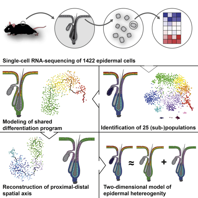
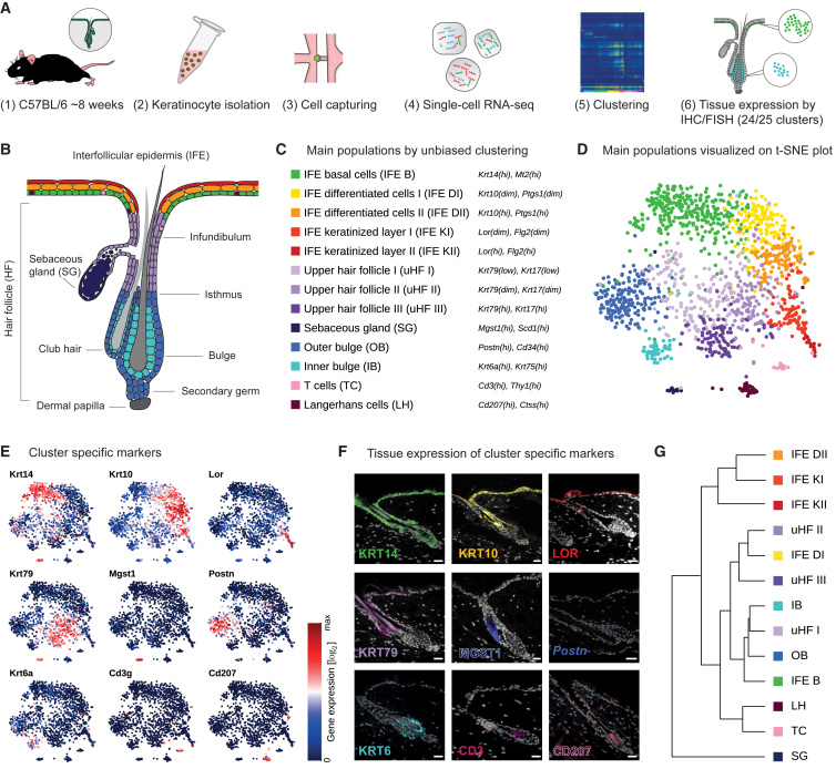
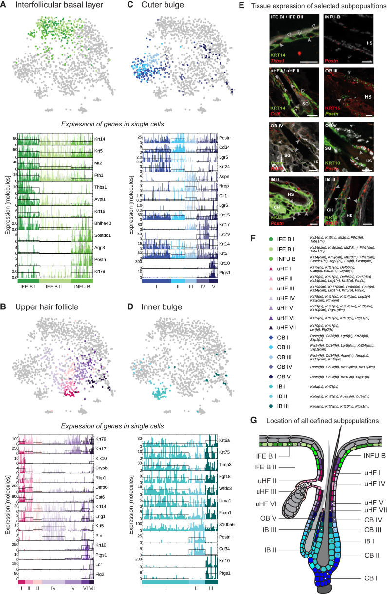
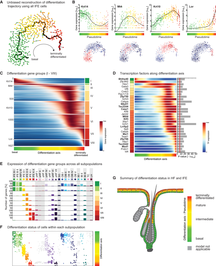
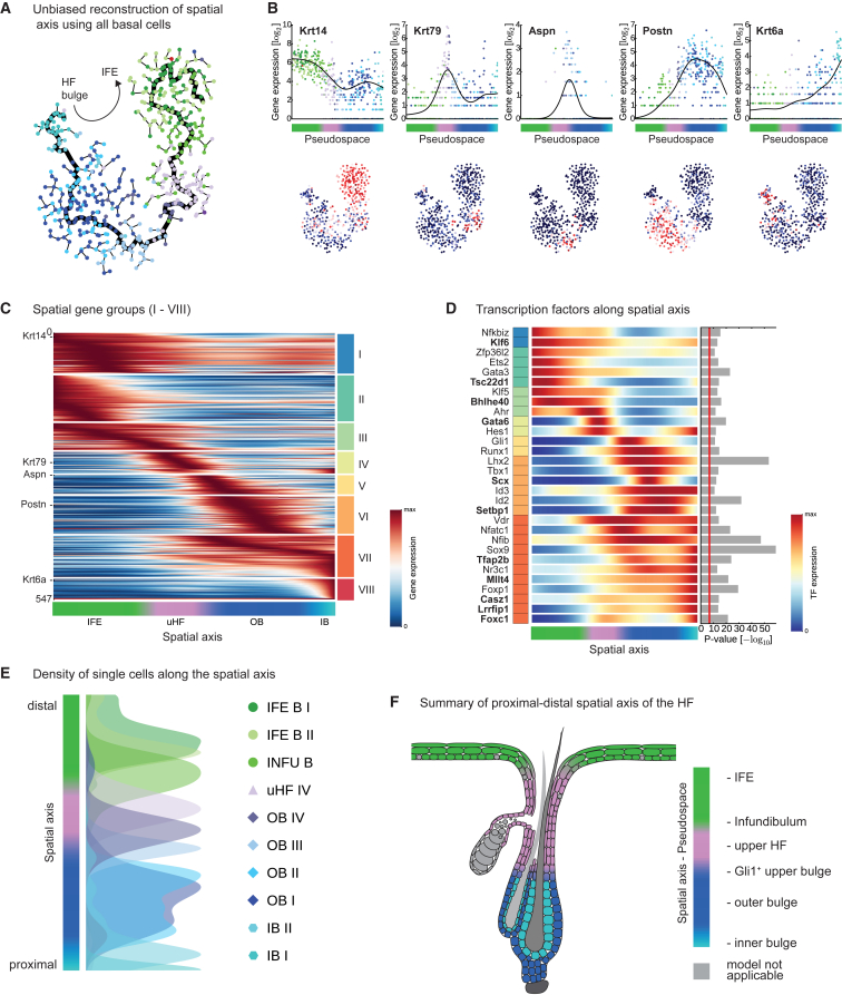
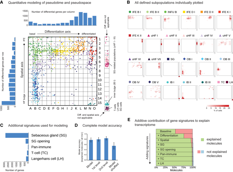
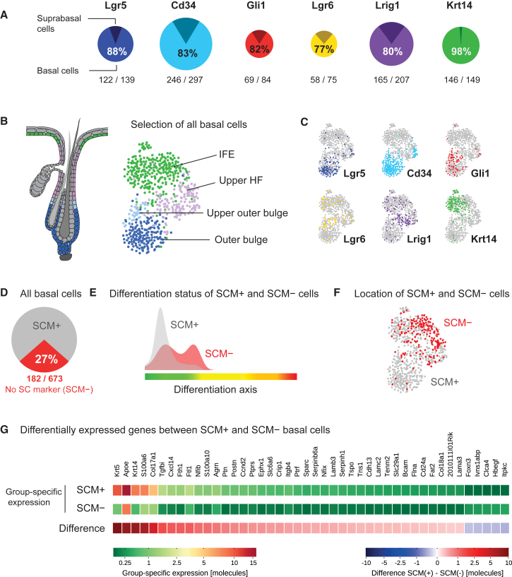
The murine epidermis with its hair follicles represents an invaluable model system for tissue regeneration and stem cell research. Here we used single-cell RNA-sequencing to reveal how cellular heterogeneity of murine telogen epidermis is tuned at the transcriptional level. Unbiased clustering of 1,422 single-cell transcriptomes revealed 25 distinct populations of interfollicular and follicular epidermal cells. Our data allowed the reconstruction of gene expression programs during epidermal differentiation and along the proximal-distal axis of the hair follicle at unprecedented resolution. Moreover, transcriptional heterogeneity of the epidermis can essentially be explained along these two axes, and we show that heterogeneity in stem cell compartments generally reflects this model: stem cell populations are segregated by spatial signatures but share a common basal-epidermal gene module. This study provides an unbiased and systematic view of transcriptional organization of adult epidermis and highlights how cellular heterogeneity can be orchestrated in vivo to assure tissue homeostasis.
Overview
The claims and statistics of this paper haven’t been checked yet.
Run the check — free, no account needed. It reads the open-access text and checks the sources it cites.
Integrity signals
Registry
Cites
Statistics
Figures
Verification
Claims the paper makes
The claims and statistics of this paper haven’t been checked yet.
Run the check — free, no account needed. It reads the open-access text and checks the sources it cites.
Claims other papers make about this one
Each is checked against this paper’s own text: does it say what the citing paper says it does?
The cited source says what the claim attributes to it.
In the citing paper
“We identified putative pilosebaceous clusters based on expression of known hair follicle markers or transcription factors (KRT15, LHX2, SOX9) and prior mouse and human studies in addition to manual cross-referencing of the Human Protein Atlas (Joost et al., 2016, Purba et al., 2015, Uhlén et al., 2015) and removed these from further analysis.”
From Multimodal Analysis of Composition and Spatial Architecture in Human Squamous Cell Carcinoma (2020) · cites this paper as Joost et al., 2016
The check wasn’t confident either way, so it gives no verdict — never disputed.
In the citing paper
“The epithelial group contained three epidermal sub-clusters: CXCL14+ basal, KRT1+ intermediate, and KRT4+ peridermal keratinocytes (Fig. 2e, Extended Data Fig. 5)22.”
From Hair-bearing human skin generated entirely from pluripotent stem cells (2020) · cites this paper as [22]
Experimental — accuracy not yet measured
Self-check
Percentages, sums and reported differences re-computed from the paper’s own numbers.
The self-check needs the paper’s full text.
Citations
Cites
Scikit-learn: Machine Learning in Python
Pedregosa, F., Varoquaux, G., Gramfort, A., Michel, V., Thirion, B., Grisel, O., Blondel, M., Müller, A., Nothman, J., Louppe, G., et al. (2012) · arXiv · doi:10.48550/arxiv.1201.0490 · Cited by 71,016
Subramanian, A., Tamayo, P., Mootha, V. K., Mukherjee, S., Ebert, B. L., Gillette, M. A., Paulovich, A., Pomeroy, S. L., Golub, T. R., Lander, E. S., et al. (2005) · Proceedings of the National Academy of Sciences · doi:10.1073/pnas.0506580102 · Cited by 53,625
(2008) · Journal of Machine Learning Research · Cited by 33,051
Exploring Network Structure, Dynamics, and Function Using NetworkX
Hagberg, A. A., Schult, D. A., & Swart, P. J. (2008) · Proceedings of the Python in Science Conference · doi:10.25080/tcwv9851 · Cited by 7,477
Trapnell, C., Cacchiarelli, D., Grimsby, J., Pokharel, P., Li, S., Morse, M., Lennon, N. J., Livak, K. J., Mikkelsen, T. S., & Rinn, J. L. (2014) · Nature Biotechnology · doi:10.1038/nbt.2859 · Cited by 7,314
Highly parallel genome-wide expression profiling of individual cells using nanoliter droplets.
Macosko, E. Z., Basu, A., Satija, R., Nemesh, J., Shekhar, K., Goldman, M., Tirosh, I., Bialas, A. R., Kamitaki, N., Martersteck, E. M., et al. (2015) · Cell · doi:10.1016/j.cell.2015.05.002 · Cited by 7,110
Clustering by Passing Messages Between Data Points
Frey, B. J., & Dueck, D. (2007) · Science · doi:10.1126/science.1136800 · Cited by 6,855
Cell types in the mouse cortex and hippocampus revealed by single-cell RNA-seq
Zeisel, A., Muñoz-Manchado, A. B., Codeluppi, S., Lönnerberg, P., La Manno, G., Juréus, A., Marques, S., Munguba, H., He, L., Betsholtz, C., et al. (2015) · Science · doi:10.1126/science.aaa1934 · Cited by 3,124
Smart-seq2 for sensitive full-length transcriptome profiling in single cells
Picelli, S., Björklund, Å. K., Faridani, O. R., Sagasser, S., Winberg, G., & Sandberg, R. (2013) · Nature Methods · doi:10.1038/nmeth.2639 · Cited by 2,358
Cotsarelis, G., Sun, T.-T., & Lavker, R. M. (1990) · Cell · doi:10.1016/0092-8674(90)90696-c · Cited by 2,276
Cited by
Sorted by citations
Single-cell transcriptional diversity is a hallmark of developmental potential
Gulati, G. S., Sikandar, S. S., Wesche, D. J., Manjunath, A., Bharadwaj, A., Berger, M. J., Ilagan, F., Kuo, A. H., Hsieh, R. W., Cai, S., et al. (2020) · Science · doi:10.1126/science.aax0249 · Cited by 1,571
Chromatin Potential Identified by Shared Single-Cell Profiling of RNA and Chromatin
Ma, S., Zhang, B., LaFave, L. M., Earl, A. S., Chiang, Z., Hu, Y., Ding, J., Brack, A., Kartha, V. K., Tay, T., et al. (2020) · Cell · doi:10.1016/j.cell.2020.09.056 · Cited by 1,146
Deciphering cell–cell interactions and communication from gene expression
Armingol, E., Officer, A., Harismendy, O., & Lewis, N. E. (2021) · Nature Reviews Genetics · doi:10.1038/s41576-020-00292-x · Cited by 1,267
Multimodal Analysis of Composition and Spatial Architecture in Human Squamous Cell Carcinoma
Ji, A. L., Rubin, A. J., Thrane, K., Jiang, S., Reynolds, D. L., Meyers, R. M., Guo, M. G., George, B. M., Mollbrink, A., Bergenstråhle, J., et al. (2020) · Cell · doi:10.1016/j.cell.2020.05.039 · Cited by 1,117
Wound healing, fibroblast heterogeneity, and fibrosis
desJardins-Park, H. E., Mascharak, S., Griffin, M., Wan, D. C., & Longaker, M. T. (2022) · Cell stem cell · doi:10.1016/j.stem.2022.07.006 · Cited by 835
Cell type–specific genetic regulation of gene expression across human tissues
Kim-Hellmuth, S., Aguet, F., Oliva, M., Muñoz-Aguirre, M., Kasela, S., Wucher, V., Castel, S. E., Hamel, A. R., Viñuela, A., Roberts, A. L., et al. (2020) · Science · doi:10.1126/science.aaz8528 · Cited by 541
Developmental cell programs are co-opted in inflammatory skin disease
Reynolds, G., Vegh, P., Fletcher, J., Poyner, E. F. M., Stephenson, E., Goh, I., Botting, R. A., Huang, N., Olabi, B., Dubois, A., et al. (2021) · Science · doi:10.1126/science.aba6500 · Cited by 529
“S6C) recapitulate previous reports in human and mouse (65).”
BackgroundGuerrero-Juarez, C. F., Dedhia, P. H., Jin, S., Ruiz-Vega, R., Ma, D., Liu, Y., Yamaga, K., Shestova, O., Gay, D. L., Yang, Z., et al. (2019) · Nature Communications · doi:10.1038/s41467-018-08247-x · Cited by 521
He, H., Suryawanshi, H., Morozov, P. S., Gay-Mimbrera, J., DUCA, E. D., Kim, H. J., Kameyama, N., Estrada, Y. D., Der, E. B., Krueger, J. G., et al. (2020) · Journal of Allergy and Clinical Immunology · doi:10.1016/j.jaci.2020.01.042 · Cited by 510
Hair-bearing human skin generated entirely from pluripotent stem cells
Lee, J., Rabbani, C. C., Gao, H., Steinhart, M. R., Woodruff, B. M., Pflum, Z. E., Kim, A., Heller, S., Liu, Y., Shipchandler, T. Z., et al. (2020) · Nature · doi:10.1038/s41586-020-2352-3 · Cited by 487
Skin and Its Regenerative Powers: An Alliance between Stem Cells and Their Niche
Gonzales, K. A. U., & Fuchs, E. (2017) · Developmental Cell · doi:10.1016/j.devcel.2017.10.001 · Cited by 414
“A uthor M anuscript A uthor M anuscript A uthor M anuscript A uthor M anuscript α6β4, KRT5/14, LGR5, SOX9, TCF3/4, NFATc1, and LHX2 (Joost et al., 2016; Yang et al., 2017).”
BackgroundSingleCellSignalR: inference of intercellular networks from single-cell transcriptomics
Cabello‐Aguilar, S., Alame, M., Kon-Sun-Tack, F., Fau, C., Lacroix, M., & Colinge, J. (2020) · Nucleic Acids Research · doi:10.1093/nar/gkaa183 · Cited by 405
Inferring spatial and signaling relationships between cells from single cell transcriptomic data
Cang, Z., & Nie, Q. H. (2020) · Nature Communications · doi:10.1038/s41467-020-15968-5 · Cited by 400
Single-Cell Transcriptomics Meets Lineage Tracing
Kester, L., & van Oudenaarden, A. (2018) · Cell Stem Cell · doi:10.1016/j.stem.2018.04.014 · Cited by 375
Peripheral and Central Mechanisms of Itch
Dong, X., & Dong, X. (2018) · Neuron · doi:10.1016/j.neuron.2018.03.023 · Cited by 391
Identification of spatial expression trends in single-cell gene expression data
Edsgärd, D., Johnsson, P., & Sandberg, R. (2018) · Nature Methods · doi:10.1038/nmeth.4634 · Cited by 331
Genetic mapping of cell type specificity for complex traits
Watanabe, K., Mirkov, M. U., de Leeuw, C., van den Heuvel, M. P., & Posthuma, D. (2019) · Nature Communications · doi:10.1038/s41467-019-11181-1 · Cited by 329
The Molecular Anatomy of Mouse Skin during Hair Growth and Rest
Joost, S., Annusver, K., Jacob, T., Sun, X., Dalessandri, T., Sivan, U., Sequeira, I., Sandberg, R., & Kasper, M. (2020) · Cell stem cell · doi:10.1016/j.stem.2020.01.012 · Cited by 333
Arisdakessian, C. G., Poirion, O., Yunits, B., Zhu, X., & Garmire, L. X. (2019) · Genome biology · doi:10.1186/s13059-019-1837-6 · Cited by 302
“We tested the accuracy of imputation on four publicly available scRNA-seq datasets (Additional file 3: Table S1): two cell lines, Jurkat and 293T (10X Genomic); one mouse neuron cells dataset (10X Genomics); and one mouse interfollicular epidermis dataset deposited in GSE67602.”
MethodInfluentialSkin Cell Heterogeneity in Development, Wound Healing, and Cancer
Rognoni, E., & Watt, F. M. (2018) · Trends in Cell Biology · doi:10.1016/j.tcb.2018.05.002 · Cited by 301
“Single cell transcriptomic analysis of mouse dorsal epidermis and cultured human epidermis has identifiedat least twodistinct IFESC transcriptional signatures, even though there appears tobea single terminal differentiation programme [18,19].”
BackgroundTranscriptional Programming of Normal and Inflamed Human Epidermis at Single-Cell Resolution
Cheng, J. B., Sedgewick, A. J., Finnegan, A. I., Harirchian, P., Lee, J., Kwon, S., Fassett, M. S., Golovato, J., Gray, M., Ghadially, R., et al. (2018) · Cell Reports · doi:10.1016/j.celrep.2018.09.006 · Cited by 284
“, 1989), and mouse sebaceous glands (MGST1; Joost et al., 2016; APOC1; Jong et al., 1998).”
InfluentialHughes, T. K., Wadsworth, M. H., Gierahn, T. M., Do, T. H., Weiss, D. I., Andrade, P. R., Ma, F., Silva, B. J. D. A., Shao, S., Tsoi, L. C., et al. (2020) · Immunity · doi:10.1016/j.immuni.2020.09.015 · Cited by 291
“We further identified distinct lineages for basal KCs (COL17A1) and cells of the hair follicle, where we detected enrichment of published hair follicle signatures (Figure 6E) (Joost et al., 2016).”
BackgroundZhu, Q., Shah, S., Dries, R., Cai, L., & Yuan, G. (2018) · Nature Biotechnology · doi:10.1038/nbt.4260 · Cited by 250
Epithelial-Mesenchymal Micro-niches Govern Stem Cell Lineage Choices
Yang, H., Adam, R. C., Ge, Y., Hua, Z. L., & Fuchs, E. V. (2017) · Cell · doi:10.1016/j.cell.2017.03.038 · Cited by 249
“Bulge HFSCs (Telo-HFSC) were homogeneous and distinguished by Cd34+Krt24+ (Figures 6B–6D), consistent with a recent study on unfractionated telogen skin cells (Joost et al., 2016).”
Identifying cell populations with scRNASeq
Andrews, T. S., & Hemberg, M. (2017) · Molecular Aspects of Medicine · doi:10.1016/j.mam.2017.07.002 · Cited by 247
“Another way to assess the reliability of cell clusters is to subsample cells and/or genes and re-analyze the result (Joost et al., 2016; Tasic et al., 2016; Zeisel et al., 2015).”
BackgroundMethodRegulation of adult stem cell quiescence and its functions in the maintenance of tissue integrity
de Morrée, A., & Rando, T. A. (2023) · Nature Reviews Molecular Cell Biology · doi:10.1038/s41580-022-00568-6 · Cited by 258
Haensel, D., Jin, S., Sun, P., Cinco, R., Dragan, M., Nguyen, Q., Cang, Z., Gong, Y., Vu, R., MacLean, A. L., et al. (2020) · Cell Reports · doi:10.1016/j.celrep.2020.02.091 · Cited by 234
Defining Adult Stem Cells by Function, not by Phenotype
Clevers, H., & Watt, F. M. (2018) · Annual Review of Biochemistry · doi:10.1146/annurev-biochem-062917-012341 · Cited by 245
A radical switch in clonality reveals a stem cell niche in the epiphyseal growth plate
Newton, P. T., Li, L., Zhou, B., Schweingruber, C., Hovořáková, M., Xie, M., Sun, X., Sandhow, L., Artemov, A. V., Ivashkin, E. G., et al. (2019) · Nature · doi:10.1038/s41586-019-0989-6 · Cited by 218
Tutorial: guidelines for the experimental design of single-cell RNA sequencing studies
Lafzi, A., Moutinho, C., Picelli, S., & Heyn, H. (2018) · Nature Protocols · doi:10.1038/s41596-018-0073-y · Cited by 218
Single cell transcriptomics of human epidermis identifies basal stem cell transition states
Wang, S., Drummond, M. L., Guerrero-Juarez, C. F., Tarapore, E., MacLean, A. L., Stabell, A. R., Wu, S. C., Gutierrez, G., That, B. T., Benavente, C. A., et al. (2020) · Nature Communications · doi:10.1038/s41467-020-18075-7 · Cited by 218
Michelson, D. A., Hase, K., Kaisho, T., Benoist, C. O., & Mathis, D. J. (2022) · Cell · doi:10.1016/j.cell.2022.05.018 · Cited by 213
Gao, S., Yan, L., Wang, R., Li, J., Yong, J., Zhou, X., Wei, Y., Wu, X., Wang, X., Fan, X., et al. (2018) · Nature Cell Biology · doi:10.1038/s41556-018-0105-4 · Cited by 208
Signaling in the stem cell niche: regulating cell fate, function and plasticity
Chacón‐Martínez, C. A., Koester, J., & Wickström, S. A. (2018) · Development · doi:10.1242/dev.165399 · Cited by 197
“In addition, a degree of transcriptional heterogeneity and the existence of an LGR6 subpopulation within the mouse back skin IFE has been observed (Füllgrabe et al., 2015; Joost et al., 2016; Snippert et al., 2010a), but the functional significance of this heterogeneity is unclear.”
BackgroundEndogenous retroviruses promote homeostatic and inflammatory responses to the microbiota
Lima-Junior, D. S., Krishnamurthy, S. R., Bouladoux, N. J., Collins, N., Han, S., Chen, Y. E., Constantinides, M. G., Link, V. M., Lim, A. I., Enamorado, M., et al. (2021) · Cell · doi:10.1016/j.cell.2021.05.020 · Cited by 204
Joost, S., Jacob, T., Sun, X., Annusver, K., Manno, G. L., Sur, I., & Kasper, M. (2018) · Cell Reports · doi:10.1016/j.celrep.2018.09.059 · Cited by 189
Mechanisms of stretch-mediated skin expansion at single-cell resolution
Aragona, M., Sifrim, A., Malfait, M., Song, Y., Herck, J. V., Dekoninck, S., Gargouri, S., Lapouge, G., Swedlund, B., Dubois, C., et al. (2020) · Nature · doi:10.1038/s41586-020-2555-7 · Cited by 193
Kernfeld, E. M., Genga, R. M. J., Neherin, K., Magaletta, M. E., Xu, P., & Maehr, R. (2018) · Immunity · doi:10.1016/j.immuni.2018.04.015 · Cited by 188
“, 2016), epidermis and hair follicle (Joost et al., 2016), and heart tissue (DeLaughter et al.”
Statistical and machine learning methods for spatially resolved transcriptomics data analysis
Zeng, Z., Li, Y., Li, Y., & Luo, Y. (2022) · Genome Biology · doi:10.1186/s13059-022-02653-7 · Cited by 186
“…spatial location reconstruction for scRNA-seq data is often performed in two steps—the feature engineering step to extract reference information from spatial transcriptomics data [50, 52, 54, 81, 82] and the model building step to infer spatial location probabilities of the cells in scRNA-seq data.”
MethodWounding induces dedifferentiation of epidermal Gata6+ cells and acquisition of stem cell properties
Donati, G., Rognoni, E., Hiratsuka, T., Liakath‐Ali, K., Hoste, E., Kar, G., Kayikci, M., Russell, R., Kretzschmar, K., Mulder, K. W., et al. (2017) · Nature Cell Biology · doi:10.1038/ncb3532 · Cited by 176
McFaline‐Figueroa, J. L., Hill, A. J., Qiu, X., Jackson, D. L., Shendure, J., & Trapnell, C. (2019) · Nature Genetics · doi:10.1038/s41588-019-0489-5 · Cited by 166
The benefits and pitfalls of machine learning for biomarker discovery
Ng, S. L.-K., Masarone, S., Watson, D., & Barnes, M. R. (2023) · Cell and Tissue Research · doi:10.1007/s00441-023-03816-z · Cited by 187
“For example, in a single-cell RNAseq (scRNAseq) study of human skin, t-SNE was used to identify clusters of cells with similar gene expression patterns, leading to the discovery of new epidermal cell types and sub-populations (Joost et al. 2016).”
MethodLef1 expression in fibroblasts maintains developmental potential in adult skin to regenerate wounds
Phan, Q. M., Fine, G. M., Salz, L., Herrera, G. G., Wildman, B., Driskell, I. M., & Driskell, R. R. (2020) · eLife · doi:10.7554/elife.60066 · Cited by 171
Gruber, F., Kremslehner, C., Eckhart, L., & Tschachler, E. (2020) · Experimental Gerontology · doi:10.1016/j.exger.2019.110780 · Cited by 166
Insulin mutations impair beta-cell development in a patient-derived iPSC model of neonatal diabetes
Balboa, D., Saarimäki‐Vire, J., Borshagovski, D., Survila, M., Lindholm, P., Galli, E., Eurola, S., Ustinov, J., Grym, H., Huopio, H., et al. (2018) · eLife · doi:10.7554/elife.38519 · Cited by 151
“…which carries a proinsulin cysteine disruption mutation (C96Y) that leads to mutant proinsulin accumulation in the ER, enlarged ER, reduction of secretory granules and mitochondrial swelling (Izumi et al., 2003; Kayo and Koizumi, 1998; Wang et al., 1999; Yoshioka et al., 1997; Zuber et al., 2004).”
BackgroundCell lineage and communication network inference via optimization for single-cell transcriptomics
Wang, S., Karikomi, M. K., MacLean, A. L., & Nie, Q. H. (2019) · Nucleic Acids Research · doi:10.1093/nar/gkz204 · Cited by 157
“We go on to apply SoptSC to skin regeneration (32) to investigate lineage relationships, cell communication networks and crosstalk between cells.”
BackgroundMethodInfluentialVu, R., Jin, S., Sun, P., Haensel, D., Nguyen, Q., Dragan, M., Kessenbrock, K., Nie, Q. H., & Dai, X. (2022) · Cell Reports · doi:10.1016/j.celrep.2022.111155 · Cited by 153
“…skin, we observed 13 clusters assigned the following identities based on each cluster’s top markers and known marker genes (Haensel et al., 2020; Joost et al., 2016) (Figures 2B and 2C; Table S2): (1) two epithelial, a basal cluster with both IFE (Krt14 + ) and HF (Krt14 + Krt17 + ) cells, and…”
InfluentialMathur, A. N., Zirak, B., Boothby, I. C., Tan, M., Cohen, J. N., Mauro, T., Mehta, P., Lowe, M. M., Abbas, A. K., Ali, N., et al. (2019) · Immunity · doi:10.1016/j.immuni.2019.02.013 · Cited by 151
“Recently, the heterogeneity of murine epidermal transcriptomes has been dissected into distinct follicular and interfollicular subsets at the single-cell level (Joost et al., 2016).”
Gellatly, K. J., Strassner, J. P., Essien, K. I., Refat, M. A., Murphy, R. L., Coffin-Schmitt, A., Pandya, A. G., Tovar‐Garza, A., Frisoli, M. L., Fan, X., et al. (2021) · Science Translational Medicine · doi:10.1126/scitranslmed.abd8995 · Cited by 142
“We identified epidermal cell types and immune cells identified in previous studies performed on skin biopsies (16, 18, 19) but were unable to detect fibroblasts and other cells found deeper in the dermis of the skin.”
BackgroundResultBMAL1-Driven Tissue Clocks Respond Independently to Light to Maintain Homeostasis
Welz, P.-S., Zinna, V. M., Symeonidi, A., Koronowski, K. B., Kinouchi, K., Smith, J. G., Guillén, I. M., Castellanos‐Martín, A., Furrow, S., Aragón, F., et al. (2019) · Cell · doi:10.1016/j.cell.2019.05.009 · Cited by 138
“We used single-cell RNA sequencing (RNA-seq) data to compare these gene expression changes to the transcriptome signatures that occur as keratinocytes progressively differentiate during epidermal stratification (Joost et al., 2016).”
Liu, X., Chen, W., Zeng, Q., Ma, B., Li, Z., Meng, T., Chen, J., Yu, N., Zhou, Z., & Long, X. (2021) · Journal of Investigative Dermatology · doi:10.1016/j.jid.2021.06.010 · Cited by 140
A cell identity switch allows residual BCC to survive Hedgehog pathway inhibition
Biehs, B., Dijkgraaf, G. J. P., Piskol, R., Alicke, B., Boumahdi, S., Peale, F., Gould, S. E., & de Sauvage, F. J. (2018) · Nature · doi:10.1038/s41586-018-0596-y · Cited by 137
Stem cells expand potency and alter tissue fitness by accumulating diverse epigenetic memories
Gonzales, K. A. U., Polak, L., Matos, I., Tierney, M. T., Gola, A., Wong, E., Infarinato, N. R., Nikolova, M., Luo, S., Liu, S., et al. (2021) · Science · doi:10.1126/science.abh2444 · Cited by 142
“S4B), Leiden clustering of all epithelial cells at day 0 revealed six distinct subsets, readily identified by established markers (16, 17) and underscoring the marked differences between bulge HFSCs and EpdSCs (Fig.”
BackgroundLou, F., Sun, Y., Xu, Z., Niu, L., Wang, Z., Deng, S., Liu, Z., Zhou, H., Bai, J., Yin, Q., et al. (2020) · Immunity · doi:10.1016/j.immuni.2020.06.004 · Cited by 135
Law, N. C., Oatley, M. J., & Oatley, J. M. (2019) · Nature Communications · doi:10.1038/s41467-019-10596-0 · Cited by 130
Comparison of Computational Methods for Imputing Single-Cell RNA-Sequencing Data
Zhang, L., & Zhang, S. (2018) · IEEE Transactions on Computational Biology and Bioinformatics · doi:10.1109/tcbb.2018.2848633 · Cited by 129
Manifold learning-based methods for analyzing single-cell RNA-sequencing data
Moon, K. R., Stanley, J. S., Burkhardt, D. B., van Dijk, D., Wolf, G., & Krishnaswamy, S. (2017) · Current Opinion in Systems Biology · doi:10.1016/j.coisb.2017.12.008 · Cited by 133
Tracing the origin of hair follicle stem cells
Morita, R., Sanzen, N., Sasaki, H., Hayashi, T., Umeda, M., Yoshimura, M., Yamamoto, T., Shibata, T., Abe, T., Kiyonari, H., et al. (2021) · Nature · doi:10.1038/s41586-021-03638-5 · Cited by 122
Mok, K., Saxena, N., Heitman, N., Grisanti, L., Srivastava, D., Muraro, M. J., Jacob, T., Sennett, R., Wang, Z., Su, Y., et al. (2018) · Developmental Cell · doi:10.1016/j.devcel.2018.11.034 · Cited by 110
Gao, Y., Yao, X., Zhai, Y., Li, L., Li, H., Sun, X., Yu, P., Xue, T., Li, Y., & Hu, Y. (2021) · Cell Death and Disease · doi:10.1038/s41419-021-03724-6 · Cited by 105
Single-Cell RNA Sequencing with Spatial Transcriptomics of Cancer Tissues
Ahmed, R., Zaman, T., Chowdhury, F., Mraiche, F., Tariq, M., Ahmad, I. S., & Hasan, A. (2022) · International Journal of Molecular Sciences · doi:10.3390/ijms23063042 · Cited by 105
“Furthermore, further advancement took place when single-cell RNA-seq was carried out on clustered cell populations in the murine epidermis, which resulted in the identification of 25 different types of cells from the tissue samples [17].”
BackgroundLong-term expansion and differentiation of adult murine epidermal stem cells in 3D organoid cultures
Boonekamp, K. E., Kretzschmar, K., Wiener, D. J., Asra, P., Derakhshan, S., Puschhof, J., López-Iglesias, C., Peters, P. J., Basak, O., & Clevers, H. (2019) · Proceedings of the National Academy of Sciences · doi:10.1073/pnas.1715272116 · Cited by 96
“Using single-cell transcriptome analysis of murine epidermis, Kasper and colleagues (54) recently showed that the various epidermal stem cell populations share a set of common genes.”
BackgroundThe Roles of YAP/TAZ and the Hippo Pathway in Healthy and Diseased Skin
Rognoni, E., & Walko, G. (2019) · Cells · doi:10.3390/cells8050411 · Cited by 106
Dynamic regulation of tissue fluidity controls skin repair during wound healing
Sarate, R. M., Hochstetter, J., Valet, M., Hallou, A., Song, Y., Bansaccal, N., Ligare, M., Aragona, M., Engelman, D., Bauduin, A., et al. (2024) · Cell · doi:10.1016/j.cell.2024.07.031 · Cited by 104
Generative adversarial networks simulate gene expression and predict perturbations in single cellsPreprint
Ghahramani, A., Watt, F. M., & Luscombe, N. M. (2018) · bioRxiv (Cold Spring Harbor Laboratory) · doi:10.1101/262501 · Cited by 99
Sexual dimorphism in skin immunity is mediated by an androgen-ILC2-dendritic cell axis
Chi, L., Liu, C., Gribonika, I., Gschwend, J., Corral, D., Han, S., Lim, A. I., Rivera, C. A., Link, V. M., Wells, A. C., et al. (2024) · Science · doi:10.1126/science.adk6200 · Cited by 109
The extracellular matrix dictates regional competence for tumour initiation
Bansaccal, N., Vieugué, P., Sarate, R. M., Song, Y., Minguijón, E., Miroshnikova, Y. A., Zeuschner, D., Collin, A. M., Allard, J., Engelman, D., et al. (2023) · Nature · doi:10.1038/s41586-023-06740-y · Cited by 96
Ge, W., Tan, S., Wang, S.-H., Li, L., Sun, X.-F., Shen, W., & Wang, X. (2020) · Theranostics · doi:10.7150/thno.44306 · Cited by 92
“For gene set 3, they enriched canonical IFE markers such as Krt10, Krt15 and the GO terms “cell junction organization, keratinocyte differentiation, and response to wounding” [41].”
BackgroundExtracellular Matrix as a Regulator of Epidermal Stem Cell Fate
Chermnykh, E., Kalabusheva, E., & Vorotelyak, E. (2018) · International Journal of Molecular Sciences · doi:10.3390/ijms19041003 · Cited by 95
Defining the Design Principles of Skin Epidermis Postnatal Growth
Dekoninck, S., Hannezo, É., Sifrim, A., Miroshnikova, Y. A., Aragona, M., Malfait, M., Gargouri, S., de Neunheuser, C., Dubois, C., Voet, T., et al. (2020) · Cell · doi:10.1016/j.cell.2020.03.015 · Cited by 85
“We performed unsupervised clustering and excluded the cell clusters of the infundibulum (Sox9, Krt17, and Krt79), sebaceous gland cells (Scd1, Mgst1, and Elovl6), as well as cells containing a high content of mitochondrial genes, such as mt-Co1 (dying cells) and performed further bioinformatics analysis on IFE cells (…”
BackgroundMethodCockburn, K., Annusver, K., Gonzalez, D. G., Ganesan, S., May, D. P., Mesa, K. R., Kawaguchi, K., Kasper, M., & Greco, V. (2022) · Nature Cell Biology · doi:10.1038/s41556-022-01021-8 · Cited by 84
Single-Cell Multi-omics: An Engine for New Quantitative Models of Gene Regulation
Packer, J. S., & Trapnell, C. (2018) · Trends in Genetics · doi:10.1016/j.tig.2018.06.001 · Cited by 85
Skin organoids: a new human model for developmental and translational research
Lee, J., & Koehler, K. R. (2021) · Experimental Dermatology · doi:10.1111/exd.14292 · Cited by 85
MIRA: joint regulatory modeling of multimodal expression and chromatin accessibility in single cells
Lynch, A. W., Theodoris, C. V., Long, H. W., Brown, M., Liu, X. S., & Meyer, C. A. (2022) · Nature Methods · doi:10.1038/s41592-022-01595-z · Cited by 80
Defining Transcriptional Signatures of Human Hair Follicle Cell States
Takahashi, R., Grzenda, A. L., Allison, T. F., Rawnsley, J. D., Balin, S. J., Sabri, S., Plath, K., & Lowry, W. E. (2019) · Journal of Investigative Dermatology · doi:10.1016/j.jid.2019.07.726 · Cited by 81
“The first single-cell mapping of murine epidermal and hair follicle cells yielded 1,422 unique transcriptome profiles, clustering into 25 unique subpopulations (Acosta et al., 2017; Joost et al., 2016).”
BackgroundKeratinocyte-intrinsic MHCII expression controls microbiota-induced Th1 cell responses
Tamoutounour, S., Han, S., Deckers, J., Constantinides, M. G., Hurabielle, C., Harrison, O. J., Bouladoux, N. J., Linehan, J. L., Link, V. M., Vujkovic-Cvijin, I., et al. (2019) · Proceedings of the National Academy of Sciences · doi:10.1073/pnas.1912432116 · Cited by 83
“epidermidis-associated MHCII and MHCII keratinocytes to different subsets of keratinocytes, lists of signature genes (versus baseline) from published single-cell RNA-seq data were downloaded (21) and overlaid with gene expression (normalized to tran-”
MethodDNA Methylation in Epidermal Differentiation, Aging, and Cancer
Köhler, F., & Rodríguez‐Paredes, M. (2019) · Journal of Investigative Dermatology · doi:10.1016/j.jid.2019.05.011 · Cited by 80
“In this regard, single-cell RNA sequencing has recently allowed the unbiased reconstruction of mouse epidermal differentiation, enabling the visualization of individual cells along a trajectory through the application of network-based approaches (Joost et al., 2016; Trapnell et al., 2014).”
BackgroundHair follicle stem cells as a skin‐organizing signaling center during adult homeostasis
Li, K. N., & Tumbar, T. (2021) · The EMBO Journal · doi:10.15252/embj.2020107135 · Cited by 81
“This information provides a platform for the more precise dissection of molecular interaction between the epithelium and the surrounding environment (Joost et al, 2016).”
Deep learning tackles single-cell analysis—a survey of deep learning for scRNA-seq analysis
Flores, M. A., Liu, Z., Zhang, T., Hasib, M. M., Chiu, Y., Ye, Z., Paniagua, K., Jo, S., Zhang, J., Gao, S., et al. (2021) · Briefings in Bioinformatics · doi:10.1093/bib/bbab531 · Cited by 73
Wang, G., Sweren, E., Andrews, W., Li, Y., Chen, J., Xue, Y., Wier, E. M., Alphonse, M. P., Luo, L., Miao, Y., et al. (2023) · Science Advances · doi:10.1126/sciadv.abo7555 · Cited by 74
Valid Post-clustering Differential Analysis for Single-Cell RNA-Seq
Zhang, J. M., Kamath, G. M., & Tse, D. N. (2019) · Cell Systems · doi:10.1016/j.cels.2019.07.012 · Cited by 72
Acosta, J. R., Joost, S., Karlsson, K., Ehrlund, A., Li, X., Aouadi, M., Kasper, M., Arner, P., Rydén, M., & Laurencikiene, J. (2017) · Stem Cell Research & Therapy · doi:10.1186/s13287-017-0701-4 · Cited by 74
“The data analysis workflow was as described in detail previously [15].”
MethodTsutsui, K., Machida, H., Nakagawa, A., Ahn, K., Morita, R., Sekiguchi, K., Miner, J. H., & Fujiwara, H. (2021) · Nature Communications · doi:10.1038/s41467-021-22881-y · Cited by 74
Hair Follicle Terminal Differentiation Is Orchestrated by Distinct Early and Late Matrix Progenitors
Mesler, A. L., Veniaminova, N. A., Lull, M. V., & Wong, S. Y. (2017) · Cell Reports · doi:10.1016/j.celrep.2017.03.077 · Cited by 69
“During catagen, the CL is eliminated, while cells immediately proximal to the CL persist in the upper reaches of the inner bulge, a region that has recently been reported to harbor at least 3 molecularly distinct subpopulations (Joost et al., 2016).”
BackgroundUSH2A is a Meissner’s corpuscle protein necessary for normal vibration sensing in mice and humans
Schwaller, F., Bégay, V., García‐García, G., Taberner, F. J., Moshourab, R. A., McDonald, B., Docter, T. A., Kühnemund, J., Ojeda‐Alonso, J., Paricio-Montesinos, R., et al. (2020) · Nature Neuroscience · doi:10.1038/s41593-020-00751-y · Cited by 69
NIX initiates mitochondrial fragmentation via DRP1 to drive epidermal differentiation
Simpson, C. L., Tokito, M. K., Uppala, R., Sarkar, M. K., Gudjonsson, J. E., & Holzbaur, E. L. F. (2021) · Cell Reports · doi:10.1016/j.celrep.2021.108689 · Cited by 74
“Using a public single-cell RNA sequence database of murine epidermis (Joost et al., 2016), we queried expression levels of known mitophagy receptors and Parkin (Figure S3A; see graphs generated from http://linnarssonlab.”
MethodConcise Review: Mechanisms of Quiescent Hair Follicle Stem Cell Regulation
Yi, R. (2017) · Stem Cells · doi:10.1002/stem.2696 · Cited by 74
Differential Evolution of the Epidermal Keratin Cytoskeleton in Terrestrial and Aquatic Mammals
Ehrlich, F., Fischer, H., Langbein, L., Praetzel‐Wunder, S., Ebner, B., Figlak, K., Weissenbacher, A., Sipos, W., Tschachler, E., & Eckhart, L. (2018) · Molecular Biology and Evolution · doi:10.1093/molbev/msy214 · Cited by 68
“K10 proteins of different sizes have been reported to arise from an indel polymorphism within exon 7 of Krt10 alleles (Korge et al. 1992) and one of these alleles was even considered to represent a distinct keratin, then named K11 (Moll et al. 1982) but later removed from the list of keratins…”
BackgroundA biomechanical switch regulates the transition towards homeostasis in oesophageal epithelium
McGinn, J., Hallou, A., Han, S., Krizic, K., Ulyanchenko, S., Iglesias‐Bartolomé, R., England, F. J., Verstreken, C. M., Chalut, K. J., Jensen, K. B., et al. (2021) · Nature Cell Biology · doi:10.1038/s41556-021-00679-w · Cited by 63
A Single-Cell Transcriptome Atlas of Cashmere Goat Hair Follicle Morphogenesis
Ge, W., Zhang, W., Zhang, Y., Zheng, Y., Li, F., Wang, S., Liu, J., Tan, S., Yan, Z., Wang, L., et al. (2021) · Genomics Proteomics & Bioinformatics · doi:10.1016/j.gpb.2021.07.003 · Cited by 67
“For clusters 0, 3, 8, 9, and 11, they expressed high levels of the epithelial lineage markers KRT14, KRT17 and KRT85 [25,26], and they were termed as epidermal, keratinocyte, epithelial, keratinocyte, and keratinocyte, respectively.”
BackgroundCircadian control of interferon-sensitive gene expression in murine skin
Greenberg, E. N., Marshall, M. E., Jin, S., Venkatesh, S., Dragan, M., Tsoi, L. C., Gudjonsson, J. E., Nie, Q. H., Takahashi, J. S., & D, B. A. M. (2020) · Proceedings of the National Academy of Sciences · doi:10.1073/pnas.1915773117 · Cited by 66
“We used Seurat (29) to cluster cells based on their gene expression profiles and t-distributed stochastic neighbor embedding (t-SNE) to display the cell clusters, which were annotated manually based on markers from previous studies (30).”
S100A6: molecular function and biomarker role
Wang, Y., Kang, X., Kang, X., & Yang, F. (2023) · Biomarker Research · doi:10.1186/s40364-023-00515-3 · Cited by 63
“With the development of single-cell sequencing technology, evidence of S100A6 expression in mouse hair follicle bulge cells and the basal layer of the hair follicle inner epidermis is more abundant [110, 111].”
BackgroundVeniaminova, N. A., Jia, Y. Y., Hartigan, A. M., Huyge, T. J., Tsai, S.-Y., Grachtchouk, M. A., Nakagawa, S., Dlugosz, A. A., Atwood, S. X., & Wong, S. Y. (2023) · Cell Reports · doi:10.1016/j.celrep.2023.113121 · Cited by 65
“…reverse transcription was performed, and the resulting Post GEM-RT product was cleaned up and the cDNA was amplified. cDNA was subjected to enzymatic fragmentation and size selection to optimize the cDNA size prior to final library construction following the manufacturer’s protocol (10x Genomics).”
Sakamoto, K., Jin, S., Goel, S., Jo, J., Voisin, B., Kim, D. Y., Nadella, V., Liang, H., Kobayashi, T., Huang, X., et al. (2021) · Immunity · doi:10.1016/j.immuni.2021.09.001 · Cited by 62
Verstappe, J., & Berx, G. (2023) · Seminars in Cancer Biology · doi:10.1016/j.semcancer.2023.02.001 · Cited by 61
Recent Highlights in Psoriasis Research
Hwang, S. T., Nijsten, T. E. C., & Elder, J. T. (2017) · Journal of Investigative Dermatology · doi:10.1016/j.jid.2016.11.007 · Cited by 63
Finnegan, A., Cho, R. J., Luu, A., Harirchian, P., Lee, J. S. H., Cheng, J. B., & Song, J. S. (2019) · Frontiers in Genetics · doi:10.3389/fgene.2019.00775 · Cited by 67
“[3] advanced this approach to the single-cell level in murine epidermis, identifying TFs varying with differentiation pseudotime and constructing gene modules using correlation-based expression similarity.”
BackgroundIdentification of cell types from single cell data using stable clustering
Peyvandipour, A., Shafi, A., Saberian, N., & Drăghici, S. (2020) · Scientific Reports · doi:10.1038/s41598-020-66848-3 · Cited by 62
Transit-Amplifying Cells in the Fast Lane from Stem Cells towards Differentiation
Rangel‐Huerta, E., & Maldonado, E. (2017) · Stem Cells International · doi:10.1155/2017/7602951 · Cited by 62
“Single-cell sequencing (S-CS) [81-83] is a powerful new tool for investigating cellular diversity in several fields of biology, including cancer research.”
InfluentialOptimized design of single-cell RNA sequencing experiments for cell-type-specific eQTL analysis
Mandric, I., Schwarz, T., Majumdar, A., Hou, K., Briscoe, L., Perez, R., Subramaniam, M., Hafemeister, C., Satija, R., Ye, C. J., et al. (2020) · Nature Communications · doi:10.1038/s41467-020-19365-w · Cited by 62
Metadata
- Authors
- Joost, S., Zeisel, A., Jacob, T., Sun, X., La Manno, G., Lönnerberg, P., Linnarsson, S., & Kasper, M.
- Affiliations
- Karolinska Institutet (SE)
- Corresponding author
- Linnarsson, Sten
- Kasper, Maria
- Venue
- Cell Systems
- Publisher
- Elsevier BV
- Published
- 2016-09
- Volume
- 3
- Issue
- 3
- Pages
- 221-237.e9
- ISSN
- 2405-4712 (ISSN-L)
- PMC
- PMC5052454
- OpenAlex
- W2489812534
- Cited by
- 429
- References
- 73
- Citations by year
- 2026: 42 · 2025: 35 · 2024: 45 · 2023: 40 · 2022: 55 · 2021: 45 · 2020: 68 · 2019: 39 · 2018: 40 · 2017: 18 · 2016: 1
- FWCI
- 16.98
- Type
- journal article
- Language
- en
- Open access
- hybrid
- doi.org
- Licence
- Locations
- Cell Systems (publishedVersion, cc-by-nc-nd)
- PubMed Central (publishedVersion, cc-by-nc-nd)
- PubMed (publishedVersion)
- Infoscience (Ecole Polytechnique Fédérale de Lausanne) (submittedVersion, cc-by-nc-nd)
- Topics
- Life Sciences › Biochemistry, Genetics and Molecular Biology › Molecular Biology › Single-cell and spatial transcriptomics
- Health Sciences › Medicine › Dermatology › Hair Growth and Disorders
- Life Sciences › Biochemistry, Genetics and Molecular Biology › Cell Biology › Skin and Cellular Biology Research
- Keywords
- epidermis, hair follicle, cellular heterogeneity, epidermal differentiation, telogen, interfollicular epidermis, proximal-distal axis, stem cell compartment, transcriptional organization, tissue homeostasis, spatial signatures, mouse skin
- MeSH terms
- Animals, Cell Differentiation, Epidermis, Female, Homeostasis, Mice, Inbred C57BL, Stem Cells, Hair Follicle, Mice, Transcriptome
About these checks
Registry, retraction, Cites and statistics results are computed, not judged, with one exception: a printed reference that no index lists is read once by a model before it counts as a citation error. Claims are extracted by a model and kept only when their quote appears verbatim in the text; each cited source is then checked, with a model’s help, for whether it says what the claim relies on. Those verdicts can be wrong — read the claim’s quote and the cited source before relying on one.
Statistics and arithmetic are recomputed, not judged.
Confidence bands are shown only for fields calibrated on our annotated test set. None is calibrated yet, so no band is shown.
What counts as a claim ›How we measure confidence ›How we check this ›
Are you an author, or do you see a problem with this page? Request a review
