

The DOI matches its Crossref record; 221 of the 248 references it cites were found in a registry or an index; 43 of the 99 sources its claims cite were checked for support.
Can MRI measure myelin? Systematic review, qualitative assessment, and meta-analysis of studies validating microstructural imaging with myelin histology
Open accessPublished version of a preprint
Abstract
Abstract Recent years have seen an increased understanding of the importance of myelination in healthy brain function and neuropsychiatric diseases. Non-invasive microstructural magnetic resonance imaging (MRI) holds the potential to expand and translate these insights to basic and clinical human research, but the sensitivity and specificity of different MR markers to myelination is a subject of debate. To consolidate current knowledge on the topic, we perform a systematic review and meta-analysis of studies that validate microstructural imaging by combining it with myelin histology. We find meta-analytic evidence for correlations between myelin histology and markers from different MRI modalities, including fractional anisotropy, radial diffusivity, macromolecular pool, magnetization transfer ratio, susceptibility and longitudinal relaxation rate, but not mean diffusivity. Meta-analytic correlation effect sizes range widely, between R 2 = 0.26 and R 2 = 0.82. However, formal comparisons between MRI-based myelin markers are limited by methodological variability, inconsistent reporting and potential for publication bias, thus preventing the establishment of a single most sensitive strategy to measure myelin with MRI. To facilitate further progress, we provide a detailed characterisation of the evaluated studies as an online resource. We also share a set of 12 recommendations for future studies validating putative MR-based myelin markers and deploying them in vivo in humans. Highlights Systematic review and meta-analysis of studies validating microstructural imaging with myelin histology We find many MR markers are sensitive to myelin, including FA, RD, MP, MTR, Susceptibility, R1, but not MD Formal comparisons between MRI-based myelin markers are limited by methodological variability, inconsistent reporting and potential for publication bias Results emphasize the advantage of using multimodal imaging when testing hypotheses related to myelin in vivo in humans.
Overview
AI snapshot
Written from the paper’s full text.
TL;DR
This systematic review and meta-analysis of 71 MRI–histology validation studies finds that many microstructural MRI markers correlate with myelin histology, but no single marker is clearly superior. The authors recommend multimodal imaging and better reporting/statistical practices for future validation and in vivo myelin studies.
Key findings
- Across 71 included validation studies, meta-analyses of 32 studies found evidence for correlations between myelin histology and FA, RD, susceptibility, R1, MP, and MTR, but not MD.
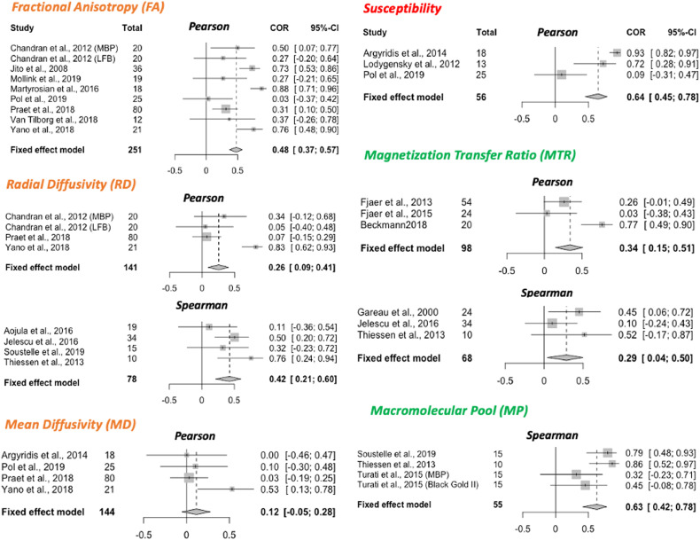
- MTR showed higher within-subject meta-analytic effect sizes (95% CI for R²: 0.60–0.84) than between-subject effect sizes (95% CI for R²: 0.15–0.51).
- No single marker had a clearly stronger effect size than others; the authors conclude that using multiple myelin-sensitive modalities in parallel is currently the best way to test myelin-related hypotheses in vivo.
- Publication-bias checks found no abnormal p-value distribution (80% of positive results had p ≤ 0.01), but a significant correlation between sample size and effect size, and trim-and-fill suggested publication bias could inflate validation effect sizes for some markers.
Methods
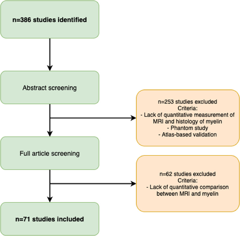
- Systematic review followed PRISMA guidelines, searching Pubmed and Scopus on February 20, 2020, plus 45 articles from the authors’ library; 386 unique articles were screened, yielding 71 included peer-reviewed validation studies.
- From each study, the authors extracted imaging modality/metrics, species, tissue state, histology methods, ROI/coregistration, statistical design, sample size, and correlation results; data are openly available.
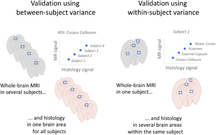
- Meta-analyses used fixed-effect models with R² as the effect size, run separately for Pearson vs. Spearman correlations and for between-subject vs. within-subject designs; only markers validated in more than two independent studies and studies with more than three subjects were included. P-curve, funnel plots, and trim-and-fill were used to assess bias.
Limitations
- The paper states that methodological variability, inconsistent reporting, and potential publication bias limit formal comparisons between MRI-based myelin markers, and that the small number of studies makes it difficult to systematically account for factors such as tissue processing, scanning temperature, and tissue type.
- The paper notes that MWF could not be meta-analyzed because 14 studies used mixed designs, unreported correlation type, or too-small samples, and that correlative validation studies do not address precision/reliability or causation.
- Evident from the text: many markers appear only once in the validation literature, only 21 of 70 studies used coregistration, 19 studies pooled ROIs across subjects without modeling nesting, and 8 studies did not allow deduction of which variance was modeled.
Model-written summary. Check it against the paper before relying on it.
Integrity signals
Registry
Cites
Statistics
Figures
Verification
Claims the paper makes
48 Own interpretation · 47 Own result · 25 No full text of the sources · 25 No basis given · 17 Partly supported · 11 Supported · 4 Supported so far · 3 Not checked yet · 2 Sources not found · 1 Own method · accuracy not yet measured
Every source the paper cites for this claim was checked and supports it.
In the paper
“While theoretical modelling can help in designing novel MR methods (Veraart et al., 2019), any model of MR signals will have intrinsic limitations given the complexity of brain tissue, including simplifying assumptions about the tissue geometry and the MR physics.”
§ Can MRI measure myelin? Systematic review, qualitative assessment, and meta-analysis of studies validating microstructural imaging with myelin histology > Introduction
What the sources say
- Supportedcites Veraart et al., 2019 source
The citation is attached to the first clause: theoretical modelling can help design MR methods. The Introduction describes biophysical modelling as the basis for quantifying tissue properties with diffusion MRI and discusses model-based MRI approaches. The subsequent statement about the intrinsic limitations of any MR-signal model is not attributed to this source. (LLM, conf=0.87)
plain- Asserted: passes — The authors put it forward as true.
- Checkable: passes — It can be checked against evidence.
- Load-bearing: passes — The paper’s conclusions depend on it.
- Based: passes — The paper gives a reason why it holds.
- Supported
The sources with a verdict support this claim; the rest weren’t checked yet, have no full text we could read, or their verdicts aren’t rated yet.
In the paper
“The same principle applies to diffusion-weighted signals, which are affected by the effects of fixation on water diffusion (Shepherd, Thelwall, Stanisz, Blackband, 2009, Sun, Neil, Liang, He, Schmidt, Hsu, Song, 2005).”
§ Can MRI measure myelin? Systematic review, qualitative assessment, and meta-analysis of studies validating microstructural imaging with myelin histology > Discussion > Challenges in acquiring MRI data for validation studies
What the sources say
- Supportedcites Shepherd, Thelwall, Stanisz, Blackband, 2009 source
The Discussion (page unavailable) reports that aldehyde fixation changed water-diffusion parameters in rat cortical slices, including an approximately 50% increase in the diffusion coefficient derived from diffusion-weighted signal attenuation. This supports the claim that fixation-induced changes in water diffusion affect diffusion-weighted signals. (LLM, conf=0.93)
- Not checked — no full text of the sourcecites Sun, Neil, Liang, He, Schmidt, Hsu, Song, 2005 source
plaindirect- Asserted: passes — The authors put it forward as true.
- Checkable: passes — It can be checked against evidence.
- Load-bearing: unclear — It is unclear whether the paper’s conclusions depend on it.
- Based: passes — The paper gives a reason why it holds.
- Supported
The cited sources support only part of this claim — read each source’s verdict below.
In the paper
“Longitudinal studies investigating tissue fixation report spatially and temporally varying effects on relaxation rates, MWF and also MP (Dawe, Bennett, Schneider, Vasireddi, Arfanakis, 2009, Seifert, 2019, Shatl, Uddin, Matsuda, Figley, 2018).”
§ Can MRI measure myelin? Systematic review, qualitative assessment, and meta-analysis of studies validating microstructural imaging with myelin histology > Discussion > Challenges in acquiring MRI data for validation studies
What the sources say
- Partly supportedcites Dawe, Bennett, Schneider, Vasireddi, Arfanakis, 2009 sourceExperimental — accuracy not yet measured
The Conclusions (page not available) report that T2 relaxation during formaldehyde fixation varies with both time and position within human brain hemispheres. The paper does not report fixation effects on myelin water fraction (MWF) or MP; its Discussion describes analysis of different water environments as future work. Thus, this source supports the relaxation component of the multiply cited sentence, but not the full list of measures. (LLM, conf=0.88)
- Supportedcites Seifert, 2019 source
The Results (page unavailable) report fixation-related changes at 1 and 31 days in MWF and the quantitative magnetization-transfer bound-pool fraction F, with different patterns in white and gray matter. The Results and Discussion (page unavailable) also report changes in relaxation times. Together, these support spatially and temporally varying effects on the measures described in the claim. (LLM, conf=0.87)
- Not matchedcites Shatl, Uddin, Matsuda, Figley, 2018
Couldn’t be matched to a paper in Crossref, so it can’t be checked.
plaindirect- Asserted: passes — The authors put it forward as true.
- Checkable: passes — It can be checked against evidence.
- Load-bearing: passes — The paper’s conclusions depend on it.
- Based: passes — The paper gives a reason why it holds.
- Partly supported
The sources with a verdict support this claim; the rest weren’t checked yet, have no full text we could read, or their verdicts aren’t rated yet.
In the paper
“many studies have already found that multiple myelin markers can provide complementary information (Callaghan, Freund, Draganski, Anderson, Cappelletti, Chowdhury, Diedrichsen, Fitzgerald, Smittenaar, Helms, Lutti, Weiskopf, 2014, Eichert et al., 2020, Kolind, Laule, Vavasour, Li, Traboulsee, Mädler, Moore, Mackay, 2008, Lipp, Jones, Bells, Sgarlata, Foster, Stickland, Davidson, Tallantyre, Rober”
§ Can MRI measure myelin? Systematic review, qualitative assessment, and meta-analysis of studies validating microstructural imaging with myelin histology > Discussion > Challenges in measuring myelin for in vivo studies in humans
What the sources say
- Supportedcites Callaghan, Freund, Draganski, Anderson, Cappelletti, Chowdhury, Diedrichsen, Fitzgerald, Smittenaar, Helms, Lutti, Weiskopf, 2014 source
The Conclusions (page unavailable) state that the paper’s multispectral MRI maps provide complementary information and identify MT and R1 as measures influenced by myelin. The Discussion (page unavailable) reports age-related demyelination evident in both MT and R1. The paper supports the general claim, though its complementary maps also measure iron. (LLM, conf=0.84)
- Supportedcites Eichert et al., 2020 source
The ‘Cortical Surface Maps of Microstructural Features’ section says four myelin-related maps are sensitive to different biophysical properties and show some differences in cortical myelin distribution. This supports the claim that multiple markers provide complementary information, while noting that their sensitivities are not fully independent. No page number is supplied in the passages. (LLM, conf=0.90)
- Not checked — no full text of the sourcecites Kolind, Laule, Vavasour, Li, Traboulsee, Mädler, Moore, Mackay, 2008 source
plaincorrelational- Asserted: passes — The authors put it forward as true.
- Checkable: passes — It can be checked against evidence.
- Load-bearing: passes — The paper’s conclusions depend on it.
- Based: passes — The paper gives a reason why it holds.
- Supported
Every source the paper cites for this claim was checked and supports it.
In the paper
“Among the studies we analysed, only a few of them assessed tissue that was scanned fresh, whereas ideally, both states should be validated (e.g. Schmierer et al., 2008).”
§ Can MRI measure myelin? Systematic review, qualitative assessment, and meta-analysis of studies validating microstructural imaging with myelin histology > Discussion > Challenges in acquiring MRI data for validation studies
What the sources say
- Supportedcites Schmierer et al., 2008 source
The citation exemplifies validation in both tissue states, not the citing authors’ count of studies scanned fresh. In Material and Methods (Samples), Schmierer et al. report scanning every brain slice before and after fixation; the Introduction states that comparing the two conditions was a key aim. (LLM, conf=0.92)
plain- Asserted: passes — The authors put it forward as true.
- Checkable: passes — It can be checked against evidence.
- Load-bearing: passes — The paper’s conclusions depend on it.
- Based: passes — The paper gives a reason why it holds.
- Supported
Every source the paper cites for this claim was checked and supports it.
In the paper
“The length of the post-mortem time indicates the advance of the tissue autolysis and therefore the microstructural intactness of the tissue (Sele et al., 2019)”
§ Can MRI measure myelin? Systematic review, qualitative assessment, and meta-analysis of studies validating microstructural imaging with myelin histology > Discussion > Challenges in obtaining ground-truth histological data for validation studies
What the sources say
- Supportedcites Sele et al., 2019 source
The Introduction states that brain-tissue ultrastructure degrades through autolysis during the post-mortem interval. The Results report that finer structures were increasingly degraded as that interval lengthened, supporting the claim that post-mortem time indicates tissue autolysis and structural intactness. (LLM, conf=0.93)
plaindirect- Asserted: passes — The authors put it forward as true.
- Checkable: passes — It can be checked against evidence.
- Load-bearing: passes — The paper’s conclusions depend on it.
- Based: passes — The paper gives a reason why it holds.
- Supported
The cited sources support only part of this claim — read each source’s verdict below.
In the paper
“This phenomenon is exemplified in Laule et al. (2006) and Peters et al. (2019), where correlation coefficients are lower in analyses including only ROIs from white matter, compared to analyses including a more heterogeneous set of ROIs.”
§ Can MRI measure myelin? Systematic review, qualitative assessment, and meta-analysis of studies validating microstructural imaging with myelin histology > Discussion > Challenges in statistical comparisons of MRI and histology
What the sources say
- Not checked — no full text of the sourcecites Laule et al. (2006) source
- Partly supportedcites Peters et al. (2019) sourceExperimental — accuracy not yet measured
The Results section (LFB and diffusion metrics; page unavailable) says that combining white-matter and perituber white-matter ROIs widened the distributions and produced a consistent negative correlation in each specimen. It does not report that correlation coefficients were lower for white-matter-only ROIs than for the combined set. (LLM, conf=0.78)
plaincorrelational- Asserted: passes — The authors put it forward as true.
- Checkable: passes — It can be checked against evidence.
- Load-bearing: unclear — It is unclear whether the paper’s conclusions depend on it.
- Based: passes — The paper gives a reason why it holds.
A result the paper reports from its own data — it cites no other source for it, so there is none to check. The quote shows it is in the paper.
In the paper
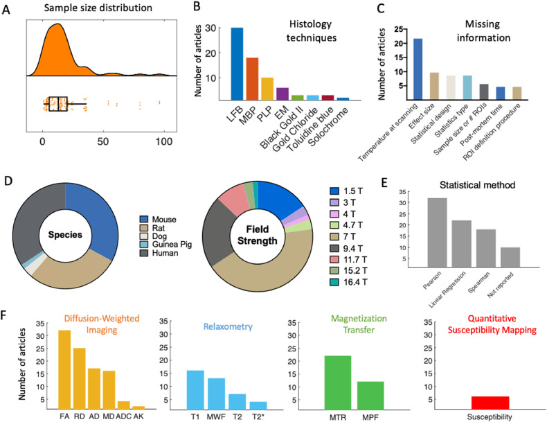
“The studies we included test the validity of a wide-range of potential myelin markers, reflecting the range of markers used in application studies.”
§ Can MRI measure myelin? Systematic review, qualitative assessment, and meta-analysis of studies validating microstructural imaging with myelin histology > Results > Exploring the features of the MR-histology validation literature
plaincorrelational- Asserted: passes — The authors put it forward as true.
- Checkable: passes — It can be checked against evidence.
- Load-bearing: unclear — It is unclear whether the paper’s conclusions depend on it.
- Based: passes — The paper gives a reason why it holds.
A result the paper reports from its own data — it cites no other source for it, so there is none to check. The quote shows it is in the paper.
In the paper
“the most commonly validated MR markers are DWI-based (such as FA), with relaxometry and MT-based studies close seconds (Fig. 3F).”
§ Can MRI measure myelin? Systematic review, qualitative assessment, and meta-analysis of studies validating microstructural imaging with myelin histology > Results > Exploring the features of the MR-histology validation literature
plain- Asserted: passes — The authors put it forward as true.
- Checkable: passes — It can be checked against evidence.
- Load-bearing: unclear — It is unclear whether the paper’s conclusions depend on it.
- Based: passes — The paper gives a reason why it holds.
A result the paper reports from its own data — it cites no other source for it, so there is none to check. The quote shows it is in the paper.
In the paper
“many markers appear only once in the literature (more specifically: Orientation Dispersion Index, Neurite Density Index, T1w/T2w, Relaxation Along a Fictitious Field in the rotating frame of rank 4, MTR from inhomogeneous MT, diffusion basis spectrum imaging - based radial diffusivity, relative semi-solid proton fraction, diffusion standard deviation index)”
§ Can MRI measure myelin? Systematic review, qualitative assessment, and meta-analysis of studies validating microstructural imaging with myelin histology > Results > Exploring the features of the MR-histology validation literature
plain- Asserted: passes — The authors put it forward as true.
- Checkable: passes — It can be checked against evidence.
- Load-bearing: unclear — It is unclear whether the paper’s conclusions depend on it.
- Based: passes — The paper gives a reason why it holds.
- Probably not a substantial claim. 50
- It may state two separate claims as one. 66
The cited sources support only part of this claim — read each source’s verdict below.
In the paper
“A systematic review was conducted using PRISMA guidelines (Shamseer et al., 2015) and incorporating best practices from the AMSTAR 2 checklist for clinical meta-analyses where applicable (e.g. searching across multiple datasets, performing study selection in duplicate) (Shea et al., 2017).”
§ Can MRI measure myelin? Systematic review, qualitative assessment, and meta-analysis of studies validating microstructural imaging with myelin histology > Methods > Systematic review: study selection
What the sources say
- Partly supportedcites Shamseer et al., 2015 sourceExperimental — accuracy not yet measured
The paper describes PRISMA-P as a guideline for documenting systematic review and meta-analysis protocols, building on the PRISMA guidelines (Abstract; no page available). It therefore supports a connection to PRISMA, but it does not establish that the citing authors conducted their completed review using PRISMA guidelines. The AMSTAR 2 portion is attributed to Shea et al., not to this source. (LLM, conf=0.85)
- Supportedcites Shea et al., 2017 source
In the “Rationale for selection of items” section (page unavailable), AMSTAR 2 includes a comprehensive literature search strategy and study selection in duplicate among its checklist items. The “Discussion” (page unavailable) also says the checklist may be used by people conducting systematic reviews. PRISMA is attributed to a separate citation, and whether the citing authors followed these practices is their own methodological statement. (LLM, conf=0.92)
plain- Asserted: passes — The authors put it forward as true.
- Checkable: passes — It can be checked against evidence.
- Load-bearing: fails — The paper’s conclusions may not depend on it.
- Based: passes — The paper gives a reason why it holds.
Probably not a substantial claim. The paper’s conclusions may not depend on it. It may state two separate claims as one.
- Partly supported
The sources with a verdict support this claim; the rest weren’t checked yet, have no full text we could read, or their verdicts aren’t rated yet.
In the paper
“these metrics are known to be influenced by both myelin and non-myelin factors, but are often used as microstructural markers (Beaulieu, 2014, De Santis, Drakesmith, Bells, Assaf, Jones, 2014, Pierpaoli, Chiro, Basser, Trace, 1996)”
§ Can MRI measure myelin? Systematic review, qualitative assessment, and meta-analysis of studies validating microstructural imaging with myelin histology > Methods > Systematic review: information extraction
What the sources say
- Not checked — no full text of the sourcecites Beaulieu, 2014 source
- Supportedcites De Santis, Drakesmith, Bells, Assaf, Jones, 2014 source
The Introduction says FA and MD are widely reported microstructural indices and lack specificity to white-matter subcomponents. It states that axonal membranes and myelination both affect FA, alongside fibre architecture, water diffusivity, and axon morphology and density. (LLM, conf=0.94)
- Not checked — no full text of the sourcecites Pierpaoli, Chiro, Basser, Trace, 1996 source
boostedcorrelational- Asserted: passes — The authors put it forward as true.
- Checkable: passes — It can be checked against evidence.
- Load-bearing: unclear — It is unclear whether the paper’s conclusions depend on it.
- Based: passes — The paper gives a reason why it holds.
It may state two separate claims as one.
The cited sources support only part of this claim — read each source’s verdict below.
In the paper
“In light of these limitations, validation is a necessary step towards making MRI-based methods biologically interpretable, and applicable to clinical and basic research (Barros, Arribarat, Combis, Chaynes, 2019, Cohen-Adad, 2018).”
§ Can MRI measure myelin? Systematic review, qualitative assessment, and meta-analysis of studies validating microstructural imaging with myelin histology > Introduction
What the sources say
- Partly supportedcites Barros, Arribarat, Combis, Chaynes, 2019 sourceExperimental — accuracy not yet measured
The Discussion (Qualitative Histological Studies and MRI; page unavailable) says that correlating MRI with iron histology helps establish what an in vivo MRI signal represents. The Conclusion (page unavailable) says such correlations support reliable brain-iron estimation, while cautioning that methodological issues must be addressed before extrapolating to in vivo MRI. This supports validation for iron-sensitive brain MRI, but not the claim's broader assertion about MRI-based methods generally. (LLM, conf=0.85)
- Not checked — no full text of the sourcecites Cohen-Adad, 2018 source
plaindirect- Asserted: passes — The authors put it forward as true.
- Checkable: fails — It may be too vague to check against evidence.
- Load-bearing: passes — The paper’s conclusions depend on it.
- Based: passes — The paper gives a reason why it holds.
Probably not a substantial claim. It may be too vague to check against evidence.
- Rests on an assumption: It is currently unclear which MR marker is the most biologically accurate non-invasive measure for myelin, and how measures differ in sensitivity to specific aspects of myelination.
- Partly supported
The cited sources support only part of this claim — read each source’s verdict below.
In the paper
“For example, different meta-analyses need to be run for papers reporting Spearman and Pearson correlation coefficients, which can differ in value even when applied to the same data, since Spearman correlation coefficients measure monotonicity and Pearson correlations measure linearity (de Winter et al., 2016).”
§ Can MRI measure myelin? Systematic review, qualitative assessment, and meta-analysis of studies validating microstructural imaging with myelin histology > Methods > Meta-analysis: study selection
What the sources say
- Partly supportedcites de Winter et al., 2016 sourceExperimental — accuracy not yet measured
Split into 2 atoms — 1 confirmed, 1 partially supported, 0 disputed, 0 misattributed, 0 unverifiable. Expand the claim to see per-atom verdicts.
plain- Asserted: passes — The authors put it forward as true.
- Checkable: passes — It can be checked against evidence.
- Load-bearing: passes — The paper’s conclusions depend on it.
- Based: passes — The paper gives a reason why it holds.
It may state two separate claims as one.
- Partly supported
The cited sources support only part of this claim — read each source’s verdict below.
In the paper
“comparable to the median of the most cited fMRI studies during the period of publication included in our meta-analysis (12), but below current median sample size (20) (Szucs and Ioannidis, 2020)”
§ Can MRI measure myelin? Systematic review, qualitative assessment, and meta-analysis of studies validating microstructural imaging with myelin histology > Results > Exploring the features of the MR-histology validation literature
What the sources say
- Partly supportedcites Szucs and Ioannidis, 2020 sourceExperimental — accuracy not yet measured
The Abstract reports a median of 12 for highly cited experimental fMRI studies, supporting the historical comparison. But it gives medians of 23 and 24 for studies published in 2017 and 2018, not 20; §4.2 places an estimated median of 19–20 around 2012–2013. The source does not assess the citing paper's validation studies. (LLM, conf=0.91)
plain- Asserted: passes — The authors put it forward as true.
- Checkable: passes — It can be checked against evidence.
- Load-bearing: fails — The paper’s conclusions may not depend on it.
- Based: passes — The paper gives a reason why it holds.
Probably not a substantial claim. The paper’s conclusions may not depend on it. It may state two separate claims as one.
- Partly supported
The cited sources support only part of this claim — read each source’s verdict below.
In the paper
“Moreover, it is known that different samples (or different anatomical areas within the same sample) are not affected homogeneously by preparation steps such as fixation or antibody penetration (Dawe, Bennett, Schneider, Vasireddi, Arfanakis, 2009, Seehaus, Roebroeck, Bastiani, Fonseca, 2015), which poses challenges in particular for between-subject validation designs.”
§ Can MRI measure myelin? Systematic review, qualitative assessment, and meta-analysis of studies validating microstructural imaging with myelin histology > Discussion > Challenges in obtaining ground-truth histological data for validation studies
What the sources say
- Partly supportedcites Dawe, Bennett, Schneider, Vasireddi, Arfanakis, 2009 sourceExperimental — accuracy not yet measured
Split into 2 atoms — 1 confirmed, 1 partially supported, 0 disputed, 0 misattributed, 0 unverifiable. Expand the claim to see per-atom verdicts.
- Partly supportedcites Seehaus, Roebroeck, Bastiani, Fonseca, 2015 sourceExperimental — accuracy not yet measured
Split into 2 atoms — 0 confirmed, 2 partially supported, 0 disputed, 0 misattributed, 0 unverifiable. Expand the claim to see per-atom verdicts.
boosted- Asserted: passes — The authors put it forward as true.
- Checkable: passes — It can be checked against evidence.
- Load-bearing: unclear — It is unclear whether the paper’s conclusions depend on it.
- Based: passes — The paper gives a reason why it holds.
It may state two separate claims as one.
- Partly supported
Every source the paper cites for this claim was checked and supports it.
In the paper
“Also, higher resolution can be achieved with ex vivo imaging, which in studies of animals with smaller brains provides more anatomically comparable detail to human studies with larger voxel sizes (Lerch et al., 2012).”
§ Can MRI measure myelin? Systematic review, qualitative assessment, and meta-analysis of studies validating microstructural imaging with myelin histology > Discussion > Challenges in acquiring MRI data for validation studies
What the sources say
- Supportedcites Lerch et al., 2012 source
The Introduction states that mouse imaging requires voxel sizes of tens to a hundred microns to obtain information comparable to common human anatomical imaging studies. It also says ex-vivo imaging permits improved resolution through longer scan times, tighter coils, and contrast agents. (LLM, conf=0.92)
plaindirect- Asserted: passes — The authors put it forward as true.
- Checkable: passes — It can be checked against evidence.
- Load-bearing: unclear — It is unclear whether the paper’s conclusions depend on it.
- Based: passes — The paper gives a reason why it holds.
It may state two separate claims as one.
- Supported
Every source the paper cites for this claim was checked and supports it.
In the paper
“This is because fixation works by linking amino acids in proteins, which means that the molecular tissue structure is inherently changed during the process (Thavarajah et al., 2012).”
§ Can MRI measure myelin? Systematic review, qualitative assessment, and meta-analysis of studies validating microstructural imaging with myelin histology > Discussion > Challenges in acquiring MRI data for validation studies
What the sources say
- Supportedcites Thavarajah et al., 2012 source
The Chemical Basics of Formaldehyde Fixation section describes formaldehyde forming covalent cross-links involving amino acid residues in proteins. It also states that these bonds alter the physicochemical state and reactivity of tissue components, supporting the claim about molecular change during formaldehyde fixation. (LLM, conf=0.92)
plaindirect- Asserted: passes — The authors put it forward as true.
- Checkable: passes — It can be checked against evidence.
- Load-bearing: passes — The paper’s conclusions depend on it.
- Based: passes — The paper gives a reason why it holds.
It may state two separate claims as one.
- Supported
The cited sources support only part of this claim — read each source’s verdict below.
In the paper
“Using staining fraction to quantify myelin could circumvent this issue, if the image segmentation intensity threshold is adjusted flexibly (Mollink et al., 2019), depending on local intensity variations.”
§ Can MRI measure myelin? Systematic review, qualitative assessment, and meta-analysis of studies validating microstructural imaging with myelin histology > Discussion > Challenges in obtaining ground-truth histological data for validation studies
What the sources say
- Partly supportedcites Mollink et al., 2019 sourceExperimental — accuracy not yet measured
Methods > Immunohistochemistry (page unavailable) describes using the stained area fraction to quantify PLP, a myelin marker, and setting intensity thresholds separately for each specimen to account for varying staining intensity. It does not describe adjusting the threshold to local intensity variations within a specimen; the local 100 × 100-pixel calculation is for stained area fraction, not threshold selection. (LLM, conf=0.86)
hedgedconditional- Asserted: passes — The authors put it forward as true.
- Checkable: passes — It can be checked against evidence.
- Load-bearing: fails — The paper’s conclusions may not depend on it.
- Based: passes — The paper gives a reason why it holds.
Probably not a substantial claim. The paper’s conclusions may not depend on it.
- Partly supported
Every source the paper cites for this claim was checked and supports it.
In the paper
“Moreover, MR relaxation relies on water protons behaving differently depending on their molecular environment, and fixation does change this environment, for example by affecting membrane permeability (Shepherd et al., 2009).”
§ Can MRI measure myelin? Systematic review, qualitative assessment, and meta-analysis of studies validating microstructural imaging with myelin histology > Discussion > Challenges in acquiring MRI data for validation studies
What the sources say
- Supportedcites Shepherd et al., 2009 source
The Materials and Methods section (Analytical Diffusion Model; page unavailable) allows intra- and extracellular water to have different relaxation times. The Abstract (page unavailable) says fixation may alter the chemical environment sampled by water and reports increased transmembrane water exchange after formaldehyde fixation, indicating increased membrane permeability. (LLM, conf=0.91)
plaindirect- Asserted: passes — The authors put it forward as true.
- Checkable: passes — It can be checked against evidence.
- Load-bearing: unclear — It is unclear whether the paper’s conclusions depend on it.
- Based: passes — The paper gives a reason why it holds.
It may state two separate claims as one.
- Supported
The sources with a verdict support this claim; the rest weren’t checked yet, have no full text we could read, or their verdicts aren’t rated yet.
In the paper
“advanced models, such as the Spherical Mean Technique (Kaden et al., 2016), neurite orientation dispersion and density imaging (NODDI; Zhang et al. (2012)), and diffusion basis spectrum imaging (Wang et al., 2011), are becoming increasingly popular compared to tensor-based metrics such as FA, but are vastly under-represented in the validation literature (Alexander, Dyrby, Nilsson, 2019, Caspers, A”
§ Can MRI measure myelin? Systematic review, qualitative assessment, and meta-analysis of studies validating microstructural imaging with myelin histology > Discussion > Challenges in measuring myelin for in vivo studies in humans
What the sources say
- Supportedcites Kaden et al., 2016 source
The citation to Kaden et al. (2016) identifies the Spherical Mean Technique as an example of an advanced diffusion MRI model. The Methods and materials section explicitly says the paper uses SMT, and the Discussion describes the multi-compartment imaging method it introduces. The later claim about popularity and representation in validation literature is not specifically attributed to this citation. (LLM, conf=0.92)
- Not checked — no full text of the sourcecites Zhang et al. (2012) source
- Supportedcites Wang et al., 2011 source
The citation to Wang et al. (2011) is attached to “diffusion basis spectrum imaging,” identifying the model rather than supporting the later claim about popularity or validation literature. The paper’s “Diffusion basis spectrum imaging” section explicitly proposes a DBSI framework. (LLM, conf=0.94)
plaincorrelational- Asserted: passes — The authors put it forward as true.
- Checkable: passes — It can be checked against evidence.
- Load-bearing: passes — The paper’s conclusions depend on it.
- Based: passes — The paper gives a reason why it holds.
It may state two separate claims as one.
- Supported
The cited sources support only part of this claim — read each source’s verdict below.
In the paper
“For studies using human brains, the option to combine in vivo scanning with ex vivo histology is rare (see Treit et al., 2019), whereas in animal studies, both in vivo and ex vivo scanning are possible.”
§ Can MRI measure myelin? Systematic review, qualitative assessment, and meta-analysis of studies validating microstructural imaging with myelin histology > Discussion > Challenges in acquiring MRI data for validation studies
What the sources say
- Partly supportedcites Treit et al., 2019 sourceExperimental — accuracy not yet measured
The Participants and Histology sections report that four patients had presurgical in vivo MRI followed by surgical resection and histology, so the paper provides a human example of combining the two. It does not establish that this opportunity is rare across studies using human brains. The animal-studies comparison is outside the clause to which this citation is attached. (LLM, conf=0.82)
plain- Asserted: passes — The authors put it forward as true.
- Checkable: passes — It can be checked against evidence.
- Load-bearing: unclear — It is unclear whether the paper’s conclusions depend on it.
- Based: passes — The paper gives a reason why it holds.
It may state two separate claims as one.
- Partly supported
Every source the paper cites for this claim was checked and supports it.
In the paper
“Advances in automatic co-registration tools could aid a more widespread implementation in validation studies (Huszar et al., 2019)”
§ Can MRI measure myelin? Systematic review, qualitative assessment, and meta-analysis of studies validating microstructural imaging with myelin histology > Discussion > Challenges in obtaining ground-truth histological data for validation studies
What the sources say
- Supportedcites Huszar et al., 2019 source
The Conclusion says the automated registration pipeline can be integrated into routine neuropathological practice and facilitates MRI–histology validation studies in much larger cohorts than previously possible. This supports the citing sentence’s qualified claim that automatic co-registration tools could enable more widespread use in validation studies. (LLM, conf=0.93)
hedgedconditional- Asserted: passes — The authors put it forward as true.
- Checkable: fails — It may be too vague to check against evidence.
- Load-bearing: fails — The paper’s conclusions may not depend on it.
- Based: passes — The paper gives a reason why it holds.
Probably not a substantial claim. It may be too vague to check against evidence. The paper’s conclusions may not depend on it.
- Supported
The cited sources support only part of this claim — read each source’s verdict below.
In the paper
“In the case of MRI metrics, repetition across time (e.g. Arshad, Stanley, Raz, 2017, Lévy, Guertin, Khatibi, Mezer, Martinu, Chen, Stikov, Rainville, Cohen-Adad, 2018), across hardware (e.g. Bane, Hectors, Wagner, Arlinghaus, Aryal, Cao, Chenevert, Fennessy, Huang, Hylton, Kalpathy-Cramer, Keenan, Malyarenko, Mulkern, Newitt, Russek, Stupic, Tudorica, Wilmes, Yankeelov, Yen, Boss, Taouli, 2018, Le”
§ Can MRI measure myelin? Systematic review, qualitative assessment, and meta-analysis of studies validating microstructural imaging with myelin histology > Discussion > Challenges in statistical comparisons of MRI and histology
What the sources say
- Supportedcites Arshad, Stanley, Raz, 2017 source
The citation to Arshad et al. is attached to repetition across time, not the separately cited hardware and sequence examples. The Introduction says test–retest reliability is important for longitudinal MRI studies involving multiple measurement occasions, and the Methods (Study Design) describe repeated acquisitions within a session. No page numbers are available in our copy. (LLM, conf=0.90)
- Partly supportedcites Bane et al., 2018 sourceExperimental — accuracy not yet measured
The Methods and Results assess T1 measurement repeatability and interplatform reproducibility across scanners, alongside accuracy and differences between protocols. This supports the hardware-reproducibility part of the claim, for which this paper is cited. The passages do not establish that repetition is a prerequisite for accuracy. (LLM, conf=0.82)
- Not matchedcites Stikov et al., 2015a
Couldn’t be matched to a paper in Crossref, so it can’t be checked.
plaindirect- Asserted: passes — The authors put it forward as true.
- Checkable: passes — It can be checked against evidence.
- Load-bearing: unclear — It is unclear whether the paper’s conclusions depend on it.
- Based: passes — The paper gives a reason why it holds.
It may state two separate claims as one.
- Supported
The cited sources support only part of this claim — read each source’s verdict below.
In the paper
“Its composition can vary between tissue types (González de San Román et al., 2018), species (Gopalakrishnan et al., 2013) and in pathologies (Wheeler et al., 2008)”
§ Can MRI measure myelin? Systematic review, qualitative assessment, and meta-analysis of studies validating microstructural imaging with myelin histology > Discussion > Challenges in obtaining ground-truth histological data for validation studies
What the sources say
- Partly supportedcites González de San Román et al., 2018 sourceExperimental — accuracy not yet measured
Only the tissue-types clause is attributed to this paper; the species and pathology clauses cite other works. The Results section on lipid imaging and the Discussion section on lipid distribution report different lipid profiles in gray matter, white matter, and cortical layers. They do not establish that the composition of myelin itself varies between tissue types (page numbers unavailable). (LLM, conf=0.78)
- Not checked — no full text of the sourcecites Gopalakrishnan et al., 2013 source
- Supportedcites Wheeler et al., 2008 source
Wheeler et al. is cited for the pathology clause, not for variation between tissue types or species. The Discussion reports that multiple sclerosis modifies the lipid composition of myelin membranes, supporting the claim that myelin composition can vary in pathology. (LLM, conf=0.93)
plaincorrelational- Asserted: passes — The authors put it forward as true.
- Checkable: passes — It can be checked against evidence.
- Load-bearing: passes — The paper’s conclusions depend on it.
- Based: passes — The paper gives a reason why it holds.
It may state two separate claims as one.
- Partly supported
The cited sources support only part of this claim — read each source’s verdict below.
In the paper
“One solution to address the limitations of these commonly used metrics would be to employ methods developed for quantification, rather than visualisation, of molecules. These methods include proton induced X-ray emission (PIXE, Stüber et al. (2014)) and lipid mass spectrometry (González de San Román et al., 2018), which could provide quantification of myelin for each imaging pixel, for example by”
§ Can MRI measure myelin? Systematic review, qualitative assessment, and meta-analysis of studies validating microstructural imaging with myelin histology > Discussion > Challenges in obtaining ground-truth histological data for validation studies
What the sources say
- Not checked — no full text of the sourcecites Stüber et al. (2014) source
- Partly supportedcites González de San Román et al., 2018 sourceExperimental — accuracy not yet measured
Split into 2 atoms — 0 confirmed, 2 partially supported, 0 disputed, 0 misattributed, 0 unverifiable. Expand the claim to see per-atom verdicts.
hedgedconditional- Asserted: passes — The authors put it forward as true.
- Checkable: passes — It can be checked against evidence.
- Load-bearing: fails — The paper’s conclusions may not depend on it.
- Based: passes — The paper gives a reason why it holds.
Probably not a substantial claim. The paper’s conclusions may not depend on it.
Every source the paper cites for this claim was checked and supports it.
In the paper
“While coregistration of MR images and histological images has unique challenges, such as the different spatial scales, changes in morphometry due to the tissue processing for histology, and potential cracks and folds in the tissue sections (Pichat et al., 2018)”
§ Can MRI measure myelin? Systematic review, qualitative assessment, and meta-analysis of studies validating microstructural imaging with myelin histology > Discussion > Challenges in obtaining ground-truth histological data for validation studies
What the sources say
- Supportedcites Pichat et al., 2018 source
The Introduction states that histology has much higher resolution than MRI. The sections on compression and distortion, changes in morphology, and cracks and holes describe shape changes and folds or cracks arising during tissue processing. Together, these passages support the stated challenges for MR–histology coregistration. (LLM, conf=0.93)
plain- Asserted: passes — The authors put it forward as true.
- Checkable: passes — It can be checked against evidence.
- Load-bearing: unclear — It is unclear whether the paper’s conclusions depend on it.
- Based: passes — The paper gives a reason why it holds.
It may state two separate claims as one.
- Supported
The cited sources support only part of this claim — read each source’s verdict below.
In the paper
“A few studies have aimed to use non-correlative approaches for validation of MR metrics, for example by demonstrating effects from a given intervention on MR and histology”
§ Can MRI measure myelin? Systematic review, qualitative assessment, and meta-analysis of studies validating microstructural imaging with myelin histology > Discussion > Challenges in statistical comparisons of MRI and histology
What the sources say
- Partly supportedcites Lodygensky et al., 2012 sourceExperimental — accuracy not yet measured
The “Myelin quantification in shiverer mice” section reports differences between mutant and control mice in both MR phase imaging and histological myelin staining, supporting a group-comparison approach alongside correlation. But the paper does not show an administered intervention producing changes in both measures: its iron-extraction experiment changed iron staining while phase contrast remained unchanged (Fig. 6). (LLM, conf=0.82)
- Supportedcites Sampaio-Baptista et al., 2013 source
The Introduction says the study combines MRI and immunohistochemistry and that histology can help validate MRI measurements. The Neuroimaging results and Histology results report differences between motor-training and control groups in both MR measures and myelin staining; the Discussion notes that the two measures did not correlate. The indexed passages provide section labels but no page numbers. (LLM, conf=0.91)
plain- Asserted: passes — The authors put it forward as true.
- Checkable: passes — It can be checked against evidence.
- Load-bearing: fails — The paper’s conclusions may not depend on it.
- Based: passes — The paper gives a reason why it holds.
Probably not a substantial claim. The paper’s conclusions may not depend on it. It may state two separate claims as one.
- Partly supported
The cited sources support only part of this claim — read each source’s verdict below.
In the paper
“With sufficient image quality and appropriate image segmentation (Stikov, Campbell, Stroh, Lavelee, Frey, Novek, Nuara, Ho, Bedell, Dougherty, Leppert, Boudreau, Narayanan, Duval, Cohen-Adad, Picard, Gasecka, Cote, Pike, 2015, Zaimi, Duval, Gasecka, Côté, Stikov, Cohen-Adad, 2016), these methods have the potential to estimate the myelin fraction of imaged tissue, as well as the packing density and”
§ Can MRI measure myelin? Systematic review, qualitative assessment, and meta-analysis of studies validating microstructural imaging with myelin histology > Discussion > Challenges in obtaining ground-truth histological data for validation studies
What the sources say
- Not matchedcites Stikov, Campbell, Stroh, Lavelee, Frey, Novek, Nuara, Ho, Bedell, Dougherty, Leppert, Boudreau, Narayanan, Duval, Cohen-Adad, Picard, Gasecka, Cote, Pike, 2015
Couldn’t be matched to a paper in Crossref, so it can’t be checked.
- Supportedcites Zaimi, Duval, Gasecka, Côté, Stikov, Cohen-Adad, 2016 source
The citation is attached to the condition of sufficient image quality and appropriate segmentation. AxonSeg is a tool for axon and myelin segmentation, and the Discussion (Image Quality and Modalities; page unavailable) says its results strongly depend on image contrast and resolution. The citation is not attached specifically to the later list of potential measurements. (LLM, conf=0.89)
hedgedconditional- Asserted: passes — The authors put it forward as true.
- Checkable: passes — It can be checked against evidence.
- Load-bearing: fails — The paper’s conclusions may not depend on it.
- Based: passes — The paper gives a reason why it holds.
Probably not a substantial claim. The paper’s conclusions may not depend on it. It may state two separate claims as one.
- Not matched
The cited sources support only part of this claim — read each source’s verdict below.
In the paper
“multiple different morphological aspects of the myelinated axon, including Nodes of Ranvier, sheath thickness, length and packing density - are all important to brain function (Almeida, Lyons, 2017, Xin, Chan, 2020).”
§ Can MRI measure myelin? Systematic review, qualitative assessment, and meta-analysis of studies validating microstructural imaging with myelin histology > Discussion > Challenges in obtaining ground-truth histological data for validation studies
What the sources say
- Partly supportedcites Almeida, Lyons, 2017 sourceExperimental — accuracy not yet measured
The section on neuronal circuit function says that nodes of Ranvier and the length, thickness, number, and distribution of myelin sheaths can affect conduction and circuit function. It does not specifically address packing density as a morphological feature, so that item in the list is not established by this source. (LLM, conf=0.82)
- Partly supportedcites Xin, Chan, 2020 sourceExperimental — accuracy not yet measured
In “Oligodendrocytes and circuit function” (page unavailable), the review links nodes of Ranvier and myelin sheath length to conduction timing in neural circuits. It also discusses changes in myelin thickness under “Experience-induced plasticity,” but the passages found do not establish the claimed functional importance of sheath thickness or packing density. (LLM, conf=0.85)
plaincorrelational- Asserted: passes — The authors put it forward as true.
- Checkable: passes — It can be checked against evidence.
- Load-bearing: fails — The paper’s conclusions may not depend on it.
- Based: passes — The paper gives a reason why it holds.
Probably not a substantial claim. The paper’s conclusions may not depend on it. It may state two separate claims as one.
- Partly supported
Every source the paper cites for this claim was checked and supports it.
In the paper
“In studies using Shiverer mice, which have a genetic mutation for the MBP-gene (Molineaux et al., 1986), MBP-immunohistochemistry is well suited to demonstrate the genetic intervention, but could lead to a biased myelin quantification when used on Shiverer mice in a validation study.”
§ Can MRI measure myelin? Systematic review, qualitative assessment, and meta-analysis of studies validating microstructural imaging with myelin histology > Discussion > Challenges in obtaining ground-truth histological data for validation studies
What the sources say
- Supportedcites Molineaux et al., 1986 source
The citation is attached to the statement that Shiverer mice have a mutation in the MBP gene. The Results section reports that the Shiverer MBP gene is partially deleted; the sentence’s subsequent discussion of immunohistochemistry is not attributed to this citation. (LLM, conf=0.94)
hedgeddirect- Asserted: passes — The authors put it forward as true.
- Checkable: passes — It can be checked against evidence.
- Load-bearing: unclear — It is unclear whether the paper’s conclusions depend on it.
- Based: passes — The paper gives a reason why it holds.
It may state two separate claims as one.
- Supported
Every source the paper cites for this claim was checked and supports it.
In the paper
“However, not all asymmetric funnel plots are due to issues that impact meta-analytic inference (Sterne et al., 2011).”
§ Can MRI measure myelin? Systematic review, qualitative assessment, and meta-analysis of studies validating microstructural imaging with myelin histology > Discussion > Challenges in statistical comparisons of MRI and histology
What the sources say
- Supportedcites Sterne et al., 2011 source
The sections ‘Implications of heterogeneity, reporting bias, and chance’ and ‘Chance’ explain that funnel plot asymmetry can arise by chance or from the choice of plotted statistics, rather than from a bias affecting the meta-analysis. No page numbers are available in our copy. (LLM, conf=0.91)
plain- Asserted: passes — The authors put it forward as true.
- Checkable: passes — It can be checked against evidence.
- Load-bearing: fails — The paper’s conclusions may not depend on it.
- Based: passes — The paper gives a reason why it holds.
Probably not a substantial claim. The paper’s conclusions may not depend on it.
- Supported
Every source the paper cites for this claim was checked and supports it.
In the paper
“Publication bias and questionable research practices have long been established as a key factor hindering pooling of evidence across studies in biomedical research (Ahmed et al., 2012).”
§ Can MRI measure myelin? Systematic review, qualitative assessment, and meta-analysis of studies validating microstructural imaging with myelin histology > Discussion > Challenges in statistical comparisons of MRI and histology
What the sources say
- Supportedcites Ahmed et al., 2012 source
The Introduction describes publication bias as a known threat to meta-analysis and identifies selective outcome reporting and reviewer selection bias as related concerns. It says these biases can leave meta-analyses synthesising an incomplete evidence base and producing potentially biased results, supporting the claim that they hinder reliable pooling of biomedical evidence. (LLM, conf=0.83)
boosteddirect- Asserted: passes — The authors put it forward as true.
- Checkable: passes — It can be checked against evidence.
- Load-bearing: fails — The paper’s conclusions may not depend on it.
- Based: passes — The paper gives a reason why it holds.
Probably not a substantial claim. The paper’s conclusions may not depend on it.
- Supported
The cited sources support only part of this claim — read each source’s verdict below.
In the paper
“For example, Jespersen et al. (2010) finds a correlation between FA and myelin stain of r = 0.78, and a correlation between FA and cell density of r = 0.72, thus questioning the specificity of the FA-myelin correlation in the study.”
§ Can MRI measure myelin? Systematic review, qualitative assessment, and meta-analysis of studies validating microstructural imaging with myelin histology > Discussion > Challenges in statistical comparisons of MRI and histology
What the sources say
- Partly supportedcites Jespersen et al. (2010) sourceExperimental — accuracy not yet measured
The Results and discussion section reports correlations between FA and AMG myelin staining, and says FA is also highly correlated with cell soma density. It also describes reasons FA may not specifically reflect myelin. The passages available from our copy do not give the claimed coefficients of r = 0.78 and r = 0.72, so those exact values cannot be verified. (LLM, conf=0.82)
plaincorrelational- Asserted: passes — The authors put it forward as true.
- Checkable: passes — It can be checked against evidence.
- Load-bearing: passes — The paper’s conclusions depend on it.
- Based: passes — The paper gives a reason why it holds.
It may state two separate claims as one.
- Partly supported
The cited sources support only part of this claim — read each source’s verdict below.
In the paper
“Conditions such as MS, or animal models such as Cuprizone-fed and Shiverer mice, are often used in validation studies, because of their known effect on myelin, but they also affect iron metabolism, due to iron’s role in myelin maintenance (e.g. Hametner, Wimmer, Haider, Pfeifenbring, Brück, Lassmann, 2013, Pandur, Pap, Varga, Jánosa, Komoly, Fórizs, Sipos, 2019, Sergeant, Vesvres, Devès, Guillou,”
§ Can MRI measure myelin? Systematic review, qualitative assessment, and meta-analysis of studies validating microstructural imaging with myelin histology > Discussion > Challenges in statistical comparisons of MRI and histology
What the sources say
- Partly supportedcites Hametner et al., 2013 sourceExperimental — accuracy not yet measured
The Discussion reports altered iron metabolism in MS lesions and normal-appearing white matter, including iron loss from myelin. The Results also describe iron accumulation in axons at MS lesion edges. The paper does not establish that cuprizone-fed or Shiverer mice have these effects or that these conditions are often used in validation studies. (LLM, conf=0.86)
- Partly supportedcites Pandur et al., 2019 sourceExperimental — accuracy not yet measured
The Discussion describes cuprizone-fed mice as a demyelination model and reports changes in iron metabolism after cuprizone treatment. The Introduction also describes axonal injuries in cuprizone-affected brain regions and disturbances of iron homeostasis in MS. Our copy does not address Shiverer mice or establish that these conditions are often used in validation studies. (LLM, conf=0.84)
- Partly supportedcites Stassart et al., 2018 sourceExperimental — accuracy not yet measured
The section on axonal pathology caused by oligodendroglial defects (page unavailable) says shiverer mice have severely abnormal CNS myelin and altered axonal calibre and cytoskeleton. It also says degenerative axonal changes are lacking in these mice. Searches for cuprizone, iron, and validation studies found no passage supporting those parts of the claim. (LLM, conf=0.86)
plaindirect- Asserted: passes — The authors put it forward as true.
- Checkable: passes — It can be checked against evidence.
- Load-bearing: passes — The paper’s conclusions depend on it.
- Based: passes — The paper gives a reason why it holds.
It may state two separate claims as one.
- Partly supported
A result the paper reports from its own data — it cites no other source for it, so there is none to check. The quote shows it is in the paper.
In the paper
“The validation literature uses a wide range of histological markers (with 9 studies using more than one marker, Fig. 3F).”
§ Can MRI measure myelin? Systematic review, qualitative assessment, and meta-analysis of studies validating microstructural imaging with myelin histology > Results > Exploring the features of the MR-histology validation literature
plain- Asserted: passes — The authors put it forward as true.
- Checkable: passes — It can be checked against evidence.
- Load-bearing: unclear — It is unclear whether the paper’s conclusions depend on it.
- Based: passes — The paper gives a reason why it holds.
It may state two separate claims as one.
The paper states this without a source or data behind it here, so there is nothing to check it against. The quote shows it is in the paper.
In the paper
“However, validation studies use varying methodologies and have varying outcomes, making it difficult to assess to what extent such justifications are valid.”
§ Can MRI measure myelin? Systematic review, qualitative assessment, and meta-analysis of studies validating microstructural imaging with myelin histology > Introduction
plaindirect- Asserted: passes — The authors put it forward as true.
- Checkable: passes — It can be checked against evidence.
- Load-bearing: passes — The paper’s conclusions depend on it.
- Based: passes — The paper gives a reason why it holds.
It may state two separate claims as one.
- Rests on an assumption: Neuroimaging studies conducted in vivo often use individual validation studies as evidence to justify employing a specific microstructural MRI measure to study myelination.
A result the paper reports from its own data — it cites no other source for it, so there is none to check. The quote shows it is in the paper.
In the paper
“Abstract screening led to exclusion of 253 studies, while full-text screening further excluded 62 studies, with a total of 71 remaining studies fitting our inclusion criteria (Table 1).”
§ Can MRI measure myelin? Systematic review, qualitative assessment, and meta-analysis of studies validating microstructural imaging with myelin histology > Results > Exploring the features of the MR-histology validation literature
plain- Asserted: passes — The authors put it forward as true.
- Checkable: passes — It can be checked against evidence.
- Load-bearing: fails — The paper’s conclusions may not depend on it.
- Based: passes — The paper gives a reason why it holds.
Probably not a substantial claim. The paper’s conclusions may not depend on it. It may state two separate claims as one.
A result the paper reports from its own data — it cites no other source for it, so there is none to check. The quote shows it is in the paper.
In the paper
“Of these articles, 294 were from Pubmed, 46 from Scopus, 45 from our expert library.”
§ Can MRI measure myelin? Systematic review, qualitative assessment, and meta-analysis of studies validating microstructural imaging with myelin histology > Results > Exploring the features of the MR-histology validation literature
plain- Asserted: passes — The authors put it forward as true.
- Checkable: passes — It can be checked against evidence.
- Load-bearing: fails — The paper’s conclusions may not depend on it.
- Based: passes — The paper gives a reason why it holds.
Probably not a substantial claim. The paper’s conclusions may not depend on it. It may state two separate claims as one.
The paper states this without a source or data behind it here, so there is nothing to check it against. The quote shows it is in the paper.
In the paper
“Formal comparisons between MRI-based myelin markers are limited by methodological variability, inconsistent reporting and potential for publication bias.”
§ Can MRI measure myelin? Systematic review, qualitative assessment, and meta-analysis of studies validating microstructural imaging with myelin histology > Abstract
plaindirect- Asserted: passes — The authors put it forward as true.
- Checkable: passes — It can be checked against evidence.
- Load-bearing: passes — The paper’s conclusions depend on it.
- Based: passes — The paper gives a reason why it holds.
It may state two separate claims as one.
A result the paper reports from its own data — it cites no other source for it, so there is none to check. The quote shows it is in the paper.
In the paper
“We find many MR markers are sensitive to myelin, including FA, RD, MP, MTR, Susceptibility, R1, but not MD.”
§ Can MRI measure myelin? Systematic review, qualitative assessment, and meta-analysis of studies validating microstructural imaging with myelin histology > Abstract
plaincorrelational- Asserted: passes — The authors put it forward as true.
- Checkable: passes — It can be checked against evidence.
- Load-bearing: passes — The paper’s conclusions depend on it.
- Based: passes — The paper gives a reason why it holds.
It may state two separate claims as one.
A result the paper reports from its own data — it cites no other source for it, so there is none to check. The quote shows it is in the paper.
In the paper
“Our search yielded 386 unique articles (Fig. 2).”
§ Can MRI measure myelin? Systematic review, qualitative assessment, and meta-analysis of studies validating microstructural imaging with myelin histology > Results > Exploring the features of the MR-histology validation literature
plain- Asserted: passes — The authors put it forward as true.
- Checkable: passes — It can be checked against evidence.
- Load-bearing: fails — The paper’s conclusions may not depend on it.
- Based: passes — The paper gives a reason why it holds.
Probably not a substantial claim. The paper’s conclusions may not depend on it.
The paper states this without a source or data behind it here, so there is nothing to check it against. The quote shows it is in the paper.
In the paper
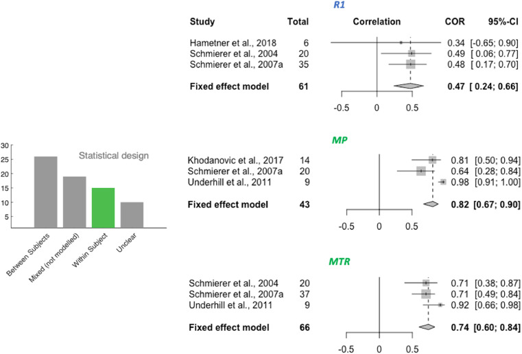
“For the three metrics R1, MP and MTR, a meta-analysis on within-subject correlations could be performed.”
§ Can MRI measure myelin? Systematic review, qualitative assessment, and meta-analysis of studies validating microstructural imaging with myelin histology > Results > Meta-analysis
hedged- Asserted: passes — The authors put it forward as true.
- Checkable: passes — It can be checked against evidence.
- Load-bearing: fails — The paper’s conclusions may not depend on it.
- Based: passes — The paper gives a reason why it holds.
Probably not a substantial claim. The paper’s conclusions may not depend on it.
A result the paper reports from its own data — it cites no other source for it, so there is none to check. The quote shows it is in the paper.
In the paper
“The remainder of the studies (n = 5) use different methodologies (within- and between-subject design) and outcome measures (Spearman and Pearson) from each other, making it difficult to pool them together.”
§ Can MRI measure myelin? Systematic review, qualitative assessment, and meta-analysis of studies validating microstructural imaging with myelin histology > Discussion > Challenges in measuring myelin for in vivo studies in humans
plaindirectn = 5- Asserted: passes — The authors put it forward as true.
- Checkable: passes — It can be checked against evidence.
- Load-bearing: passes — The paper’s conclusions depend on it.
- Based: passes — The paper gives a reason why it holds.
It may state two separate claims as one.
A result the paper reports from its own data — it cites no other source for it, so there is none to check. The quote shows it is in the paper.
In the paper
“A notable absence from our meta-analyses is Myelin Water Fraction (MWF).”
§ Can MRI measure myelin? Systematic review, qualitative assessment, and meta-analysis of studies validating microstructural imaging with myelin histology > Discussion > Challenges in measuring myelin for in vivo studies in humans
plain- Asserted: passes — The authors put it forward as true.
- Checkable: passes — It can be checked against evidence.
- Load-bearing: passes — The paper’s conclusions depend on it.
- Based: passes — The paper gives a reason why it holds.
Probably not a substantial claim.
A result the paper reports from its own data — it cites no other source for it, so there is none to check. The quote shows it is in the paper.
In the paper
“Except for mean diffusivity, we find meta-analytic evidence for correlations between histological markers and all markers investigated, with no clear marker having a stronger effect size than others.”
§ Can MRI measure myelin? Systematic review, qualitative assessment, and meta-analysis of studies validating microstructural imaging with myelin histology > Results > Meta-analysis
plaincorrelational- Asserted: passes — The authors put it forward as true.
- Checkable: passes — It can be checked against evidence.
- Load-bearing: passes — The paper’s conclusions depend on it.
- Based: passes — The paper gives a reason why it holds.
It may state two separate claims as one.
A result the paper reports from its own data — it cites no other source for it, so there is none to check. The quote shows it is in the paper.
In the paper
“While 14 studies in total examine MWF, a high number of these studies use a mixed within- and between- subject design (n = 7), do not report the type of static measure used (n = 1), or use sample sizes too small for inclusion (n = 1), which is why they did not fit our inclusion criteria for the meta-analysis.”
§ Can MRI measure myelin? Systematic review, qualitative assessment, and meta-analysis of studies validating microstructural imaging with myelin histology > Discussion > Challenges in measuring myelin for in vivo studies in humans
plaindirectn = 7- Asserted: passes — The authors put it forward as true.
- Checkable: passes — It can be checked against evidence.
- Load-bearing: passes — The paper’s conclusions depend on it.
- Based: passes — The paper gives a reason why it holds.
It may state two separate claims as one.
The paper states this without a source or data behind it here, so there is nothing to check it against. The quote shows it is in the paper.
In the paper
“In the majority of studies we assessed, the fixation process ranged from a few days to a few weeks, which means the MR signal was likely very different between studies.”
§ Can MRI measure myelin? Systematic review, qualitative assessment, and meta-analysis of studies validating microstructural imaging with myelin histology > Discussion > Challenges in acquiring MRI data for validation studies
hedgedconditional- Asserted: passes — The authors put it forward as true.
- Checkable: passes — It can be checked against evidence.
- Load-bearing: passes — The paper’s conclusions depend on it.
- Based: passes — The paper gives a reason why it holds.
It may state two separate claims as one.
A result the paper reports from its own data — it cites no other source for it, so there is none to check. The quote shows it is in the paper.
In the paper
“we find that the studies in our review validate MR metrics with either in vivo or ex vivo MR scanning, each of which has advantages and disadvantages.”
§ Can MRI measure myelin? Systematic review, qualitative assessment, and meta-analysis of studies validating microstructural imaging with myelin histology > Discussion > Challenges in acquiring MRI data for validation studies
plaincorrelational- Asserted: passes — The authors put it forward as true.
- Checkable: passes — It can be checked against evidence.
- Load-bearing: unclear — It is unclear whether the paper’s conclusions depend on it.
- Based: passes — The paper gives a reason why it holds.
It may state two separate claims as one.
A result the paper reports from its own data — it cites no other source for it, so there is none to check. The quote shows it is in the paper.
In the paper
“A total of 32 studies were selected for inclusion in a quantitative meta-analysis based on this selection process (24 reporting Pearson only, 7 reporting Spearman only, and 1 reporting both; 22 using between-subject design, 10 using within-subject design).”
§ Can MRI measure myelin? Systematic review, qualitative assessment, and meta-analysis of studies validating microstructural imaging with myelin histology > Results > Meta-analysis
plain- Asserted: passes — The authors put it forward as true.
- Checkable: passes — It can be checked against evidence.
- Load-bearing: unclear — It is unclear whether the paper’s conclusions depend on it.
- Based: passes — The paper gives a reason why it holds.
It may state two separate claims as one.
A result the paper reports from its own data — it cites no other source for it, so there is none to check. The quote shows it is in the paper.
In the paper
“We find no evidence of abnormal p-value distribution, with a left-skewed p-curve and 80% of positive results providing p-values of 0.01 or lower (Supplementary Fig. S1).”
§ Can MRI measure myelin? Systematic review, qualitative assessment, and meta-analysis of studies validating microstructural imaging with myelin histology > Results > P-value distribution bias and publication bias
plain- Asserted: passes — The authors put it forward as true.
- Checkable: passes — It can be checked against evidence.
- Load-bearing: unclear — It is unclear whether the paper’s conclusions depend on it.
- Based: passes — The paper gives a reason why it holds.
It may state two separate claims as one.
A result the paper reports from its own data — it cites no other source for it, so there is none to check. The quote shows it is in the paper.
In the paper
“Our meta-analyses of 7 commonly used MR markers (FA, RD, MD, susceptibility, R1, MTR and MP) show that there is evidence all these markers correlate with myelin, with the exception of MD.”
§ Can MRI measure myelin? Systematic review, qualitative assessment, and meta-analysis of studies validating microstructural imaging with myelin histology > Discussion > Many MRI markers correlate with myelin
plaincorrelational- Asserted: passes — The authors put it forward as true.
- Checkable: passes — It can be checked against evidence.
- Load-bearing: passes — The paper’s conclusions depend on it.
- Based: passes — The paper gives a reason why it holds.
It may state two separate claims as one.
The paper states this without a source or data behind it here, so there is nothing to check it against. The quote shows it is in the paper.
In the paper
“Most validation studies aim to quantify overall myelin content in a tissue, with the rationale that this is most closely related to the signals measured with MRI, and that for many applications in basic science and clinical translation, total myelin content measures can already be hugely beneficial.”
§ Can MRI measure myelin? Systematic review, qualitative assessment, and meta-analysis of studies validating microstructural imaging with myelin histology > Discussion > Challenges in obtaining ground-truth histological data for validation studies
plaincorrelational- Asserted: passes — The authors put it forward as true.
- Checkable: passes — It can be checked against evidence.
- Load-bearing: fails — The paper’s conclusions may not depend on it.
- Based: passes — The paper gives a reason why it holds.
Probably not a substantial claim. The paper’s conclusions may not depend on it. It may state two separate claims as one.
The paper states this without a source or data behind it here, so there is nothing to check it against. The quote shows it is in the paper.
In the paper
“Many of the studies in our review focus on accuracy, i.e. the measured parameter being in agreement with the true underlying biological parameter of interest.”
§ Can MRI measure myelin? Systematic review, qualitative assessment, and meta-analysis of studies validating microstructural imaging with myelin histology > Discussion > Challenges in statistical comparisons of MRI and histology
plain- Asserted: passes — The authors put it forward as true.
- Checkable: passes — It can be checked against evidence.
- Load-bearing: fails — The paper’s conclusions may not depend on it.
- Based: passes — The paper gives a reason why it holds.
Probably not a substantial claim. The paper’s conclusions may not depend on it.
A result the paper reports from its own data — it cites no other source for it, so there is none to check. The quote shows it is in the paper.
In the paper
“Most of the assessed validation papers try to achieve spatial correspondence without coregistration (only 21 out of 70 studies use coregistration), but rather by manually outlining ROIs in the same anatomical location, identifying these ROIs based on landmarks.”
§ Can MRI measure myelin? Systematic review, qualitative assessment, and meta-analysis of studies validating microstructural imaging with myelin histology > Discussion > Challenges in obtaining ground-truth histological data for validation studies
plain- Asserted: passes — The authors put it forward as true.
- Checkable: passes — It can be checked against evidence.
- Load-bearing: unclear — It is unclear whether the paper’s conclusions depend on it.
- Based: passes — The paper gives a reason why it holds.
It may state two separate claims as one.
A result the paper reports from its own data — it cites no other source for it, so there is none to check. The quote shows it is in the paper.
In the paper
“In our review, processing of the tissue for histology varied considerably across studies, with both paraffin embedding and cryosectioning being used with about equal frequency.”
§ Can MRI measure myelin? Systematic review, qualitative assessment, and meta-analysis of studies validating microstructural imaging with myelin histology > Discussion > Challenges in obtaining ground-truth histological data for validation studies
plain- Asserted: passes — The authors put it forward as true.
- Checkable: passes — It can be checked against evidence.
- Load-bearing: unclear — It is unclear whether the paper’s conclusions depend on it.
- Based: passes — The paper gives a reason why it holds.
It may state two separate claims as one.
A result the paper reports from its own data — it cites no other source for it, so there is none to check. The quote shows it is in the paper.
In the paper
“Our meta-analysis finds evidence for correlations between myelin histology and a range of MR markers: FA, RD, susceptibility, R1, MP and MTR, but not MD.”
§ Can MRI measure myelin? Systematic review, qualitative assessment, and meta-analysis of studies validating microstructural imaging with myelin histology > Discussion > Conclusions and future directions
plaincorrelational- Asserted: passes — The authors put it forward as true.
- Checkable: passes — It can be checked against evidence.
- Load-bearing: passes — The paper’s conclusions depend on it.
- Based: passes — The paper gives a reason why it holds.
It may state two separate claims as one.
- Rests on an assumption: Myelin histology provides a valid reference standard for myelin content in the validation studies included in the meta-analysis.
A result the paper reports from its own data — it cites no other source for it, so there is none to check. The quote shows it is in the paper.
In the paper
“Within the studies selected for our systematic review, only a few (n = 7) performed iron histology, while half of the studies considered axons (n = 35).”
§ Can MRI measure myelin? Systematic review, qualitative assessment, and meta-analysis of studies validating microstructural imaging with myelin histology > Discussion > Challenges in statistical comparisons of MRI and histology
plainn = 7- Asserted: passes — The authors put it forward as true.
- Checkable: passes — It can be checked against evidence.
- Load-bearing: passes — The paper’s conclusions depend on it.
- Based: passes — The paper gives a reason why it holds.
It may state two separate claims as one.
The paper states this without a source or data behind it here, so there is nothing to check it against. The quote shows it is in the paper.
In the paper
“Second, we find that heterogeneous and inconsistent reporting makes it difficult to assess the quality of the studies, and the factors driving differences in results.”
§ Can MRI measure myelin? Systematic review, qualitative assessment, and meta-analysis of studies validating microstructural imaging with myelin histology > Discussion > Conclusions and future directions
plaindirect- Asserted: passes — The authors put it forward as true.
- Checkable: passes — It can be checked against evidence.
- Load-bearing: passes — The paper’s conclusions depend on it.
- Based: passes — The paper gives a reason why it holds.
Probably not a substantial claim.
In the paper
“Some microstructural features of the brain, such as iron and axonal density, are related to myelin and have similar effects as myelin on MR signals (Möller et al., 2019).”
§ Can MRI measure myelin? Systematic review, qualitative assessment, and meta-analysis of studies validating microstructural imaging with myelin histology > Methods > Systematic review: information extraction
What the sources say
- Not checked — no full text of the sourcecites Möller et al., 2019 source
plaincorrelational- Asserted: passes — The authors put it forward as true.
- Checkable: passes — It can be checked against evidence.
- Load-bearing: passes — The paper’s conclusions depend on it.
- Based: passes — The paper gives a reason why it holds.
Probably not a substantial claim. It may state two separate claims as one.
What the paper did — it cites no source for this, so there is none to check. The quote shows it is in the paper.
In the paper
“As most studies were excluded at this stage, abstract screening was independently run by two investigators (I.L. and A.L.) and discordant decisions on article inclusions were resolved by discussion and consensus-finding between the investigators.”
§ Can MRI measure myelin? Systematic review, qualitative assessment, and meta-analysis of studies validating microstructural imaging with myelin histology > Methods > Systematic review: study selection
plain- Asserted: passes — The authors put it forward as true.
- Checkable: passes — It can be checked against evidence.
- Load-bearing: fails — The paper’s conclusions may not depend on it.
- Based: passes — The paper gives a reason why it holds.
Probably not a substantial claim. The paper’s conclusions may not depend on it. It may state two separate claims as one.
A conclusion the paper draws from its own results — it cites no other source for it, so there is none to check. The quote shows it is in the paper.
In the paper
“Taken together, these factors highlight that the validation literature has considered different field strengths, species, and MRI and histology approaches to quantify myelin, making common results across papers highly generalizable.”
§ Can MRI measure myelin? Systematic review, qualitative assessment, and meta-analysis of studies validating microstructural imaging with myelin histology > Results > Exploring the features of the MR-histology validation literature
plaindirect- Asserted: passes — The authors put it forward as true.
- Checkable: passes — It can be checked against evidence.
- Load-bearing: unclear — It is unclear whether the paper’s conclusions depend on it.
- Based: passes — The paper gives a reason why it holds.
It may state two separate claims as one.
In the paper
“As stage and type of tissue fixation both affect MR parameters (Dusek et al., 2019), we extracted information on the state of the tissue during MR imaging.”
§ Can MRI measure myelin? Systematic review, qualitative assessment, and meta-analysis of studies validating microstructural imaging with myelin histology > Methods > Systematic review: information extraction
What the sources say
- Not checked — no full text of the sourcecites Dusek et al., 2019 source
plaindirect- Asserted: passes — The authors put it forward as true.
- Checkable: passes — It can be checked against evidence.
- Load-bearing: passes — The paper’s conclusions depend on it.
- Based: passes — The paper gives a reason why it holds.
Probably not a substantial claim. It may state two separate claims as one.
In the paper
“Funnel plots are a common tool to estimate publication bias in meta-analyses (Egger et al., 1997).”
§ Can MRI measure myelin? Systematic review, qualitative assessment, and meta-analysis of studies validating microstructural imaging with myelin histology > Methods > Meta-analyses: quantifying and correcting for publication bias in the literature
What the sources say
- Not checked — no full text of the sourcecites Egger et al., 1997 source
plain- Asserted: passes — The authors put it forward as true.
- Checkable: passes — It can be checked against evidence.
- Load-bearing: fails — The paper’s conclusions may not depend on it.
- Based: passes — The paper gives a reason why it holds.
Probably not a substantial claim. The paper’s conclusions may not depend on it.
In the paper
“Last but not least, we report how tissue was treated for histology (cryosectioning or paraffin embedding), which can affect tissue staining success and intensity (Werner et al., 2000).”
§ Can MRI measure myelin? Systematic review, qualitative assessment, and meta-analysis of studies validating microstructural imaging with myelin histology > Methods > Systematic review: information extraction
What the sources say
- Not checked — no full text of the sourcecites Werner et al., 2000 source
plainconditional- Asserted: passes — The authors put it forward as true.
- Checkable: passes — It can be checked against evidence.
- Load-bearing: fails — The paper’s conclusions may not depend on it.
- Based: passes — The paper gives a reason why it holds.
Probably not a substantial claim. The paper’s conclusions may not depend on it.
The paper states this without a source or data behind it here, so there is nothing to check it against. The quote shows it is in the paper.
In the paper
“Moreover, studies using within-subject and between-subject designs need to be considered in different analyses.”
§ Can MRI measure myelin? Systematic review, qualitative assessment, and meta-analysis of studies validating microstructural imaging with myelin histology > Methods > Meta-analysis: study selection
plain- Asserted: passes — The authors put it forward as true.
- Checkable: passes — It can be checked against evidence.
- Load-bearing: passes — The paper’s conclusions depend on it.
- Based: passes — The paper gives a reason why it holds.
Probably not a substantial claim.
The paper states this without a source or data behind it here, so there is nothing to check it against. The quote shows it is in the paper.
In the paper
“In these two types of designs, variability is driven by different factors: in within-subject designs, variability is driven by anatomical and tissue differences in myelin content; whereas in between-subject designs, variability is driven by external factors (such as pathology, age or genotype).”
§ Can MRI measure myelin? Systematic review, qualitative assessment, and meta-analysis of studies validating microstructural imaging with myelin histology > Methods > Systematic review: information extraction
plaindirect- Asserted: passes — The authors put it forward as true.
- Checkable: passes — It can be checked against evidence.
- Load-bearing: passes — The paper’s conclusions depend on it.
- Based: passes — The paper gives a reason why it holds.
Probably not a substantial claim. It may state two separate claims as one.
A claim is checked only against the cited paper’s full text, never its abstract, and none is available for its sources.
In the paper
“As temperature has an effect on MR relaxation times (Birkl et al., 2016) and diffusion (Dhital et al., 2016), tissue temperature during scanning was extracted.”
§ Can MRI measure myelin? Systematic review, qualitative assessment, and meta-analysis of studies validating microstructural imaging with myelin histology > Methods > Systematic review: information extraction
What the sources say
plaindirect- Asserted: passes — The authors put it forward as true.
- Checkable: passes — It can be checked against evidence.
- Load-bearing: passes — The paper’s conclusions depend on it.
- Based: passes — The paper gives a reason why it holds.
Probably not a substantial claim. It may state two separate claims as one.
A conclusion the paper draws from its own results — it cites no other source for it, so there is none to check. The quote shows it is in the paper.
In the paper
“If the temperature varied across samples or across ROIs within the same sample, this could induce unwanted variability in the MRI metrics.”
§ Can MRI measure myelin? Systematic review, qualitative assessment, and meta-analysis of studies validating microstructural imaging with myelin histology > Discussion > Challenges in acquiring MRI data for validation studies
hedgedconditional- Asserted: passes — The authors put it forward as true.
- Checkable: passes — It can be checked against evidence.
- Load-bearing: fails — The paper’s conclusions may not depend on it.
- Based: passes — The paper gives a reason why it holds.
Probably not a substantial claim. The paper’s conclusions may not depend on it.
The paper states this without a source or data behind it here, so there is nothing to check it against. The quote shows it is in the paper.
In the paper
“Tissue fixation is a necessary step for stopping tissue decomposition in ex vivo scans, but it also has a major influence on MR signals.”
§ Can MRI measure myelin? Systematic review, qualitative assessment, and meta-analysis of studies validating microstructural imaging with myelin histology > Discussion > Challenges in acquiring MRI data for validation studies
plaindirect- Asserted: passes — The authors put it forward as true.
- Checkable: passes — It can be checked against evidence.
- Load-bearing: passes — The paper’s conclusions depend on it.
- Based: passes — The paper gives a reason why it holds.
It may state two separate claims as one.
A conclusion the paper draws from its own results — it cites no other source for it, so there is none to check. The quote shows it is in the paper.
In the paper
“Some markers are only validated with one type of statistical design, which limits their potential applications.”
§ Can MRI measure myelin? Systematic review, qualitative assessment, and meta-analysis of studies validating microstructural imaging with myelin histology > Discussion > Challenges in measuring myelin for in vivo studies in humans
plaindirect- Asserted: passes — The authors put it forward as true.
- Checkable: passes — It can be checked against evidence.
- Load-bearing: passes — The paper’s conclusions depend on it.
- Based: passes — The paper gives a reason why it holds.
It may state two separate claims as one.
A conclusion the paper draws from its own results — it cites no other source for it, so there is none to check. The quote shows it is in the paper.
In the paper
“In vivo scanning has the advantage that it better resembles the parameters of in vivo human studies, while ex vivo scanning has the advantage that the tissue is scanned and processed for histology in a similar state.”
§ Can MRI measure myelin? Systematic review, qualitative assessment, and meta-analysis of studies validating microstructural imaging with myelin histology > Discussion > Challenges in acquiring MRI data for validation studies
plain- Asserted: passes — The authors put it forward as true.
- Checkable: passes — It can be checked against evidence.
- Load-bearing: fails — The paper’s conclusions may not depend on it.
- Based: passes — The paper gives a reason why it holds.
Probably not a substantial claim. The paper’s conclusions may not depend on it. It may state two separate claims as one.
In the paper
“Staining intensity is often assumed to scale linearly with myelin content. However, this is not always true, for example in cases where staining saturation can take place (e.g. in silver staining of white matter; Pistorio et al., 2006).”
§ Can MRI measure myelin? Systematic review, qualitative assessment, and meta-analysis of studies validating microstructural imaging with myelin histology > Discussion > Challenges in obtaining ground-truth histological data for validation studies
What the sources say
- Not checked — no full text of the sourcecites Pistorio et al., 2006 source
plain- Asserted: passes — The authors put it forward as true.
- Checkable: passes — It can be checked against evidence.
- Load-bearing: unclear — It is unclear whether the paper’s conclusions depend on it.
- Based: passes — The paper gives a reason why it holds.
It may state two separate claims as one.
A conclusion the paper draws from its own results — it cites no other source for it, so there is none to check. The quote shows it is in the paper.
In the paper
“However, both metrics come with limitations.”
§ Can MRI measure myelin? Systematic review, qualitative assessment, and meta-analysis of studies validating microstructural imaging with myelin histology > Discussion > Challenges in obtaining ground-truth histological data for validation studies
plain- Asserted: passes — The authors put it forward as true.
- Checkable: passes — It can be checked against evidence.
- Load-bearing: unclear — It is unclear whether the paper’s conclusions depend on it.
- Based: passes — The paper gives a reason why it holds.
It may state two separate claims as one.
A conclusion the paper draws from its own results — it cites no other source for it, so there is none to check. The quote shows it is in the paper.
In the paper
“Importantly, the tissue temperature could unintentionally change throughout the scanning session due to gradient heating, making it important to monitor scanning temperature in ex vivo scans.”
§ Can MRI measure myelin? Systematic review, qualitative assessment, and meta-analysis of studies validating microstructural imaging with myelin histology > Discussion > Challenges in acquiring MRI data for validation studies
hedgeddirect- Asserted: passes — The authors put it forward as true.
- Checkable: passes — It can be checked against evidence.
- Load-bearing: fails — The paper’s conclusions may not depend on it.
- Based: passes — The paper gives a reason why it holds.
Probably not a substantial claim. The paper’s conclusions may not depend on it. It may state two separate claims as one.
A conclusion the paper draws from its own results — it cites no other source for it, so there is none to check. The quote shows it is in the paper.
In the paper
“Their advantage is that they allow imaging large fields of view, which is often needed in a validation study to compensate for the comparatively low spatial resolution of MRI relative to microscopy.”
§ Can MRI measure myelin? Systematic review, qualitative assessment, and meta-analysis of studies validating microstructural imaging with myelin histology > Discussion > Challenges in obtaining ground-truth histological data for validation studies
plaindirect- Asserted: passes — The authors put it forward as true.
- Checkable: passes — It can be checked against evidence.
- Load-bearing: fails — The paper’s conclusions may not depend on it.
- Based: passes — The paper gives a reason why it holds.
Probably not a substantial claim. The paper’s conclusions may not depend on it. It may state two separate claims as one.
The paper states this without a source or data behind it here, so there is nothing to check it against. The quote shows it is in the paper.
In the paper
“First, validity depends on accuracy and accuracy depends on precision.”
§ Can MRI measure myelin? Systematic review, qualitative assessment, and meta-analysis of studies validating microstructural imaging with myelin histology > Discussion > Challenges in statistical comparisons of MRI and histology
plaindirect- Asserted: passes — The authors put it forward as true.
- Checkable: fails — It may be too vague to check against evidence.
- Load-bearing: passes — The paper’s conclusions depend on it.
- Based: fails — The paper may give no reason why it holds.
Probably not a substantial claim. It may be too vague to check against evidence. The paper may give no reason why it holds. It may state two separate claims as one.
A claim is checked only against the cited paper’s full text, never its abstract, and none is available for its sources.
In the paper
“While they are not as common in the validation literature, and often not feasible over large areas of the brain, these methods provide more detailed information compared to stainings or immunohistochemistry, and could provide further validation for microstructural MR markers (Sternberger, Itoyama, Kies, Webster DeF., 1978, Vincze, Mázló, Seress, Komoly, Ábrahám, 2008).”
§ Can MRI measure myelin? Systematic review, qualitative assessment, and meta-analysis of studies validating microstructural imaging with myelin histology > Discussion > Challenges in obtaining ground-truth histological data for validation studies
What the sources say
hedgeddirect- Asserted: passes — The authors put it forward as true.
- Checkable: passes — It can be checked against evidence.
- Load-bearing: unclear — It is unclear whether the paper’s conclusions depend on it.
- Based: passes — The paper gives a reason why it holds.
It may state two separate claims as one.
A claim is checked only against the cited paper’s full text, never its abstract, and none is available for its sources.
In the paper
“For example, LFB is likely attracted by the basic amino acids of myelins proteins and by phospholipids”
§ Can MRI measure myelin? Systematic review, qualitative assessment, and meta-analysis of studies validating microstructural imaging with myelin histology > Discussion > Challenges in obtaining ground-truth histological data for validation studies
What the sources say
hedgedconditional- Asserted: passes — The authors put it forward as true.
- Checkable: passes — It can be checked against evidence.
- Load-bearing: fails — The paper’s conclusions may not depend on it.
- Based: passes — The paper gives a reason why it holds.
Probably not a substantial claim. The paper’s conclusions may not depend on it. It may state two separate claims as one.
A claim is checked only against the cited paper’s full text, never its abstract, and none is available for its sources.
In the paper
“Myelin is a highly complex substrate, with the dry mass consisting of about 70% lipids and 30% proteins”
§ Can MRI measure myelin? Systematic review, qualitative assessment, and meta-analysis of studies validating microstructural imaging with myelin histology > Discussion > Challenges in obtaining ground-truth histological data for validation studies
What the sources say
plain- Asserted: passes — The authors put it forward as true.
- Checkable: passes — It can be checked against evidence.
- Load-bearing: fails — The paper’s conclusions may not depend on it.
- Based: passes — The paper gives a reason why it holds.
Probably not a substantial claim. The paper’s conclusions may not depend on it.
A claim is checked only against the cited paper’s full text, never its abstract, and none is available for its sources.
In the paper
“whereas Gold atoms present in Gold stains are chemically reduced by myelin lipids in formaldehyde fixed tissue, leading to the black appearance of gold staining in myelin fibres”
§ Can MRI measure myelin? Systematic review, qualitative assessment, and meta-analysis of studies validating microstructural imaging with myelin histology > Discussion > Challenges in obtaining ground-truth histological data for validation studies
What the sources say
plaindirect- Asserted: passes — The authors put it forward as true.
- Checkable: passes — It can be checked against evidence.
- Load-bearing: fails — The paper’s conclusions may not depend on it.
- Based: passes — The paper gives a reason why it holds.
Probably not a substantial claim. The paper’s conclusions may not depend on it.
A conclusion the paper draws from its own results — it cites no other source for it, so there is none to check. The quote shows it is in the paper.
In the paper
“The myelin structure in the stained tissue is therefore inherently affected by how it was processed for histology and may not be comparable across studies that used different processing strategies (Recommendation 2.1).”
§ Can MRI measure myelin? Systematic review, qualitative assessment, and meta-analysis of studies validating microstructural imaging with myelin histology > Discussion > Challenges in obtaining ground-truth histological data for validation studies
hedgedconditional- Asserted: passes — The authors put it forward as true.
- Checkable: passes — It can be checked against evidence.
- Load-bearing: passes — The paper’s conclusions depend on it.
- Based: passes — The paper gives a reason why it holds.
It may state two separate claims as one.
In the paper
“and automatic atlas-based ROI definitions are recommended practice in the neuroimaging field (Nichols et al., 2017).”
§ Can MRI measure myelin? Systematic review, qualitative assessment, and meta-analysis of studies validating microstructural imaging with myelin histology > Discussion > Challenges in obtaining ground-truth histological data for validation studies
What the sources say
- Not checked — no full text of the sourcecites Nichols et al., 2017 source
plain- Asserted: passes — The authors put it forward as true.
- Checkable: passes — It can be checked against evidence.
- Load-bearing: fails — The paper’s conclusions may not depend on it.
- Based: passes — The paper gives a reason why it holds.
Probably not a substantial claim. The paper’s conclusions may not depend on it.
A conclusion the paper draws from its own results — it cites no other source for it, so there is none to check. The quote shows it is in the paper.
In the paper
“First, placing ROIs may vary depending on the experimenter performing it”
§ Can MRI measure myelin? Systematic review, qualitative assessment, and meta-analysis of studies validating microstructural imaging with myelin histology > Discussion > Challenges in obtaining ground-truth histological data for validation studies
hedgedconditional- Asserted: passes — The authors put it forward as true.
- Checkable: passes — It can be checked against evidence.
- Load-bearing: fails — The paper’s conclusions may not depend on it.
- Based: passes — The paper gives a reason why it holds.
Probably not a substantial claim. The paper’s conclusions may not depend on it.
None of the sources cited for this claim have been checked yet.
In the paper
“While tissue fixation or freezing do not have a strong effect on lipids (Carriel et al., 2017a), extraction by solvents, such as used in paraffin embedding, extracts most lipids and only retains those covalently bound to protein (Carriel, Campos, Aneiros-Fernandez, Kiernan, 2017, Kiernan, 2007).”
§ Can MRI measure myelin? Systematic review, qualitative assessment, and meta-analysis of studies validating microstructural imaging with myelin histology > Discussion > Challenges in obtaining ground-truth histological data for validation studies
What the sources say
plaindirect- Asserted: passes — The authors put it forward as true.
- Checkable: passes — It can be checked against evidence.
- Load-bearing: passes — The paper’s conclusions depend on it.
- Based: passes — The paper gives a reason why it holds.
It may state two separate claims as one.
The paper states this without a source or data behind it here, so there is nothing to check it against. The quote shows it is in the paper.
In the paper
“Likewise, correlation does not equal causation and factors that affect both MRI and histology (such as post-mortem times) may artificially inflate or deflate correlation coefficients.”
§ Can MRI measure myelin? Systematic review, qualitative assessment, and meta-analysis of studies validating microstructural imaging with myelin histology > Discussion > Challenges in statistical comparisons of MRI and histology
hedgedconditional- Asserted: passes — The authors put it forward as true.
- Checkable: passes — It can be checked against evidence.
- Load-bearing: unclear — It is unclear whether the paper’s conclusions depend on it.
- Based: passes — The paper gives a reason why it holds.
It may state two separate claims as one.
A conclusion the paper draws from its own results — it cites no other source for it, so there is none to check. The quote shows it is in the paper.
In the paper
“it has the advantage that ROI definition can be performed more reproducibly.”
§ Can MRI measure myelin? Systematic review, qualitative assessment, and meta-analysis of studies validating microstructural imaging with myelin histology > Discussion > Challenges in obtaining ground-truth histological data for validation studies
plaindirect- Asserted: passes — The authors put it forward as true.
- Checkable: passes — It can be checked against evidence.
- Load-bearing: fails — The paper’s conclusions may not depend on it.
- Based: passes — The paper gives a reason why it holds.
Probably not a substantial claim. The paper’s conclusions may not depend on it.
A conclusion the paper draws from its own results — it cites no other source for it, so there is none to check. The quote shows it is in the paper.
In the paper
“However, precision, i.e. low variability in repeated measurements, and thus high reliability, is also crucial to validity of a metric.”
§ Can MRI measure myelin? Systematic review, qualitative assessment, and meta-analysis of studies validating microstructural imaging with myelin histology > Discussion > Challenges in statistical comparisons of MRI and histology
plaindirect- Asserted: passes — The authors put it forward as true.
- Checkable: passes — It can be checked against evidence.
- Load-bearing: passes — The paper’s conclusions depend on it.
- Based: passes — The paper gives a reason why it holds.
Probably not a substantial claim.
A conclusion the paper draws from its own results — it cites no other source for it, so there is none to check. The quote shows it is in the paper.
In the paper
“Second, coregistration may greatly improve the spatial correspondence between MR and histological images.”
§ Can MRI measure myelin? Systematic review, qualitative assessment, and meta-analysis of studies validating microstructural imaging with myelin histology > Discussion > Challenges in obtaining ground-truth histological data for validation studies
hedgedconditional- Asserted: passes — The authors put it forward as true.
- Checkable: passes — It can be checked against evidence.
- Load-bearing: fails — The paper’s conclusions may not depend on it.
- Based: passes — The paper gives a reason why it holds.
Probably not a substantial claim. The paper’s conclusions may not depend on it.
The paper states this without a source or data behind it here, so there is nothing to check it against. The quote shows it is in the paper.
In the paper
“Second, correlative approaches are key to assessing accuracy in histological validation studies, because for most MR metrics, there is no mathematical model capable of converting the units of the metric to readouts from myelin histology.”
§ Can MRI measure myelin? Systematic review, qualitative assessment, and meta-analysis of studies validating microstructural imaging with myelin histology > Discussion > Challenges in statistical comparisons of MRI and histology
plaindirect- Asserted: passes — The authors put it forward as true.
- Checkable: passes — It can be checked against evidence.
- Load-bearing: passes — The paper’s conclusions depend on it.
- Based: passes — The paper gives a reason why it holds.
It may state two separate claims as one.
A conclusion the paper draws from its own results — it cites no other source for it, so there is none to check. The quote shows it is in the paper.
In the paper
“and therefore different visualization strategies may be useful for different scenarios.”
§ Can MRI measure myelin? Systematic review, qualitative assessment, and meta-analysis of studies validating microstructural imaging with myelin histology > Discussion > Challenges in obtaining ground-truth histological data for validation studies
hedgedconditional- Asserted: passes — The authors put it forward as true.
- Checkable: fails — It may be too vague to check against evidence.
- Load-bearing: fails — The paper’s conclusions may not depend on it.
- Based: passes — The paper gives a reason why it holds.
Probably not a substantial claim. It may be too vague to check against evidence. The paper’s conclusions may not depend on it.
A conclusion the paper draws from its own results — it cites no other source for it, so there is none to check. The quote shows it is in the paper.
In the paper
“However, it remains poorly understood to what extent staining fraction values are comparable across different staining methods.”
§ Can MRI measure myelin? Systematic review, qualitative assessment, and meta-analysis of studies validating microstructural imaging with myelin histology > Discussion > Challenges in obtaining ground-truth histological data for validation studies
plain- Asserted: passes — The authors put it forward as true.
- Checkable: passes — It can be checked against evidence.
- Load-bearing: fails — The paper’s conclusions may not depend on it.
- Based: fails — The paper may give no reason why it holds.
Probably not a substantial claim. The paper’s conclusions may not depend on it. The paper may give no reason why it holds.
A conclusion the paper draws from its own results — it cites no other source for it, so there is none to check. The quote shows it is in the paper.
In the paper
“Another disadvantage of traditional histology methods is that what they gain in field of view size they lose in resolution.”
§ Can MRI measure myelin? Systematic review, qualitative assessment, and meta-analysis of studies validating microstructural imaging with myelin histology > Discussion > Challenges in obtaining ground-truth histological data for validation studies
plaindirect- Asserted: passes — The authors put it forward as true.
- Checkable: passes — It can be checked against evidence.
- Load-bearing: fails — The paper’s conclusions may not depend on it.
- Based: passes — The paper gives a reason why it holds.
Probably not a substantial claim. The paper’s conclusions may not depend on it. It may state two separate claims as one.
A conclusion the paper draws from its own results — it cites no other source for it, so there is none to check. The quote shows it is in the paper.
In the paper
“Tackling these issues will be crucial to improving our strategies to measure myelin in humans.”
§ Can MRI measure myelin? Systematic review, qualitative assessment, and meta-analysis of studies validating microstructural imaging with myelin histology > Discussion > Conclusions and future directions
plainconditional- Asserted: passes — The authors put it forward as true.
- Checkable: fails — It may be too vague to check against evidence.
- Load-bearing: passes — The paper’s conclusions depend on it.
- Based: passes — The paper gives a reason why it holds.
Probably not a substantial claim. It may be too vague to check against evidence.
In the paper
“Iron occurs in high concentrations in oligodendrocytes and is important for the production and maintenance of myelin (Möller et al., 2019).”
§ Can MRI measure myelin? Systematic review, qualitative assessment, and meta-analysis of studies validating microstructural imaging with myelin histology > Discussion > Challenges in statistical comparisons of MRI and histology
What the sources say
- Not checked — no full text of the sourcecites Möller et al., 2019 source
plaindirect- Asserted: passes — The authors put it forward as true.
- Checkable: passes — It can be checked against evidence.
- Load-bearing: unclear — It is unclear whether the paper’s conclusions depend on it.
- Based: passes — The paper gives a reason why it holds.
It may state two separate claims as one.
A claim is checked only against the cited paper’s full text, never its abstract, and none is available for its sources.
In the paper
“It affects some MRI metrics similarly to myelin (Möller et al., 2019) and its distribution resembles myeloarchitecture (Fukunaga et al., 2010).”
§ Can MRI measure myelin? Systematic review, qualitative assessment, and meta-analysis of studies validating microstructural imaging with myelin histology > Discussion > Challenges in statistical comparisons of MRI and histology
What the sources say
plaincorrelational- Asserted: passes — The authors put it forward as true.
- Checkable: passes — It can be checked against evidence.
- Load-bearing: unclear — It is unclear whether the paper’s conclusions depend on it.
- Based: passes — The paper gives a reason why it holds.
It may state two separate claims as one.
A conclusion the paper draws from its own results — it cites no other source for it, so there is none to check. The quote shows it is in the paper.
In the paper
“This suggests that for the time being, using multiple microstructural imaging markers in parallel may be the best way to test hypotheses related to myelin in humans in vivo.”
§ Can MRI measure myelin? Systematic review, qualitative assessment, and meta-analysis of studies validating microstructural imaging with myelin histology > Discussion > Conclusions and future directions
hedgedconditional- Asserted: passes — The authors put it forward as true.
- Checkable: fails — It may be too vague to check against evidence.
- Load-bearing: passes — The paper’s conclusions depend on it.
- Based: passes — The paper gives a reason why it holds.
Probably not a substantial claim. It may be too vague to check against evidence.
- Rests on an assumption: Myelin histology provides a valid reference standard for myelin content in the validation studies included in the meta-analysis.
The paper states this without a source or data behind it here, so there is nothing to check it against. The quote shows it is in the paper.
In the paper
“In healthy tissue, myelin content is often related to iron and axon density.”
§ Can MRI measure myelin? Systematic review, qualitative assessment, and meta-analysis of studies validating microstructural imaging with myelin histology > Discussion > Challenges in statistical comparisons of MRI and histology
hedgedcorrelational- Asserted: passes — The authors put it forward as true.
- Checkable: passes — It can be checked against evidence.
- Load-bearing: passes — The paper’s conclusions depend on it.
- Based: passes — The paper gives a reason why it holds.
It may state two separate claims as one.
A conclusion the paper draws from its own results — it cites no other source for it, so there is none to check. The quote shows it is in the paper.
In the paper
“While this provides the correlation with more power, spurious correlations are a concern when dealing with multiple groups.”
§ Can MRI measure myelin? Systematic review, qualitative assessment, and meta-analysis of studies validating microstructural imaging with myelin histology > Discussion > Challenges in statistical comparisons of MRI and histology
plaincorrelational- Asserted: passes — The authors put it forward as true.
- Checkable: passes — It can be checked against evidence.
- Load-bearing: passes — The paper’s conclusions depend on it.
- Based: passes — The paper gives a reason why it holds.
It may state two separate claims as one.
The paper states this without a source or data behind it here, so there is nothing to check it against. The quote shows it is in the paper.
In the paper
“Asymmetric funnel plots can be driven by many factors. Selective outcome reporting and selective analysis reporting are the most common.”
§ Can MRI measure myelin? Systematic review, qualitative assessment, and meta-analysis of studies validating microstructural imaging with myelin histology > Discussion > Challenges in statistical comparisons of MRI and histology
plaindirect- Asserted: passes — The authors put it forward as true.
- Checkable: passes — It can be checked against evidence.
- Load-bearing: fails — The paper’s conclusions may not depend on it.
- Based: passes — The paper gives a reason why it holds.
Probably not a substantial claim. The paper’s conclusions may not depend on it. It may state two separate claims as one.
A conclusion the paper draws from its own results — it cites no other source for it, so there is none to check. The quote shows it is in the paper.
In the paper
“This limits the interpretability of the results, and fails to take the nested structure of the dataset into account.”
§ Can MRI measure myelin? Systematic review, qualitative assessment, and meta-analysis of studies validating microstructural imaging with myelin histology > Discussion > Challenges in statistical comparisons of MRI and histology
plaindirectn = 19- Asserted: passes — The authors put it forward as true.
- Checkable: passes — It can be checked against evidence.
- Load-bearing: passes — The paper’s conclusions depend on it.
- Based: passes — The paper gives a reason why it holds.
It may state two separate claims as one.
The paper states this without a source or data behind it here, so there is nothing to check it against. The quote shows it is in the paper.
In the paper
“For instance, in some circumstances sampling variation and chance can lead to an asymmetric funnel plot without real publication bias.”
§ Can MRI measure myelin? Systematic review, qualitative assessment, and meta-analysis of studies validating microstructural imaging with myelin histology > Discussion > Challenges in statistical comparisons of MRI and histology
plaindirect- Asserted: passes — The authors put it forward as true.
- Checkable: passes — It can be checked against evidence.
- Load-bearing: fails — The paper’s conclusions may not depend on it.
- Based: passes — The paper gives a reason why it holds.
Probably not a substantial claim. The paper’s conclusions may not depend on it.
A conclusion the paper draws from its own results — it cites no other source for it, so there is none to check. The quote shows it is in the paper.
In the paper
“Therefore, using pre-registration may help establish more accurate effect sizes for the correlations between MR metrics and myelin histology”
§ Can MRI measure myelin? Systematic review, qualitative assessment, and meta-analysis of studies validating microstructural imaging with myelin histology > Discussion > Challenges in statistical comparisons of MRI and histology
hedgedconditional- Asserted: passes — The authors put it forward as true.
- Checkable: fails — It may be too vague to check against evidence.
- Load-bearing: passes — The paper’s conclusions depend on it.
- Based: passes — The paper gives a reason why it holds.
Probably not a substantial claim. It may be too vague to check against evidence.
A conclusion the paper draws from its own results — it cites no other source for it, so there is none to check. The quote shows it is in the paper.
In the paper
“Taken together, these observations suggest that measures to prevent publication bias may be useful for future validation studies.”
§ Can MRI measure myelin? Systematic review, qualitative assessment, and meta-analysis of studies validating microstructural imaging with myelin histology > Discussion > Challenges in statistical comparisons of MRI and histology
hedgedconditional- Asserted: passes — The authors put it forward as true.
- Checkable: fails — It may be too vague to check against evidence.
- Load-bearing: passes — The paper’s conclusions depend on it.
- Based: passes — The paper gives a reason why it holds.
Probably not a substantial claim. It may be too vague to check against evidence.
A conclusion the paper draws from its own results — it cites no other source for it, so there is none to check. The quote shows it is in the paper.
In the paper
“These results need to be interpreted with caution”
§ Can MRI measure myelin? Systematic review, qualitative assessment, and meta-analysis of studies validating microstructural imaging with myelin histology > Discussion > Challenges in statistical comparisons of MRI and histology
plain- Asserted: passes — The authors put it forward as true.
- Checkable: passes — It can be checked against evidence.
- Load-bearing: passes — The paper’s conclusions depend on it.
- Based: passes — The paper gives a reason why it holds.
Probably not a substantial claim.
In the paper
“Pre-registered protocols with pre-specified power analyses can strengthen the robustness of effect size estimates, and even provide more accurate estimates than meta-analytic analyses themselves (Kvarven et al., 2020).”
§ Can MRI measure myelin? Systematic review, qualitative assessment, and meta-analysis of studies validating microstructural imaging with myelin histology > Discussion > Challenges in statistical comparisons of MRI and histology
What the sources say
- Not checked — no full text of the sourcecites Kvarven et al., 2020 source
plaindirect- Asserted: passes — The authors put it forward as true.
- Checkable: passes — It can be checked against evidence.
- Load-bearing: passes — The paper’s conclusions depend on it.
- Based: passes — The paper gives a reason why it holds.
It may state two separate claims as one.
The paper relies on this without support.
It is currently unclear which MR marker is the most biologically accurate non-invasive measure for myelin, and how measures differ in sensitivity to specific aspects of myelination.
“Currently, it is not clear which of these MR markers is the most biologically accurate non-invasive measure for myelin, and how the measures differ in their sensitivity to specific aspects of myelination.”
The paper relies on this without support.
Neuroimaging studies conducted in vivo often use individual validation studies as evidence to justify employing a specific microstructural MRI measure to study myelination.
“Neuroimaging studies conducted in vivo often use individual validation studies of this kind as evidence to justify employing a specific microstructural MRI measure to study myelination.”
The paper relies on this without support.
The gold standard for validation studies aiming to assess the accuracy of MR markers is to compare MRI and underlying myelin content within the same tissue.
“For this reason, the gold standard for validation studies aiming to assess the accuracy of MR markers is to compare MRI and underlying myelin content within the same tissue.”
The paper relies on this without support.
Myelin histology provides a valid reference standard for myelin content in the validation studies included in the meta-analysis.
“Our meta-analysis focussed on studies using a correlative approach to measure the accuracy of MR metrics for validation.”
Self-check
Percentages, sums and reported differences re-computed from the paper’s own numbers.
Each contradiction between two passages listed here was confirmed by an independent check.
Rated minor: it would not touch a result — a value no result rests on, a p-value that stays on the same side of the significance level, or a conflict that is clerical, resolved elsewhere in the paper, or outside its own findings.
In the paper
§ Can MRI measure myelin? Systematic review, qualitative assessment, and meta-analysis of studies validating microstructural imaging with myelin histology > Results > Exploring the features of the MR-histology validation literature
“The validation literature uses a wide range of histological markers (with 9 studies using more than one marker, Fig. 3F).”
§ Can MRI measure myelin? Systematic review, qualitative assessment, and meta-analysis of studies validating microstructural imaging with myelin histology > Results > Exploring the features of the MR-histology validation literature
“[F] MR markers most commonly used in the literature, subdivided based on MR technique they are based on.”
Citations
Cites
Bias in meta-analysis detected by a simple, graphical test
Egger, M., Smith, G. D., Schneider, M., & Minder, C. (1997) · BMJ · doi:10.1136/bmj.315.7109.629 · Cited by 56,722
Duval, S., & Tweedie, R. (2000) · Biometrics · doi:10.1111/j.0006-341x.2000.00455.x · Cited by 14,099
Shamseer, L., Moher, D., Clarke, M., Ghersi, D., Liberati, A., Petticrew, M., Shekelle, P., Stewart, L. A., & the PRISMA-P Group (2015) · BMJ · doi:10.1136/bmj.g7647 · Cited by 13,120
Shea, B. J., Reeves, B. C., Wells, G., Thuku, M., Hamel, C., Moran, J., Moher, D., Tugwell, P., Welch, V., Kristjansson, E., et al. (2017) · BMJ · doi:10.1136/bmj.j4008 · Cited by 10,636
Sterne, J. A. C., Sutton, A. J., Ioannidis, J. P. A., Terrin, N., Jones, D. R., Lau, J., Carpenter, J., Rucker, G., Harbord, R. M., Schmid, C. H., et al. (2011) · BMJ · doi:10.1136/bmj.d4002 · Cited by 7,709
Power failure: why small sample size undermines the reliability of neuroscience
Button, K. S., Ioannidis, J. P. A., Mokrysz, C., Nosek, B. A., Flint, J., Robinson, E. S. J., & Munafò, M. R. (2013) · Nature Reviews Neuroscience · doi:10.1038/nrn3475 · Cited by 7,641
The basis of anisotropic water diffusion in the nervous system—A technical review.
Beaulieu, C. (2002) · NMR in Biomedicine · doi:10.1002/nbm.782 · Cited by 4,628
Measurement in Medicine: The Analysis of Method Comparison Studies
Altman, D. G., & Bland, J. M. (1983) · The Statistician · doi:10.2307/2987937 · Cited by 4,300
NODDI: Practical in vivo neurite orientation dispersion and density imaging of the human brain
Zhang, H., Schneider, T., Wheeler-Kingshott, C. A., & Alexander, D. C. (2012) · NeuroImage · doi:10.1016/j.neuroimage.2012.03.072 · Cited by 3,288
Diffusion tensor MR imaging of the human brain
Pierpaoli, C., Jezzard, P., Basser, P. J., Barnett, A., & Di Chiro, G. (1996) · Radiology · doi:10.1148/radiology.201.3.8939209 · Cited by 2,664
A Method for the Combined Staining of Cells and Fibers in the Nervous System
Klüver, H., & Barrera, E. (1953) · Journal of Neuropathology & Experimental Neurology · doi:10.1097/00005072-195312040-00008 · Cited by 1,724
Patra, P. P., Chikkaraddy, R., Tripathi, R. P. N., Dasgupta, A., & Kumar, G. V. P. (2014) · Nature Communications · doi:10.1038/ncomms5357 · Cited by 1,723
Plasticity in gray and white: neuroimaging changes in brain structure during learning
Zatorre, R. J., Fields, R. D., & Johansen-Berg, H. (2012) · Nature Neuroscience · doi:10.1038/nn.3045 · Cited by 1,685
Oligodendroglia metabolically support axons and contribute to neurodegeneration
Lee, Y., Morrison, B. M., Li, Y., Lengacher, S., Farah, M. H., Hoffman, P. N., Liu, Y., Tsingalia, A., Jin, L., Zhang, P.-W., et al. (2012) · Nature · doi:10.1038/nature11314 · Cited by 1,664
Glycolytic oligodendrocytes maintain myelin and long-term axonal integrity
Fünfschilling, U., Supplie, L. M., Mahad, D., Boretius, S., Saab, A. S., Edgar, J., Brinkmann, B. G., Kassmann, C. M., Tzvetanova, I. D., Möbius, W., et al. (2012) · Nature · doi:10.1038/nature11007 · Cited by 1,523
Magnetization transfer contrast (MTC) and tissue water proton relaxation in vivo
Wolff, S. D., & Balaban, R. S. (1989) · Magnetic Resonance in Medicine · doi:10.1002/mrm.1910100113 · Cited by 1,482
Mapping Human Cortical Areas In Vivo Based on Myelin Content as Revealed by T1- and T2-Weighted MRI
Glasser, M. F., & Van Essen, D. C. (2011) · The Journal of Neuroscience · doi:10.1523/jneurosci.2180-11.2011 · Cited by 1,447
The Extent and Consequences of P-Hacking in Science
Head, M. L., Holman, L., Lanfear, R., Kahn, A. T., & Jennions, M. D. (2015) · PLOS Biology · doi:10.1371/journal.pbio.1002106 · Cited by 1,388 · cited 2×
“Mottershead et al. (2003) Relaxometry PD, T1, T2 Human fresh LFB -”
Backgroundde Winter, J. C. F., Gosling, S. D., & Potter, J. (2016) · Psychological Methods · doi:10.1037/met0000079 · Cited by 1,201
Motor skill learning requires active central myelination
McKenzie, I. A., Ohayon, D., Li, H., Paes de Faria, J., Emery, B., Tohyama, K., & Richardson, W. D. (2014) · Science · doi:10.1126/science.1254960 · Cited by 1,177
Feitosa, M. F., Kraja, A. T., Chasman, D. I., Sung, Y. J., Winkler, T. W., Ντάλλα, Ι., Guo, X., Franceschini, N., Cheng, C., Sim, X., et al. (2018) · PLoS ONE · doi:10.1371/journal.pone.0198166 · Cited by 1,169
Inflammatory cortical demyelination in early multiple sclerosis.
Lucchinetti, C. F., Popescu, B. F. G., Bunyan, R. F., Moll, N. M., Roemer, S. F., Lassmann, H., Brück, W., Parisi, J. E., Scheithauer, B. W., Giannini, C., et al. (2011) · New England Journal of Medicine · doi:10.1056/nejmoa1100648 · Cited by 1,103
A theory of the effects of fibre size in medullated nerve
Rushton, W. A. H. (1951) · The Journal of Physiology · doi:10.1113/jphysiol.1951.sp004655 · Cited by 1,075
Zhang, M., Zhou, Q., Luo, Y., Nguyen, T., Rosenblatt, M. I., & Guaiquil, V. H. (2018) · PLoS ONE · doi:10.1371/journal.pone.0191962 · Cited by 990
A critical period for social experience-dependent oligodendrocyte maturation and myelination
Makinodan, M., Rosen, K. M., Ito, S., & Corfas, G. (2012) · Science · doi:10.1126/science.1220845 · Cited by 922
In vivo visualization of myelin water in brain by magnetic resonance
MacKay, A. L., Whittall, K. P., Adler, J., Li, D. K. B., Paty, D. W., & Graeb, D. A. (1994) · Magnetic Resonance in Medicine · doi:10.1002/mrm.1910310614 · Cited by 916
Walk on the Wild Side: Estimating the Global Magnitude of Visits to Protected Areas
Balmford, A. P., Green, J., Anderson, M., Beresford, J., Huang, C., Naidoo, R., Walpole, M., & Manica, A. (2015) · PLoS Biology · doi:10.1371/journal.pbio.1002074 · Cited by 869
Lipid composition of the normal human brain: gray matter, white matter, and myelin
O'Brien, J. S., & Sampson, E. L. (1965) · Journal of Lipid Research · doi:10.1016/s0022-2275(20)39619-x · Cited by 848
Doing Meta-Analysis with R: A Hands-On Guide
Harrer, M., Cuijpers, P. J., Furukawa, T. A., & Ebert, D. D. (2021) · VU Research Portal · Cited by 840
Magnetization transfer ratio and myelin in postmortem multiple sclerosis brain
Schmierer, K., Scaravilli, F., Altmann, D. R., Barker, G. J., & Miller, D. H. (2004) · Annals of Neurology · doi:10.1002/ana.20202 · Cited by 777
Quantitative interpretation of magnetization transfer
Henkelman, R. M., Huang, X., Xiang, Q., Stanisz, G. J., Swanson, S. D., & Bronskill, M. J. (1993) · Magnetic Resonance in Medicine · doi:10.1002/mrm.1910290607 · Cited by 734
p -Curve and Effect Size: Correcting for Publication Bias Using Only Significant Results
Simonsohn, U., Nelson, L. D., & Simmons, J. P. (2014) · Perspectives on Psychological Science · doi:10.1177/1745691614553988 · Cited by 717 · cited 1×
“To test whether the validation literature in our meta-analyses is biased, we perform a p-curve analysis (Simonsohn et al., 2014) through the dmetar R package.”
MethodBest practices in data analysis and sharing in neuroimaging using MRI
Nichols, T. E., Das, S., Eickhoff, S. B., Evans, A. C., Glatard, T., Hanke, M., Kriegeskorte, N., Milham, M. P., Poldrack, R. A., Poline, J.-B., et al. (2017) · Nature Neuroscience · doi:10.1038/nn.4500 · Cited by 714
Impaired adult myelination in the prefrontal cortex of socially isolated mice
Liu, J., Dietz, K., DeLoyht, J. M., Pedre, X., Kelkar, D., Kaur, J., Vialou, V., Lobo, M. K., Dietz, D. M., Nestler, E. J., et al. (2012) · Nature Neuroscience · doi:10.1038/nn.3263 · Cited by 685
Myelin and iron concentration in the human brain: a quantitative study of MRI contrast
Stüber, C., Morawski, M., Schäfer, A., Labadie, C., Wähnert, M., Leuze, C., Streicher, M., Barapatre, N., Reimann, K., Geyer, S., et al. (2014) · NeuroImage · doi:10.1016/j.neuroimage.2014.02.026 · Cited by 635
Determinants of conduction velocity in myelinated nerve fibers
Waxman, S. G. (1980) · Muscle & Nerve · doi:10.1002/mus.880030207 · Cited by 629
Effect of formalin tissue fixation and processing on immunohistochemistry
Werner, M., Chott, A., Fabiano, A., & Battifora, H. (2000) · The American Journal of Surgical Pathology · doi:10.1097/00000478-200007000-00014 · Cited by 598
Oligodendroglial NMDA receptors regulate glucose import and axonal energy metabolism
Saab, A. S., Tzvetavona, I. D., Trevisiol, A., Baltan, S., Dibaj, P., Kusch, K., Möbius, W., Goetze, B., Jahn, H. M., Huang, W., et al. (2016) · Neuron · doi:10.1016/j.neuron.2016.05.016 · Cited by 590
Quantifying brain microstructure with diffusion MRI: Theory and parameter estimation
Novikov, D. S., Fieremans, E., Jespersen, S. N., & Kiselev, V. G. (2019) · NMR in Biomedicine · doi:10.1002/nbm.3998 · Cited by 581
The International Weight Management in Pregnancy (i-WIP) Collaborative Group (2017) · BMJ · doi:10.1136/bmj.j3119 · Cited by 529
Iron and neurodegeneration in the multiple sclerosis brain
Hametner, S., Wimmer, I., Haider, L., Pfeifenbring, S., Brück, W., & Lassmann, H. (2013) · Annals of Neurology · doi:10.1002/ana.23974 · Cited by 519
Chemical and physical basics of routine formaldehyde fixation
Thavarajah, R., Mudimbaimannar, V. K., Elizabeth, J., Rao, U. K., & Ranganathan, K. (2012) · Journal of Oral and Maxillofacial Pathology · doi:10.4103/0973-029x.102496 · Cited by 509
Myelin water imaging in multiple sclerosis: quantitative correlations with histopathology
Laule, C., Leung, E., Li, D. K., Traboulsee, A. L., Paty, D. W., MacKay, A. L., & Moore, G. R. (2006) · Multiple Sclerosis Journal · doi:10.1177/1352458506070928 · Cited by 489
Understanding Correlation: Factors That Affect the Size of r
Goodwin, L. D., & Leech, N. L. (2006) · The Journal of Experimental Education · doi:10.3200/jexe.74.3.249-266 · Cited by 477
Lifelong cortical myelin plasticity and age-related degeneration in the live mammalian brain
Hill, R. A., Li, A. M., & Grutzendler, J. (2018) · Nature Neuroscience · doi:10.1038/s41593-018-0120-6 · Cited by 467 · cited 1×
“Underhill et al. (2011) MT MP(’BPF’), MTR Rat in vivo LFB +”
Imaging brain microstructure with diffusion MRI: practicality and applications
Alexander, D. C., Dyrby, T. B., Nilsson, M., & Zhang, H. G. (2017) · NMR in Biomedicine · doi:10.1002/nbm.3841 · Cited by 456
Szucs, D., & Ioannidis, J. P. (2020) · NeuroImage · doi:10.1016/j.neuroimage.2020.117164 · Cited by 453
Lifespan maturation and degeneration of human brain white matter
Yeatman, J. D., Wandell, B. A., & Mezer, A. (2014) · Nature Communications · doi:10.1038/ncomms5932 · Cited by 446
Disruption of oligodendrogenesis impairs memory consolidation in adult mice
Steadman, P. E., Xia, F., Ahmed, M., Mocle, A. J., Penning, A. R. A., Geraghty, A. C., Steenland, H. W., Monje, M., Josselyn, S. A., & Frankland, P. W. (2020) · Neuron · doi:10.1016/j.neuron.2019.10.013 · Cited by 426
Motor skill learning induces changes in white matter microstructure and myelination
Sampaio-Baptista, C., Khrapitchev, A. A., Foxley, S., Schlagheck, T., Scholz, J., Jbabdi, S., DeLuca, G. C., Miller, K. L., Taylor, A., Thomas, N., et al. (2013) · The Journal of Neuroscience · doi:10.1523/jneurosci.3048-13.2013 · Cited by 417
Layer-specific variation of iron content in cerebral cortex as a source of MRI contrast
Fukunaga, M., Li, T.-Q., van Gelderen, P., de Zwart, J. A., Shmueli, K., Yao, B., Lee, J., Maric, D., Aronova, M. A., Zhang, G., et al. (2010) · Proceedings of the National Academy of Sciences · doi:10.1073/pnas.0911177107 · Cited by 408
Ahmed, I., Sutton, A. J., & Riley, R. D. (2012) · BMJ · doi:10.1136/bmj.d7762 · Cited by 404
Diffusion tensor imaging of post mortem multiple sclerosis brain
Schmierer, K., Wheeler-Kingshott, C. A. M., Boulby, P. A., Scaravilli, F., Altmann, D. R., Barker, G. J., Tofts, P. S., & Miller, D. H. (2007) · NeuroImage · doi:10.1016/j.neuroimage.2006.12.010 · Cited by 395
Quantification of increased cellularity during inflammatory demyelination
Wang, Y., Wang, Q., Haldar, J. P., Yeh, F.-C., Xie, M., Sun, P., Tu, T.-W., Trinkaus, K., Klein, R. S., Cross, A. H., et al. (2011) · Brain · doi:10.1093/brain/awr307 · Cited by 384
Myelin water imaging of multiple sclerosis at 7 T: Correlations with histopathology
Laule, C., Kozłowski, P., Leung, E., Li, D. K. B., MacKay, A. L., & Moore, G. R. W. (2007) · NeuroImage · doi:10.1016/j.neuroimage.2007.12.008 · Cited by 379 · cited 3×
“This pheomenon is exemplified in Laule et al. (2006) and Peters et al.”
BackgroundMethodInfluentialPreservation of a remote fear memory requires new myelin formation
Pan, S., Mayoral, S. R., Choi, H. S., Chan, J. R., & Kheirbek, M. A. (2020) · Nature Neuroscience · doi:10.1038/s41593-019-0582-1 · Cited by 371
Changes in Prefrontal Axons May Disrupt the Network in Autism
Zikopoulos, B., & Barbas, H. (2010) · The Journal of Neuroscience · doi:10.1523/jneurosci.2257-10.2010 · Cited by 357
Multi-compartment microscopic diffusion imaging
Kaden, E., Kelm, N. D., Carson, R. P., Does, M. D., & Alexander, D. C. (2016) · NeuroImage · doi:10.1016/j.neuroimage.2016.06.002 · Cited by 345
Myelin plasticity: sculpting circuits in learning and memory
Xin, W., & Chan, J. R. (2020) · Nature Reviews Neuroscience · doi:10.1038/s41583-020-00379-8 · Cited by 329
Aldehyde fixative solutions alter the water relaxation and diffusion properties of nervous tissue
Shepherd, T. M., Thelwall, P. E., Stanisz, G. J., & Blackband, S. J. (2009) · Magnetic Resonance in Medicine · doi:10.1002/mrm.21977 · Cited by 305
Multiple sclerosis normal‐appearing white matter: Pathology–imaging correlations
Moll, N. M., Rietsch, A. M., Thomas, S., Ransohoff, A. J., Lee, J., Fox, R., Chang, A., Ransohoff, R. M., & Fisher, E. (2011) · Annals of Neurology · doi:10.1002/ana.22521 · Cited by 297
Quantitative Magnetization Transfer Imaging in Postmortem Multiple Sclerosis Brain
Schmierer, K., Tozer, D., Scaravilli, F., Altmann, D., Barker, G. J., Tofts, P., & Miller, D. H. (2007) · Journal of Magnetic Resonance Imaging · doi:10.1002/jmri.20984 · Cited by 296
High field MRI correlates of myelin content and axonal density in multiple sclerosis
Mottershead, J. P., Schmierer, K., Clemence, M., Thornton, J. S., Scaravilli, F., Barker, G. J., Tofts, P. S., Newcombe, J., Cuzner, M. L., Ordidge, R. J., et al. (2003) · Journal of Neurology · doi:10.1007/s00415-003-0192-3 · Cited by 296
Callaghan, M. F., Freund, P., Draganski, B., Anderson, E., Cappelletti, M., Chowdhury, R., Diedrichsen, J., FitzGerald, T. H. B., Smittenaar, P., Helms, G., et al. (2014) · Neurobiology of Aging · doi:10.1016/j.neurobiolaging.2014.02.008 · Cited by 292 · cited 1×
“9 Callaghan et al., 2014; Eichert et al., 2020; Kolind et al., 2008; Lipp t al., 2019; Yeatman et al., 2014 ). Another crucial aspect highlighted by our results is the need to balnce evidence from theoretical models with evidence from validation tudies, when selecting MR markers for a human in vivo study on myeliation…”
BackgroundInfluentialNeurite dispersion: a new marker of multiple sclerosis spinal cord pathology?
Grussu, F., Schneider, T., Tur, C., Yates, R. L., Tachrount, M., Ianuş, A., Yiannakas, M. C., Newcombe, J., Zhang, H., Alexander, D. C., et al. (2017) · Annals of Clinical and Translational Neurology · doi:10.1002/acn3.445 · Cited by 291
Jespersen, S. N., Bjarkam, C. R., Nyengaard, J. R., Chakravarty, M. M., Hansen, B., Vosegaard, T., Østergaard, L., Yablonskiy, D., Nielsen, N. C., & Vestergaard-Poulsen, P. (2010) · NeuroImage · doi:10.1016/j.neuroimage.2009.08.053 · Cited by 290
MRI‐based myelin water imaging: A technical review
Alonso‐Ortiz, E., Levesque, I. R., & Pike, G. B. (2014) · Magnetic Resonance in Medicine · doi:10.1002/mrm.25198 · Cited by 285
On the accuracy of T1 mapping: Searching for common ground
Stikov, N., Boudreau, M., Levesque, I. R., Tardif, C. L., Barral, J. K., & Pike, G. B. (2015) · Magnetic Resonance in Medicine · doi:10.1002/mrm.25135 · Cited by 285
Quantitative magnetic resonance of postmortem multiple sclerosis brain before and after fixation
Schmierer, K., Wheeler‐Kingshott, C. A. M., Tozer, D. J., Boulby, P. A., Parkes, H. G., Yousry, T. A., Scaravilli, F., Barker, G. J., Tofts, P. S., & Miller, D. H. (2008) · Magnetic Resonance in Medicine · doi:10.1002/mrm.21487 · Cited by 281
Mezer, A., Yeatman, J. D., Stikov, N., Kay, K., Cho, N., Dougherty, R. F., Perry, L. M., Parvizi, J., Hua, L. H., Pauly, K. B., et al. (2013) · Nature Medicine · doi:10.1038/nm.3390 · Cited by 276 · cited 1×
“In recent years a growing number of MR techniques have been successfully applied to study brain microstructure, and biophysical multi-compartment models have been developed to explicitly attempt to capture and quantify myelin-specific signals (MacKay et al., 2009; Mezer et al., 2013; Alonso-Ortiz et al., 2015; Campbel…”
BackgroundGoldman, L., & Albus, J. S. (1968) · Biophysical Journal · doi:10.1016/s0006-3495(68)86510-5 · Cited by 275
Palmer, D. J., Sullivan, T., Gold, M., Prescott, S. L., Heddle, R., Gibson, R. A., & Makrides, M. (2012) · BMJ · doi:10.1136/bmj.e184 · Cited by 272
Comparing meta-analyses and preregistered multiple-laboratory replication projects
Kvarven, A., Strømland, E., & Johannesson, M. (2019) · Nature Human Behaviour · doi:10.1038/s41562-019-0787-z · Cited by 271
Sternberger, N. H., Itoyama, Y., Kies, M. W., & Webster, H. D. (1978) · Proceedings of the National Academy of Sciences · doi:10.1073/pnas.75.5.2521 · Cited by 270
The Axon-Myelin Unit in Development and Degenerative Disease
Stassart, R. M., Möbius, W., Nave, K.-A., & Edgar, J. M. (2018) · Frontiers in Neuroscience · doi:10.3389/fnins.2018.00467 · Cited by 246
The Biological Basis of Diffusion Anisotropy
Beaulieu, C. (2009) · Elsevier eBooks · doi:10.1016/b978-0-12-374709-9.00006-7 · Cited by 240
Takagi, T., Nakamura, M., Yamada, M., Hikishima, K., Momoshima, S., Fujiyoshi, K., Shibata, S., Okano, H. J., Toyama, Y., & Okano, H. (2008) · NeuroImage · doi:10.1016/j.neuroimage.2008.09.022 · Cited by 237
Non‐parametric combination and related permutation tests for neuroimaging
Winkler, A. M., Webster, M., Brooks, J. C. W., Tracey, I., Smith, S. M., & Nichols, T. E. (2016) · Human Brain Mapping · doi:10.1002/hbm.23115 · Cited by 237
De Santis, S., Drakesmith, M., Bells, S., Assaf, Y., & Jones, D. K. (2014) · NeuroImage · doi:10.1016/j.neuroimage.2013.12.003 · Cited by 231
Magnetization transfer: theory and clinical applications in neuroradiology.
Grossman, R. I., Gomori, J. M., Ramer, K., Lexa, F. J., & Schnall, M. D. (1994) · Radiographics · doi:10.1148/radiographics.14.2.8190954 · Cited by 231
On Myelinated Axon Plasticity and Neuronal Circuit Formation and Function
Almeida, R. G., & Lyons, D. A. (2017) · The Journal of Neuroscience · doi:10.1523/jneurosci.3185-16.2017 · Cited by 221
(2004) · Cited by 214
Pink, J., Lane, S., Pirmohamed, M., & Hughes, D. A. (2011) · BMJ · doi:10.1136/bmj.d6333 · Cited by 212
The effects of normal aging on myelinated nerve fibers in monkey central nervous system
Peters, A. (2009) · Frontiers in Neuroanatomy · doi:10.3389/neuro.05.011.2009 · Cited by 211
A survey of methods for 3D histology reconstruction
Pichat, J., Iglesias, J. E., Yousry, T., Ourselin, S., & Modat, M. (2018) · Medical Image Analysis · doi:10.1016/j.media.2018.02.004 · Cited by 207
Iron, myelin, and the brain: neuroimaging meets neurobiology
Möller, H. E., Bossoni, L., Connor, J. R., Crichton, R. R., Does, M. D., Ward, R. J., Zecca, L., Zucca, F. A., & Ronen, I. (2019) · Trends in Neurosciences · doi:10.1016/j.tins.2019.03.009 · Cited by 207
Formalin fixation alters water diffusion coefficient magnitude but not anisotropy in infarcted brain
Sun, S., Neil, J. J., Liang, H., He, Y. Y., Schmidt, R. E., Hsu, C. Y., & Song, S. (2005) · Magnetic Resonance in Medicine · doi:10.1002/mrm.20488 · Cited by 204
meta: General Package for Meta-Analysis
Schwarzer, G. (2006) · CRAN: Contributed Packages · doi:10.32614/cran.package.meta · Cited by 202
Advances in MRI-based computational neuroanatomy
Weiskopf, N., Mohammadi, S., Lutti, A., & Callaghan, M. F. (2015) · Current Opinion in Neurology · doi:10.1097/wco.0000000000000222 · Cited by 197
Jelescu, I. O., Zurek, M., Winters, K. V., Veraart, J., Rajaratnam, A., Kim, N. S., Babb, J. S., Shepherd, T. M., Novikov, D. S., Kim, S. G., et al. (2016) · NeuroImage · doi:10.1016/j.neuroimage.2016.02.004 · Cited by 188
Chang, E. H., Argyelan, M., Aggarwal, M., Chandon, T.-S. S., Karlsgodt, K. H., Mori, S., & Malhotra, A. K. (2017) · NeuroImage · doi:10.1016/j.neuroimage.2016.11.068 · Cited by 186
MEG Signatures of a Perceived Match or Mismatch between Individual and Group Opinions
Zubarev, I., Klucharev, V. A., Ossadtchi, A. E., Moiseeva, V., & Шестакова, А. Н. (2017) · Frontiers in Neuroscience · doi:10.3389/fnins.2017.00010 · Cited by 185
Behavioural relevance of variation in white matter microstructure
Johansen‐Berg, H. (2010) · Current Opinion in Neurology · doi:10.1097/wco.0b013e32833b7631 · Cited by 179
Hametner, S., Endmayr, V., Deistung, A., Palmrich, P., Prihoda, M., Haimburger, E., Menard, C., Feng, X., Haider, T. T., Leisser, M., et al. (2018) · NeuroImage · doi:10.1016/j.neuroimage.2018.06.007 · Cited by 175 · cited 2×
“Hametner et al. (2018) Relaxometry T1, R2 ∗ Human in situ LFB -”
BackgroundDiffusely Abnormal White Matter in Chronic Multiple Sclerosis
Seewann, A., Vrenken, H., van der Valk, P., Blezer, E. L. A., Knol, D. L., Castelijns, J. A., Polman, C. H., Pouwels, P. J. W., Barkhof, F., & Geurts, J. J. G. (2009) · Archives of Neurology · doi:10.1001/archneurol.2009.57 · Cited by 174
Bootrapping robust estimates of regression
Salibián‐Barrera, M., & Zamar, R. H. (2002) · The Annals of Statistics · doi:10.1214/aos/1021379865 · Cited by 172
Is multicomponent T2 a good measure of myelin content in peripheral nerve?
Webb, S., Munro, C. A., Midha, R., & Stanisz, G. J. (2003) · Magnetic Resonance in Medicine · doi:10.1002/mrm.10411 · Cited by 172
Wheeler, D., Bandaru, V. V. R., Calabresi, P. A., Nath, A., & Haughey, N. J. (2008) · Brain · doi:10.1093/brain/awn190 · Cited by 171
An interactive meta-analysis of MRI biomarkers of myelin
Mancini, M., Karakuzu, A., Cohen-Adad, J., Cercignani, M., Nichols, T. E., & Stikov, N. (2020) · eLife · doi:10.7554/elife.61523 · Cited by 168
Meta-Analysis: A Comparison of Approaches
Tomasic, D. M. (2005) · Medicine & Science in Sports & Exercise · doi:10.1097/00005768-200503000-00032 · Cited by 166
Wang, Y., Sun, P., Wang, Q., Trinkaus, K. M., Schmidt, R. E., Naismith, R. T., Cross, A. H., & Song, S. (2015) · Brain · doi:10.1093/brain/awv046 · Cited by 166
Postmortem MRI of human brain hemispheres: T 2 relaxation times during formaldehyde fixation
Dawe, R. J., Bennett, D. A., Schneider, J. A., Vasireddi, S. K., & Arfanakis, K. (2009) · Magnetic Resonance in Medicine · doi:10.1002/mrm.21909 · Cited by 162
Schmierer, K., Parkes, H. G., So, P., An, S., Brandner, S., Ordidge, R. J., Yousry, T. A., & Miller, D. H. (2010) · Brain · doi:10.1093/brain/awp335 · Cited by 154
Histological validation of high-resolution DTI in human post mortem tissue
Seehaus, A., Roebroeck, A., Bastiani, M., Fonseca, L., Bratzke, H., Lori, N., Vilanova, A., Goebel, R., & Galuske, R. (2015) · Frontiers in Neuroanatomy · doi:10.3389/fnana.2015.00098 · Cited by 149
Quantitative MRI and ultrastructural examination of the cuprizone mouse model of demyelination
Thiessen, J. D., Zhang, Y., Zhang, H., Wang, L., Buist, R., Del Bigio, M. R., Kong, J., Li, X., & Martin, M. (2013) · NMR in Biomedicine · doi:10.1002/nbm.2992 · Cited by 148
MRI diffusion coefficients in spinal cord correlate with axon morphometry
Schwartz, E. D., Cooper, E. T., Fan, Y., Jawad, A. F., Chin, C., Nissanov, J., & Hackney, D. B. (2004) · Neuroreport · doi:10.1097/00001756-200501190-00017 · Cited by 148
Bot, J., Blezer, E. L. A., Kamphorst, W., Nijeholt, G. J. L. À., Adèr, H. J., Castelijns, J. A., Nicolay, K., Bergers, E., Ravid, R., Polman, C. H., et al. (2004) · Radiology · doi:10.1148/radiol.2332031572 · Cited by 147 · cited 3×
“Bot et al. (2004) Relaxometry T1, T2 Human fixed LFB -”
BackgroundMolineaux, S. M., Engh, H., de Ferra, F., Hudson, L., & Lazzarini, R. A. (1986) · Proceedings of the National Academy of Sciences · doi:10.1073/pnas.83.19.7542 · Cited by 141
On the scaling behavior of water diffusion in human brain white matter
Veraart, J., Fieremans, E., & Novikov, D. S. (2019) · NeuroImage · doi:10.1016/j.neuroimage.2018.09.075 · Cited by 141
Advances in noninvasive myelin imaging
Heath, F., Hurley, S. A., Johansen‐Berg, H., & Sampaio‐Baptista, C. (2017) · Developmental Neurobiology · doi:10.1002/dneu.22552 · Cited by 139
BOLD-f MRI response to single-pulse transcranial magnetic stimulation (TMS)
Bohning, D. E., Shastri, A., Wassermann, E. M., Ziemann, U., Lorberbaum, J. P., Nahas, Z. H., Lomarev, M. P., & George, M. S. (2000) · Journal of Magnetic Resonance Imaging · doi:10.1002/1522-2586(200006)11:6 · Cited by 138
MR properties of excised neural tissue following experimentally induced demyelination
Odrobina, E., Lam, T. Y. J., Pun, T., Midha, R., & Stanisz, G. (2005) · NMR in Biomedicine · doi:10.1002/nbm.951 · Cited by 138
Visualization of the medial forebrain bundle using diffusion tensor imaging
Hana, A., Hana, A., Dooms, G., Boecher-Schwarz, H., & Hertel, F. (2015) · Frontiers in Neuroanatomy · doi:10.3389/fnana.2015.00139 · Cited by 137
Inferring brain tissue composition and microstructure via MR relaxometry
Does, M. D. (2018) · NeuroImage · doi:10.1016/j.neuroimage.2017.12.087 · Cited by 137
Underhill, H. R., Rostomily, R., Mikheev, A. M., et al. (2010) · NeuroImage · doi:10.1016/j.neuroimage.2010.10.065 · Cited by 136
Myelin plasticity in the central nervous system
Purger, D. A., Gibson, E. M., & Monje, M. (2015) · Neuropharmacology · doi:10.1016/j.neuropharm.2015.08.001 · Cited by 133 · cited 1×
“, 2016) or behavioural interventions through experience-dependent myelin plasticity (Purger et al., 2016), is an emerging focus in clinical trials.”
BackgroundPraet, J., Manyakov, N. V., Muchene, L., Mai, Z., Terzopoulos, V., de Backer, S., Torremans, A., Guns, P.-J., Van De Casteele, T., Bottelbergs, A., et al. (2018) · Alzheimer's Research & Therapy · doi:10.1186/s13195-017-0329-8 · Cited by 133
Arshad, M., Stanley, J. A., & Raz, N. (2017) · Human Brain Mapping · doi:10.1002/hbm.23481 · Cited by 131 · cited 1×
“Bagnato et al. (2018) Relaxometry R2 ∗ Human fixed LFB and PLP -”
BackgroundBeckmann, N., Giorgetti, E., Neuhaus, A., Zurbruegg, S., Accart, N., Smith, P., Perdoux, J., Perrot, L., Nash, M., Desrayaud, S., et al. (2018) · Acta Neuropathologica Communications · doi:10.1186/s40478-018-0510-8 · Cited by 131
Characterizing White Matter Damage in Rat Spinal Cord with Quantitative MRI and Histology
Kozlowski, P., Raj, D., Liu, J., Lam, C., Yung, A. C., & Tetzlaff, W. (2008) · Journal of Neurotrauma · doi:10.1089/neu.2007.0462 · Cited by 129 · cited 1×
“In the few papers that used multiple histological methods, validation results were not always the same for the different histological markers used (e.g. Kozlowski et al. (2008); Laule et al. (2011)).”
MethodPromise and pitfalls of g-ratio estimation with MRI
Campbell, J. S. W., Leppert, I. R., Narayanan, S., Boudreau, M., Duval, T., Cohen-Adad, J., Pike, G. B., & Stikov, N. (2018) · NeuroImage · doi:10.1016/j.neuroimage.2017.08.038 · Cited by 129
Ohara, S. (2009) · Frontiers in Neuroanatomy · doi:10.3389/neuro.05.001.2009 · Cited by 122
Microstructural imaging of human neocortex in vivo
Edwards, L., Kirilina, E. P., Mohammadi, S., & Weiskopf, N. (2018) · NeuroImage · doi:10.1016/j.neuroimage.2018.02.055 · Cited by 122
Stedehouder, J., & Kushner, S. A. (2016) · Molecular Psychiatry · doi:10.1038/mp.2016.147 · Cited by 120
Righart, R., Biberacher, V., Jonkman, L. E., Klaver, R., Schmidt, P. J., Buck, D., Berthele, A., Kirschke, J. S., Zimmer, C., Hemmer, B., et al. (2017) · Annals of Neurology · doi:10.1002/ana.25020 · Cited by 120
Ziegler, G., Hauser, T. U., Moutoussis, M., Bullmore, E. T., Goodyer, I. M., Fonagy, P., Jones, P. B., Lindenberger, U., & Dolan, R. J. (2019) · Nature Neuroscience · doi:10.1038/s41593-019-0394-3 · Cited by 120
Janve, V. A., Zu, Z., Yao, S.-Y., Li, K., Zhang, F. L., Wilson, K. J., Ou, X., Does, M. D., Subramaniam, S., & Gochberg, D. F. (2013) · NeuroImage · doi:10.1016/j.neuroimage.2013.02.034 · Cited by 119 · cited 2×
“Janve et al. (2013) MT MP (’PSR’) Rat perfusion fixed LFB -”
BackgroundEffects of formalin fixation and temperature on MR relaxation times in the human brain
Birkl, C., Langkammer, C., Golob‐Schwarzl, N., Leoni, M., Haybaeck, J., Goessler, W., Fazekas, F., & Ropele, S. (2016) · NMR in Biomedicine · doi:10.1002/nbm.3477 · Cited by 111
Bane, O., Hectors, S. J., Wagner, M., Arlinghaus, L. L., Aryal, M. P., Cao, Y., Chenevert, T. L., Fennessy, F., Huang, W., Hylton, N. M., et al. (2018) · Magnetic Resonance in Medicine · doi:10.1002/mrm.26903 · Cited by 109
Wanted dead or alive? The tradeoff between in-vivo versus ex-vivo MR brain imaging in the mouse
Lerch, J. P., Gazdzinski, L., Germann, J., Sled, J. G., Henkelman, R. M., & Nieman, B. J. (2012) · Frontiers in Neuroinformatics · doi:10.3389/fninf.2012.00006 · Cited by 104
Comparing dependent robust correlations
Wilcox, R. R. (2016) · British Journal of Mathematical and Statistical Psychology · doi:10.1111/bmsp.12069 · Cited by 101
Tu, T., Williams, R. A., Lescher, J. D., Jikaria, N. R., Turtzo, L. C., & Frank, J. A. (2016) · Annals of Neurology · doi:10.1002/ana.24641 · Cited by 100
Sound-Evoked Activity Influences Myelination of Brainstem Axons in the Trapezoid Body
Sinclair, J. L., Fischl, M. J., Alexandrova, O., Heβ, M., Grothe, B., Leibold, C., & Kopp‐Scheinpflug, C. (2017) · Journal of Neuroscience · doi:10.1523/jneurosci.3728-16.2017 · Cited by 100
Leutritz, T., Seif, M., Helms, G., et al. (2020) · Human Brain Mapping · doi:10.1002/hbm.25122 · Cited by 98
Schmued, L., Bowyer, J., Cozart, M., Heard, D., Binienda, Z., & Paule, M. (2008) · Brain Research · doi:10.1016/j.brainres.2008.06.129 · Cited by 97
Khodanovich, M. Y., Sorokina, I. V., Glazacheva, V., Евгеньевич, А. А., Nemirovich-Danchenko, N. M., Romashchenko, A. V., Tolstikova, T. G., Mustafina, L. R., & Yarnykh, V. L. (2017) · Scientific Reports · doi:10.1038/srep46686 · Cited by 97
MR Relaxation in Multiple Sclerosis
MacKay, A. L., Vavasour, I. M., Rauscher, A., Kolind, S., Mädler, B., Moore, G. R. W., Traboulsee, A. L., Li, D. K. B., & Laule, C. (2008) · Neuroimaging Clinics of North America · doi:10.1016/j.nic.2008.09.007 · Cited by 95
Staining Methods for Normal and Regenerative Myelin in the Nervous System
Carriel, V., Campos, A., Alaminos, M., et al. (2017) · Methods in molecular biology · doi:10.1007/978-1-4939-6788-9_15 · Cited by 95
In vivo assessment of myelination by phase imaging at high magnetic field
Lodygensky, G. A., Marques, J. P., Maddage, R., Perroud, E., Sizonenko, S. V., Hüppi, P. S., & Gruetter, R. (2012) · NeuroImage · doi:10.1016/j.neuroimage.2011.09.057 · Cited by 91 · cited 1×
“Lodygensky et al. (2012) QSM Susceptibility Rat in vivo Black Gold II +”
BackgroundMapping Human Laryngeal Motor Cortex during Vocalization
Eichert, N., Papp, D., Mars, R. B., & Watkins, K. E. (2020) · Cerebral Cortex · doi:10.1093/cercor/bhaa182 · Cited by 89
van Tilborg, E., Achterberg, E. J. M., van Kammen, C. M., van der Toorn, A., Groenendaal, F., Dijkhuizen, R. M., Heijnen, C. J., Vanderschuren, L. J. M. J., Benders, M. J. N. L., & Nijboer, C. H. A. (2017) · Glia · doi:10.1002/glia.23216 · Cited by 88
Shatil, A. S., Uddin, M. N., Matsuda, K. M., & Figley, C. R. (2018) · Frontiers in Medicine · doi:10.3389/fmed.2018.00031 · Cited by 88
The Spectrum of Long-Term Behavioral Disturbances and Provided Care After Traumatic Brain Injury
Timmer, M. L., Jacobs, B., Schönherr, M. C., et al. (2020) · Frontiers in Neurology · doi:10.3389/fneur.2020.00246 · Cited by 88
Probing myelin content of the human brain with MRI: A review
Piredda, G. F., Hilbert, T., Thiran, J., & Kober, T. (2020) · Magnetic Resonance in Medicine · doi:10.1002/mrm.28509 · Cited by 88
Schmued, L., & Slikker, W. (1999) · Brain Research · doi:10.1016/s0006-8993(99)01624-8 · Cited by 85
Vincze, A., Mázló, M., Seress, L., Komoly, S., & Ábrahám, H. (2008) · International Journal of Developmental Neuroscience · doi:10.1016/j.ijdevneu.2008.05.003 · Cited by 85
Evaluation of diffusion kurtosis imaging in ex vivo hypomyelinated mouse brains
Kelm, N. D., West, K. L., Carson, R. P., Gochberg, D. F., Ess, K. C., & Does, M. D. (2015) · NeuroImage · doi:10.1016/j.neuroimage.2015.09.028 · Cited by 84 · cited 3×
“Kelm et al. (2016) MT MP (’PSR’) Mouse perfusion fixed EM -”
BackgroundInfluentialTurati, L., Moscatelli, M. E. M., Mastropietro, A., Dowell, N. G., Zucca, I., Erbetta, A., Cordiglieri, C., Brenna, G., Bianchi, B., Mantegazza, R., et al. (2015) · NMR in Biomedicine · doi:10.1002/nbm.3253 · Cited by 84
A modified technique for high-resolution staining of myelin
Pistorio, A. L., Hendry, S. H., & Wang, X. (2006) · Journal of Neuroscience Methods · doi:10.1016/j.jneumeth.2005.10.014 · Cited by 83
Levesque, I. R., & Pike, G. B. (2009) · Magnetic Resonance in Medicine · doi:10.1002/mrm.22131 · Cited by 82
Myelin volume fraction imaging with MRI
West, K. L., Kelm, N. D., Carson, R. P., Gochberg, D. F., Ess, K. C., & Does, M. D. (2016) · NeuroImage · doi:10.1016/j.neuroimage.2016.12.067 · Cited by 80
Duhamel, G., Prévost, V., Cayre, M., Hertanu, A., Mchinda, S., Carvalho, V., Varma, G., Durbec, P., Alsop, D. C., & Girard, O. M. (2019) · NeuroImage · doi:10.1016/j.neuroimage.2019.05.061 · Cited by 79
Laule, C., Vavasour, I. M., Leung, E., Li, D. K., Kozlowski, P., Traboulsee, A. L., Oger, J., MacKay, A. L., & Moore, G. W. (2011) · Multiple Sclerosis Journal · doi:10.1177/1352458510384008 · Cited by 77
What can we learn from T2* maps of the cortex?
Cohen‐Adad, J. (2013) · NeuroImage · doi:10.1016/j.neuroimage.2013.01.023 · Cited by 75
Kiernan, J. A. (2007) · Journal of Histotechnology · doi:10.1179/his.2007.30.2.87 · Cited by 72
Generalized Laminar Population Analysis (gLPA) for Interpretation of Multielectrode Data from Cortex
Głąbska, H., Norheim, E. S., Devor, A., Dale, A. M., Einevoll, G. T., & Wojcik, D. K. (2016) · Frontiers in Neuroinformatics · doi:10.3389/fninf.2016.00001 · Cited by 70
Warntjes, J. B. M., Persson, A., Berge, J., & Zech, W. (2017) · American Journal of Neuroradiology · doi:10.3174/ajnr.a5168 · Cited by 69
Lipidome and proteome map of myelin membranes
Gopalakrishnan, G., Awasthi, A., Belkaid, W., De Faria, O., Liazoghli, D., Colman, D. R., & Dhaunchak, A. S. (2013) · Journal of Neuroscience Research · doi:10.1002/jnr.23157 · Cited by 68
The Biological Basis of Diffusion Anisotropy
Beaulieu, C. (2014) · Diffusion MRI · doi:10.1016/b978-0-12-396460-1.00008-1 · Cited by 66 · cited 1×
“…on diffusion models (these metrics are known to be influenced by both myelin and non-myelin factors, but are often used as microstructural markers (Beaulieu, 2014; Pierpaoli et al., 1996; De Santis et al., 2014) c) MTI: acquisitions applying off-resonance pulses to induce magnetization transfer…”
BackgroundChandran, P., Upadhyay, J., Markosyan, S., Lisowski, A. R., Buck, W. R., Chin, C.-L., Fox, G. B., Luo, F. R., & Day, M. (2011) · Neuroscience · doi:10.1016/j.neuroscience.2011.10.051 · Cited by 62
Argyridis, I., Li, W., Johnson, G. A., & Liu, C. (2013) · NeuroImage · doi:10.1016/j.neuroimage.2013.11.026 · Cited by 61
Deep Gray Matter Demyelination Detected by Magnetization Transfer Ratio in the Cuprizone Model
Fjær, S., Bø, L., Lundervold, A., Myhr, K., Pavlin, T., Torkildsen, Ø., & Wergeland, S. (2013) · PLoS ONE · doi:10.1371/journal.pone.0084162 · Cited by 60
Untangling the R2* contrast in multiple sclerosis: A combined MRI-histology study at 7.0 Tesla
Bagnato, F. R., Hametner, S., Boyd, E. C., Endmayr, V., Shi, Y., Ikonomidou, V. N., Chen, G., Pawate, S., Lassmann, H., Smith, S. A., et al. (2018) · PLoS ONE · doi:10.1371/journal.pone.0193839 · Cited by 59
AxonSeg: open source software for axon and myelin segmentation and morphometric analysis
Zaimi, A., Duval, T., Gasecka, A., Côté, D., Stikov, N., & Cohen-Adad, J. (2016) · Frontiers in Neuroinformatics · doi:10.3389/fninf.2016.00037 · Cited by 57
Mangeat, G., Govindarajan, S. T., Mainero, C., & Cohen‐Adad, J. (2015) · NeuroImage · doi:10.1016/j.neuroimage.2015.06.033 · Cited by 54
In Vivo Longitudinal MRI and Behavioral Studies in Experimental Spinal Cord Injury
Sundberg, L. M., Herrera, J. J., & Narayana, P. A. (2010) · Journal of Neurotrauma · doi:10.1089/neu.2010.1369 · Cited by 54
Comparing MRI metrics to quantify white matter microstructural damage in multiple sclerosis
Lipp, I., Jones, D. K., Bells, S., Sgarlata, E., Foster, C., Stickland, R., Davidson, A. E., Tallantyre, E. C., Robertson, N. P., Wise, R. G., et al. (2019) · Human Brain Mapping · doi:10.1002/hbm.24568 · Cited by 54
Self-citationKhodanovich, M. Y., Pishchelko, A. O., Glazacheva, V., Pan, E. S., Евгеньевич, А. А., Светлик, М. В., Tyumentseva, Y., Anan’ina, T. V., & Yarnykh, V. L. (2019) · Cells · doi:10.3390/cells8101204 · Cited by 53
Staining Methods for Normal and Regenerative Myelin in the Nervous System
García‐García, Ó. D., Weiss, T., Chato‐Astrain, J., Raimondo, S., & Carriel, V. (2022) · Methods in molecular biology · doi:10.1007/978-1-0716-2675-7_15 · Cited by 53
Stikov, N., Campbell, J. S. W., Stroh, T., Lavelée, M., Frey, S., Novek, J., Nuara, S. G., Ho, M., Bedell, B. J., Dougherty, R. F., et al. (2015) · Data in Brief · doi:10.1016/j.dib.2015.05.019 · Cited by 52
Hurst, L. D., Ghanbarian, A. T., Forrest, A. R. R., & Huminiecki, Ł. (2015) · PLoS Biology · doi:10.1371/journal.pbio.1002315 · Cited by 52
Correlating Quantitative MR Imaging with Histopathology in X-Linked Adrenoleukodystrophy
van der Voorn, J. P., Pouwels, P. J. W., Powers, J. M., Kamphorst, W., Martin, J. J., Troost, D., Spreeuwenberg, M. D., Barkhof, F., & van der Knaap, M. S. (2011) · American Journal of Neuroradiology · doi:10.3174/ajnr.a2327 · Cited by 51
Microstructural imaging in the spinal cord and validation strategies
Cohen-Adad, J. (2018) · NeuroImage · doi:10.1016/j.neuroimage.2018.04.009 · Cited by 50
Jensen, S. K., & Yong, V. W. (2016) · Trends in Neurosciences · doi:10.1016/j.tins.2016.04.003 · Cited by 47
Wang, S., Wu, E. X., Cai, K., Lau, H.-F., Cheung, P., & Khong, P. L. (2009) · American Journal of Neuroradiology · doi:10.3174/ajnr.a1697 · Cited by 46 · cited 3×
“Chang et al. (2017a) DWI FA, AD, RD, MD Mouse perfusion fixed MBP -”
BackgroundInfluentialYano, R., Hata, J., Abe, Y., Seki, F., Yoshida, K., Komaki, Y., Okano, H., & Tanaka, K. F. (2017) · Neurochemistry International · doi:10.1016/j.neuint.2017.10.004 · Cited by 46 · cited 1×
“Yano et al. (2018) DWI FA, MD, RD Mouse perfusion fixed PLP +”
Kolind, S. H., Laule, C., Vavasour, I. M., Li, D. K. B., Traboulsee, A. L., Mädler, B., Moore, G. R. W., & MacKay, A. L. (2008) · NeuroImage · doi:10.1016/j.neuroimage.2007.11.033 · Cited by 44
Formalin tissue fixation biases myelin – sensitive MRI
Seifert, A. C., Umphlett, M., Hefti, M., Fowkes, M., & Xu, J. (2019) · Magnetic Resonance in Medicine · doi:10.1002/mrm.27821 · Cited by 44
Reeves, C., Tachrount, M., Thomas, D. L., Michalak, Z., Liu, J. Y. W., Ellis, M. J., Diehl, B., Miserocchi, A., McEvoy, A. W., Eriksson, S. H., et al. (2015) · Brain Pathology · doi:10.1111/bpa.12298 · Cited by 43
Hakkarainen, H., Sierra, A., Mangia, S., et al. (2015) · Magnetic Resonance in Medicine · doi:10.1002/mrm.25590 · Cited by 43
Salthouse, T. N. (1962) · Stain Technology · doi:10.3109/10520296209114492 · Cited by 40
Jito, J., Nakasu, S., Ito, R., Fukami, T., Morikawa, S., & Inubushi, T. (2008) · Journal of Magnetic Resonance Imaging · doi:10.1002/jmri.21496 · Cited by 40
Chen, H., Holmes, N., Liu, J., Tetzlaff, W., & Kozłowski, P. (2017) · NeuroImage · doi:10.1016/j.neuroimage.2017.03.065 · Cited by 40
Soustelle, L., Antal, M. C., Lamy, J., Rousseau, F., Armspach, J., & de Sousa, P. L. (2019) · NMR in Biomedicine · doi:10.1002/nbm.4116 · Cited by 36
Lévy, S., Guertin, M., Khatibi, A., Mezer, A., Martinu, K., Chen, J.-I., Stikov, N., Rainville, P., & Cohen‐Adad, J. (2018) · PLoS ONE · doi:10.1371/journal.pone.0189944 · Cited by 35
Tissue Fixation and Processing for the Histological Identification of Lipids
Carriel, V., Campos, F., Aneiros-Fernández, J., & Kiernan, J. A. (2017) · Methods in Molecular Biology · doi:10.1007/978-1-4939-6788-9_14 · Cited by 34 · cited 2×
“While tissue fixation or freezing do not have a strong effect on lipids (Carriel et al., 2017a), extraction by solvents, such as used in paraffin embedding, extracts most lipids and only retains those covalently bound to protein (Kiernan, 2007; Carriel et al., 2017b).”
BackgroundLynn, J. D., Anand, C., Arshad, M., Homayouni, R., Rosenberg, D. R., Ofen, N., Raz, N., & Stanley, J. A. (2021) · Cerebral Cortex · doi:10.1093/cercor/bhaa272 · Cited by 33
Aojula, A., Botfield, H. F., McAllister, J. P., Gonzalez, A. M., Abdullah, O., Logan, A., & Sinclair, A. J. (2016) · Fluids and Barriers of the CNS · doi:10.1186/s12987-016-0033-2 · Cited by 32
Matching ex vivo MRI With Iron Histology: Pearls and Pitfalls
De Barros, A., Arribarat, G., Combis, J., Chaynes, P., & Péran, P. (2019) · Frontiers in Neuroanatomy · doi:10.3389/fnana.2019.00068 · Cited by 31
Kozłowski, P., Rosicka, P., Liu, J., Yung, A. C., & Tetzlaff, W. (2014) · Magnetic Resonance Imaging · doi:10.1016/j.mri.2013.12.006 · Cited by 29
Sele, M., Wernitznig, S., Lipovšek, S., Radulović, S., Haybaeck, J., Birkl-Toeglhofer, A. M., Wodlej, C., Kleinegger, F., Sygulla, S., Leoni, M., et al. (2019) · Acta Neuropathologica Communications · doi:10.1186/s40478-019-0794-3 · Cited by 29
Histochemistry of Single Molecules
Rostron, K., & Lawrence, C. L. (2017) · Methods in molecular biology · doi:10.1007/978-1-4939-6788-9 · Cited by 28
Decoding the microstructural correlate of diffusion MRI
Caspers, S., & Axer, M. (2017) · NMR in Biomedicine · doi:10.1002/nbm.3779 · Cited by 28
White matter changes in the perforant path area in patients with amyotrophic lateral sclerosis
Mollink, J., Hiemstra, M., Miller, K. L., Huszar, I. N., Jenkinson, M., Raaphorst, J., Wiesmann, M., Ansorge, O., Pallebage‐Gamarallage, M., & van Cappellen van Walsum, A. M. (2019) · Neuropathology and Applied Neurobiology · doi:10.1111/nan.12555 · Cited by 28
Fjær, S., Bø, L., Myhr, K., Torkildsen, Ø., & Wergeland, S. (2015) · Neurochemistry International · doi:10.1016/j.neuint.2015.02.006 · Cited by 27
Histological and magnetic resonance analysis of sciatic nerves in the tellurium model of neuropathy
Pun, T., Odrobina, E. E., Xu, Q., Lam, T. Y. J., Munro, C. A., Midha, R., & Stanisz, G. J. (2005) · Journal of the Peripheral Nervous System · doi:10.1111/j.1085-9489.2005.10107.x · Cited by 27
SCALING LAWS FOR MYELINATED AXONS DERIVED FROM AN ELECTROTONIC CORE-CONDUCTOR MODEL
Basser, P. J. (2004) · Journal of Integrative Neuroscience · doi:10.1142/s0219635204000427 · Cited by 27 · cited 1×
“Early studies found that myelin provides insulation and facilitates electrical conduction in neural circuits (Basser, 2004; Waxman, 1980; Goldman and Albus, 1968; Rushton, 1951).”
BackgroundWei, P., Leong, D., Calabrese, E., WHITE, L. E., Pierce, T. T., Platt, S. R., & Provenzale, J. (2013) · The Neuroradiology Journal · doi:10.1177/197140091302600502 · Cited by 27
Modeling T1 and T2 relaxation in bovine white matter
Barta, R. A., Kalantari, S., Laule, C., et al. (2015) · Journal of Magnetic Resonance · doi:10.1016/j.jmr.2015.08.001 · Cited by 26
Chang, E. H., Árgyelán, M., Aggarwal, M., Chandon, T.-S. S., Karlsgodt, K. H., Mori, S., & Malhotra, A. K. (2016) · Data in Brief · doi:10.1016/j.dib.2016.12.018 · Cited by 26
Cited by
Sorted by citations
Quantitative magnetic resonance imaging of brain anatomy and in vivo histology
Weiskopf, N., Edwards, L., Helms, G., Mohammadi, S., & Kirilina, E. P. (2021) · Nature Reviews Physics · doi:10.1038/s42254-021-00326-1 · Cited by 321
Beck, D., de Lange, A.-M. G., Maximov, I. I., Richard, G., Andreassen, O. A., Nordvik, J. E., & Westlye, L. T. (2021) · NeuroImage · doi:10.1016/j.neuroimage.2020.117441 · Cited by 209
Jelescu, I. O., de Skowronski, A., Geffroy, F., Palombo, M., & Novikov, D. S. (2022) · NeuroImage · doi:10.1016/j.neuroimage.2022.119277 · Cited by 177
“In myelinated structures, the exchange time could also become a strong proxy for the myelin thickness, which is at the heart of several “in vivo histology” efforts (Brusini et al., 2019; Hill et al., 2021; Lazari and Lipp, 2021; Mancini et al., 2020).”
BackgroundNorbom, L. B., Ferschmann, L., Parker, N., Agartz, I., Andreassen, O. A., Paus, T., Westlye, L. T., & Tamnes, C. K. (2021) · Progress in Neurobiology · doi:10.1016/j.pneurobio.2021.102109 · Cited by 129
Post mortem mapping of connectional anatomy for the validation of diffusion MRI
Yendiki, A., Aggarwal, M., Axer, M., Howard, A. F. D., van Walsum, A. V. C., & Haber, S. N. (2022) · NeuroImage · doi:10.1016/j.neuroimage.2022.119146 · Cited by 116
Metadata
- Authors
- Lazari, A., & Lipp, I.
- Affiliations
- University of Oxford (GB)
- Max Planck Institute for Human Cognitive and Brain Sciences (DE)
- Corresponding author
- Lipp, Ilona
- Venue
- NeuroImage
- Publisher
- Elsevier BV
- Published
- 2021-04
- Volume
- 230
- Pages
- 117744
- ISSN
- 1053-8119 (ISSN-L)
- PMC
- PMC8063174
- OpenAlex
- W3126596385
- Cited by
- 176
- References
- 248
- Citations by year
- 2026: 15 · 2025: 30 · 2024: 38 · 2023: 32 · 2022: 41 · 2021: 18
- FWCI
- 6.55
- Type
- journal article
- Language
- en
- Open access
- gold
- doi.org
- Licence
- Locations
- NeuroImage (publishedVersion, cc-by)
- bioRxiv (Cold Spring Harbor Laboratory) (acceptedVersion, cc-by)
- PubMed (publishedVersion)
- PubMed Central (submittedVersion, cc-by)
- DOAJ (DOAJ: Directory of Open Access Journals) (submittedVersion, cc-by-sa)
- MPG.PuRe (Max Planck Society) (submittedVersion, other-oa)
- Funders
- Wellcome Trust (109062/Z/15/Z)
- Topics
- Health Sciences › Medicine › Radiology, Nuclear Medicine and Imaging › Advanced Neuroimaging Techniques and Applications
- Health Sciences › Medicine › Radiology, Nuclear Medicine and Imaging › Advanced MRI Techniques and Applications
- Life Sciences › Neuroscience › Cognitive Neuroscience › Functional Brain Connectivity Studies
- Keywords
- myelin, fractional anisotropy, radial diffusivity, magnetization transfer ratio, susceptibility, longitudinal relaxation rate, mean diffusivity, white matter microstructure, publication bias
- MeSH terms
- Brain, Humans, Magnetic Resonance Imaging, Myelin Proteins, Myelin Sheath, Reproducibility of Results, Qualitative Research
About these checks
Registry, retraction, Cites and statistics results are computed, not judged, with one exception: a printed reference that no index lists is read once by a model before it counts as a citation error. Claims are extracted by a model and kept only when their quote appears verbatim in the text; each cited source is then checked, with a model’s help, for whether it says what the claim relies on. Those verdicts can be wrong — read the claim’s quote and the cited source before relying on one.
A language model drew the claims from the paper on 2026-10-13, under version 3 of the claim definition (model deepseek-v4.1-flash, prompt extract-v3).
A second model labelled each claim on 2026-10-13 (model jev-1.13-20260917, definition revision gold-guideline-v0.2).
Cited sources are checked with a language model’s help (gpt-6-sol).
Statistics and arithmetic are recomputed, not judged.
Confidence bands are shown only for fields calibrated on our annotated test set. None is calibrated yet, so no band is shown.
What counts as a claim ›How we measure confidence ›How we check this ›
Are you an author, or do you see a problem with this page? Request a review
