

The DOI matches its Crossref record; 26 of the 26 references it cites were found in a registry or an index.
Myelin lipid metabolism and its role in myelination and myelin maintenance
Overview
The claims and statistics of this paper haven’t been checked yet.
Run the check — free, no account needed. It reads the open-access text and checks the sources it cites.
Integrity signals
Registry
Cites
Statistics
Figures
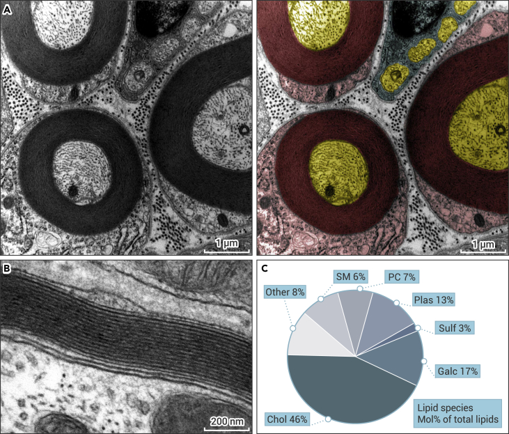
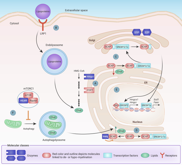
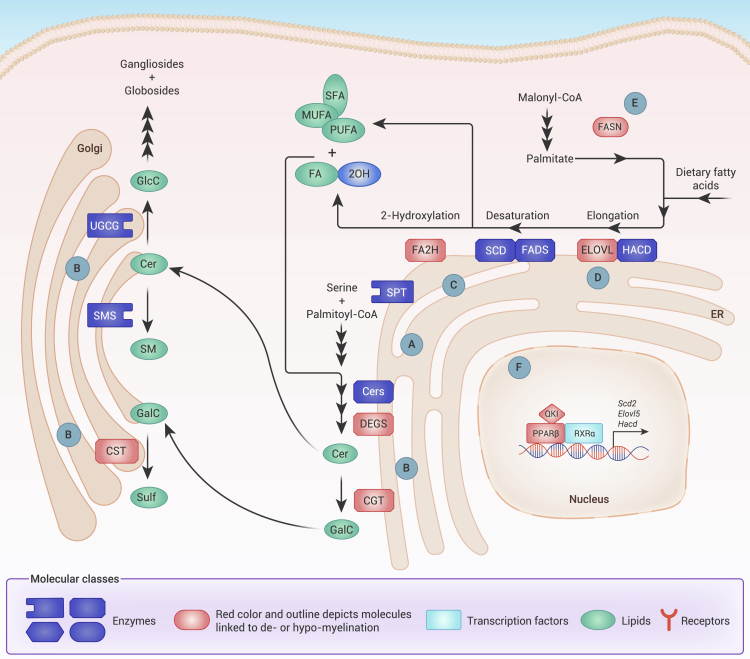
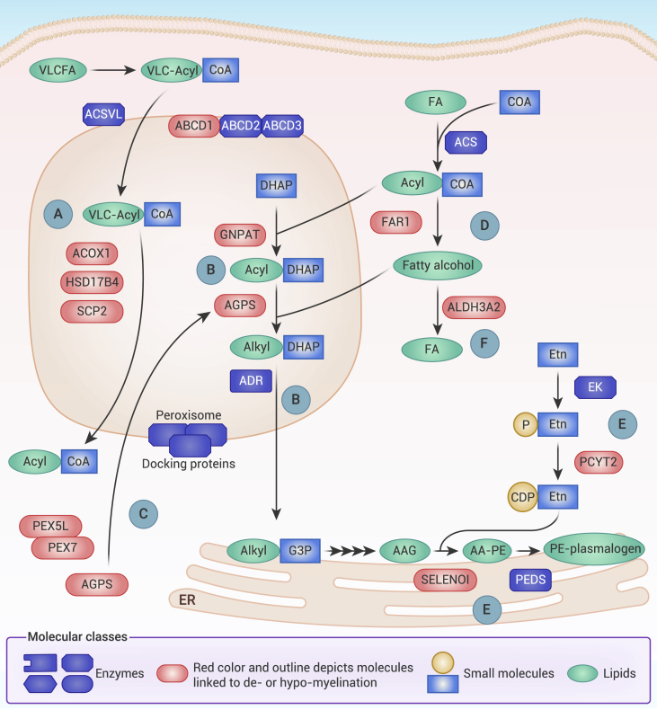
Verification
Claims the paper makes
The claims and statistics of this paper haven’t been checked yet.
Run the check — free, no account needed. It reads the open-access text and checks the sources it cites.
Claims other papers make about this one
Each is checked against this paper’s own text: does it say what the citing paper says it does?
In the citing paper
“More than 70% of myelin is composed of lipids, with cholesterol being one of the most abundant lipid species, ensuring the stability, integrity, and compaction of the myelin sheath.1”
From Neurons sequester the cholesterol-inhibiting TFEB in oligodendrocyte cytoplasm to safeguard myelination and neural function (2025) · cites this paper as [1]
Self-check
Percentages, sums and reported differences re-computed from the paper’s own numbers.
The self-check needs the paper’s full text.
Citations
Cites
mTOR Signaling in Growth, Metabolism, and Disease
Saxton, R. A., & Sabatini, D. M. (2017) · Cell · doi:10.1016/j.cell.2017.02.004 · Cited by 7,555
SREBPs: activators of the complete program of cholesterol and fatty acid synthesis in the liver
Horton, J. D., Goldstein, J. L., & Brown, M. S. (2002) · Journal of Clinical Investigation · doi:10.1172/jci0215593 · Cited by 4,092
mTOR at the nexus of nutrition, growth, ageing and disease
Liu, G. Y., & Sabatini, D. M. (2020) · Nature Reviews Molecular Cell Biology · doi:10.1038/s41580-019-0199-y · Cited by 2,737
Braissant, O., Foufelle, F., Scotto, C., Dauça, M., & Wahli, W. (1996) · Endocrinology · doi:10.1210/endo.137.1.8536636 · Cited by 2,217
mTOR is a key modulator of ageing and age-related disease
Johnson, S. C., Rabinovitch, P. S., & Kaeberlein, M. (2013) · Nature · doi:10.1038/nature11861 · Cited by 1,676
Glycolytic oligodendrocytes maintain myelin and long-term axonal integrity
Fünfschilling, U., Supplie, L. M., Mahad, D., Boretius, S., Saab, A. S., Edgar, J., Brinkmann, B. G., Kassmann, C. M., Tzvetanova, I. D., Möbius, W., et al. (2012) · Nature · doi:10.1038/nature11007 · Cited by 1,523
Disruption of Cnp1 uncouples oligodendroglial functions in axonal support and myelination
Lappe-Siefke, C., Goebbels, S., Gravel, M., Nicksch, E., Lee, J., Braun, P. E., Griffiths, I. R., & Nave, K.-A. (2003) · Nature Genetics · doi:10.1038/ng1095 · Cited by 1,036
High cholesterol level is essential for myelin membrane growth
Saher, G., Brügger, B., Lappe-Siefke, C., Möbius, W., Tozawa, R.-I., Wehr, M. C., Wieland, F., Ishibashi, S., & Nave, K.-A. (2005) · Nature Neuroscience · doi:10.1038/nn1426 · Cited by 799
Han, X., M. Holtzman, D., W. McKeel, D., Kelley, J., & Morris, J. C. (2002) · Journal of Neurochemistry · doi:10.1046/j.1471-4159.2002.00997.x · Cited by 626
Age-related myelin degradation burdens the clearance function of microglia during aging
Safaiyan, S., Kannaiyan, N., Snaidero, N., Brioschi, S., Biber, K., Yona, S., Edinger, A. L., Jung, S., Rossner, M. J., & Simons, M. (2016) · Nature Neuroscience · doi:10.1038/nn.4325 · Cited by 604
Research works printed but not linked: 204
- Tasaki, 1939, The electro-saltatory transmission of the nerve impulse and the effect of narcosis upon the nerve fiber, American Journal of Physiology-Legacy Content, 127, 211, 10.1152/ajplegacy.1939.127.2.211 · DOI
- Huxley, 1949, Evidence for saltatory conduction in peripheral myelinated nerve fibres, J. Physiol., 108, 315, 10.1113/jphysiol.1949.sp004335 · DOI
- Pease-Raissi, 2021, Building a (w)rapport between neurons and oligodendroglia: reciprocal interactions underlying adaptive myelination, Neuron, 109, 1258, 10.1016/j.neuron.2021.02.003 · DOI
- Harty, 2017, Unwrapping the unappreciated: recent progress in Remak Schwann cell biology, Curr. Opin. Neurobiol., 47, 131, 10.1016/j.conb.2017.10.003 · DOI
- Stierli, 2019, Schwann cell plasticity-roles in tissue homeostasis, regeneration, and disease, Glia, 67, 2203, 10.1002/glia.23643 · DOI
- Stierli, 2018, The regulation of the homeostasis and regeneration of peripheral nerve is distinct from the CNS and independent of a stem cell population, Development, 145, dev170316, 10.1242/dev.170316 · DOI
- Norton, 1973, Myelination in rat brain: changes in myelin composition during brain maturation, J. Neurochem., 21, 759, 10.1111/j.1471-4159.1973.tb07520.x · DOI
- O'Brien, 1965, Lipid composition of the normal human brain: gray matter, white matter, and myelin, J. Lipid Res., 6, 537, 10.1016/S0022-2275(20)39619-X · DOI · Paper page
- O'Brien, 1967, Lipid composition of myelin from the peripheral nervous system. Intradural spinal roots, J. Neurochem., 14, 357, 10.1111/j.1471-4159.1967.tb09532.x · DOI
- Schmitt, 2015, Metabolism and functions of lipids in myelin, Biochim. Biophys. Acta, 1851, 999, 10.1016/j.bbalip.2014.12.016 · DOI
- Lingwood, 2010, Lipid rafts as a membrane-organizing principle, Science, 327, 46, 10.1126/science.1174621 · DOI · Paper page
- Korade, 2008, Lipid rafts, cholesterol, and the brain, Neuropharmacology, 55, 1265, 10.1016/j.neuropharm.2008.02.019 · DOI · Paper page
- Saher, 2011, Cholesterol: a novel regulatory role in myelin formation, Neuroscientist, 17, 79, 10.1177/1073858410373835 · DOI · Paper page
- Woelk, 1973, Lipid and fatty acid composition of myelin purified from normal and MS brains, Eur. Neurol., 10, 250, 10.1159/000114281 · DOI
- Yang, 2016, The role of cholesterol in membrane fusion, Chem. Phys. Lipids, 199, 136, 10.1016/j.chemphyslip.2016.05.003 · DOI · Paper page
- Saher, 2010, Cholesterol and myelin biogenesis, Subcell. Biochem., 51, 489, 10.1007/978-90-481-8622-8_18 · DOI
- Saher, 2009, Cholesterol regulates the endoplasmic reticulum exit of the major membrane protein P0 required for peripheral myelin compaction, J. Neurosci., 29, 6094, 10.1523/JNEUROSCI.0686-09.2009 · DOI
- Saher, 2015, Cholesterol in myelin biogenesis and hypomyelinating disorders, Biochim. Biophys. Acta, 1851, 1083, 10.1016/j.bbalip.2015.02.010 · DOI · Paper page
- Klugmann, 1997, Assembly of CNS myelin in the absence of proteolipid protein, Neuron, 18, 59, 10.1016/S0896-6273(01)80046-5 · DOI
- Werner, 2013, A critical role for the cholesterol-associated proteolipids PLP and M6B in myelination of the central nervous system, Glia, 61, 567, 10.1002/glia.22456 · DOI
- Zhou, 2019, PMP22 regulates cholesterol trafficking and ABCA1-mediated cholesterol efflux, J. Neurosci., 39, 5404, 10.1523/JNEUROSCI.2942-18.2019 · DOI
- Miron, 2009, Statin therapy inhibits remyelination in the central nervous system, Am. J. Pathol., 174, 1880, 10.2353/ajpath.2009.080947 · DOI · Paper page
- Camargo, 2009, SREBPs: SREBP function in glia-neuron interactions, FEBS J., 276, 628, 10.1111/j.1742-4658.2008.06808.x · DOI
- Verheijen, 2003, Local regulation of fat metabolism in peripheral nerves, Genes Dev., 17, 2450, 10.1101/gad.1116203 · DOI · Paper page
- Leblanc, 2005, Regulation of cholesterol/lipid biosynthetic genes by Egr2/Krox20 during peripheral nerve myelination, J. Neurochem., 93, 737, 10.1111/j.1471-4159.2005.03056.x · DOI
- Suzuki, 2013, Reduction of the cholesterol sensor SCAP in the brains of mice causes impaired synaptic transmission and altered cognitive function, PLoS Biol., 11, 10.1371/journal.pbio.1001532 · DOI
- Verheijen, 2009, SCAP is required for timely and proper myelin membrane synthesis, Proc. Natl. Acad. Sci. USA, 106, 21383, 10.1073/pnas.0905633106 · DOI · Paper page
- Camargo, 2012, High-fat diet ameliorates neurological deficits caused by defective astrocyte lipid metabolism, Faseb. J., 26, 4302, 10.1096/fj.12-205807 · DOI
- Wattenberg, 2019, Intra- and intercellular trafficking in sphingolipid metabolism in myelination, Adv. Biol. Regul., 71, 97, 10.1016/j.jbior.2018.11.002 · DOI
- Yamaji, 2015, Sphingolipid metabolism and interorganellar transport: localization of sphingolipid enzymes and lipid transfer proteins, Traffic, 16, 101, 10.1111/tra.12239 · DOI
- O'Brien, 1964, The fatty acid composition of brain sphingolipids: sphingomyelin, ceramide, cerebroside, and cerebroside sulfate, J. Lipid Res., 5, 339, 10.1016/S0022-2275(20)40202-0 · DOI
- Bakhti, 2014, Myelin architecture: zippering membranes tightly together, Cell. Mol. Life Sci., 71, 1265, 10.1007/s00018-013-1492-0 · DOI
- Boggs, 2010, Participation of galactosylceramide and sulfatide in glycosynapses between oligodendrocyte or myelin membranes, FEBS Lett., 584, 1771, 10.1016/j.febslet.2009.11.074 · DOI
- Grassi, 2016, The role of 3-O-sulfogalactosylceramide, sulfatide, in the lateral organization of myelin membrane, Neurochem. Res., 41, 130, 10.1007/s11064-015-1747-2 · DOI
- Kulkarni, 1999, Adhesion between cerebroside bilayers, Biochemistry, 38, 15264, 10.1021/bi991725m · DOI
- Bosio, 1996, Functional breakdown of the lipid bilayer of the myelin membrane in central and peripheral nervous system by disrupted galactocerebroside synthesis, Proc. Natl. Acad. Sci. USA, 93, 13280, 10.1073/pnas.93.23.13280 · DOI
- Coetzee, 1996, Myelination in the absence of galactocerebroside and sulfatide: normal structure with abnormal function and regional instability, Cell, 86, 209, 10.1016/S0092-8674(00)80093-8 · DOI
- Marcus, 2006, Sulfatide is essential for the maintenance of CNS myelin and axon structure, Glia, 53, 372, 10.1002/glia.20292 · DOI
- Dupree, 1998, Myelin abnormalities in mice deficient in galactocerebroside and sulfatide, J. Neurocytol., 27, 649, 10.1023/A:1006908013972 · DOI
- Honke, 2002, Paranodal junction formation and spermatogenesis require sulfoglycolipids, Proc. Natl. Acad. Sci. USA, 99, 4227, 10.1073/pnas.032068299 · DOI
- Ishibashi, 2002, A myelin galactolipid, sulfatide, is essential for maintenance of ion channels on myelinated axon but not essential for initial cluster formation, J. Neurosci., 22, 6507, 10.1523/JNEUROSCI.22-15-06507.2002 · DOI
- Hayashi, 2013, Sulfatide decrease in myelin influences formation of the paranodal axo-glial junction and conduction velocity in the sciatic nerve, Glia, 61, 466, 10.1002/glia.22447 · DOI
- Ozgen, 2014, The lateral membrane organization and dynamics of myelin proteins PLP and MBP are dictated by distinct galactolipids and the extracellular matrix, PLoS One, 9, 10.1371/journal.pone.0101834 · DOI
- Palavicini, 2016, Novel molecular insights into the critical role of sulfatide in myelin maintenance/function, J. Neurochem., 139, 40, 10.1111/jnc.13738 · DOI
- Pomicter, 2013, Nfasc155H and MAG are specifically susceptible to detergent extraction in the absence of the myelin sphingolipid sulfatide, Neurochem. Res., 38, 2490, 10.1007/s11064-013-1162-5 · DOI
- Suzuki, 2004, Paranodal axoglial junction is required for the maintenance of the Nav1.6-type sodium channel in the node of Ranvier in the optic nerves but not in peripheral nerve fibers in the sulfatide-deficient mice, Glia, 46, 274, 10.1002/glia.20008 · DOI
- Suzuki, 1999, Drastically abnormal gluco- and galactosylceramide composition does not affect ganglioside metabolism in the brain of mice deficient in galactosylceramide synthase, Neurochem. Res., 24, 471, 10.1023/A:1022571410445 · DOI
- Zöller, 2005, Oligodendrocyte-specific ceramide galactosyltransferase (CGT) expression phenotypically rescues CGT-deficient mice and demonstrates that CGT activity does not limit brain galactosylceramide level, Glia, 52, 190, 10.1002/glia.20230 · DOI
- Hama, 2010, Fatty acid 2-Hydroxylation in mammalian sphingolipid biology, Biochim. Biophys. Acta, 1801, 405, 10.1016/j.bbalip.2009.12.004 · DOI
- Alderson, 2006, FA2H-dependent fatty acid 2-hydroxylation in postnatal mouse brain, J. Lipid Res., 47, 2772, 10.1194/jlr.M600362-JLR200 · DOI
- Eckhardt, 2005, A mammalian fatty acid hydroxylase responsible for the formation of alpha-hydroxylated galactosylceramide in myelin, Biochem. J., 388, 245, 10.1042/BJ20041451 · DOI
- Maldonado, 2008, FA2H is responsible for the formation of 2-hydroxy galactolipids in peripheral nervous system myelin, J. Lipid Res., 49, 153, 10.1194/jlr.M700400-JLR200 · DOI
- Zöller, 2008, Absence of 2-hydroxylated sphingolipids is compatible with normal neural development but causes late-onset axon and myelin sheath degeneration, J. Neurosci., 28, 9741, 10.1523/JNEUROSCI.0458-08.2008 · DOI
- Potter, 2011, Central nervous system dysfunction in a mouse model of FA2H deficiency, Glia, 59, 1009, 10.1002/glia.21172 · DOI
- Saadat, 2010, Absence of oligodendroglial glucosylceramide synthesis does not result in CNS myelin abnormalities or alter the dysmyelinating phenotype of CGT-deficient mice, Glia, 58, 391, 10.1002/glia.20930 · DOI · Paper page
- Meixner, 2011, Myelination in the absence of UDP-galactose:ceramide galactosyl-transferase and fatty acid 2 -hydroxylase, BMC Neurosci., 12, 22, 10.1186/1471-2202-12-22 · DOI
- Braverman, 2012, Functions of plasmalogen lipids in health and disease, Biochim. Biophys. Acta, 1822, 1442, 10.1016/j.bbadis.2012.05.008 · DOI · Paper page
- Nagan, 2001, Plasmalogens: biosynthesis and functions, Prog. Lipid Res., 40, 199, 10.1016/S0163-7827(01)00003-0 · DOI
- Rapport, 1959, The structure of plasmalogens. IV. Lipids in normal and neoplastic tissues of man and in normal tissues of rabbit and rat, Biochim. Biophys. Acta, 33, 319, 10.1016/0006-3002(59)90119-2 · DOI
- Heymans, 1983, Severe plasmalogen deficiency in tissues of infants without peroxisomes (Zellweger syndrome), Nature, 306, 69, 10.1038/306069a0 · DOI
- Webster, 1960, Studies on the plasmalogens of nervous tissue, Biochim. Biophys. Acta, 44, 109, 10.1016/0006-3002(60)91529-8 · DOI
- Balakrishnan, 1961, The distribution of phosphorus-containing lipid compounds in the human brain, J. Neurochem., 8, 276, 10.1111/j.1471-4159.1961.tb13553.x · DOI
- Norton, 1966, The lipid composition of purified bovine brain myelin, J. Neurochem., 13, 213, 10.1111/j.1471-4159.1966.tb06794.x · DOI
- Cumings, 1958, Lipids in the brains of infants and children, J. Neurochem., 2, 289, 10.1111/j.1471-4159.1958.tb12377.x · DOI
- Han, 1990, Plasmenylcholine and phosphatidylcholine membrane bilayers possess distinct conformational motifs, Biochemistry, 29, 4992, 10.1021/bi00472a032 · DOI
- Rubio, 2018, Regulation of phagocytosis in macrophages by membrane ethanolamine plasmalogens, Front. Immunol., 9, 1723, 10.3389/fimmu.2018.01723 · DOI
- Dean, 2018, Structural and functional roles of ether lipids, Protein Cell, 9, 196, 10.1007/s13238-017-0423-5 · DOI
- Luoma, 2015, Plasmalogen phospholipids protect internodal myelin from oxidative damage, Free Radic. Biol. Med., 84, 296, 10.1016/j.freeradbiomed.2015.03.012 · DOI · Paper page
- Sindelar, 1999, The protective role of plasmalogens in iron-induced lipid peroxidation, Free Radic. Biol. Med., 26, 318, 10.1016/S0891-5849(98)00221-4 · DOI
- Zou, 2020, Plasticity of ether lipids promotes ferroptosis susceptibility and evasion, Nature, 585, 603, 10.1038/s41586-020-2732-8 · DOI
- Gross, 1984, High plasmalogen and arachidonic acid content of canine myocardial sarcolemma: a fast atom bombardment mass spectroscopic and gas chromatography-mass spectroscopic characterization, Biochemistry, 23, 158, 10.1021/bi00296a026 · DOI
- da Silva, 2014, Peripheral nervous system plasmalogens regulate Schwann cell differentiation and myelination, J. Clin. Invest., 124, 2560, 10.1172/JCI72063 · DOI
- Malheiro, 2019, Leukodystrophy caused by plasmalogen deficiency rescued by glyceryl 1-myristyl ether treatment, Brain Pathol., 29, 622, 10.1111/bpa.12710 · DOI · Paper page
- Brites, 2003, Impaired neuronal migration and endochondral ossification in Pex7 knockout mice: a model for rhizomelic chondrodysplasia punctata, Hum. Mol. Genet., 12, 2255, 10.1093/hmg/ddg236 · DOI · Paper page
- Rodemer, 2003, Inactivation of ether lipid biosynthesis causes male infertility, defects in eye development and optic nerve hypoplasia in mice, Hum. Mol. Genet., 12, 1881, 10.1093/hmg/ddg191 · DOI
- Teigler, 2009, Defects in myelination, paranode organization and Purkinje cell innervation in the ether lipid-deficient mouse cerebellum, Hum. Mol. Genet., 18, 1897, 10.1093/hmg/ddp110 · DOI · Paper page
- Dorninger, 2015, Homeostasis of phospholipids - the level of phosphatidylethanolamine tightly adapts to changes in ethanolamine plasmalogens, Biochim. Biophys. Acta, 1851, 117, 10.1016/j.bbalip.2014.11.005 · DOI
- Hoxha, 2021, Elovl5 is required for proper action potential conduction along peripheral myelinated fibers, Glia, 69, 2419, 10.1002/glia.24048 · DOI
- Montani, 2018, De novo fatty acid synthesis by Schwann cells is essential for peripheral nervous system myelination, J. Cell Biol., 217, 1353, 10.1083/jcb.201706010 · DOI
- Dimas, 2019, CNS myelination and remyelination depend on fatty acid synthesis by oligodendrocytes, Elife, 8, 10.7554/eLife.44702 · DOI · Paper page
- Berger, 2002, The mechanisms of action of PPARs, Annu. Rev. Med., 53, 409, 10.1146/annurev.med.53.082901.104018 · DOI · Paper page
- Krey, 1993, Xenopus peroxisome proliferator activated receptors: genomic organization, response element recognition, heterodimer formation with retinoid X receptor and activation by fatty acids, J. Steroid Biochem. Mol. Biol., 47, 65, 10.1016/0960-0760(93)90058-5 · DOI
- Berger, 2002, Physiological and therapeutic roles of peroxisome proliferator-activated receptors, Diabetes Technol. Therapeut., 4, 163, 10.1089/15209150260007381 · DOI
- Mandrekar-Colucci, 2013, PPAR agonists as therapeutics for CNS trauma and neurological diseases, ASN Neuro, 5, 10.1042/AN20130030 · DOI
- Zolezzi, 2017, PPARs in the central nervous system: roles in neurodegeneration and neuroinflammation, Biol. Rev. Camb. Phil. Soc., 92, 2046, 10.1111/brv.12320 · DOI
- Agarwal, 2017, Peroxisome proliferator-activated receptors (PPARs) as therapeutic target in neurodegenerative disorders, Biochem. Biophys. Res. Commun., 483, 1166, 10.1016/j.bbrc.2016.08.043 · DOI
- Braissant, 1998, Differential expression of peroxisome proliferator-activated receptor-alpha, -beta, and -gamma during rat embryonic development, Endocrinology, 139, 2748, 10.1210/endo.139.6.6049 · DOI
- Granneman, 1998, Member of the peroxisome proliferator-activated receptor family of transcription factors is differentially expressed by oligodendrocytes, J. Neurosci. Res., 51, 563, 10.1002/(SICI)1097-4547(19980301)51:5<563::AID-JNR3>3.0.CO;2-D · DOI
- Higashiyama, 2007, Expression profiling of liver receptor homologue 1 (LRH-1) in mouse tissues using tissue microarray, J. Mol. Histol., 38, 45, 10.1007/s10735-007-9077-6 · DOI
- Woods, 2003, Localization of PPARdelta in murine central nervous system: expression in oligodendrocytes and neurons, Brain Res., 975, 10, 10.1016/S0006-8993(03)02515-0 · DOI
- Saluja, 2001, PPAR delta agonists stimulate oligodendrocyte differentiation in tissue culture, Glia, 33, 191, 10.1002/1098-1136(200103)33:3<191::AID-GLIA1018>3.0.CO;2-M · DOI
- Jana, 2012, Gemfibrozil, a lipid-lowering drug, increases myelin genes in human oligodendrocytes via peroxisome proliferator-activated receptor-beta, J. Biol. Chem., 287, 34134, 10.1074/jbc.M112.398552 · DOI
- Vittoria Simonini, 2010, Regulation of oligodendrocyte progenitor cell maturation by PPARdelta: effects on bone morphogenetic proteins, ASN Neuro, 2
- Almad, 2010, Chronic expression of PPAR-delta by oligodendrocyte lineage cells in the injured rat spinal cord, J. Comp. Neurol., 518, 785, 10.1002/cne.22242 · DOI
- Peters, 2000, Growth, adipose, brain, and skin alterations resulting from targeted disruption of the mouse peroxisome proliferator-activated receptor beta(delta), Mol. Cell Biol., 20, 5119, 10.1128/MCB.20.14.5119-5128.2000 · DOI
- Zhou, 2020, Mature myelin maintenance requires Qki to coactivate PPARbeta-RXRalpha-mediated lipid metabolism, J. Clin. Invest., 130, 2220, 10.1172/JCI131800 · DOI
- Cermenati, 2015, Lack of sterol regulatory element binding factor-1c imposes glial Fatty Acid utilization leading to peripheral neuropathy, Cell Metabol., 21, 571, 10.1016/j.cmet.2015.02.016 · DOI
- DeBose-Boyd, 2018, SREBPs in lipid metabolism, insulin signaling, and beyond, Trends Biochem. Sci., 43, 358, 10.1016/j.tibs.2018.01.005 · DOI
- Monnerie, 2017, Reduced sterol regulatory element-binding protein (SREBP) processing through site-1 protease (S1P) inhibition alters oligodendrocyte differentiation in vitro, J. Neurochem., 140, 53, 10.1111/jnc.13721 · DOI
- Shin, 2021, Qki activates Srebp2-mediated cholesterol biosynthesis for maintenance of eye lens transparency, Nat. Commun., 12, 3005, 10.1038/s41467-021-22782-0 · DOI
- Zhou, 2021, Qki regulates myelinogenesis through Srebp2-dependent cholesterol biosynthesis, Elife, 10, e60467, 10.7554/eLife.60467 · DOI
- Eid, 2017, mTORC1 activates SREBP-2 by suppressing cholesterol trafficking to lysosomes in mammalian cells, Proc. Natl. Acad. Sci. USA, 114, 7999, 10.1073/pnas.1705304114 · DOI
- Sherman, 2012, Arrest of myelination and reduced axon growth when Schwann cells lack mTOR, J. Neurosci., 32, 1817, 10.1523/JNEUROSCI.4814-11.2012 · DOI
- Wahl, 2014, Mammalian target of rapamycin promotes oligodendrocyte differentiation, initiation and extent of CNS myelination, J. Neurosci., 34, 4453, 10.1523/JNEUROSCI.4311-13.2014 · DOI
- Norrmén, 2014, mTORC1 controls PNS myelination along the mTORC1-RXRgamma-SREBP-lipid biosynthesis axis in Schwann cells, Cell Rep., 9, 646, 10.1016/j.celrep.2014.09.001 · DOI
- Bercury, 2014, Conditional ablation of raptor or rictor has differential impact on oligodendrocyte differentiation and CNS myelination, J. Neurosci., 34, 4466, 10.1523/JNEUROSCI.4314-13.2014 · DOI
- Lebrun-Julien, 2014, Balanced mTORC1 activity in oligodendrocytes is required for accurate CNS myelination, J. Neurosci., 34, 8432, 10.1523/JNEUROSCI.1105-14.2014 · DOI
- Dai, 2014, Interaction of mTOR and Erk1/2 signaling to regulate oligodendrocyte differentiation, Glia, 62, 2096, 10.1002/glia.22729 · DOI
- Khandker, 2022, Cholesterol biosynthesis defines oligodendrocyte precursor heterogeneity between brain and spinal cord, Cell Rep., 38, 10.1016/j.celrep.2022.110423 · DOI
- Zou, 2014, Oligodendrocyte precursor cell-intrinsic effect of Rheb1 controls differentiation and mediates mTORC1-dependent myelination in brain, J. Neurosci., 34, 15764, 10.1523/JNEUROSCI.2267-14.2014 · DOI
- Carson, 2015, Hypomyelination following deletion of Tsc2 in oligodendrocyte precursors, Ann. Clin. Transl. Neurol., 2, 1041, 10.1002/acn3.254 · DOI
- Shi, 2018, mTORC1 activation by loss of Tsc1 in myelinating glia causes downregulation of quaking and neurofascin 155 leading to paranodal domain disorganization, Front. Cell. Neurosci., 12, 201, 10.3389/fncel.2018.00201 · DOI
- Ercan, 2017, Neuronal CTGF/CCN2 negatively regulates myelination in a mouse model of tuberous sclerosis complex, J. Exp. Med., 214, 681, 10.1084/jem.20160446 · DOI
- Lewis, 2013, Impaired language pathways in tuberous sclerosis complex patients with autism spectrum disorders, Cerebr. Cortex, 23, 1526, 10.1093/cercor/bhs135 · DOI
- Peters, 2013, Diffusion tensor imaging and related techniques in tuberous sclerosis complex: review and future directions, Future Neurol., 8, 583, 10.2217/fnl.13.37 · DOI
- Ruppe, 2014, Developmental brain abnormalities in tuberous sclerosis complex: a comparative tissue analysis of cortical tubers and perituberal cortex, Epilepsia, 55, 539, 10.1111/epi.12545 · DOI
- Feng, 2010, The star family member QKI and cell signaling, Adv. Exp. Med. Biol., 693, 25, 10.1007/978-1-4419-7005-3_2 · DOI
- Larocque, 2005, QUAKING KH domain proteins as regulators of glial cell fate and myelination, RNA Biol., 2, 37, 10.4161/rna.2.2.1603 · DOI
- Sidman, 1964, Mutant mice (quaking and jimpy) with deficient myelination in the central nervous system, Science, 144, 309, 10.1126/science.144.3616.309 · DOI
- Ebersole, 1996, The quaking gene product necessary in embryogenesis and myelination combines features of RNA binding and signal transduction proteins, Nat. Genet., 12, 260, 10.1038/ng0396-260 · DOI
- Darbelli, 2016, Quaking regulates neurofascin 155 expression for myelin and axoglial junction maintenance, J. Neurosci., 36, 4106, 10.1523/JNEUROSCI.3529-15.2016 · DOI
- Larocque, 2002, Nuclear retention of MBP mRNAs in the quaking viable mice, Neuron, 36, 815, 10.1016/S0896-6273(02)01055-3 · DOI
- Li, 2000, Destabilization and mislocalization of myelin basic protein mRNAs in quaking dysmyelination lacking the QKI RNA-binding proteins, J. Neurosci., 20, 4944, 10.1523/JNEUROSCI.20-13-04944.2000 · DOI
- Aberg, 2006, Human QKI, a potential regulator of mRNA expression of human oligodendrocyte-related genes involved in schizophrenia, Proc. Natl. Acad. Sci. USA, 103, 7482, 10.1073/pnas.0601213103 · DOI · Paper page
- Lavon, 2019, QKI-V5 is downregulated in CNS inflammatory demyelinating diseases, Mult. Scler. Relat. Disord., 39
- Richard, 2010, Reaching for the stars: linking RNA binding proteins to diseases, Adv. Exp. Med. Biol., 693, 142, 10.1007/978-1-4419-7005-3_10 · DOI
- Tripathi, 2017, Remarkable stability of myelinating oligodendrocytes in mice, Cell Rep., 21, 316, 10.1016/j.celrep.2017.09.050 · DOI
- Lüders, 2019, Maintenance of high proteolipid protein level in adult central nervous system myelin is required to preserve the integrity of myelin and axons, Glia, 67, 634, 10.1002/glia.23549 · DOI
- Fornasiero, 2018, Precisely measured protein lifetimes in the mouse brain reveal differences across tissues and subcellular fractions, Nat. Commun., 9, 4230, 10.1038/s41467-018-06519-0 · DOI
- Lin, 2014, Impaired eukaryotic translation initiation factor 2B activity specifically in oligodendrocytes reproduces the pathology of vanishing white matter disease in mice, J. Neurosci., 34, 12182, 10.1523/JNEUROSCI.1373-14.2014 · DOI
- Ando, 2003, Turnover of myelin lipids in aging brain, Neurochem. Res., 28, 5, 10.1023/A:1021635826032 · DOI
- Clarke, 2019, The Ormdl genes regulate the sphingolipid synthesis pathway to ensure proper myelination and neurologic function in mice, Elife, 8, 10.7554/eLife.51067 · DOI
- Viader, 2013, Aberrant Schwann cell lipid metabolism linked to mitochondrial deficits leads to axon degeneration and neuropathy, Neuron, 77, 886, 10.1016/j.neuron.2013.01.012 · DOI
- Beirowski, 2014, Metabolic regulator LKB1 is crucial for Schwann cell-mediated axon maintenance, Nat. Neurosci., 17, 1351, 10.1038/nn.3809 · DOI
- Moser, 2005, Adrenoleukodystrophy: new approaches to a neurodegenerative disease, JAMA, 294, 3131, 10.1001/jama.294.24.3131 · DOI · Paper page
- Ferdinandusse, 2006, Clinical and biochemical spectrum of D-bifunctional protein deficiency, Ann. Neurol., 59, 92, 10.1002/ana.20702 · DOI
- Horvath, 2015, SCP2 mutations and neurodegeneration with brain iron accumulation, Neurology, 85, 1909, 10.1212/WNL.0000000000002157 · DOI
- Chung, 2020, Loss- or gain-of-function mutations in ACOX1 cause axonal loss via different mechanisms, Neuron, 106, 589, 10.1016/j.neuron.2020.02.021 · DOI · Paper page
- Huyghe, 2006, Peroxisomal multifunctional protein-2 deficiency causes motor deficits and glial lesions in the adult central nervous system, Am. J. Pathol., 168, 1321, 10.2353/ajpath.2006.041220 · DOI
- Bams-Mengerink, 2006, MRI of the brain and cervical spinal cord in rhizomelic chondrodysplasia punctata, Neurology, 66, 798, 10.1212/01.wnl.0000205594.34647.d0 · DOI · Paper page
- Sztriha, 2000, Abnormal myelin formation in rhizomelic chondrodysplasia punctata type 2 (DHAPAT-deficiency), Dev. Med. Child Neurol., 42, 492
- Wanders, 2005, Peroxisomal disorders I: biochemistry and genetics of peroxisome biogenesis disorders, Clin. Genet., 67, 107, 10.1111/j.1399-0004.2004.00329.x · DOI
- Waterham, 2012, Genetics and molecular basis of human peroxisome biogenesis disorders, Biochim. Biophys. Acta, 1822, 1430, 10.1016/j.bbadis.2012.04.006 · DOI
- Barøy, 2015, A novel type of rhizomelic chondrodysplasia punctata, RCDP5, is caused by loss of the PEX5 long isoform, Hum. Mol. Genet., 24, 5845, 10.1093/hmg/ddv305 · DOI · Paper page
- Buchert, 2014, A peroxisomal disorder of severe intellectual disability, epilepsy, and cataracts due to fatty acyl-CoA reductase 1 deficiency, Am. J. Hum. Genet., 95, 602, 10.1016/j.ajhg.2014.10.003 · DOI · Paper page
- Ferdinandusse, 2021, An autosomal dominant neurological disorder caused by de novo variants in FAR1 resulting in uncontrolled synthesis of ether lipids, Genet. Med., 23, 740, 10.1038/s41436-020-01027-3 · DOI
- Ahmed, 2017, A mutation of EPT1 (SELENOI) underlies a new disorder of Kennedy pathway phospholipid biosynthesis, Brain, 140, 547
- Horibata, 2018, EPT1 (selenoprotein I) is critical for the neural development and maintenance of plasmalogen in humans, J. Lipid Res., 59, 1015, 10.1194/jlr.P081620 · DOI
- Vaz, 2019, Mutations in PCYT2 disrupt etherlipid biosynthesis and cause a complex hereditary spastic paraplegia, Brain, 142, 3382, 10.1093/brain/awz291 · DOI
- Kryukov, 2003, Characterization of mammalian selenoproteomes, Science, 300, 1439, 10.1126/science.1083516 · DOI
- Nakashima, 1997, Cloning of a human cDNA for CTP-phosphoethanolamine cytidylyltransferase by complementation in vivo of a yeast mutant, J. Biol. Chem., 272, 9567, 10.1074/jbc.272.14.9567 · DOI
- Edvardson, 2008, Mutations in the fatty acid 2-hydroxylase gene are associated with leukodystrophy with spastic paraparesis and dystonia, Am. J. Hum. Genet., 83, 643, 10.1016/j.ajhg.2008.10.010 · DOI
- Kruer, 2010, Defective FA2H leads to a novel form of neurodegeneration with brain iron accumulation (NBIA), Ann. Neurol., 68, 611, 10.1002/ana.22122 · DOI
- Kitamura, 2017, Phytosphingosine degradation pathway includes fatty acid alpha-oxidation reactions in the endoplasmic reticulum, Proc. Natl. Acad. Sci. USA, 114, E2616, 10.1073/pnas.1700138114 · DOI
- Staps, 2020, Disturbed brain ether lipid metabolism and histology in Sjogren-Larsson syndrome, J. Inherit. Metab. Dis., 43, 1265, 10.1002/jimd.12275 · DOI
- Karsai, 2019, DEGS1-associated aberrant sphingolipid metabolism impairs nervous system function in humans, J. Clin. Invest., 129, 1229, 10.1172/JCI124159 · DOI
- Pant, 2019, Loss of the sphingolipid desaturase DEGS1 causes hypomyelinating leukodystrophy, J. Clin. Invest., 129, 1240, 10.1172/JCI123959 · DOI
- Mahmood, 2010, Metachromatic leukodystrophy: a case of triplets with the late infantile variant and a systematic review of the literature, J. Child Neurol., 25, 572, 10.1177/0883073809341669 · DOI
- Komatsuzaki, 2019, Clinical characteristics of 248 patients with Krabbe disease: quantitative natural history modeling based on published cases, Genet. Med., 21, 2208, 10.1038/s41436-019-0480-7 · DOI
- Wenger, 2000, Krabbe disease: genetic aspects and progress toward therapy, Mol. Genet. Metabol., 70, 1, 10.1006/mgme.2000.2990 · DOI
- Pareyson, 2017, New developments in Charcot-Marie-Tooth neuropathy and related diseases, Curr. Opin. Neurol., 30, 471, 10.1097/WCO.0000000000000474 · DOI
- Rossor, 2013, Clinical implications of genetic advances in Charcot-Marie-Tooth disease, Nat. Rev. Neurol., 9, 562, 10.1038/nrneurol.2013.179 · DOI
- Barreto, 2016, Epidemiologic study of charcot-marie-tooth disease: a systematic review, Neuroepidemiology, 46, 157, 10.1159/000443706 · DOI
- Berger, 2006, Schwann cells and the pathogenesis of inherited motor and sensory neuropathies (Charcot-Marie-Tooth disease), Glia, 54, 243, 10.1002/glia.20386 · DOI
- Murakami, 2019, Schwann cell and the pathogenesis of charcot-marie-tooth disease, Adv. Exp. Med. Biol., 1190, 301, 10.1007/978-981-32-9636-7_19 · DOI
- Fledrich, 2012, A rat model of Charcot-Marie-Tooth disease 1A recapitulates disease variability and supplies biomarkers of axonal loss in patients, Brain, 135, 72, 10.1093/brain/awr322 · DOI
- Fledrich, 2018, Targeting myelin lipid metabolism as a potential therapeutic strategy in a model of CMT1A neuropathy, Nat. Commun., 9, 3025, 10.1038/s41467-018-05420-0 · DOI
- Visigalli, 2020, Exploiting sphingo- and glycerophospholipid impairment to select effective drugs and biomarkers for CMT1A, Front. Neurol., 11, 903, 10.3389/fneur.2020.00903 · DOI
- Zhou, 2019, A neutral lipid-enriched diet improves myelination and alleviates peripheral nerve pathology in neuropathic mice, Exp. Neurol., 321, 10.1016/j.expneurol.2019.113031 · DOI
- Lee, 2014, PMP22 is critical for actin-mediated cellular functions and for establishing lipid rafts, J. Neurosci., 34, 16140, 10.1523/JNEUROSCI.1908-14.2014 · DOI
- Zhou, 2020, Subcellular diversion of cholesterol by gain- and loss-of-function mutations in PMP22, Glia, 68, 2300, 10.1002/glia.23840 · DOI
- Simons, 2002, Overexpression of the myelin proteolipid protein leads to accumulation of cholesterol and proteolipid protein in endosomes/lysosomes: implications for Pelizaeus-Merzbacher disease, J. Cell Biol., 157, 327, 10.1083/jcb.200110138 · DOI · Paper page
- Saher, 2012, Therapy of Pelizaeus-Merzbacher disease in mice by feeding a cholesterol-enriched diet, Nat. Med., 18, 1130, 10.1038/nm.2833 · DOI · Paper page
- de Faria, 2021, Periods of synchronized myelin changes shape brain function and plasticity, Nat. Neurosci., 24, 1508, 10.1038/s41593-021-00917-2 · DOI
- Svennerholm, 1991, Membrane lipids in the aging human brain, J. Neurochem., 56, 2051, 10.1111/j.1471-4159.1991.tb03466.x · DOI
- Cheng, 2010, Apolipoprotein E mediates sulfatide depletion in animal models of Alzheimer's disease, Neurobiol. Aging, 31, 1188, 10.1016/j.neurobiolaging.2008.07.020 · DOI · Paper page
- Han, 2001, Plasmalogen deficiency in early Alzheimer's disease subjects and in animal models: molecular characterization using electrospray ionization mass spectrometry, J. Neurochem., 77, 1168, 10.1046/j.1471-4159.2001.00332.x · DOI
- Kaya, 2020, Brain region-specific amyloid plaque-associated myelin lipid loss, APOE deposition and disruption of the myelin sheath in familial Alzheimer's disease mice, J. Neurochem., 154, 84, 10.1111/jnc.14999 · DOI
- Bartzokis, 2003, White matter structural integrity in healthy aging adults and patients with Alzheimer disease: a magnetic resonance imaging study, Arch. Neurol., 60, 393, 10.1001/archneur.60.3.393 · DOI
- Ringman, 2007, Diffusion tensor imaging in preclinical and presymptomatic carriers of familial Alzheimer's disease mutations, Brain, 130, 1767, 10.1093/brain/awm102 · DOI · Paper page
- Luo, 2019, Application of T1-/T2-weighted ratio mapping to elucidate intracortical demyelination process in the alzheimer's disease continuum, Front. Neurosci., 13, 904, 10.3389/fnins.2019.00904 · DOI
- Mitew, 2010, Focal demyelination in Alzheimer's disease and transgenic mouse models, Acta Neuropathol., 119, 567, 10.1007/s00401-010-0657-2 · DOI · Paper page
- Mathys, 2019, Single-cell transcriptomic analysis of Alzheimer's disease, Nature, 570, 332, 10.1038/s41586-019-1195-2 · DOI · Paper page
- Zhou, 2020, Human and mouse single-nucleus transcriptomics reveal TREM2-dependent and TREM2-independent cellular responses in Alzheimer's disease, Nat. Med., 26, 131, 10.1038/s41591-019-0695-9 · DOI · Paper page
- Berghoff, 2017, Dietary cholesterol promotes repair of demyelinated lesions in the adult brain, Nat. Commun., 8, 10.1038/ncomms14241 · DOI · Paper page
- Stumpf, 2019, Ketogenic diet ameliorates axonal defects and promotes myelination in Pelizaeus-Merzbacher disease, Acta Neuropathol., 138, 147, 10.1007/s00401-019-01985-2 · DOI
- Raas, 2021, Metabolic rerouting via SCD1 induction impacts X-linked adrenoleukodystrophy, J. Clin. Invest., 131, 10.1172/JCI142500 · DOI
- Hill, 2018, Lifelong cortical myelin plasticity and age-related degeneration in the live mammalian brain, Nat. Neurosci., 21, 683, 10.1038/s41593-018-0120-6 · DOI · Paper page
- Lamming, 2013, Rapalogs and mTOR inhibitors as anti-aging therapeutics, J. Clin. Invest., 123, 980, 10.1172/JCI64099 · DOI · Paper page
- Watkins, 1995, Distinction between peroxisomal bifunctional enzyme and acyl-CoA oxidase deficiencies, Ann. Neurol., 38, 472, 10.1002/ana.410380322 · DOI
- Ferdinandusse, 2006, Mutational spectrum of D-bifunctional protein deficiency and structure-based genotype-phenotype analysis, Am. J. Hum. Genet., 78, 112, 10.1086/498880 · DOI · Paper page
- Schaumburg, 1975, Adrenoleukodystrophy. A clinical and pathological study of 17 cases, Arch. Neurol., 32, 577, 10.1001/archneur.1975.00490510033001 · DOI · Paper page
- Dick, 2008, A novel locus for an autosomal recessive hereditary spastic paraplegia (SPG35) maps to 16q21-q23, Neurology, 71, 248, 10.1212/01.wnl.0000319610.29522.8a · DOI
- Lossos, 2006, Phenotypic variability among adult siblings with Sjogren-Larsson syndrome, Arch. Neurol., 63, 278, 10.1001/archneur.63.2.278 · DOI
- Sjogren, 1957, Oligophrenia in combination with congenital ichthyosis and spastic disorders; a clinical and genetic study, Acta Psychiatr. Neurol. Scand. Suppl., 113, 1
- Willemsen, 2000, Juvenile macular dystrophy associated with deficient activity of fatty aldehyde dehydrogenase in Sjogren-Larsson syndrome, Am. J. Ophthalmol., 130, 782, 10.1016/S0002-9394(00)00576-6 · DOI
- MacFaul, 1982, Metachromatic leucodystrophy: review of 38 cases, Arch. Dis. Child., 57, 168, 10.1136/adc.57.3.168 · DOI
- Zafeiriou, 1999, Neurophysiology and MRI in late-infantile metachromatic leukodystrophy, Pediatr. Neurol., 21, 843, 10.1016/S0887-8994(99)00093-4 · DOI
- Dolgin, 2019, DEGS1 variant causes neurological disorder, Eur. J. Hum. Genet., 27, 1668, 10.1038/s41431-019-0444-z · DOI
- Anstey, 1999, Characterization of photosensitivity in the Smith-Lemli-Opitz syndrome: a new congenital photosensitivity syndrome, Br. J. Dermatol., 141, 406, 10.1046/j.1365-2133.1999.03032.x · DOI
200 of 204 shown
Cited by
Sorted by citations
Microglia and Astrocytes in Alzheimer’s Disease: Significance and Summary of Recent Advances
Deng, Q., Wu, C., Parker, E., Liu, T. C.-Y., Duan, R., & Yang, L. (2023) · Aging and disease · doi:10.14336/ad.2023.0907 · Cited by 102
Cholesterol Metabolism in CNS Diseases: The Potential of SREBP2 and LXR as Therapeutic Targets
Wang, N.-Q., Sun, P.-X., Shen, Q.-Q., & Deng, M.-Y. (2025) · Molecular Neurobiology · doi:10.1007/s12035-024-04672-w · Cited by 19
Spatial lipidomics reveals demyelination and remyelination dynamics in the mouse brain
Opielka, M., Urbanowicz, K., Konieczna-Wolska, K., Müller, E., Krajewski, O., Feucherolles, M., Zhou, Q., Bienkowski, M., Frache, G., Hopf, C., et al. (2025) · Journal of Lipid Research · doi:10.1016/j.jlr.2025.100912 · Cited by 8
Zhang, Y., Cheng, Z., Huang, S., Sharma, T., Barbosa, D., Yu, G., & Sun, L. O. (2025) · Cell Reports · doi:10.1016/j.celrep.2025.116252 · Cited by 4
Li, H., Zhao, Z., Zhu, L., Tan, Y., Zhang, Z., Zhang, Z., Kang, J., Lu, H., Peng, W., & Wu, Q. (2026) · Journal of Advanced Research · doi:10.1016/j.jare.2025.08.044 · Cited by 3
Gutierrez-Kuri, E., Marie Garcia-Rogers, J. L., Perez, J., Smith, S., Kenwood, M. R., Archuleta, K. S., Xiao, Y., Campos, G., Barannikov, S., Wang, H., et al. (2026) · doi:10.64898/2026.07.13.738356
Metadata
- Authors
- Barnes-Vélez, J. A., Aksoy Yasar, F. B., & Hu, J.
- Venue
- The Innovation
- Publisher
- Elsevier BV
- Published
- 2023-01
- Volume
- 4
- Issue
- 1
- Pages
- 100360
- ISSN
- 2666-6758
- PMC
- PMC9800635
- Cited by
- 73
- References
- 26
- Language
- en
About these checks
Registry, retraction, Cites and statistics results are computed, not judged, with one exception: a printed reference that no index lists is read once by a model before it counts as a citation error. Claims are extracted by a model and kept only when their quote appears verbatim in the text; each cited source is then checked, with a model’s help, for whether it says what the claim relies on. Those verdicts can be wrong — read the claim’s quote and the cited source before relying on one.
Statistics and arithmetic are recomputed, not judged.
Confidence bands are shown only for fields calibrated on our annotated test set. None is calibrated yet, so no band is shown.
What counts as a claim ›How we measure confidence ›How we check this ›
Are you an author, or do you see a problem with this page? Request a review
