

The DOI matches its Crossref record; 63 of the 65 references it cites were found in a registry or an index.
Neuronal activity increases translocator protein (TSPO) levels
Open accessAbstract
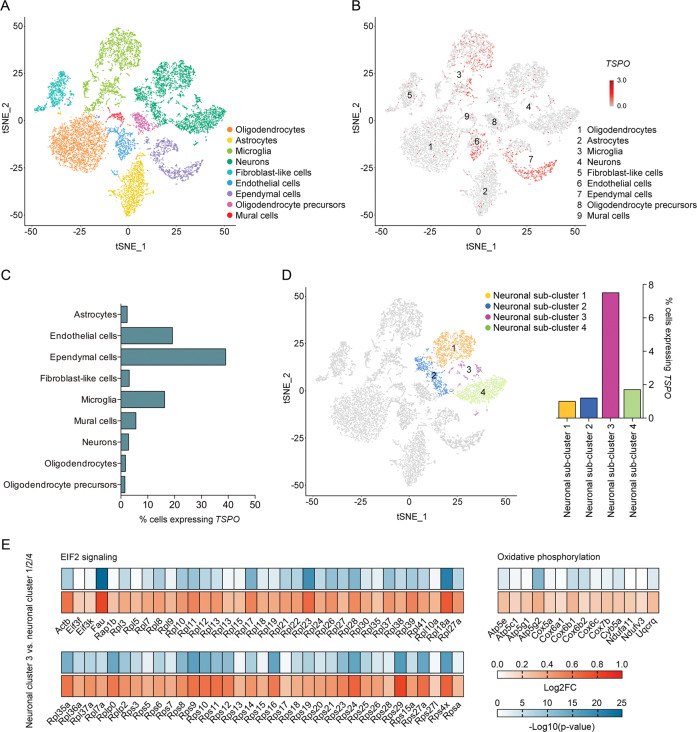
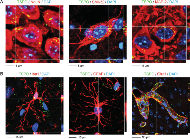
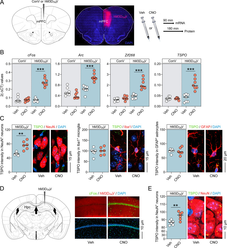
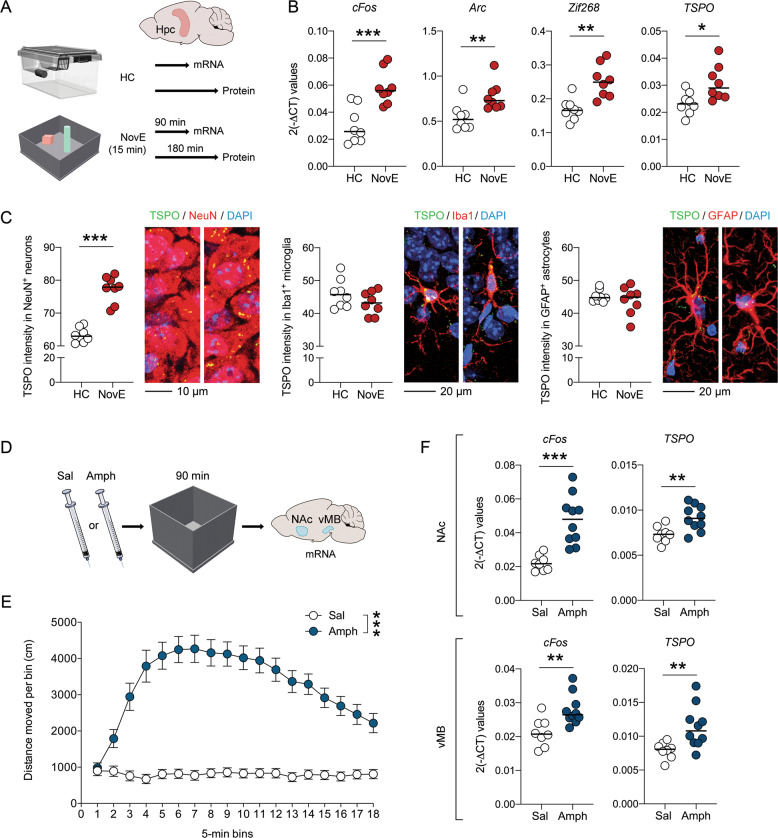
The mitochondrial protein, translocator protein (TSPO), is a widely used biomarker of neuroinflammation, but its non-selective cellular expression pattern implies roles beyond inflammatory processes. In the present study, we investigated whether neuronal activity modifies TSPO levels in the adult central nervous system. First, we used single-cell RNA sequencing to generate a cellular landscape of basal TSPO gene expression in the hippocampus of adult (12 weeks old) C57BL6/N mice, followed by confocal laser scanning microscopy to verify TSPO protein in neuronal and non-neuronal cell populations. We then quantified TSPO mRNA and protein levels after stimulating neuronal activity with distinct stimuli, including designer receptors exclusively activated by designer drugs (DREADDs), exposure to a novel environment and acute treatment with the psychostimulant drug, amphetamine. Single-cell RNA sequencing demonstrated a non-selective and multi-cellular gene expression pattern of TSPO at basal conditions in the adult mouse hippocampus. Confocal laser scanning microscopy confirmed that TSPO protein is present in neuronal and non-neuronal (astrocytes, microglia, vascular endothelial cells) cells of cortical (medial prefrontal cortex) and subcortical (hippocampus) brain regions. Stimulating neuronal activity through chemogenetic (DREADDs), physiological (novel environment exposure) or psychopharmacological (amphetamine treatment) approaches led to consistent increases in TSPO gene and protein levels in neurons, but not in microglia or astrocytes. Taken together, our findings show that neuronal activity has the potential to modify TSPO levels in the adult central nervous system. These findings challenge the general assumption that altered TSPO expression or binding unequivocally mirrors ongoing neuroinflammation and emphasize the need to consider non-inflammatory interpretations in some physiological or pathological contexts.
Overview
The claims and statistics of this paper haven’t been checked yet.
Run the check — free, no account needed. It reads the open-access text and checks the sources it cites.
Integrity signals
Registry
Cites
Statistics
Figures
Verification
Claims the paper makes
The claims and statistics of this paper haven’t been checked yet.
Run the check — free, no account needed. It reads the open-access text and checks the sources it cites.
Claims other papers make about this one
Each is checked against this paper’s own text: does it say what the citing paper says it does?
The cited source says what the claim attributes to it.
In the citing paper
“Given that more recent evidence confirms TSPO expression in neurons [44]”
From Pharmacological modulation of TSPO in microglia/macrophages and neurons in a chronic neurodegenerative model of prion disease (2023) · cites this paper as [44]
Self-check
Percentages, sums and reported differences re-computed from the paper’s own numbers.
The self-check needs the paper’s full text.
Citations
Cites
Analysis of Relative Gene Expression Data Using Real-Time Quantitative PCR and the 2−ΔΔCT Method
Livak, K. J., & Schmittgen, T. D. (2001) · Methods · doi:10.1006/meth.2001.1262 · Cited by 180,860
Comprehensive Integration of Single-Cell Data
Stuart, T., Butler, A., Hoffman, P., Hafemeister, C., Papalexi, E., Mauck, W. M., Hao, Y., Stoeckius, M., Smibert, P., & Satija, R. (2019) · Cell · doi:10.1016/j.cell.2019.05.031 · Cited by 15,453
Gene Expression Omnibus: NCBI gene expression and hybridization array data repository
Edgar, R. (2002) · Nucleic Acids Research · doi:10.1093/nar/30.1.207 · Cited by 13,288
Integrating single-cell transcriptomic data across different conditions, technologies, and species
Butler, A., Hoffman, P., Smibert, P., Papalexi, E., & Satija, R. (2018) · Nature Biotechnology · doi:10.1038/nbt.4096 · Cited by 13,174
Zhang, Y., Chen, K., Sloan, S. A., Bennett, M. L., Scholze, A. R., O'Keeffe, S., Phatnani, H. P., Guarnieri, P., Caneda, C., Ruderisch, N., et al. (2014) · The Journal of Neuroscience · doi:10.1523/jneurosci.1860-14.2014 · Cited by 4,875
Armbruster, B. N., Li, X., Pausch, M. H., Herlitze, S., & Roth, B. L. (2007) · Proceedings of the National Academy of Sciences · doi:10.1073/pnas.0700293104 · Cited by 2,046
Roth, B. L. (2016) · Neuron · doi:10.1016/j.neuron.2016.01.040 · Cited by 1,742
PanglaoDB: a web server for exploration of mouse and human single-cell RNA sequencing data
Franzén, O., Gan, L.-M., & Björkegren, J. L. M. (2019) · Database · doi:10.1093/database/baz046 · Cited by 1,551 · cited 1×
“Cell-type assignments were predicted by matching representative expressed features to published cell markers data [21].”
BackgroundMicroglia in physiology and disease
Wolf, S. A., Boddeke, H. W. G. M., & Kettenmann, H. (2017) · Annual Review of Physiology · doi:10.1146/annurev-physiol-022516-034406 · Cited by 1,426
Brain on stress: how the social environment gets under the skin
McEwen, B. S. (2012) · Proceedings of the National Academy of Sciences · doi:10.1073/pnas.1121254109 · Cited by 1,200
Cited by
Sorted by citations
The Semantics of Microglia Activation: Neuroinflammation, Homeostasis, and Stress
Woodburn, S. C., Bollinger, J. L., & Wohleb, E. S. (2021) · Journal of Neuroinflammation · doi:10.1186/s12974-021-02309-6 · Cited by 783
Inflammation-Related Functional and Structural Dysconnectivity as a Pathway to Psychopathology
Goldsmith, D. R., Bekhbat, M., Mehta, N. D., & Felger, J. C. (2022) · Biological Psychiatry · doi:10.1016/j.biopsych.2022.11.003 · Cited by 229
“competition from activated peripheral immune cells (37, 38), rather than a reduced inflammatory”
Comer, A. L., Carrier, M., Tremblay, M., & Cruz‐Martín, A. (2020) · Frontiers in Cellular Neuroscience · doi:10.3389/fncel.2020.00274 · Cited by 212
“day 11-15) (prenatal exposure) cerebellum myelin density, cerebellum iron levels, RNAseq of cerebellum increased inflammation signaling; increased iron inclusions; myelin sheath damage Klocke et al., 2018 PM (2.5 um) mice adults (exposure in utero) western blot, ELISA and IHC in temporal cortex deficits in spatial mem…”
BackgroundInfluentialPET Imaging of Neuroinflammation in Alzheimer’s Disease
Zhou, R., Ji, B., Kong, Y., Qin, L., Ren, W., Guan, Y., & Ni, R. (2021) · Frontiers in Immunology · doi:10.3389/fimmu.2021.739130 · Cited by 157
Neuroinflammation in schizophrenia: the role of nuclear factor kappa B
Murphy, C. E., Walker, A. K., & Weickert, C. S. (2021) · Translational Psychiatry · doi:10.1038/s41398-021-01607-0 · Cited by 128
Aiding and Abetting Anhedonia: Impact of Inflammation on the Brain and Pharmacological Implications
Lucido, M. J., Bekhbat, M., Goldsmith, D. R., Treadway, M. T., Haroon, E., Felger, J. C., & Miller, A. H. (2021) · Pharmacological Reviews · doi:10.1124/pharmrev.120.000043 · Cited by 133
“TSPO ligands also bind to other activated cells in the brain, including astrocytes, vascular endothelial cells, and neurons, making interpretation of the results challenging (Notter et al., 2020).”
BackgroundLasselin, J., Lekander, M., Benson, S., Schedlowski, M., & Engler, H. (2020) · Molecular Psychiatry · doi:10.1038/s41380-020-00869-2 · Cited by 121
Guilarte, T. R., Rodichkin, A. N., McGlothan, J. L., de la Rocha, A. A., & Azzam, D. J. (2021) · Pharmacology & Therapeutics · doi:10.1016/j.pharmthera.2021.108048 · Cited by 116
Adolescence is a sensitive period for prefrontal microglia to act on cognitive development
Schalbetter, S. M., von Arx, A. S., Cruz-Ochoa, N. A., Dawson, K. L. D., Ivanov, A., Mueller, F. S., Lin, H.-Y., Amport, R., Mildenberger, W., Mattei, D., et al. (2022) · Science Advances · doi:10.1126/sciadv.abi6672 · Cited by 115
“The stereotaxic surgery was performed using methods established and validated before ( 51 , 52 ).”
InfluentialSelf-citationMartins, D., Giacomel, A., Williams, S., Turkheimer, F., Dipasquale, O., & Veronese, M. (2021) · Cell Reports · doi:10.1016/j.celrep.2021.110173 · Cited by 97
Turkheimer, F., Althubaity, N., Schubert, J. J., Nettis, M. A., Cousins, O., Dima, D., Mondelli, V., Bullmore, E. T., Pariante, C. M., & Veronese, M. (2020) · Brain Behavior and Immunity · doi:10.1016/j.bbi.2020.10.025 · Cited by 89
Kokkosis, A. G., Madeira, M. M., Hage, Z., Valais, K., Koliatsis, D., Resutov, E., & Tsirka, S. E. (2023) · Glia · doi:10.1002/glia.24464 · Cited by 91
“…reduced the TSPO protein levels in the non-stressed PLX56-Con (compared to the non-treated Con group), it did not reach zero levels, consistent with expression of TSPO by other cell types besides microglia (endothelial cells, astrocytes, and neurons) (Supplementary Figure 3) (Notter et al., 2021).”
ResultPresent and future of microglial pharmacology
Šimončičová, E., de Andrade, E. G., Vecchiarelli, H. A., Awogbindin, I. O., Delage, C. I., & Tremblay, M. (2022) · Trends in Pharmacological Sciences · doi:10.1016/j.tips.2021.11.006 · Cited by 79
Advancing an Inflammatory Subtype of Major Depression
Miller, A. H. (2025) · American Journal of Psychiatry · doi:10.1176/appi.ajp.20250289 · Cited by 93
“TSPO binding in the brain is a putative marker of activated microglia, although its expression on multiple other cell types in the brain has limited interpretation of imaging results (30).”
Imaging Neuroinflammation in Neurodegenerative Disorders
Masdeu, J. C., Pascual, B., & Fujita, M. (2022) · Journal of Nuclear Medicine · doi:10.2967/jnumed.121.263200 · Cited by 77
“Postmortem studies on AD showed increased P2X7 (104,105) and decreased P2Y12 (106), consistent with greater detrimental than neuroprotective effects of neuroinflammation in moderate-advanced stages of AD.”
BackgroundInfluentialNaoi, M., Maruyama, W., & Shamoto‐Nagai, M. (2022) · International Journal of Molecular Sciences · doi:10.3390/ijms231911059 · Cited by 75
“macological (amphetamine) stimuli [241].”
Rupprecht, R., Wetzel, C. H., Dorostkar, M. M., Herms, J. W., Albert, N. L., Schwarzbach, J. V., Schumacher, M., & Neumann, I. D. (2022) · Molecular Psychiatry · doi:10.1038/s41380-022-01561-3 · Cited by 66
“Moreover, TSPO expression should not unequivocally be considered as a marker of neuroinflammation, since also neuronal activation may increase TSPO levels [48].”
BackgroundControl of Neuroinflammation through Radiation-Induced Microglial Changes
Boyd, A., Byrne, S., Middleton, R. J., Banati, R. B., & Liu, G. J. (2021) · Cells · doi:10.3390/cells10092381 · Cited by 65
Annual Research Review: Neuroimmune network model of depression: a developmental perspective
Nusslock, R., Alloy, L. B., Brody, G. H., & Miller, G. E. (2024) · Journal of Child Psychology and Psychiatry · doi:10.1111/jcpp.13961 · Cited by 67
“This latter finding should also be interpreted with caution, however, as PET studies of TSPO are unable to distinguish the many states and functions of microglia, which include synaptic pruning and dendritic arborization that are unrelated to inflammation (Notter et al., 2021).”
Inflammatory Biomarkers in Addictive Disorders
Morcuende, Á., Navarrete, F., Nieto, E. O., Manzanares, J., & Femenía, T. (2021) · Biomolecules · doi:10.3390/biom11121824 · Cited by 55
“However, neuronal activity can modify TSPO levels, suggesting that non-inflammatory interpretations should be considered in some physiological or pathological contexts [70].”
BackgroundRupprecht, R., Pradhan, A. K., Kufner, M., Brunner, L.-M., Nothdurfter, C., Wein, S., Schwarzbach, J. V., Puig-Bosch, X., Rupprecht, C., & Rammes, G. (2022) · European Archives of Psychiatry and Clinical Neuroscience · doi:10.1007/s00406-022-01532-3 · Cited by 45
“However, it should be emphasized that TSPO expression should not just be considered as a marker of microglia activation, because it has been demonstrated that neuronal activation also may increase TSPO expression [20].”
BackgroundBarron, A. M., Higuchi, M., Hattori, S., Kito, S., Suhara, T., & Ji, B. (2020) · Molecular Neurobiology · doi:10.1007/s12035-020-02136-5 · Cited by 44
“Interestingly, neuronal activity has been shown to upregulate hippocampal and cortical neuronal TSPO expression, although the functional significance of neuronal TSPO was not addressed [34].”
BackgroundSforzini, L., Cattaneo, A., Ferrari, C., Turner, L., Mariani, N., Enache, D., Hastings, C. N. M., Lombardo, G., Nettis, M. A., Nikkheslat, N., et al. (2023) · Translational Psychiatry · doi:10.1038/s41398-023-02438-x · Cited by 41
“However, studies using positron emission tomography (PET) of translocator protein (TSPO), a widely used in vivo measure of microglial activation, have found no correlation between serum CRP levels and PET binding of TSPO, in both subjects with MDD and controls [51, 52], although TSPO expression may reflect other non-i…”
BackgroundMicroglial contribution to the pathology of neurodevelopmental disorders in humans
Matulevičiūtė, R., Akinluyi, E. T., Muntslag, T. A. O., Dewing, J. M., Long, K. R., Vernon, A. C., Tremblay, M., & Menassa, D. A. (2023) · Acta Neuropathologica · doi:10.1007/s00401-023-02629-2 · Cited by 41
“Furthermore, concerns have been raised about TSPO specificity for microglia, given it is also altered in other cell types, including astrocytes, under inflammation conditions, and neurons [140, 141, 205].”
BackgroundEffects of Antipsychotic Drugs: Cross Talk Between the Nervous and Innate Immune System
Dinesh, A. A., Islam, J., Khan, J., Turkheimer, F., & Vernon, A. C. (2020) · CNS Drugs · doi:10.1007/s40263-020-00765-x · Cited by 40
Neuroprotective effect of mitochondrial translocator protein ligand in a mouse model of tauopathy
Fairley, L. H., Sahara, N., Aoki, I., Ji, B., Suhara, T., Higuchi, M., & Barron, A. M. (2021) · Journal of Neuroinflammation · doi:10.1186/s12974-021-02122-1 · Cited by 37
“Although TSPO is highly expressed in activated microglia [28, 33], recent studies have also demonstrated that neurons express TSPO, albeit at low levels, and may too be a direct target for TSPO therapeutics [7, 36].”
BackgroundZhu, Y., Webster, M. J., Murphy, C. E., Middleton, F. A., Massa, P. T., Liu, C., Dai, R., & Weickert, C. S. (2022) · Frontiers in Neuroscience · doi:10.3389/fnins.2022.858989 · Cited by 39
Schalbetter, S. M., Mueller, F. S., Scarborough, J., Richetto, J., Weber‐Stadlbauer, U., Meyer, U., & Notter, T. (2021) · Lab Animal · doi:10.1038/s41684-021-00723-0 · Cited by 34
Self-citationMitochondrial Translocator Protein (TSPO) Expression in the Brain After Whole Body Gamma Irradiation
Betlazar, C., Middleton, R. J., Howell, N. R., Storer, B., Davis, E., Davies, J. B., Banati, R. B., & Liu, G.-J. (2021) · Frontiers in Cell and Developmental Biology · doi:10.3389/fcell.2021.715444 · Cited by 38
Vicente-Rodríguez, M., Singh, N., Turkheimer, F., Peris-Yague, A., Randall, K., Veronese, M., Simmons, C., Karim Haji-Dheere, A., Bordoloi, J., Sander, K., et al. (2021) · Brain, Behavior, and Immunity · doi:10.1016/j.bbi.2021.05.025 · Cited by 28
“, 1977); astrocytes which proliferate and reconfigure during inflammation (Liddelow and Barres, 2017); and even neurons whose increased activation was recently reported to increase TSPO (Notter et al., 2020).”
BackgroundThe (neuro)inflammatory system in anxiety disorders and PTSD: Potential treatment targets
Sah, A., & Singewald, N. (2025) · Pharmacology & Therapeutics · doi:10.1016/j.pharmthera.2025.108825 · Cited by 39
Sexually dimorphic neuroimmune response to chronic opioid treatment and withdrawal
Kumar, M., Rainville, J. R., Williams, K., Lile, J. A., Hodes, G. E., Vassoler, F. M., & Turner, J. R. (2021) · Neuropharmacology · doi:10.1016/j.neuropharm.2021.108469 · Cited by 27
“However, while TSPO has been considered a marker for microglia activation and neuroimmune response, a recent study showed that TSPO can also be expressed in neurons, astrocytes, and endothelial cells (Notter et al., 2020).”
BackgroundTranslocator protein in the rise and fall of central nervous system neurons
Cheung, G., Lin, Y. C., & Papadopoulos, V. (2023) · Frontiers in Cellular Neuroscience · doi:10.3389/fncel.2023.1210205 · Cited by 33
“In adult hippocampal neurons, an increase in TSPO was found in response to increased neuronal activity (Notter et al., 2020).”
BackgroundKim, E., Figueiredo, I., Simmons, C., Randall, K., Gonzalez, L. R., Wood, T., Ranieri, B., Sureda-Gibert, P., Howes, O. D., Pariante, C. M., et al. (2023) · Brain Behavior and Immunity · doi:10.1016/j.bbi.2023.07.010 · Cited by 28
Cakmak, J. D., Liu, L., Poirier, S. E., Schaefer, B., Poolacherla, R., Burhan, A. M., Sabesan, P., Lawrence, K. S., Théberge, J., Hicks, J. W., et al. (2022) · Journal of Psychiatry and Neuroscience · doi:10.1503/jpn.210124 · Cited by 27
Guo, J., Qiu, T., Wang, L., Shi, L., Ai, M., Xia, Z., Peng, Z., Zheng, A., Li, X., & Kuang, L. (2022) · Frontiers in Cellular Neuroscience · doi:10.3389/fncel.2022.802192 · Cited by 26
Shraim, M. A., Massé‐Alarie, H., Farrell, M. J., Cavaleri, R., Loggia, M. L., & Hodges, P. W. (2024) · European Journal of Pain · doi:10.1002/ejp.2313 · Cited by 29
“Recent in vitro animal data suggest increased TSPO expression in activated neurons (Notter et al., 2021).”
Adams, C., Perry, N., Conigrave, J. H., Hurzeler, T. P., Stevens, J., Dunbar, K. P. Y., Sweeney, A., Lee, K. S., Sutherland, G. T., Haber, P., et al. (2023) · Alcoholism Clinical and Experimental Research · doi:10.1111/acer.14992 · Cited by 28
The involvement of the mitochondrial membrane in drug delivery
Huang, Y., Ji, W., Zhang, J., Huang, Z. H., Ding, A., Bai, H., Peng, B., Huang, K., Du, W., Zhao, T., et al. (2024) · Acta Biomaterialia · doi:10.1016/j.actbio.2024.01.027 · Cited by 24
Rupprecht, R., Rupprecht, C., Benedetto, B. D., & Rammes, G. (2021) · The World Journal of Biological Psychiatry · doi:10.1080/15622975.2021.1961503 · Cited by 24
“However, TSPO expression should not unequivocally be considered as a marker of neuroinflammation, since also neuronal activation may increase TSPO levels (Notter et al. 2020).”
BackgroundLiao, X., Chen, M., & Li, Y. (2023) · Frontiers in Neuroendocrinology · doi:10.1016/j.yfrne.2023.101064 · Cited by 25
Liu, Y.-D., Chang, Y., Xie, X., Wang, X., Ma, H.-Y., Liu, M., & Zhang, H. (2025) · Molecular Neurobiology · doi:10.1007/s12035-025-05177-w · Cited by 31
“Additionally, increased neuronal activity [40] and the migration of endothelial cells [41] or peripheral myeloid cells to the brain can all promote TSPO expression.”
The mitochondrial translocator protein (TSPO): a key multifunctional molecule in the nervous system
Chemali, L. E., Akwa, Y., & Massaad-Massade, L. (2022) · Biochemical Journal · doi:10.1042/bcj20220050 · Cited by 30
Advances in the study of the molecular biological mechanisms of radiation-induced brain injury.
Chen, G., Li, W. J., Xu, J., Pang, L., Tang, L., Yu, S., Li, A., Ge, H., Huang, R., & Cheng, H. (2023) · PubMed Central · Cited by 24
The translocator protein (TSPO) is prodromal to mitophagy loss in neurotoxicity
Frison, M., Faccenda, D., Abeti, R., Rigon, M., Strobbe, D., England-Rendon, B. S., Cash, D., Barnes, K., Sadeghian, M., Sajic, M., et al. (2021) · Molecular Psychiatry · doi:10.1038/s41380-021-01050-z · Cited by 23
Uncovering microglial pathways driving sex-specific neurobiological effects in stress and depression
Bollinger, J. L. (2021) · Brain Behavior & Immunity - Health · doi:10.1016/j.bbih.2021.100320 · Cited by 23
“In fact, recent data show that induced neuronal activity increases TSPO in neurons, but not glia (Notter et al., 2020).”
BackgroundInflammation in migraine…or not…: A critical evaluation of the evidence
Charles, A. C., Nwaobi, S. E., & Goadsby, P. J. (2021) · Headache The Journal of Head and Face Pain · doi:10.1111/head.14224 · Cited by 20
Herrero, F., Mueller, F. S., Gruchot, J., Küry, P., Weber‐Stadlbauer, U., & Meyer, U. (2022) · Brain Behavior and Immunity · doi:10.1016/j.bbi.2022.10.006 · Cited by 21
Self-citationFundamental Neurochemistry Review: Lipids across microglial states
Traetta, M. E., Vecchiarelli, H. A., & Tremblay, M. (2024) · Journal of Neurochemistry · doi:10.1111/jnc.16259 · Cited by 21
Butler, T., Wang, X. H., Chiang, G. C.-Y., Li, Y., Zhou, L., Xi, K., Wickramasuriya, N., Tanzi, E. B., Spector, E. B., Özşahin, İ., et al. (2023) · American Journal of Neuroradiology · doi:10.3174/ajnr.a7903 · Cited by 18
Pradhan, A. K., Neumüller, T., Klug, C., Fuchs, S., Schlegel, M., Ballmann, M., Tartler, K. J., Pianos, A., Garcia, M. S., Lière, P., et al. (2023) · Translational Psychiatry · doi:10.1038/s41398-023-02630-z · Cited by 19
“Additionally, in contrast to previous ideas of TSPO levels being in fl uenced only in in fl ammatory conditions or in glial cells, recent fi ndings suggest that neuronal activity can in fl uence the TSPO levels in the brain [70, 71].”
ResultRadiotracers for Imaging of Inflammatory Biomarkers TSPO and COX-2 in the Brain and in the Periphery
Uzuegbunam, B. C., Rummel, C. D., Librizzi, D., Culmsee, C., & Yousefi, B. H. (2023) · International Journal of Molecular Sciences · doi:10.3390/ijms242417419 · Cited by 20
“Consequently, the detection of neuroinflammation via TSPO detection may not be cell type-specific and may also involve non-inflammatory neuronal activity [17].”
BackgroundVicente-Rodríguez, M., Mancuso, R., Peris-Yague, A., Simmons, C., NIMA Consortium, Wlazly, D., Dickinson, A., Foster, A., Knight, C., Leckey, C., et al. (2023) · Journal of Neuroinflammation · doi:10.1186/s12974-023-02769-y · Cited by 17
“Given that more recent evidence confirms TSPO expression in neurons [44], along with the established presence in the glial and endothelial cells [26, 45], we were keen to examine the contribution of neurons, glia and endothelial cells to TSPO signal in ME7 mice.”
BackgroundExploring [11C]CPPC as a CSF1R-targeted PET imaging marker for early Parkinson’s disease severity
Mills, K. A., Du, Y., Coughlin, J. M., Foss, C. A., Horti, A. G., Jenkins, K. R., Skorobogatova, Y., Spiro, E., Motley, C., Dannals, R. F., et al. (2025) · Journal of Clinical Investigation · doi:10.1172/jci186591 · Cited by 17
von Arx, A. S., Dawson, K. L. D., Lin, H.-Y., Mattei, D., Notter, T., Meyer, U., & Schalbetter, S. M. (2023) · Brain Behavior and Immunity · doi:10.1016/j.bbi.2023.04.007 · Cited by 16
“Quantitative real-time polymerase chain reaction (qRT-PCR) was performed according to previously established protocols (Richetto et al., 2015; Notter et al., 2021; Schalbetter et al., 2021, 2022).”
Self-citationDowner, O. M., Marcus, R. E. G., Zürcher, N. R., & Hooker, J. M. (2020) · ACS Chemical Neuroscience · doi:10.1021/acschemneuro.0c00362 · Cited by 15
Trost, A., Motloch, K. A., Koller, A., Bruckner, D., Runge, C., Schroedl, F., Bogner, B., Kaser-Eichberger, A., Strohmaier, C., Ladek, A., et al. (2021) · Experimental Eye Research · doi:10.1016/j.exer.2021.108806 · Cited by 15
Tremolanti, C., Germelli, L., Barresi, E., Pozzo, E. D., Simorini, F., Castellano, S., Taliani, S., Settimo, F. D., Martini, C., & Costa, B. (2022) · Current Medicinal Chemistry · doi:10.2174/0929867329666220415120820 · Cited by 15
Immunometabolism Reframes Alzheimer’s Disease: From Systemic Dysmetabolism to Glial Rewiring
Bach, D., & Nguyen, L. L. (2026) · Cellular and Molecular Neurobiology · doi:10.1007/s10571-026-01691-0 · Cited by 15
“Early human studies show regional overlap between CSF1R-PET signal and hypometabolic regions, reinforcing immune-metabolic coupling; TSPO biology itself warrants caution because it is not microglia-exclusive (Nam et al. 2023 ; Notter et al. 2021 ).”
Allopregnanolone and its antagonist modulate neuroinflammation and neurological impairment
Bäckström, T. C., Doverskog, M., Blackburn, T. P., Scharschmidt, B. F., & Felipo, V. (2024) · Neuroscience & Biobehavioral Reviews · doi:10.1016/j.neubiorev.2024.105668 · Cited by 14
“An increase of TSPO levels occurs, for instance, with increased neuronal activity (Notter et al., 2021).”
BackgroundIn vivo translocator protein in females with autism spectrum disorder: a pilot study
Tseng, C.-E. J., Canales, C., Marcus, R. E. G., Parmar, A. J., Hightower, B. G., Mullett, J. E., Makary, M. M., Tassone, A. U., Saro, H. K., Hickey, P. C., et al. (2024) · Neuropsychopharmacology · doi:10.1038/s41386-024-01859-6 · Cited by 13
Martins, D., Giacomel, A., Williams, S., Turkheimer, F., Dipasquale, O., Veronese, M., & PET templates working group (2021) · bioRxiv (Cold Spring Harbor Laboratory) · doi:10.1101/2021.06.18.448872 · Cited by 13
Topalis, V., Voros, C., & Ziaka, M. (2025) · Journal of Geriatric Psychiatry and Neurology · doi:10.1177/08919887251361578 · Cited by 14
Fairley, L. H., Lai, K. O., Grimm, A., Eckert, A., & Barron, A. M. (2024) · Biochimie · doi:10.1016/j.biochi.2024.07.003 · Cited by 14
Holzgreve, A., Pötter, D., Brendel, M., Orth, M., Weidner, L., Gold, L., Kirchner, M. A., Bartos, L. M., Unterrainer, L. M., Unterrainer, M., et al. (2022) · Biomedicines · doi:10.3390/biomedicines10040738 · Cited by 14
“Further cell types such as activated astrocytes and endothelial cells express TSPO in the context of brain diseases and thus are likely to contribute to a certain degree to the TSPO PET signal in brain tumors [46,47].”
BackgroundRiebel, M., Brunner, L.-M., Nothdurfter, C., Wein, S., Schwarzbach, J. V., Lière, P., Schumacher, M., & Rupprecht, R. (2024) · European Archives of Psychiatry and Clinical Neuroscience · doi:10.1007/s00406-024-01843-7 · Cited by 12
“However, TSPO expression should not unequivocally be considered as a marker of neuroinflammation, since also neuronal activa - tion may increase TSPO levels [ 67 ].”
Hernández‐Martín, N., Martínez, M. G., Bascuñana, P., de la Rosa, R. F., García‐García, L., Gómez-Oliver, F., Solas, M., Martín, E. D., & Pozo, M. A. (2024) · Glia · doi:10.1002/glia.24607 · Cited by 11
“Nevertheless, other studies have demonstrated that neuronal activity can independently alter TSPO levels in the brain (Notter et al., 2021).”
Andrew, P. M., MacMahon, J. A., Bernardino, P. N., Tsai, Y., Hobson, B. A., Porter, V. A., Huddleston, S. L., Luo, A., Bruun, D. A., Saito, N. H., et al. (2024) · Journal of Neuroinflammation · doi:10.1186/s12974-024-03272-8 · Cited by 13
Nwaubani, P., Cercignani, M., & Colasanti, A. (2022) · Clinical and Experimental Immunology · doi:10.1093/cei/uxac058 · Cited by 10
“Furthermore, recent evidence confirmed that TSPO is also expressed in neurons [37, 38].”
BackgroundMicroglial translocator protein and stressor-related disorder
Kikutani, K., Giga, H., Hosokawa, K., Shime, N., & Aizawa, H. (2020) · Neurochemistry International · doi:10.1016/j.neuint.2020.104855 · Cited by 10
Raymann, S., Schalbetter, S. M., Schaer, R., Bernhardt, A. C., Mueller, F. S., Meyer, U., & Weber‐Stadlbauer, U. (2022) · Cerebral Cortex · doi:10.1093/cercor/bhac207 · Cited by 10
Self-citationGlial overexpression of Tspo extends lifespan and protects against frataxin deficiency in Drosophila
Jullian, E., Russi, M., Turki, E., Bouvelot, M., Tixier, L., Middendorp, S., Martin, E., & Monnier, V. (2024) · Biochimie · doi:10.1016/j.biochi.2024.05.003 · Cited by 9
Microglial contribution to synaptic uptake in the prefrontal cortex in schizophrenia
Tzioras, M., Stevenson, A. J., Boche, D., & Spires‐Jones, T. L. (2020) · Neuropathology and Applied Neurobiology · doi:10.1111/nan.12660 · Cited by 10
Morrissey, N. A., Beall, C., & Ellacott, K. L. J. (2021) · Journal of Neuroendocrinology · doi:10.1111/jne.13027 · Cited by 7
Varlow, C., Knight, A. C., Mcquade, P. S., & Vasdev, N. (2022) · Brain Communications · doi:10.1093/braincomms/fcac019 · Cited by 9
Pignataro, A., Krashia, P., Introna, M. D., Nobili, A., Sabetta, A., Stabile, F., Barbera, L. L., D’Addario, S. L., Ventura, R., Cecconi, F., et al. (2023) · Translational Psychiatry · doi:10.1038/s41398-023-02357-x · Cited by 7
Tang, J. M., McClennan, A., Liu, L., Hadway, J. A., Ronald, J. A., Hicks, J. W., Hoffman, L., & Anazodo, U. (2023) · International Journal of Molecular Sciences · doi:10.3390/ijms24087522 · Cited by 7
“However, it should be noted that the percentage of cells expressing TSPO are reported to be ~7 times higher for microglia than for astrocytes, as measured by scRNA-seq—suggesting a preference for the TSPO radiotracer to bind to microglia [71].”
BackgroundMicroglia matters: visualizing the immune battle in Parkinson’s disease
Lee, S. J., Wang, C., & Hooker, J. M. (2025) · Journal of Clinical Investigation · doi:10.1172/jci192919 · Cited by 7
“However, interpreting TSPOrelated bio logical mechanisms is complex because TSPO expression is not specific to a single cell type (9, 10), and TSPO reflects various functional changes such as steroidogenesis, cytokine release, and reactive oxygen gen eration (11).”
Cazuza, R. A., Zagrai, S. M., Grieco, A. R., Avery, T. D., Abell, A. D., Wey, H., Loggia, M. L., & Grace, P. M. (2024) · Brain Behavior and Immunity · doi:10.1016/j.bbi.2024.08.057 · Cited by 6
“These cell types were selected because they express TSPO in naïve animals (Notter et al., 2021).”
BackgroundResultFelger, J. C. (2023) · Advances in neurobiology · doi:10.1007/978-3-031-21054-9_16 · Cited by 6
Structure-based drug design for TSPO: Challenges and opportunities
Giladi, M., Montgomery, A. P., Kassiou, M., & Danon, J. J. (2024) · Biochimie · doi:10.1016/j.biochi.2024.05.018 · Cited by 6
Microglial Positron Emission Tomography Imaging In Vivo
Ottoy, J., Picker, L. D., & Kang, M. S. (2024) · Advances in neurobiology · doi:10.1007/978-3-031-55529-9_32 · Cited by 6
Dirks, M., Buchert, R., Wirries, A., Pflugrad, H., Große, G. M., Petrusch, C., Schütze, C., Wilke, F., Mamach, M., Hamann, L., et al. (2021) · European Journal of Nuclear Medicine and Molecular Imaging · doi:10.1007/s00259-021-05398-w · Cited by 6
Regulation of astrocyte metabolism by mitochondrial translocator protein 18 kDa
Firth, W., Robb, J. L., Stewart, D., Pye, K. R., Bamford, R., Oguro‐Ando, A., Beall, C., & Ellacott, K. L. J. (2024) · Journal of Neurochemistry · doi:10.1111/jnc.16089 · Cited by 5
“Interestingly, expression of TSPO in neurons positively correlates with neuronal activity and oxidative phosphorylation (Guilarte et al., 2022; Notter et al., 2021 ), supporting a link between TSPO and regulation of cellular metabolism under ‘basal’ conditions in these cells.”
González‐Blanco, L., Bermejo-Millo, J. C., Oliveira, G. L., Potes, Y., Antuña, E., Menéndez‐Valle, I., Vega‐Naredo, I., Coto‐Montes, A. M., & Caballero, B. (2021) · Cells · doi:10.3390/cells10102784 · Cited by 6
“Interestingly, damaged neurons also show increased TSPO levels in those specific zones affected by brain injury [10], as well as under cell stress conditions in zones such as the brain cortex and hippocampus [11].”
BackgroundParametric Mapping for TSPO PET Imaging with Spectral Analysis Impulsive Response Function
Veronese, M., Tuosto, M., Marques, T. R., Howes, O. D., Pascual, B., Yu, M., Masdeu, J. C., Turkheimer, F., Bertoldo, A., & Zanotti‐Fregonara, P. (2021) · Molecular Imaging and Biology · doi:10.1007/s11307-020-01575-9 · Cited by 5
“TSPO is a five-transmembrane domain protein localised on the outer mitochondrial membrane in different cell types including microglia, astrocytes, endothelial cells, and, in small density, even in neurons [3].”
BackgroundSchaer, R., Wenger, N., Steiner, S., Notter, T., & Meyer, U. (2026) · Neuropsychopharmacology · doi:10.1038/s41386-026-02373-7 · Cited by 4
“In addition, immunohistochemical analyses of c-Fos expression were conducted to assess neuronal activation patterns [64, 81] 90min after MPH or VEH treatment.”
Self-citationBlaschke, S. J., Rautenberg, N., Endepols, H., Jendro, A., Konrad, J., Vlachakis, S., Wiedermann, D., Schroeter, M., Hoffmann, B., Merkel, R., et al. (2024) · International Journal of Molecular Sciences · doi:10.3390/ijms25094837 · Cited by 4
“…in microglia, have been widely used to monitor neuroinflammation in, e.g., stroke [51] or neurodegeneration [52], but other sources of TSPO accumulation, e.g., increased neuronal activity [53], reactive astrocytes [54], or endothelial cells [55], have been debated to contribute to the PET signal.”
BackgroundBeaino, W., Kooijman, E. J. M., Werry, E. L., Vellinga, R. J., van den Hoek, J., Sohler, G., Cumbers, G. A., Genetzakis, E., Harvey-Latham, E. D., Schuit, R. C., et al. (2025) · European Journal of Nuclear Medicine and Molecular Imaging · doi:10.1007/s00259-025-07109-1 · Cited by 4
“Notably, TSPO has been used as a biomarker of neuroinflammation due to its increased expression during inflammatory processes, particularly in activated microglia [2, 3] but also in astrocytes [4, 5] and neurons [6].”
Neuroglia in hepatic encephalopathy
Zielińska, M., Popek, M. P., & Albrecht, J. (2025) · Handbook of clinical neurology · doi:10.1016/b978-0-443-19102-2.00011-9 · Cited by 4
Neurodevelopmental and Neuropsychiatric Disorders
Traetta, M. E., Filho, A. M. C., Akinluyi, E. T., & Tremblay, M. (2024) · Advances in neurobiology · doi:10.1007/978-3-031-55529-9_26 · Cited by 4
Saleem, A., Shah, S. I. A., Mangar, S. A., Coello, C. S., Wall, M. B., Rizzo, G., Jones, T., & Price, P. (2023) · Prostate Cancer · doi:10.1155/2023/6641707 · Cited by 5
“…marker of microglial 8 Prostate Cancer activity in the brain, TSPO protein is present in neuronal and non-neuronal cells of cortical and subcortical brain regions and increased neuronal activity has demonstrated consistent increases in TSPO gene and protein levels in neurons but not microglia [55].”
BackgroundTollefson, S., Himes, M. L., Kozinski, K. M., Lopresti, B. J., Mason, N. S., Hibbeln, J. R., Muldoon, M. F., & Narendran, R. (2021) · Biological Psychiatry Cognitive Neuroscience and Neuroimaging · doi:10.1016/j.bpsc.2021.09.005 · Cited by 4
Mahmud, M., Wade, C., Jawad, S., Hadi, Z., Otoul, C., Kaminski, R. M., Muglia, P., Kadiu, I., Rabiner, E. A., Maguire, R. P., et al. (2023) · Frontiers in Neuroimaging · doi:10.3389/fnimg.2023.1142463 · Cited by 3
Refining seizure foci localization: the potential of TSPO-PET
Wang, Y., Chen, Y., Xu, S., & Wu, X. (2025) · Acta Epileptologica · doi:10.1186/s42494-025-00234-2 · Cited by 3
“Neurons, under certain conditions, may also express small amounts of TSPO [19, 65].”
Shedenkova, M. O., Gurianova, A., Krasilnikova, I. A., Sudina, A. K., Karpulevich, E., Maksimov, Y., Samburova, M. V., Guguchkin, E., Nefedova, Z., Babenko, V. A., et al. (2025) · Cells · doi:10.3390/cells14231915 · Cited by 2
Tournier, B. B., Snoeijs, C., Tsartsalis, S., Amossé, Q., Farchoukh, R., Kövari, E., Ceyzériat, K., & Millet, P. (2022) · Aging Brain · doi:10.1016/j.nbas.2022.100045 · Cited by 2
“Introduction lar smooth muscle cells and some neurons [11,12,23].”
BackgroundMicroglial physiology and function
Verkhratsky, A., & Butt, A. M. (2023) · Elsevier eBooks · doi:10.1016/b978-0-12-821565-4.00002-x · Cited by 2
Maalouf, E., Khasawneh, H., Karbhari, A., AlAsfoor, S., Breen‐Lyles, M. K., Bernard, C. E., Rajan, E., Farrugia, G., Lowe, V. J., Goenka, A. H., et al. (2024) · Neurogastroenterology & Motility · doi:10.1111/nmo.14762 · Cited by 2
Translocator protein and neurodegeneration: insights from Alzheimer’s disease
Pradhan, A. K., Rupprecht, R., & Rammes, G. (2024) · Neural Regeneration Research · doi:10.4103/nrr.nrr-d-24-00246 · Cited by 1
“However, in most cases, changes in the expression profile of TSPO upon non-inflammatory stimuli such as changes due to neuronal activity are often neglected (Notter et al., 2021).”
Background
Metadata
- Authors
- Notter, T., Schalbetter, S. M., Clifton, N. E., Mattei, D., Richetto, J., Thomas, K., Meyer, U., & Hall, J.
- Affiliations
- Cardiff University (GB)
- University of Zurich (CH)
- Corresponding author
- Notter, Tina
- ORCID
- Venue
- Molecular Psychiatry
- Published
- 2020-05-12
- Volume
- 26
- Issue
- 6
- Pages
- 2025-2037
- ISSN
- 1359-4184 (ISSN-L)
- 1476-5578
- PMC
- PMC8440208
- OpenAlex
- W3016299128
- Cited by
- 118
- References
- 65
- Citations by year
- 2026: 9 · 2025: 13 · 2024: 24 · 2023: 20 · 2022: 20 · 2021: 24 · 2020: 8
- FWCI
- 6.97
- Type
- journal article
- Language
- en
- Open access
- hybrid
- doi.org
- Locations
- Molecular Psychiatry (publishedVersion, cc-by)
- Europe PMC (PubMed Central) (publishedVersion, other-oa)
- PubMed Central (publishedVersion, cc-by)
- PubMed (publishedVersion)
- ORCA Online Research @Cardiff (Cardiff University) (acceptedVersion)
- Zurich Open Repository and Archive (University of Zurich) (acceptedVersion, cc-by)
- Zurich Open Repository and Archive (University of Zurich) (submittedVersion, cc-by)
- Zurich Open Repository and Archive (University of Zurich)
- Funders
- National Science Foundation
- Schweizerischer Nationalfonds zur Förderung der Wissenschaftlichen Forschung (PZ00P3_18009/1, 188524, 174868, 310030)
- Universität Zürich
- Medical Research Council (MR/L010305/1)
- Topics
- Life Sciences › Neuroscience › Neurology › Neuroinflammation and Neurodegeneration Mechanisms
- Life Sciences › Pharmacology, Toxicology and Pharmaceutics › Pharmacology › Pharmacological Receptor Mechanisms and Effects
- Life Sciences › Neuroscience › Cellular and Molecular Neuroscience › Neuroscience and Neuropharmacology Research
- Keywords
- translocator protein (TSPO), neuronal activity, neuroinflammation biomarkers, hippocampus, medial prefrontal cortex, amphetamine, astrocytes, microglia, vascular endothelial cells, single-cell RNA sequencing
- MeSH terms
- Animals, Neurons, Microglia, Receptors, GABA, Endothelial Cells, Positron-Emission Tomography, Mice
About these checks
Registry, retraction, Cites and statistics results are computed, not judged, with one exception: a printed reference that no index lists is read once by a model before it counts as a citation error. Claims are extracted by a model and kept only when their quote appears verbatim in the text; each cited source is then checked, with a model’s help, for whether it says what the claim relies on. Those verdicts can be wrong — read the claim’s quote and the cited source before relying on one.
Statistics and arithmetic are recomputed, not judged.
Confidence bands are shown only for fields calibrated on our annotated test set. None is calibrated yet, so no band is shown.
What counts as a claim ›How we measure confidence ›How we check this ›
Are you an author, or do you see a problem with this page? Request a review
