

The DOI matches its Crossref record; 131 of the 131 references it cites were found in a registry or an index.
Single cell transcriptomics of human epidermis identifies basal stem cell transition states
Open accessPublished version of a preprint
Abstract
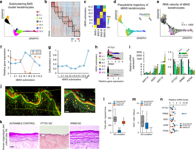
How stem cells give rise to epidermis is unclear despite the crucial role the epidermis plays in barrier and appendage formation. Here we use single cell-RNA sequencing to interrogate basal stem cell heterogeneity of human interfollicular epidermis and find four spatially distinct stem cell populations at the top and bottom of rete ridges and transitional positions between the basal and suprabasal epidermal layers. Cell-cell communication modeling suggests that basal cell populations serve as crucial signaling hubs to maintain epidermal communication. Combining pseudotime, RNA velocity, and cellular entropy analyses point to a hierarchical differentiation lineage supporting multi-stem cell interfollicular epidermal homeostasis models and suggest that transitional basal stem cells are stable states essential for proper stratification. Finally, alterations in differentially expressed transitional basal stem cell genes result in severe thinning of human skin equivalents, validating their essential role in epidermal homeostasis and reinforcing the critical nature of basal stem cell heterogeneity.
Overview
The claims and statistics of this paper haven’t been checked yet.
Run the check — free, no account needed. It reads the open-access text and checks the sources it cites.
Integrity signals
Registry
Cites
Statistics
Figures
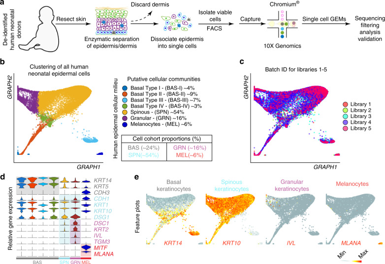
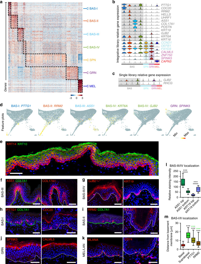
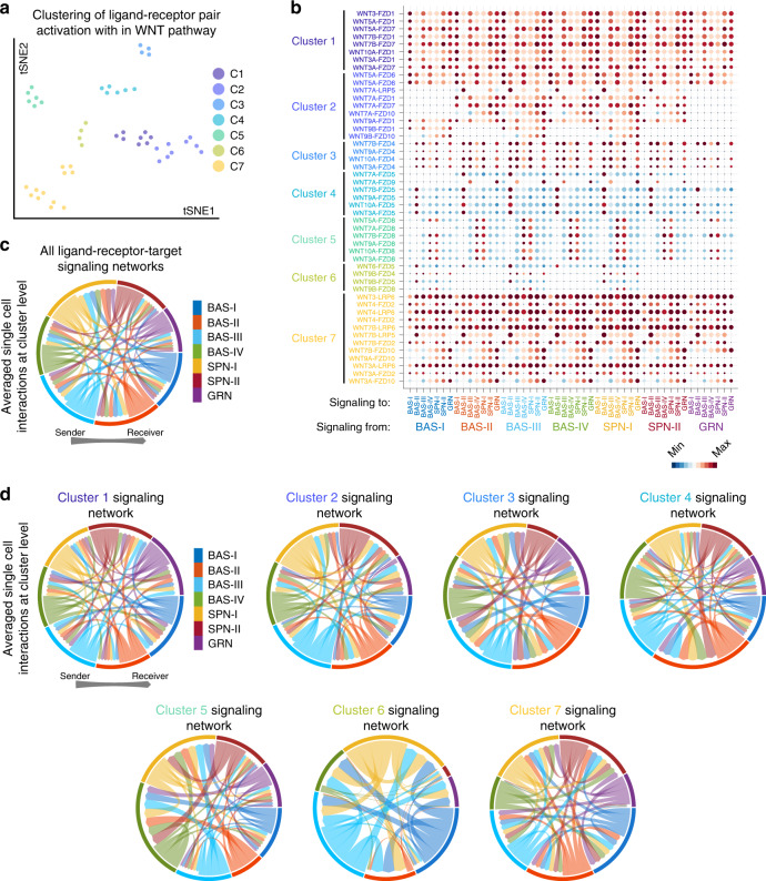
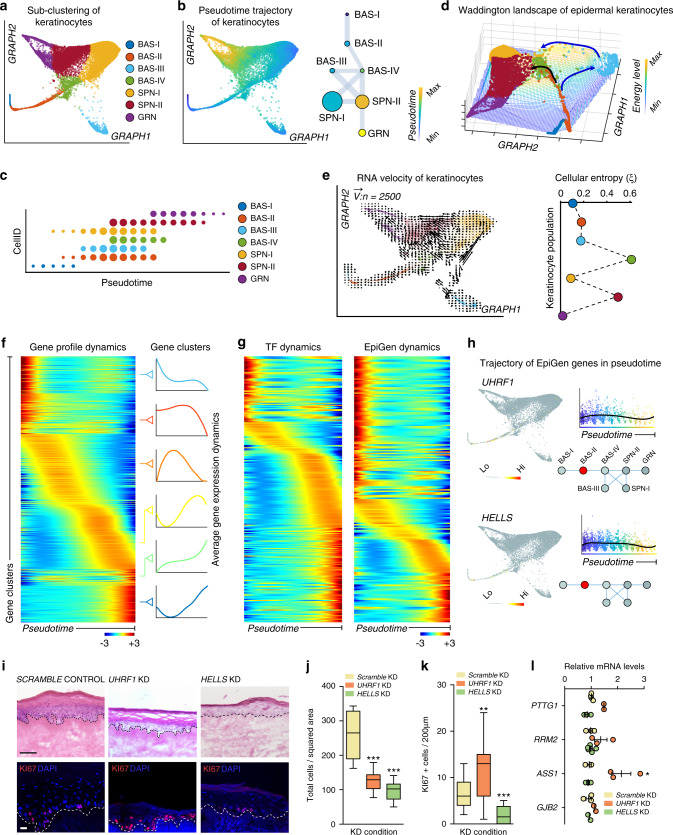
Verification
Claims the paper makes
The claims and statistics of this paper haven’t been checked yet.
Run the check — free, no account needed. It reads the open-access text and checks the sources it cites.
Claims other papers make about this one
Each is checked against this paper’s own text: does it say what the citing paper says it does?
The source backs part of the claim, not all of it — or backs it for a different population, condition or comparison.
In the citing paper
“It enables both profiling of gene expression measurements at a single-cell resolution and identification of reliable cellular heterogeneity, allowing for the identification of previously unrecognized levels of cellular heterogeneity, revealing regulatory relationships between genes, and tracking the trajectories of distinct cell lineages in the same or different developmental stages [63–66].”
From Diversity of human skin three-dimensional organotypic cultures (2024) · cites this paper as [66]
Experimental — accuracy not yet measuredThe source backs part of the claim, not all of it — or backs it for a different population, condition or comparison.
In the citing paper
“we spatially resolved four distinct basal stem cell populations within human IFE and delineated multiple spinous and granular cell populations that contributed to a hierarchical differentiation lineage”
From Single-cell transcriptomics of human-skin-equivalent organoids (2023) · cites this paper as [27]
Experimental — accuracy not yet measuredThe cited source says what the claim attributes to it.
In the citing paper
“human epidermis is more heterogeneous than previously thought.”
From Single-cell transcriptomics of human-skin-equivalent organoids (2023) · cites this paper as [27]
The cited source says what the claim attributes to it.
In the citing paper
“BAS-III cells typically sit atop the rete ridges in vivo, whereas BAS-IV cells lie at the bottom of rete ridges.27”
From Single-cell transcriptomics of human-skin-equivalent organoids (2023) · cites this paper as [27]
Self-check
Percentages, sums and reported differences re-computed from the paper’s own numbers.
The self-check needs the paper’s full text.
Citations
Cites
Subramanian, A., Tamayo, P., Mootha, V. K., Mukherjee, S., Ebert, B. L., Gillette, M. A., Paulovich, A., Pomeroy, S. L., Golub, T. R., Lander, E. S., et al. (2005) · Proceedings of the National Academy of Sciences · doi:10.1073/pnas.0506580102 · Cited by 53,625
Enrichr: a comprehensive gene set enrichment analysis web server 2016 update
Kuleshov, M. V., Jones, M. R., Rouillard, A. D., Fernandez, N. F., Duan, Q., Wang, Z., Koplev, S., Jenkins, S. L., Jagodnik, K. M., Lachmann, A., et al. (2016) · Nucleic Acids Research · doi:10.1093/nar/gkw377 · Cited by 10,799
Enrichr: interactive and collaborative HTML5 gene list enrichment analysis tool
Chen, E. Y., Tan, C. M., Kou, Y., Duan, Q., Wang, Z., Meirelles, G. V., Clark, N. R., & Ma’ayan, A. (2013) · BMC Bioinformatics · doi:10.1186/1471-2105-14-128 · Cited by 7,814
Trapnell, C., Cacchiarelli, D., Grimsby, J., Pokharel, P., Li, S., Morse, M., Lennon, N. J., Livak, K. J., Mikkelsen, T. S., & Rinn, J. L. (2014) · Nature Biotechnology · doi:10.1038/nbt.2859 · Cited by 7,314
Highly parallel genome-wide expression profiling of individual cells using nanoliter droplets.
Macosko, E. Z., Basu, A., Satija, R., Nemesh, J., Shekhar, K., Goldman, M., Tirosh, I., Bialas, A. R., Kamitaki, N., Martersteck, E. M., et al. (2015) · Cell · doi:10.1016/j.cell.2015.05.002 · Cited by 7,110
Wnt/β-Catenin Signaling in Development and Disease
Clevers, H. (2006) · Cell · doi:10.1016/j.cell.2006.10.018 · Cited by 5,568
Wnt/β-catenin signaling and disease
Clevers, H., & Nusse, R. (2012) · Cell · doi:10.1016/j.cell.2012.05.012 · Cited by 5,554
Reversed graph embedding resolves complex single-cell trajectories
Qiu, X., Mao, Q., Tang, Y., Wang, L., Chawla, R., Pliner, H. A., & Trapnell, C. (2017) · Nature Methods · doi:10.1038/nmeth.4402 · Cited by 5,070
Dissecting the multicellular ecosystem of metastatic melanoma by single-cell RNA-seq
Tirosh, I., Izar, B., Prakadan, S. M., Wadsworth, M. H., Treacy, D., Trombetta, J. J., Rotem, A., Rodman, C., Lian, C., Murphy, G., et al. (2016) · Science · doi:10.1126/science.aad0501 · Cited by 4,944
Wnt/β-Catenin Signaling, Disease, and Emerging Therapeutic Modalities
Nusse, R., & Clevers, H. (2017) · Cell · doi:10.1016/j.cell.2017.05.016 · Cited by 4,342
Cited by
Sorted by citations
Items 101–200 of 221
Liu, L., Zheng, D., Xu, Z.-F., Zhou, H.-C., Wang, Y.-C., Zhou, H., Ge, J.-Y., Sako, D., Li, M., Akimoto, K., et al. (2021) · Cells · doi:10.3390/cells11010070 · Cited by 10
Yakupu, A., Zhang, D., Guan, H., Jiang, M., Dong, J., Niu, Y., Tang, J., Liu, Y., Ma, X., & Lu, S. (2023) · Frontiers in Genetics · doi:10.3389/fgene.2023.1072995 · Cited by 10
Low temperature and mTOR inhibition favor stem cell maintenance in human keratinocyte cultures
Nanba, D., Sakabe, J., Mosig, J., Brouard, M., Toki, F., Shimokawa, M., Kamiya, M., Braschler, T. M., Azzabi, F., Lathion, S., et al. (2023) · EMBO Reports · doi:10.15252/embr.202255439 · Cited by 9
Pontiggia, L., Ahuja, A. K., Yosef, H. K., Rütsche, D., Ernst Reichmann, A., Moehrlen, U., & Biedermann, T. (2022) · Cells · doi:10.3390/cells11142156 · Cited by 9
Lotti, R., Palazzo, E., Quadri, M., Dumas, M., Schnebert, S., Biondini, D., Bianchini, M. A., Nizard, C., Pincelli, C., & Marconi, A. (2022) · Stem Cells · doi:10.1093/stmcls/sxac060 · Cited by 10
Wedemeyer, M. A., Ding, T., Garfinkle, E. A. R., Westfall, J. J., Navarro, J. B., Gonzalez, M. E. H., Varga, E. A., Witman, P. M., Mardis, E. R., Cottrell, C. E., et al. (2024) · Scientific Reports · doi:10.1038/s41598-024-72167-8 · Cited by 9
Overmiller, A. M., Uchiyama, A., Hope, E., Nayak, S., O’Neill, C. G., Hasneen, K., Chen, Y., Naz, F., Dell’Orso, S., Brooks, S. R., et al. (2024) · JCI Insight · doi:10.1172/jci.insight.182844 · Cited by 11
Skin aging: mechanisms, evaluation, and rejuvenation
Li, R., Zhang, J., Mao, K., Meng, D., & Han, J.-D. J. (2026) · The EMBO Journal · doi:10.1038/s44318-026-00810-3 · Cited by 13
FitDevo: accurate inference of single-cell developmental potential using sample-specific gene weight
Zhang, F., Yang, C., Wang, Y., Jiao, H., Wang, Z., Shen, J., & Li, L. (2022) · Briefings in Bioinformatics · doi:10.1093/bib/bbac293 · Cited by 10
Mining cell–cell signaling in single-cell transcriptomics atlases
Deng, M., Wang, Y., & Yan, Y. (2022) · Current Opinion in Cell Biology · doi:10.1016/j.ceb.2022.102101 · Cited by 10
A transit-amplifying progenitor with biphasic behavior contributes to epidermal renewal
Ghuwalewala, S., Jiang, K., Ragi, S. D., Shalloway, D. I., & Tumbar, T. (2024) · Development · doi:10.1242/dev.202389 · Cited by 8
DeVore, S. B., Schuetz, M., Alvey, L., Lujan, H., Ochayon, D. E., Williams, L., Chang, W. C., Filuta, A. L., Ruff, B. P., Kothari, A., et al. (2024) · Cell Reports · doi:10.1016/j.celrep.2024.114589 · Cited by 8
Winkler, R., Quaas, M., Glasmacher, S., Uwe Wolfrum, B., Thalheim, T., Galle, J., Krohn, K., Magin, T. M., & Aust, G. (2022) · Cells · doi:10.3390/cells11193151 · Cited by 7
Dynamic regulation of human epidermal differentiation by adhesive and mechanical forces
Zijl, S., Salameti, V., Louis, B., Negri, V. A., & Watt, F. M. (2022) · Current topics in developmental biology/Current Topics in Developmental Biology · doi:10.1016/bs.ctdb.2022.03.004 · Cited by 8
Li, J., Xia, Y., Kong, S., YANG, K. Y., Chen, H., Zhang, Y., Liu, D., Chen, L., & Sun, X. (2023) · Journal of the European Academy of Dermatology and Venereology · doi:10.1111/jdv.19289 · Cited by 8
Adult tissue-specific stem cell interaction: novel technologies and research advances
Luo, X., Liu, Z., & Xu, R. (2023) · Frontiers in Cell and Developmental Biology · doi:10.3389/fcell.2023.1220694 · Cited by 7
Exploring the Functions of Mutant p53 through TP53 Knockout in HaCaT Keratinocytes
Romashin, D. D., Rusanov, A. L., Arzumanian, V. A., Varshaver, A., Poverennaya, E. V., Vakhrushev, I. V., Netrusov, A. I., & Luzgina, N. G. (2024) · Current Issues in Molecular Biology · doi:10.3390/cimb46020094 · Cited by 6
Yang, J., Wang, Z., Zhou, H., Xiong, Y., Li, Y., Zheng, Y., & Liu, L. (2025) · iScience · doi:10.1016/j.isci.2025.112373 · Cited by 6
Petty, A. J., Cardones, A. R., Jin, Y. J., Jain, V., Hocke, E., Pathak, H. B., Mitra, A., Gregory, S. G., Selim, M. A., & Zhang, J. Y. (2025) · JID Innovations · doi:10.1016/j.xjidi.2025.100373 · Cited by 6
Emerging Roles of Ferroptosis in Skin Pathophysiology
Reznik, A. S., Vats, K., Singh, K., Mizes, A., Jaklitsch, E. L., Bayır, H., Kagan, V. E., & Bunimovich, Y. L. (2025) · Journal of Investigative Dermatology · doi:10.1016/j.jid.2025.06.1289 · Cited by 7
Rete ridges form via evolutionarily distinct mechanisms in mammalian skin
Thompson, S. M., Yaple, V., Searle, G. H., Phan, Q. M., Makkar, J., Cheng, X., Liu, R., Puławska-Czub, A., Yanke, C., Williams, N. M., et al. (2026) · Nature · doi:10.1038/s41586-025-10055-5 · Cited by 7
Self-citationBlank Spots in the Map of Human Skin: The Challenge for Xenotransplantation
Cherkashina, O. L., Morgun, E. I., Rippa, A. L., Kosykh, A. V., Alekhnovich, A. V., Stoliarzh, A. B., Terskikh, V. V., Vorotelyak, E. A., & Kalabusheva, E. P. (2023) · International Journal of Molecular Sciences · doi:10.3390/ijms241612769 · Cited by 6
Emerging Techniques in Spatial Multiomics: Fundamental Principles and Applications to Dermatology
Jia, B. B., Sun, B. K., Lee, E. Y., & Ren, B. (2024) · Journal of Investigative Dermatology · doi:10.1016/j.jid.2024.09.006 · Cited by 8
Innovative approaches in skin therapy: The rise of organoid cultivation
Zhou, R., Luo, X., Chen, S., Jia, K., Yu, H., Huang, Z., Tan, Y., Ma, M., Zhao, J., Xing, J., et al. (2025) · Chemical Engineering Journal · doi:10.1016/j.cej.2025.162248 · Cited by 6
Skin Explorer: An Interactive Single-Cell RNA-Sequencing Resource for Healthy Human Skin
Yuan, H., Sanz, N. C., Wagner, R. E., Roessler, L., Heinmäe, E., Yeerken, A., Are, A., Annusver, K., & Kasper, M. (2025) · Journal of Investigative Dermatology · doi:10.1016/j.jid.2025.08.025 · Cited by 7
Alpert, A., Nahman, O., Starosvetsky, E., Hayun, M., Curiel, T. J., Ofran, Y., & Shen-Orr, S. S. (2021) · Cell Systems · doi:10.1016/j.cels.2021.09.003 · Cited by 8
Ali, D., Alhattab, D. M., Jafar, H. D., Alzubide, M., Sharar, N., Bdour, S. M., & Awidi, A. (2021) · International Journal of Molecular Sciences · doi:10.3390/ijms221910810 · Cited by 6
Retinoic Acid Signaling Alters the Balance of Epidermal Stem Cell Populations in the Skin
Dumrongphuttidecha, T., Ishikawa, M., Cabezas‐Wallscheid, N., & Sada, A. (2025) · Journal of Investigative Dermatology · doi:10.1016/j.jid.2025.09.008 · Cited by 6
Wang, L.-P., Li, C.-Y., Wu, Y., Chen, M., Hsieh, Y.-P., Huang, T., Hong, T.-M., & Chen, Y. (2025) · Diagnostics · doi:10.3390/diagnostics15192477 · Cited by 5
Ishikawa, M., Phung, H. M., Dumrongphuttidecha, T., & Sada, A. (2025) · Current Opinion in Cell Biology · doi:10.1016/j.ceb.2025.102594 · Cited by 5
Yang, B., Xu, Z., Chen, H., Ma, T., Zhao, Y., Pang, M., & Wang, J. (2024) · Animals · doi:10.3390/ani14060922 · Cited by 5
DeBerg, H. A., Fahning, M. L., Varkhande, S. R., Schlenker, J. D., Schmitt, W. P., Gupta, A., Singh, A. K., Gratz, I. K., Carlin, J. S., Campbell, D., et al. (2025) · Journal of Investigative Dermatology · doi:10.1016/j.jid.2025.03.033 · Cited by 5
Zheng, T., Yuan, R., Zhang, Y., Zou, Q., Wang, Y., Li, Y., Gong, Z., Chen, Z., & Jiang, Y. (2025) · BMC Biology · doi:10.1186/s12915-025-02390-w · Cited by 7
Guo, F., Lin, F., Shen, B., Wang, S., Li, Y., Guo, J., Chen, Y., Liu, Y., Lu, Y., Hu, R., et al. (2024) · Nanophotonics · doi:10.1515/nanoph-2023-0722 · Cited by 5
Self-citationTanahashi, K., Kono, M., Yoshikawa, T., Suzuki, Y., Inoie, M., Kuwatsuka, Y., Kinoshita, F., Takeichi, T., & Akiyama, M. (2024) · British Journal of Dermatology · doi:10.1093/bjd/ljae193 · Cited by 5
Cell-laden biomimetic microneedles reconstruct skin rete ridge and stem cell niche
Zhao, X., Wu, Z., Guo, Y., Pu, L., Pei, Z., Liu, Y., Hou, B., Xie, S., Luo, G., & Zhan, R. (2025) · Journal of Nanobiotechnology · doi:10.1186/s12951-025-03430-x · Cited by 7
李, Q.-C. 啟. L., 林, H. 海. L., Wang, P., 董, Q. 秋. D., 王, K. 坤. W., 帅, J.-W. 建. S., & Ye, F.-F. (2025) · Chinese Physics B · doi:10.1088/1674-1056/ade427 · Cited by 4
Molina, M. A., Biswas, S., Jiménez-Vázquez, O., & Bodily, J. M. (2025) · Tumour Virus Research · doi:10.1016/j.tvr.2025.200315 · Cited by 4
Sousa, P., Lopes, B., Sousa, A. C., Moreira, A. D. S., Rêma, A., Alvites, R., Geuna, S., Alves, N. M., & Maurício, A. C. (2025) · International Journal of Molecular Sciences · doi:10.3390/ijms26115081 · Cited by 4
Katsuki, T., Kusumoto, D., Akiba, Y., Kimura, M., Komuro, J., Nakamura, T., Hashimoto, H., Kouka, T., Sugai, K., Katsumata, Y., et al. (2025) · JACC Basic to Translational Science · doi:10.1016/j.jacbts.2025.05.003 · Cited by 5
Spatiotemporal Switches in Progenitor Cell Fate Govern Upper Hair Follicle Growth and Maintenance
Andersen, M. S., Ulyanchenko, S., Schweiger, P. J., Hannezo, É., Simons, B. D., & Jensen, K. B. (2025) · Journal of Investigative Dermatology · doi:10.1016/j.jid.2025.01.034 · Cited by 3
Harris, A., Burnham, K., Pradhyumnan, R., Jaishankar, A., Häkkinen, L., Rosero, R. E. G., Piazza, Y., Andl, C. D., & Andl, T. (2025) · International Journal of Molecular Sciences · doi:10.3390/ijms26073144 · Cited by 3
Zhao, Q., Ma, R., Huang, K., Wang, J., Zhang, D., Wang, J., Ding, X., Chen, F., Zhao, S., Ni, N., et al. (2025) · hLife · doi:10.1016/j.hlife.2025.10.003 · Cited by 3
Unraveling the Functional Heterogeneity of Human Skin at Single-Cell Resolution
Sol, S., Boncimino, F., Todorova, K. A., & Mandinova, A. (2024) · Hematology/Oncology Clinics of North America · doi:10.1016/j.hoc.2024.05.001 · Cited by 3
Hirase, R., Fujita, T., Miyai, T., Kawasaki, H., & Koseki, H. (2024) · Experimental Dermatology · doi:10.1111/exd.70000 · Cited by 3
Sousa, P., Lopes, B., Sousa, A. C., Coelho, A., Moreira, A. D. S., Rêma, A., Gonçalves-Maia, M., Amorim, I., Alvites, R., Alves, N. M., et al. (2024) · Biomedicines · doi:10.3390/biomedicines12122854 · Cited by 4
Stage of Keratinocyte Differentiation Is a Key Determinant of Viral Susceptibility in Human Skin
Moran, M. C., Chinchilli, E., Kenney, H. M., Pope, E. M., Scott, G., Brewer, M. G., & Beck, L. A. (2023) · Journal of Investigative Dermatology · doi:10.1016/j.jid.2023.03.1656 · Cited by 3
Mangez, C., Roux, P., Stamatas, G. N., Oddos, T., & Brun, C. (2021) · Experimental Dermatology · doi:10.1111/exd.14503 · Cited by 3
Guo, Y., Wu, W., Chen, H., Wang, X., Zhang, Y., Li, S., & Yang, X. (2025) · Frontiers in Cell and Developmental Biology · doi:10.3389/fcell.2025.1475334 · Cited by 2
Fan, W., Ishikawa, M., Raja, E., Hegazy, A. M., Date, H., Ngo, Y. X., Sato, Y., Yamagata, K., Yanagisawa, H., & Sada, A. (2026) · Aging Cell · doi:10.1111/acel.70483 · Cited by 2
Ishikawa, M., Ngo, Y. X., Nishikawa, I., Kato, H., Maeda, R., Mizuno, R., Mizuno, J., Izumi, K., Yanagisawa, H., & Sada, A. (2026) · Journal of Investigative Dermatology · doi:10.1016/j.jid.2026.02.014 · Cited by 5
Chromatin organization of muscle stem cell
Santarelli, P., Rosti, V., Vivo, M., & Lanzuolo, C. (2024) · Current topics in developmental biology/Current Topics in Developmental Biology · doi:10.1016/bs.ctdb.2024.01.014 · Cited by 2
Vinasco‐Sandoval, T., Lemaı̂tre, G., Soularue, P., Martin, M. T., & Fortunel, N. O. (2024) · International Journal of Molecular Sciences · doi:10.3390/ijms25063353 · Cited by 2
The Recovery of Epidermal Proliferation Pattern in Human Skin Xenograft
Черкашина, О. С., Tsitrina, A. A., Abolin, D. S., Morgun, E., Kosykh, A. V., Sabirov, M. S., Vorotelyak, E. A., & Kalabusheva, E. P. (2025) · Cells · doi:10.3390/cells14060448 · Cited by 2
Unraveling the Transcription Factor Code of Odontoblast Differentiation
Lavický, J., Lopez, M. G., Chochola, V., Kompaníková, P., Rakultsev, V., Raška, J., Winchester, E. W., Tuaima, H., Englmaier, L., Verner, J., et al. (2025) · Journal of Dental Research · doi:10.1177/00220345251348148 · Cited by 2
Age‐related decrease in responsiveness of CD271 ‐positive skin stem cells to growth factors
Akamatsu, H., Yamada, T., Sanada, A., Ishii, Y., Iwata, Y., Arima, M., Hasegawa, S., & Sugiura, K. (2022) · Experimental Dermatology · doi:10.1111/exd.14601 · Cited by 2
Smits, J. G. A., Cunha, D. L., Amini, M., Bertolin, M., Laberthonnìère, C., Qu, J., Owen, N., Latta, L., Seitz, B., Roux, L. N., et al. (2022) · bioRxiv (Cold Spring Harbor Laboratory) · doi:10.1101/2022.07.13.499857 · Cited by 2
Burks, H. E., Pokorny, J. L., Koetsier, J. L., Roth‐Carter, Q. R., Arnette, C. R., Gerami, P., Seykora, J. T., Johnson, J. L., & Green, K. J. (2022) · bioRxiv (Cold Spring Harbor Laboratory) · doi:10.1101/2022.12.01.518424 · Cited by 2
Regenerative cell turnover coupled to a tumor-hostile niche protects naked mole-rats from cancerPreprint
Behm, M., Baeza-Centurión, P., Weser, S., Busa, V. F., Zafferani, P., Penso-Dolfin, L., Botey-Bataller, J., Delaunay, S., Hirschmüller, N., Berthet, B., et al. (2023) · bioRxiv (Cold Spring Harbor Laboratory) · doi:10.1101/2023.10.10.561212 · Cited by 2
Ishikawa, M., Ngo, Y. X., Nishikawa, I., Kato, H., Maeda, R., Mizuno, R., Mizuno, J., Izumi, K., Yanagisawa, H., & Sada, A. (2025) · bioRxiv (Cold Spring Harbor Laboratory) · doi:10.1101/2025.01.21.634195 · Cited by 2
Inflammatory IL-1 signaling remodels epidermal stem cell compartments by suppressing Wnt activityPreprint
Phung, H. M., Nishikawa, I., Nguyen, N. T. K., Yesbolatova, A. K., Hegazy, A. M., Kosasih, T., Aoi, J., Fukushima, S., Hiroyasu, S., Takizawa, H., et al. (2026) · bioRxiv (Cold Spring Harbor Laboratory) · doi:10.64898/2026.02.06.704488 · Cited by 2
Li, Q., Chan, H., Zhou, J., Zheng, W., Yang, B., & Liu, Z. (2025) · Journal of the European Academy of Dermatology and Venereology · doi:10.1111/jdv.20870 · Cited by 1
Droll, S. H., Xue, C., Dewar, E. I. O., Chamberlain, N. K., Zhang, B. J., Levine, M. C., & Bao, X. (2025) · Journal of Investigative Dermatology · doi:10.1016/j.jid.2025.07.022 · Cited by 1
The cell as a token: high-dimensional geometry in language models and cell embeddings
Gilpin, W. (2025) · Bioinformatics · doi:10.1093/bioinformatics/btaf595 · Cited by 3
Zhu, S., Yan, Y., Jia, M., Li, H., Han, B., Shi, T., Xu, L., Wang, X., Zhang, Q., Zheng, W., et al. (2025) · iMeta · doi:10.1002/imt2.70100 · Cited by 2
Liu, W., Cao, Y. W., Chen, Y., Lu, Y.-F., Xu, C.-Y., Yan-Hong, Z. H. A. I., Wang, C., & Cao, Z. (2026) · Annals of Medicine · doi:10.1080/07853890.2026.2627742 · Cited by 1
Transcriptional and epigenetic mechanisms governing epidermal stem cell regulation
Lamb, S., Omari, Z., & Seldin, L. (2026) · Frontiers in Cell and Developmental Biology · doi:10.3389/fcell.2026.1733472 · Cited by 1
Miura, A., Kobayashi, Y., Hirose, Y., Azuma, T., Shimbo, T., Ouchi, Y., Kitayama, T., Yamamoto, R., Takaki, E., Yamazaki, S., et al. (2026) · Nature Communications · doi:10.1038/s41467-026-75278-0 · Cited by 1
Single-cell genetics approach in ophthalmology
Leung, A., Shinozaki, Y., Koizumi, S., & Ohnuma, S. (2022) · Elsevier eBooks · doi:10.1016/b978-0-12-819291-7.00013-7 · Cited by 1
Application of Single-Cell Sequencing on Stem Cell Research
Paul, S., Umar, T. P., Paul, M. K., Tiwari, H., Shukla, M. K., Kandasami, L., Joliholi, A., Dey, D., Choudhary, D. K., Govarthanan, K., et al. (2023) · Springer eBooks · doi:10.1007/978-981-99-0846-2_43-1 · Cited by 1
Spatio-temporal regulation of gene expression defines subpopulations of epidermal stem cells
Aruketty, M., & Kurinna, S. M. (2020) · Biochemical Society Transactions · doi:10.1042/bst20200740 · Cited by 1
Guerrero‐Juarez, C. F., Lee, G. H., Liu, Y., Wang, S., Sha, Y., Chow, R. Y., Nguyen, T. T. L., Aasi, S. Z., Karikomi, M. K., Drummond, M. L., et al. (2021) · bioRxiv (Cold Spring Harbor Laboratory) · doi:10.1101/2021.10.07.463571 · Cited by 1
Self-citationEpidermal basal domains organization highlights skin robustness to environmental exposurePreprint
Ghuwalewala, S., Lee, S. A., Jiang, K., Baidya, J., Chovatiya, G. L., Kaur, P., Shalloway, D. I., & Tumbar, T. (2022) · bioRxiv (Cold Spring Harbor Laboratory) · doi:10.1101/2022.02.23.481662 · Cited by 1
Miny, S., Runel, G., Chlasta, J., Lapart, J.-A., & Bonod‐Bidaud, C. (2025) · bioRxiv (Cold Spring Harbor Laboratory) · doi:10.1101/2025.02.12.637845 · Cited by 1
Amakor, J. K., Rajan, A. A. N., Kamaraj, M., Jacobs, K. A., Hutchins, E. J., Wittmann, T., & Kutys, M. L. (2026) · bioRxiv (Cold Spring Harbor Laboratory) · doi:10.64898/2026.03.26.714483 · Cited by 1
Transit-amplifying progenitor with maturation-dependent behavior contributes to epidermal renewalPreprint
Ghuwalewala, S., Jiang, K., Ragi, S. D., Shalloway, D. I., & Tumbar, T. (2022) · bioRxiv (Cold Spring Harbor Laboratory) · doi:10.1101/2022.06.12.495812
Ober-Reynolds, B., Wang, C., Ko, J., Rios, E. J., Aasi, S., Davis, M. M., Oro, A., & Greenleaf, W. (2022) · bioRxiv · doi:10.1101/2022.09.28.509620
Paige, J. T., Lightell, D., Douglas, H. F., Klingenberg, N. C., Pham, T., & Woods, T. C. (2023) · Experimental Dermatology · doi:10.1111/exd.14845 · Cited by 2
Endothelial-fibroblast interactions during Scarb1 accelerate heart failurePreprint
Katsuki, T., Kusumoto, D., Akiba, Y., Kimura, M., Komuro, J., Nakamura, T., Hashimoto, H., Kouka, T., Suzuki, Y., Kubota, Y., et al. (2023) · bioRxiv (Cold Spring Harbor Laboratory) · doi:10.1101/2023.09.15.557661
Dysregulation of the Hippo pathway enhances PD-L2 transcription to promote cancer immune evasionPreprint
Ando, T., Okamoto, K., Sato, K., Goto, Y., Izumi, H., Kataoka, N., Ueda, Y., Iglesias‐Bartolomé, R., Yoshimoto, T., Shintani, T., et al. (2024) · Research Square · doi:10.21203/rs.3.rs-4330731/v1
Application of Single-Cell Sequencing on Stem Cell Research
Paul, S., Umar, T. P., Paul, M. K., Tiwari, H., Shukla, M. K., Kandasami, L., Joliholi, A., Dey, D., Choudhary, D. K., Govarthanan, K., et al. (2024) · Springer eBooks · doi:10.1007/978-981-99-7119-0_43
Filatov, N. S., Bilyalov, A. I., Gazizova, G. R., Bilyalova, A., Shagimardanova, E. I., Vorontsova, M. V., Kiyasov, A. P., Gusev, O., & Kozlova, O. (2024) · Russian Journal of Genetics · doi:10.1134/s1022795424700686
Novel Approaches to 3D Skin Cancer Models Usability in Research
Levkovych, I., & Sobiepanek, A. (2024) · Interdisciplinary cancer research · doi:10.1007/16833_2024_481
Comprehensive Enumeration of Cancer Stem-like Cell Heterogeneity Using Deep Neural NetworkPreprint
Chowdhury, D., Priyadarshi, S., Biswas, S., Neekhra, B., Gupta, D., & Haldar, S. (2024) · bioRxiv (Cold Spring Harbor Laboratory) · doi:10.1101/2024.11.26.625418
Filatov, N. S., Bilyalov, A. I., Gazizova, G. R., Bilyalova, A., Shagimardanova, E. I., Vorontsova, M. V., Kiyasov, A. P., Gusev, O., & Kozlova, O. (2024) · Генетика · doi:10.31857/s0016675824090033
Singh, S., Wei, L., Samiei, E., Gaber, K., Gao, Q., Persad, A. H., Veres, T., & Günther, A. (2025) · bioRxiv (Cold Spring Harbor Laboratory) · doi:10.1101/2025.05.16.654553
Enhanced differentiation potential of pigmented human epidermal equivalents
Jia, Y. Y., Bajsert, J., Kurdian, A. I., Shiu, J., Pérez‐Aso, M., & Atwood, S. X. (2026) · Scientific Reports · doi:10.1038/s41598-026-63611-y
Self-citationMcCluskey, A., Kettlewell, T., Smith, A. M., Kundu, R., Gunn, D. A., & Otto, T. D. (2026) · BMC Methods · doi:10.1186/s44330-026-00082-7
Düz, T., Törőcsik, D., Momnougui, S. M. E., Oubounyt, M., Al, B., Gallinat, S., Baumbach, J., & Holzscheck, N. (2025) · bioRxiv (Cold Spring Harbor Laboratory) · doi:10.1101/2025.09.09.675235
Excessive glycine loop variations in the keratin 10 tail domain and implications for skin fragility
Mi, Z., Yu, Y., Wang, Z., Bai, F., Yu, G., Li, B., Dai, Y., Lin, Y., Sang, X., Han, J., et al. (2025) · British Journal of Dermatology · doi:10.1093/bjd/ljaf436
Thalheim, T., & Schneider, M. R. (2024) · Figshare · doi:10.6084/m9.figshare.26676416
Thalheim, T., & Schneider, M. R. (2024) · Figshare · doi:10.6084/m9.figshare.26676422
Fu, Y. C., Das, A., Wang, D., Braun, R., & Yi, R. (2024) · Figshare · doi:10.6084/m9.figshare.26094289
Fu, Y. C., Das, A., Wang, D., Braun, R., & Yi, R. (2024) · Figshare · doi:10.6084/m9.figshare.26094505
Thalheim, T., & Schneider, M. R. (2024) · Figshare · doi:10.6084/m9.figshare.26676419
Thalheim, T., & Schneider, M. R. (2024) · Figshare · doi:10.6084/m9.figshare.26676425
Fu, Y. C., Das, A., Wang, D., Braun, R., & Yi, R. (2024) · Figshare · doi:10.6084/m9.figshare.26738433
The stem cell compartment in human oral mucosa and its activation in oral lichen planusPreprint
Schreurs, O., Fedele, S., Porter, S. R., Kjølle, G. K., Schenck, K., Søland, T. M., & Walko, G. (2026) · bioRxiv (Cold Spring Harbor Laboratory) · doi:10.64898/2026.03.04.709521
Cell-Laden Gel Biomimetic Skin Promotes Full-Thickness Skin Wound Regeneration
Zhang, P., Chen, Q., Pu, Y., Liu, M., Ma, M., Wu, Y., Zhang, Y., & Yang, X. (2026) · Gels · doi:10.3390/gels12030258
A single-cell multi-omics atlas of human eyelid skin
Ma, W., Xu, Y., Chen, J., Yuan, Y., Wei, X., Deng, Q., Li, R., Du, J., Zhang, Z., Zhou, Z., et al. (2026) · Frontiers in Genetics · doi:10.3389/fgene.2026.1780660
Metadata
- Authors
- Wang, S., Drummond, M. L., Guerrero-Juarez, C. F., Tarapore, E., MacLean, A. L., Stabell, A. R., Wu, S. C., Gutierrez, G., That, B. T., Benavente, C. A., Nie, Q., & Atwood, S. X.
- Affiliations
- University of California, Irvine (US)
- Corresponding author
- Nie, Qing
- Atwood, Scott X.
- ORCID
- Venue
- Nature Communications
- Published
- 2020-08-25
- Volume
- 11
- Issue
- 1
- Pages
- 4239-4239
- ISSN
- 2041-1723 (ISSN-L)
- PMC
- PMC7447770
- OpenAlex
- W3081219157
- Cited by
- 218
- References
- 131
- Citations by year
- 2026: 32 · 2025: 40 · 2024: 47 · 2023: 38 · 2022: 37 · 2021: 18 · 2020: 6
- FWCI
- 28.17
- Type
- journal article
- Language
- en
- Open access
- gold
- doi.org
- Locations
- Nature Communications (publishedVersion, cc-by)
- bioRxiv (Cold Spring Harbor Laboratory) (acceptedVersion)
- PubMed Central (publishedVersion, cc-by)
- PubMed (publishedVersion)
- RePEc: Research Papers in Economics (submittedVersion)
- DOAJ (DOAJ: Directory of Open Access Journals) (submittedVersion, cc-by-sa)
- eScholarship (California Digital Library) (submittedVersion)
- eScholarship (California Digital Library) (submittedVersion, cc-by)
- Funders
- Concern Foundation (CF204525)
- U.S. Department of Health & Human Services | National Institutes of Health (P30AR075047)
- Topics
- Health Sciences › Medicine › Dermatology › Hair Growth and Disorders
- Life Sciences › Biochemistry, Genetics and Molecular Biology › Molecular Biology › Single-cell and spatial transcriptomics
- Life Sciences › Biochemistry, Genetics and Molecular Biology › Cell Biology › Skin and Cellular Biology Research
- Keywords
- basal stem cells, interfollicular epidermis, epidermal stratification, epidermal homeostasis, intercellular communication, pseudotime, RNA velocity, skin equivalents, rete ridges
- MeSH terms
- Epidermal Cells, Cell Communication, Cell Differentiation, Epidermis, Gene Expression Regulation, Homeostasis, Humans, Infant, Newborn, Male, Models, Biological, Stem Cells, Signal Transduction, Keratinocytes, Cell Lineage, Gene Expression Profiling, Foreskin
About these checks
Registry, retraction, Cites and statistics results are computed, not judged, with one exception: a printed reference that no index lists is read once by a model before it counts as a citation error. Claims are extracted by a model and kept only when their quote appears verbatim in the text; each cited source is then checked, with a model’s help, for whether it says what the claim relies on. Those verdicts can be wrong — read the claim’s quote and the cited source before relying on one.
Statistics and arithmetic are recomputed, not judged.
Confidence bands are shown only for fields calibrated on our annotated test set. None is calibrated yet, so no band is shown.
What counts as a claim ›How we measure confidence ›How we check this ›
Are you an author, or do you see a problem with this page? Request a review
