

The DOI matches its Crossref record; 131 of the 131 references it cites were found in a registry or an index.
Single cell transcriptomics of human epidermis identifies basal stem cell transition states
Open accessPublished version of a preprint
Abstract
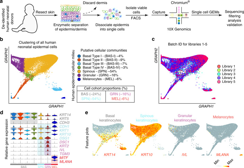
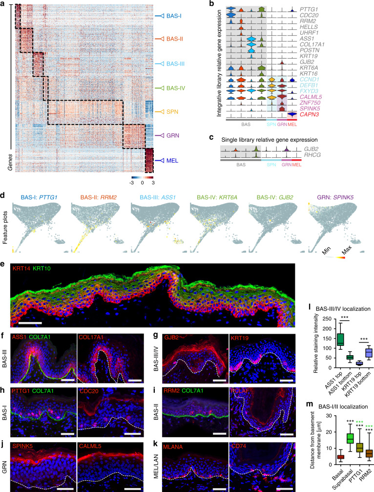
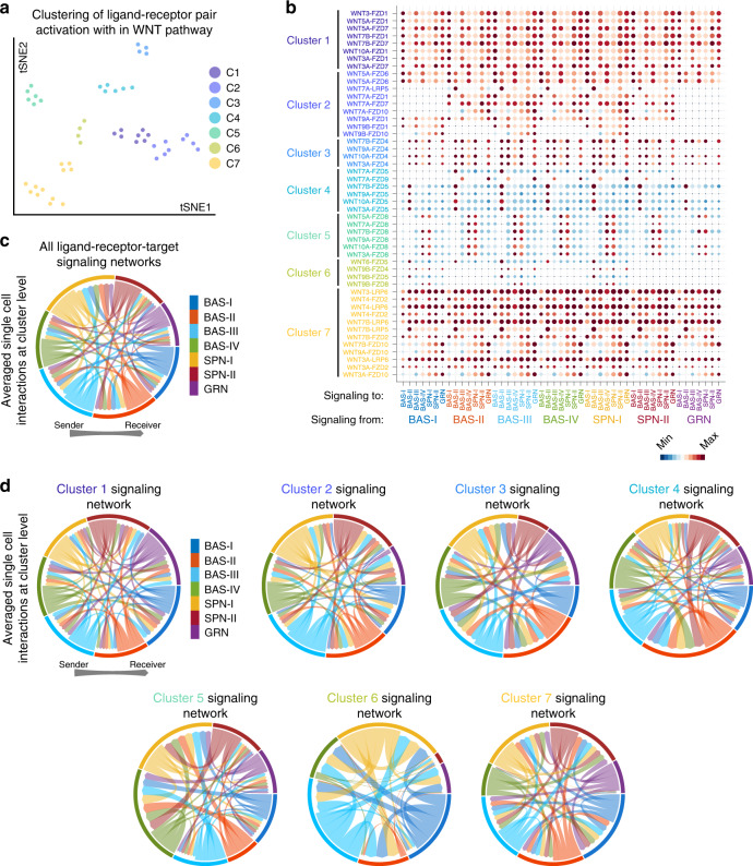
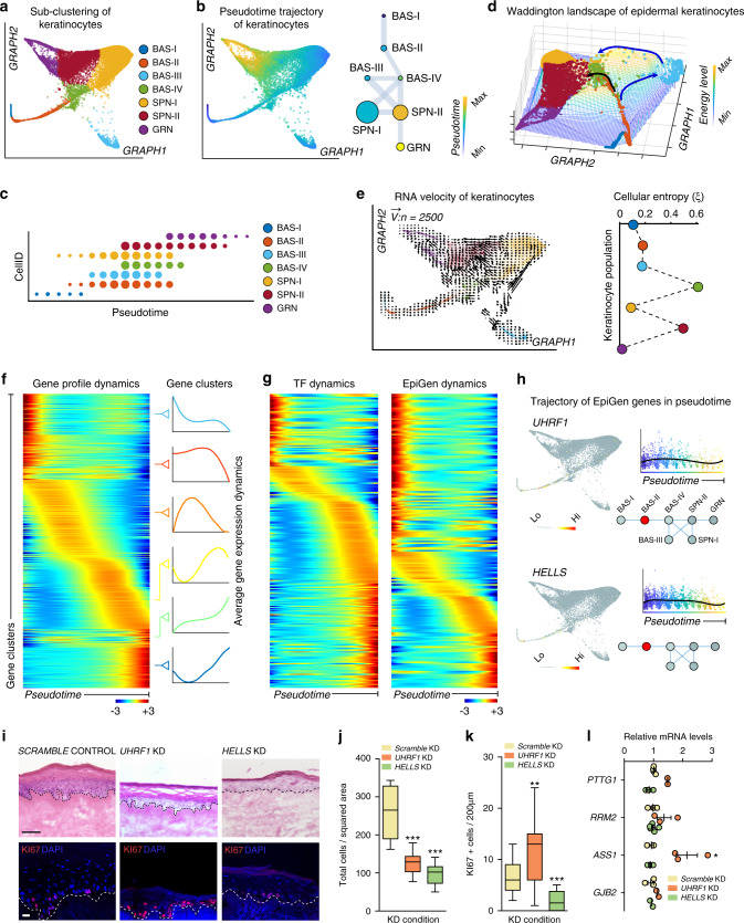
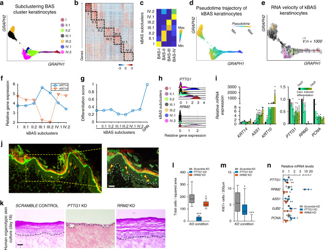
How stem cells give rise to epidermis is unclear despite the crucial role the epidermis plays in barrier and appendage formation. Here we use single cell-RNA sequencing to interrogate basal stem cell heterogeneity of human interfollicular epidermis and find four spatially distinct stem cell populations at the top and bottom of rete ridges and transitional positions between the basal and suprabasal epidermal layers. Cell-cell communication modeling suggests that basal cell populations serve as crucial signaling hubs to maintain epidermal communication. Combining pseudotime, RNA velocity, and cellular entropy analyses point to a hierarchical differentiation lineage supporting multi-stem cell interfollicular epidermal homeostasis models and suggest that transitional basal stem cells are stable states essential for proper stratification. Finally, alterations in differentially expressed transitional basal stem cell genes result in severe thinning of human skin equivalents, validating their essential role in epidermal homeostasis and reinforcing the critical nature of basal stem cell heterogeneity.
Overview
The claims and statistics of this paper haven’t been checked yet.
Run the check — free, no account needed. It reads the open-access text and checks the sources it cites.
Integrity signals
Registry
Cites
Statistics
Figures
Verification
Claims the paper makes
The claims and statistics of this paper haven’t been checked yet.
Run the check — free, no account needed. It reads the open-access text and checks the sources it cites.
Claims other papers make about this one
Each is checked against this paper’s own text: does it say what the citing paper says it does?
The source backs part of the claim, not all of it — or backs it for a different population, condition or comparison.
In the citing paper
“It enables both profiling of gene expression measurements at a single-cell resolution and identification of reliable cellular heterogeneity, allowing for the identification of previously unrecognized levels of cellular heterogeneity, revealing regulatory relationships between genes, and tracking the trajectories of distinct cell lineages in the same or different developmental stages [63–66].”
From Diversity of human skin three-dimensional organotypic cultures (2024) · cites this paper as [66]
Experimental — accuracy not yet measuredThe source backs part of the claim, not all of it — or backs it for a different population, condition or comparison.
In the citing paper
“we spatially resolved four distinct basal stem cell populations within human IFE and delineated multiple spinous and granular cell populations that contributed to a hierarchical differentiation lineage”
From Single-cell transcriptomics of human-skin-equivalent organoids (2023) · cites this paper as [27]
Experimental — accuracy not yet measuredThe cited source says what the claim attributes to it.
In the citing paper
“human epidermis is more heterogeneous than previously thought.”
From Single-cell transcriptomics of human-skin-equivalent organoids (2023) · cites this paper as [27]
The cited source says what the claim attributes to it.
In the citing paper
“BAS-III cells typically sit atop the rete ridges in vivo, whereas BAS-IV cells lie at the bottom of rete ridges.27”
From Single-cell transcriptomics of human-skin-equivalent organoids (2023) · cites this paper as [27]
Self-check
Percentages, sums and reported differences re-computed from the paper’s own numbers.
The self-check needs the paper’s full text.
Citations
Cites
Subramanian, A., Tamayo, P., Mootha, V. K., Mukherjee, S., Ebert, B. L., Gillette, M. A., Paulovich, A., Pomeroy, S. L., Golub, T. R., Lander, E. S., et al. (2005) · Proceedings of the National Academy of Sciences · doi:10.1073/pnas.0506580102 · Cited by 53,625
Enrichr: a comprehensive gene set enrichment analysis web server 2016 update
Kuleshov, M. V., Jones, M. R., Rouillard, A. D., Fernandez, N. F., Duan, Q., Wang, Z., Koplev, S., Jenkins, S. L., Jagodnik, K. M., Lachmann, A., et al. (2016) · Nucleic Acids Research · doi:10.1093/nar/gkw377 · Cited by 10,799
Enrichr: interactive and collaborative HTML5 gene list enrichment analysis tool
Chen, E. Y., Tan, C. M., Kou, Y., Duan, Q., Wang, Z., Meirelles, G. V., Clark, N. R., & Ma’ayan, A. (2013) · BMC Bioinformatics · doi:10.1186/1471-2105-14-128 · Cited by 7,814
Trapnell, C., Cacchiarelli, D., Grimsby, J., Pokharel, P., Li, S., Morse, M., Lennon, N. J., Livak, K. J., Mikkelsen, T. S., & Rinn, J. L. (2014) · Nature Biotechnology · doi:10.1038/nbt.2859 · Cited by 7,314
Highly parallel genome-wide expression profiling of individual cells using nanoliter droplets.
Macosko, E. Z., Basu, A., Satija, R., Nemesh, J., Shekhar, K., Goldman, M., Tirosh, I., Bialas, A. R., Kamitaki, N., Martersteck, E. M., et al. (2015) · Cell · doi:10.1016/j.cell.2015.05.002 · Cited by 7,110
Wnt/β-Catenin Signaling in Development and Disease
Clevers, H. (2006) · Cell · doi:10.1016/j.cell.2006.10.018 · Cited by 5,568
Wnt/β-catenin signaling and disease
Clevers, H., & Nusse, R. (2012) · Cell · doi:10.1016/j.cell.2012.05.012 · Cited by 5,554
Reversed graph embedding resolves complex single-cell trajectories
Qiu, X., Mao, Q., Tang, Y., Wang, L., Chawla, R., Pliner, H. A., & Trapnell, C. (2017) · Nature Methods · doi:10.1038/nmeth.4402 · Cited by 5,070
Dissecting the multicellular ecosystem of metastatic melanoma by single-cell RNA-seq
Tirosh, I., Izar, B., Prakadan, S. M., Wadsworth, M. H., Treacy, D., Trombetta, J. J., Rotem, A., Rodman, C., Lian, C., Murphy, G., et al. (2016) · Science · doi:10.1126/science.aad0501 · Cited by 4,944
Wnt/β-Catenin Signaling, Disease, and Emerging Therapeutic Modalities
Nusse, R., & Clevers, H. (2017) · Cell · doi:10.1016/j.cell.2017.05.016 · Cited by 4,342
circlize implements and enhances circular visualization in R
Gu, Z., Gu, L., Eils, R., Schlesner, M., & Brors, B. (2014) · Bioinformatics · doi:10.1093/bioinformatics/btu393 · Cited by 4,290
La Manno, G., Soldatov, R., Zeisel, A., Braun, E., Hochgerner, H., Petukhov, V., Lidschreiber, K., Kastriti, M. E., Lönnerberg, P., Furlan, A., et al. (2018) · Nature · doi:10.1038/s41586-018-0414-6 · Cited by 4,118
Puram, S. V., Tirosh, I., Parikh, A. S., Patel, A. P., Yizhak, K., Gillespie, S., Rodman, C., Luo, C. L., Mroz, E. A., Emerick, K. S., et al. (2017) · Cell · doi:10.1016/j.cell.2017.10.044 · Cited by 2,557
Cotsarelis, G., Sun, T.-T., & Lavker, R. M. (1990) · Cell · doi:10.1016/0092-8674(90)90696-c · Cited by 2,276
Defining the Epithelial Stem Cell Niche in Skin
Tumbar, T., Guasch, G., Greco, V., Blanpain, C., Lowry, W. E., Rendl, M., & Fuchs, E. (2004) · Science · doi:10.1126/science.1092436 · Cited by 2,031
p63 is a p53 homologue required for limb and epidermal morphogenesis
Mills, A. A., Zheng, B., Wang, X., Vogel, H. S., Roop, D. R., & Bradley, A. (1999) · Nature · doi:10.1038/19531 · Cited by 2,015
Blanpain, C., Lowry, W. E., Geoghegan, A., Polak, L., & Fuchs, E. (2004) · Cell · doi:10.1016/j.cell.2004.08.012 · Cited by 1,428
Intra- and Inter-cellular Rewiring of the Human Colon during Ulcerative Colitis
Smillie, C. S., Biton, M., Ordovás-Montañés, J., Sullivan, K. M., Burgin, G., Graham, D. B., Herbst, R. H., Rogel, N., Slyper, M., Waldman, J., et al. (2019) · Cell · doi:10.1016/j.cell.2019.06.029 · Cited by 1,417
Diffusion pseudotime robustly reconstructs lineage branching
Haghverdi, L., Büttner, M., Wolf, F. A., Buettner, F., & Theis, F. J. (2016) · Nature Methods · doi:10.1038/nmeth.3971 · Cited by 1,385
Capturing and profiling adult hair follicle stem cells
Morris, R. J., Liu, Y., Marles, L., Yang, Z., Trempus, C., Li, S., Lin, J. S., Sawicki, J. A., & Cotsarelis, G. (2004) · Nature Biotechnology · doi:10.1038/nbt950 · Cited by 1,283
WNT Signals Are Required for the Initiation of Hair Follicle Development
Andl, T., Reddy, S. T., Gaddapara, T., & Millar, S. E. (2002) · Developmental Cell · doi:10.1016/s1534-5807(02)00167-3 · Cited by 1,088
Notch Signaling in Development, Tissue Homeostasis, and Disease
Siebel, C., & Lendahl, U. (2017) · Physiological Reviews · doi:10.1152/physrev.00005.2017 · Cited by 1,001
A draft network of ligand–receptor-mediated multicellular signalling in human
Ramilowski, J. A., Goldberg, T., Harshbarger, J., Kloppmann, E., Lizio, M., Satagopam, V. P., Itoh, M., Kawaji, H., Carninci, P., Rost, B., et al. (2015) · Nature Communications · doi:10.1038/ncomms8866 · Cited by 887
Notch signaling is a direct determinant of keratinocyte growth arrest and entry into differentiation
Rangarajan, A., Talora, C., Okuyama, R., Nicolas, M., Mammucari, C., Oh, H., Aster, J. C., Krishna, S., METZGER, D., Chambon, P., et al. (2001) · The EMBO Journal · doi:10.1093/emboj/20.13.3427 · Cited by 867
A single type of progenitor cell maintains normal epidermis
Clayton, E., Doupé, D. P., Klein, A. M., Winton, D. J., Simons, B. D., & Jones, P. H. (2007) · Nature · doi:10.1038/nature05574 · Cited by 853
Schiebinger, G., Shu, J., Tabaka, M., Cleary, B., Subramanian, V., Solomon, A., Gould, J., Liu, S., Lin, S., Berube, P., et al. (2019) · Cell · doi:10.1016/j.cell.2019.01.006 · Cited by 829
Klf4 is a transcription factor required for establishing the barrier function of the skin
Segre, J. A., Bauer, C. R., & Fuchs, E. V. (1999) · Nature Genetics · doi:10.1038/11926 · Cited by 810
A Two-Step Mechanism for Stem Cell Activation during Hair Regeneration
Greco, V., Chen, T., Rendl, M., Schober, M., Pasolli, H. A., Stokes, N., dela Cruz-Racelis, J., & Fuchs, E. (2009) · Cell Stem Cell · doi:10.1016/j.stem.2008.12.009 · Cited by 800
Lgr6 Marks Stem Cells in the Hair Follicle That Generate All Cell Lineages of the Skin
Snippert, H. J., Haegebarth, A., Kasper, M., Jaks, V., van Es, J. H., Barker, N., van de Wetering, M., van den Born, M., Begthel, H., Vries, R. G., et al. (2010) · Science · doi:10.1126/science.1184733 · Cited by 751
Signaling Receptors for TGF-β Family Members
Heldin, C.-H., & Moustakas, A. (2016) · Cold Spring Harbor Perspectives in Biology · doi:10.1101/cshperspect.a022053 · Cited by 711
Ezh2 Orchestrates Gene Expression for the Stepwise Differentiation of Tissue-Specific Stem Cells
Ezhkova, E., Pasolli, H. A., Parker, J. S., Stokes, N., Su, I.-H., Hannon, G. J., Tarakhovsky, A., & Fuchs, E. V. (2009) · Cell · doi:10.1016/j.cell.2008.12.043 · Cited by 613
Distinct contribution of stem and progenitor cells to epidermal maintenance
Mascré, G., Dekoninck, S., Drogat, B., Youssef, K. K., Brohée, S., Sotiropoulou, P. A., Simons, B. D., & Blanpain, C. (2012) · Nature · doi:10.1038/nature11393 · Cited by 586
Morris, R. J., Bortner, C. D., Cotsarelis, G., Reece, J. M., Trempus, C. S., Faircloth, R. S., & Tennant, R. W. (2003) · Journal of Investigative Dermatology · doi:10.1046/j.1523-1747.2003.12088.x · Cited by 567
Lyle, S. R., Christofidou‐Solomidou, M., Liu, Y., Elder, D. E., Albelda, S., & Cotsarelis, G. (1998) · Journal of Cell Science · doi:10.1242/jcs.111.21.3179 · Cited by 538
Lrig1 Expression Defines a Distinct Multipotent Stem Cell Population in Mammalian Epidermis
Jensen, K. B., Collins, C. A., Nascimento, E. M., Tan, D. W., Frye, M., Itami, S., & Watt, F. M. (2009) · Cell stem cell · doi:10.1016/j.stem.2009.04.014 · Cited by 523
Guerrero-Juarez, C. F., Dedhia, P. H., Jin, S., Ruiz-Vega, R., Ma, D., Liu, Y., Yamaga, K., Shestova, O., Gay, D. L., Yang, Z., et al. (2019) · Nature Communications · doi:10.1038/s41467-018-08247-x · Cited by 521
Self-citationNetPath: a public resource of curated signal transduction pathways
Kandasamy, K., Mohan, S. S., Raju, R., Keerthikumar, S., Kumar, G. S. S., Venugopal, A. K., Telikicherla, D., Navarro, J. D., Mathivanan, S., Pecquet, C., et al. (2010) · Genome biology · doi:10.1186/gb-2010-11-1-r3 · Cited by 499
Karaayvaz, M., Cristea, S., Gillespie, S. M., Patel, A. P., Mylvaganam, R., Luo, C. C., Specht, M. C., Bernstein, B., Michor, F., & Ellisen, L. W. (2018) · Nature Communications · doi:10.1038/s41467-018-06052-0 · Cited by 481
TGF-β Promotes Heterogeneity and Drug Resistance in Squamous Cell Carcinoma
Oshimori, N., Oristian, D., & Fuchs, E. (2015) · Cell · doi:10.1016/j.cell.2015.01.043 · Cited by 471
THE EPIDERMAL PROLIFERATIVE UNIT: THE POSSIBLE ROLE OF THE CENTRAL BASAL CELL
Potten, C. S. (1974) · Cell Proliferation · doi:10.1111/j.1365-2184.1974.tb00401.x · Cited by 454
Philippeos, C., Telerman, S. B., Oulès, B., Pisco, A. O., Shaw, T. J., Elgueta, R., Lombardi, G., Driskell, R. R., Soldin, M., Lynch, M. D., et al. (2018) · Journal of Investigative Dermatology · doi:10.1016/j.jid.2018.01.016 · Cited by 454
Canonical notch signaling functions as a commitment switch in the epidermal lineagePreprint
Blanpain, C., Lowry, W. E., Pasolli, H. A., & Fuchs, E. V. (2006) · Genes & Development · doi:10.1101/gad.1477606 · Cited by 444
DNMT1 maintains progenitor function in self-renewing somatic tissue
Sen, G. L., Reuter, J., Webster, D. E., Zhu, L., & Khavari, P. A. (2010) · Nature · doi:10.1038/nature08683 · Cited by 432
Joost, S., Zeisel, A., Jacob, T., Sun, X., La Manno, G., Lönnerberg, P., Linnarsson, S., & Kasper, M. (2016) · Cell Systems · doi:10.1016/j.cels.2016.08.010 · Cited by 429
Blimp1 Defines a Progenitor Population that Governs Cellular Input to the Sebaceous Gland
Horsley, V., O'Carroll, D., Tooze, R., Ohinata, Y., Saitou, M., Obukhanych, T., Nussenzweig, M., Tarakhovsky, A., & Fuchs, E. (2006) · Cell · doi:10.1016/j.cell.2006.06.048 · Cited by 426
Kratochwil, K., Dull, M., Fariñas, I., Galcerán, J., & Grosschedl, R. (1996) · Genes & Development · doi:10.1101/gad.10.11.1382 · Cited by 425
Kumar, M. P., Du, J., Lagoudas, G., Yang, J., Sawyer, A. J., Drummond, D. C., Lauffenburger, D. A., & Raue, A. (2018) · Cell Reports · doi:10.1016/j.celrep.2018.10.047 · Cited by 417
Skin and Its Regenerative Powers: An Alliance between Stem Cells and Their Niche
Gonzales, K. A. U., & Fuchs, E. (2017) · Developmental Cell · doi:10.1016/j.devcel.2017.10.001 · Cited by 414
Heterogeneity in Epidermal Basal Keratinocytes: Morphological and Functional Correlations
Lavker, R. M., & Sun, T. (1982) · Science · doi:10.1126/science.7058342 · Cited by 400
Epidermal stem cells: markers, patterning and the control of stem cell fate
Watt, F. M. (1998) · Philosophical Transactions of the Royal Society B Biological Sciences · doi:10.1098/rstb.1998.0247 · Cited by 400
Epidermal stem cells: Properties, markers, and location
Lavker, R. M., & Sun, T. (2000) · Proceedings of the National Academy of Sciences · doi:10.1073/pnas.250380097 · Cited by 388
Profiling human breast epithelial cells using single cell RNA sequencing identifies cell diversity
Nguyen, Q., Pervolarakis, N., Blake, K., Ma, D., Davis, R. T., James, N., Phung, A. T., Willey, E., Kumar, R., Jabart, E., et al. (2018) · Nature Communications · doi:10.1038/s41467-018-04334-1 · Cited by 388
Dual SMAD Signaling Inhibition Enables Long-Term Expansion of Diverse Epithelial Basal Cells
Mou, H., Vinarsky, V., Tata, P. R., Brazauskas, K., Choi, S. H., Crooke, A. K., Zhang, B., Solomon, G. M., Turner, B., Bihler, H. J., et al. (2016) · Cell stem cell · doi:10.1016/j.stem.2016.05.012 · Cited by 387
Keratin 15 Promoter Targets Putative Epithelial Stem Cells in the Hair Follicle Bulge
Liu, Y., Lyle, S. R., Yang, Z., & Cotsarelis, G. (2003) · Journal of Investigative Dermatology · doi:10.1046/j.1523-1747.2003.12600.x · Cited by 384
Jensen, U. B., Lowell, S., & Watt, F. M. (1999) · Development · doi:10.1242/dev.126.11.2409 · Cited by 383
Live imaging of stem cell and progeny behaviour in physiological hair-follicle regeneration
Rompolas, P., Deschene, E. R., Zito, G., Gonzalez, D. G., Saotome, I., Haberman, A. M., & Greco, V. (2012) · Nature · doi:10.1038/nature11218 · Cited by 379
Zhang, Y., Tomann, P., Andl, T., Gallant, N. M., Huelsken, J., Jerchow, B., Birchmeier, W., Paus, R., Piccolo, S., Mikkola, M. L., et al. (2009) · Developmental Cell · doi:10.1016/j.devcel.2009.05.011 · Cited by 370
Interfollicular Epidermal Stem Cells Self-Renew via Autocrine Wnt Signaling
Lim, X., Tan, S. H., Koh, W. L. C., Chau, R. M. W., Yan, K. S., Kuo, C. J., van Amerongen, R., Klein, A. M., & Nusse, R. (2013) · Science · doi:10.1126/science.1239730 · Cited by 357
Single-cell entropy for accurate estimation of differentiation potency from a cell’s transcriptome
Teschendorff, A. E., & Enver, T. (2017) · Nature Communications · doi:10.1038/ncomms15599 · Cited by 346
Calcium regulation of keratinocyte differentiation
Bikle, D. D., Xie, Z., & Tu, C. (2012) · Expert Review of Endocrinology & Metabolism · doi:10.1586/eem.12.34 · Cited by 345
SFRP2/DPP4 and FMO1/LSP1 Define Major Fibroblast Populations in Human Skin
Tabib, T., Morse, C., Wang, T., Chen, W., & Lafyatis, R. (2017) · Journal of Investigative Dermatology · doi:10.1016/j.jid.2017.09.045 · Cited by 345
Choi, Y. S., Zhang, Y., Xu, M., Yang, Y., Ito, M., Peng, T., Cui, Z., Nagy, A., Hadjantonakis, A., Lang, R. A., et al. (2013) · Cell stem cell · doi:10.1016/j.stem.2013.10.003 · Cited by 332
The status of Wnt signalling regulates neural and epidermal fates in the chick embryo
Wilson, S., Rydström, A., Trimborn, T., Willert, K., Nusse, R., Jessell, T. M., & Edlund, H. (2001) · Nature · doi:10.1038/35077115 · Cited by 323
The Epidermis Comprises Autonomous Compartments Maintained by Distinct Stem Cell Populations
Page, M. E., Lombard, P., Ng, F., Göttgens, B., & Jensen, K. B. (2013) · Cell Stem Cell · doi:10.1016/j.stem.2013.07.010 · Cited by 320
A Homolog of Drosophila grainy head Is Essential for Epidermal Integrity in Mice
Ting, S. B., Caddy, J., Hislop, N. R., Wilanowski, T., Auden, A., Zhao, L., Ellis, S. L., Kaur, P., Uchida, Y., Holleran, W. M., et al. (2005) · Science · doi:10.1126/science.1107511 · Cited by 310
Cytokines and the Skin Barrier
Hänel, K., Cornelissen, C., Lüscher, B., & Baron, J. (2013) · International Journal of Molecular Sciences · doi:10.3390/ijms14046720 · Cited by 305
Lu, C. P., Polak, L., Rocha, A. S., Pasolli, H. A., Chen, S.-C., Sharma, N., Blanpain, C., & Fuchs, E. (2012) · Cell · doi:10.1016/j.cell.2012.04.045 · Cited by 301
Involucrin and Other Markers of Keratinocyte Terminal Differentiation
Watt, F. M. (1983) · Journal of Investigative Dermatology · doi:10.1111/1523-1747.ep12540786 · Cited by 289
Aird, K. M., Zhang, G., Li, H., Tu, Z., Bitler, B. G., Garipov, A., Wu, H., Wei, Z., Wagner, S. N., Herlyn, M., et al. (2013) · Cell Reports · doi:10.1016/j.celrep.2013.03.004 · Cited by 285
Transcriptional Programming of Normal and Inflamed Human Epidermis at Single-Cell Resolution
Cheng, J. B., Sedgewick, A. J., Finnegan, A. I., Harirchian, P., Lee, J., Kwon, S., Fassett, M. S., Golovato, J., Gray, M., Ghadially, R., et al. (2018) · Cell Reports · doi:10.1016/j.celrep.2018.09.006 · Cited by 284
Control of differentiation in a self-renewing mammalian tissue by the histone demethylase JMJD3Preprint
Sen, G. L., Webster, D. E., Barragan, D. I., Chang, H. Y., & Khavari, P. A. (2008) · Genes & Development · doi:10.1101/gad.1673508 · Cited by 268
Wnt Signaling in Skin Development, Homeostasis, and Disease
Lim, X., & Nusse, R. (2013) · Cold Spring Harbor Perspectives in Biology · doi:10.1101/cshperspect.a008029 · Cited by 268
Epithelial-Mesenchymal Micro-niches Govern Stem Cell Lineage Choices
Yang, H., Adam, R. C., Ge, Y., Hua, Z. L., & Fuchs, E. V. (2017) · Cell · doi:10.1016/j.cell.2017.03.038 · Cited by 249
Hdac1 and Hdac2 Act Redundantly to Control p63 and p53 Functions in Epidermal Progenitor Cells
LeBoeuf, M. R., Terrell, A. M., Trivedi, S., Sinha, S., Epstein, J. A., Olson, E. N., Morrisey, E. E., & Millar, S. E. (2010) · Developmental Cell · doi:10.1016/j.devcel.2010.10.015 · Cited by 246
Identity Noise and Adipogenic Traits Characterize Dermal Fibroblast Aging
Salzer, M. C., Lafzi, A., Berenguer, A., Youssif, C., Castellanos‐Martín, A., Solanas, G., Peixoto, F. O., Attolini, C. S., Prats, N., Aguilera, M., et al. (2018) · Cell · doi:10.1016/j.cell.2018.10.012 · Cited by 246
Spatiotemporal coordination of stem cell commitment during epidermal homeostasis
Rompolas, P., Mesa, K. R., Kawaguchi, K., Park, S., Gonzalez, D. G., Brown, S., Boucher, J. D., Klein, A. M., & Greco, V. (2016) · Science · doi:10.1126/science.aaf7012 · Cited by 228
Nijhof, J. G. W., Braun, K. M., Giangreco, A., van Pelt, C., Kawamoto, H., Boyd, R. L., Willemze, R., Mullenders, L. H. F., Watt, F. M., de Gruijl, F. R., et al. (2006) · Development · doi:10.1242/dev.02443 · Cited by 211
Homeostatic Epidermal Stem Cell Self-Renewal Is Driven by Local Differentiation
Mesa, K. R., Kawaguchi, K., Cockburn, K., Gonzalez, D. G., Boucher, J. D., Xin, T., Klein, A. M., & Greco, V. (2018) · Cell stem cell · doi:10.1016/j.stem.2018.09.005 · Cited by 207
Defining the cellular lineage hierarchy in the interfollicular epidermis of adult skin
Sada, A., Jacob, F., Leung, E. Y., Wang, S. S., White, B. S., Shalloway, D. I., & Tumbar, T. (2016) · Nature Cell Biology · doi:10.1038/ncb3359 · Cited by 197
Self-citationA LncRNA-MAF:MAFB transcription factor network regulates epidermal differentiation
Lopez-Pajares, V., Qu, K., Zhang, J., Webster, D. E., Barajas, B. C., Siprashvili, Z., Zarnegar, B. J., Boxer, L. D., Rios, E. J., Tao, S., et al. (2015) · Developmental Cell · doi:10.1016/j.devcel.2015.01.028 · Cited by 194
Keyes, W. M., Pecoraro, M., Aranda, V., Vernersson-Lindahl, E., Li, W., Vogel, H., Guo, X., Garcia, E. L., Michurina, T. V., Enikolopov, G., et al. (2011) · Cell Stem Cell · doi:10.1016/j.stem.2010.12.009 · Cited by 193
Joost, S., Jacob, T., Sun, X., Annusver, K., Manno, G. L., Sur, I., & Kasper, M. (2018) · Cell Reports · doi:10.1016/j.celrep.2018.09.059 · Cited by 189
Dong, J., Hu, Y., Fan, X., Wu, X., Mao, Y., Hu, B., Guo, H., Wen, L., & Tang, F. (2018) · Genome Biology · doi:10.1186/s13059-018-1416-2 · Cited by 187
Jensen, U. B., Yan, X., Triel, C., Woo, S., Christensen, R., & Owens, D. M. (2008) · Journal of Cell Science · doi:10.1242/jcs.025502 · Cited by 180
Molecular Biology of Keratinocyte Differentiation
Eckert, R. L., & Rorke, E. A. (1989) · Environmental Health Perspectives · doi:10.1289/ehp.8980109 · Cited by 174
Sic Transit Gloria: Farewell to the Epidermal Transit Amplifying Cell?
Jones, P. H., Simons, B. D., & Watt, F. M. (2007) · Cell stem cell · doi:10.1016/j.stem.2007.09.014 · Cited by 172
Targeting JAK/STAT signalling in inflammatory skin diseases with small molecule inhibitors
Welsch, K., Holstein, J., Laurence, A. D. J., & Ghoreschi, K. (2017) · European Journal of Immunology · doi:10.1002/eji.201646680 · Cited by 172
Fessing, M. Y., Mardaryev, A. N., Gdula, M. R., Sharov, A. A., Sharova, T. Y., Rapisarda, V., Gordon, K. B., Smorodchenko, A., Poterlowicz, K., Ferone, G., et al. (2011) · The Journal of Cell Biology · doi:10.1083/jcb.201101148 · Cited by 169
Cell lineage and communication network inference via optimization for single-cell transcriptomics
Wang, S., Karikomi, M. K., MacLean, A. L., & Nie, Q. H. (2019) · Nucleic Acids Research · doi:10.1093/nar/gkz204 · Cited by 157
Self-citationMultiple Roles of Notch Signaling in the Regulation of Epidermal Development
Moriyama, M., Durham, A., Moriyama, H., Hasegawa, K., Nishikawa, S., Radtke, F., & Osawa, M. (2008) · Developmental Cell · doi:10.1016/j.devcel.2008.01.017 · Cited by 150
Correction of aberrant growth preserves tissue homeostasis
Brown, S., Pineda, C. M., Xin, T., Boucher, J. D., Suozzi, K. C., Park, S., Matte-Martone, C. C., Gonzalez, D. G., Rytlewski, J. A., Beronja, S., et al. (2017) · Nature · doi:10.1038/nature23304 · Cited by 149
Mackenzie, I. C. (1997) · Journal of Investigative Dermatology · doi:10.1111/1523-1747.ep12336255 · Cited by 138
Organization of Stem Cells and Their Progeny in Human Epidermis
Ghazizadeh, S., & Taichman, L. B. (2005) · Journal of Investigative Dermatology · doi:10.1111/j.0022-202x.2004.23599.x · Cited by 133
Xu, M., Horrell, J., Snitow, M. E., Cui, J., Gochnauer, H. R., Syrett, C. M., Kallish, S., Seykora, J. T., Liu, F., Gaillard, D., et al. (2017) · Nature Communications · doi:10.1038/ncomms15397 · Cited by 133
Epigenetic Regulation of Gene Expression in Keratinocytes
Botchkarev, V. A., Gdula, M. R., Mardaryev, A. N., Sharov, A. A., & Fessing, M. Y. (2012) · Journal of Investigative Dermatology · doi:10.1038/jid.2012.182 · Cited by 131
Lee, B., VILLARREAL-PONCE, A. P., Fallahi, M., Ovadia, J., Sun, P., Yu, Q., Ito, S., Sinha, S., Nie, Q. H., & Dai, X. (2014) · Developmental Cell · doi:10.1016/j.devcel.2014.03.005 · Cited by 123
Self-citationGupta, K., Levinsohn, J. L., Linderman, G. C., Chen, D., Sun, T. Y., Dong, D., Taketo, M. M., Bosenberg, M., Kluger, Y., Choate, K. A., et al. (2018) · Developmental Cell · doi:10.1016/j.devcel.2018.11.032 · Cited by 117
Indra, A. K., Dupé, V., Bornert, J., Messaddeq, N., Yaniv, M., Mark, M. P., Chambon, P., & METZGER, D. (2005) · Development · doi:10.1242/dev.02019 · Cited by 116
Mok, K., Saxena, N., Heitman, N., Grisanti, L., Srivastava, D., Muraro, M. J., Jacob, T., Sennett, R., Wang, Z., Su, Y., et al. (2018) · Developmental Cell · doi:10.1016/j.devcel.2018.11.034 · Cited by 110
Gomez, C., Chua, W., Miremadi, A., Quist, S., Headon, D. J., & Watt, F. M. (2013) · Stem Cell Reports · doi:10.1016/j.stemcr.2013.04.001 · Cited by 107
Lopez, R. G., Garcia-Silva, S., Moore, S. J., Bereshchenko, O., Martinez-Cruz, A. B., Ermakova, O., Kurz, E., Paramio, J. M., & Nerlov, C. (2009) · Nature Cell Biology · doi:10.1038/ncb1960 · Cited by 106
Kashiwagi, M., Morgan, B. A., & Georgopoulos, K. (2007) · Development · doi:10.1242/dev.001750 · Cited by 94
The elastic embedding algorithm for dimensionality reduction
Carreira-Perpiñán, M. Á. (2010) · Cited by 94
Lsh, a SNF2 family member, is required for normal murine development
Geiman, T. M., Tessarollo, L., Anver, M. R., Kopp, J. B., Ward, J. M., & Muegge, K. (2001) · Biochimica et Biophysica Acta (BBA) - General Subjects · doi:10.1016/s0304-4165(01)00129-5 · Cited by 91
Ito, M., & Kizawa, K. (2001) · Journal of Investigative Dermatology · doi:10.1046/j.0022-202x.2001.01369.x · Cited by 89
Li, J., Jiang, T., Hughes, M. W., Wu, P., Widelitz, R. B., Fan, G., & Chuong, C. (2012) · Journal of Investigative Dermatology · doi:10.1038/jid.2012.206 · Cited by 88
Lipid defect underlies selective skin barrier impairment of an epidermal-specific deletion of Gata-3
Strong, C. D. G., Wertz, P. W., Wang, C., Yang, F., Meltzer, P. S., Andl, T., Millar, S. E., Ho, I., Pai, S., & Segre, J. A. (2006) · The Journal of Cell Biology · doi:10.1083/jcb.200605057 · Cited by 84
Taniguchi, K., Arima, K., 増岡, 美., Ohta, S., Shiraishi, H., Ontsuka, K., Suzuki, S., Inamitsu, M., Yamamoto, K.-I., Simmons, O., et al. (2013) · Journal of Investigative Dermatology · doi:10.1038/jid.2013.500 · Cited by 83
Bamberger, C., Schärer, A., Antsiferova, M. A., Tychsen, B., Pankow, S., Müller, M. R., Rülicke, T., Paus, R., & Werner, S. (2005) · American Journal Of Pathology · doi:10.1016/s0002-9440(10)62047-0 · Cited by 81
c-MYC-Induced Sebaceous Gland Differentiation Is Controlled by an Androgen Receptor/p53 Axis
Cottle, D. L., Kretzschmar, K., Schweiger, P. J., Quist, S. R., Gollnick, H. P., Natsuga, K., Aoyagi, S., & Watt, F. M. (2013) · Cell Reports · doi:10.1016/j.celrep.2013.01.013 · Cited by 80
Heterogeneous Stem Cells in Skin Homeostatis and Wound Repair
Meilana, A., Dewi, N. M., & Wijaya, A. (2015) · The Indonesian Biomedical Journal · doi:10.18585/inabj.v7i2.74 · Cited by 78
Ordered Structure of The Epidermis
Mackenzie, I. C. (1975) · Journal of Investigative Dermatology · doi:10.1111/1523-1747.ep12598037 · Cited by 77
Jarid2 regulates mouse epidermal stem cell activation and differentiation
Mejetta, S., Morey, L., Pascual, G., Kuebler, B., Mysliwiec, M. R., Lee, Y., Shiekhattar, R., Croce, L. D., & Benitah, S. A. (2011) · The EMBO Journal · doi:10.1038/emboj.2011.265 · Cited by 77
Mardaryev, A. N., Liu, B., Rapisarda, V., Poterlowicz, K., Malashchuk, I., Rudolf, J., Sharov, A. A., Jahoda, C. A. B., Fessing, M. Y., Benitah, S. A., et al. (2015) · The Journal of Cell Biology · doi:10.1083/jcb.201506065 · Cited by 70
Ito, M., Kizawa, K., Toyoda, M., & Morohashi, M. (2002) · Journal of Investigative Dermatology · doi:10.1046/j.1523-1747.2002.19644.x · Cited by 69
Oh, J. W., Hsi, T., Guerrero‐Juarez, C. F., Ramos, R., & Plikus, M. V. (2013) · Journal of Investigative Dermatology · doi:10.1038/jid.2013.387 · Cited by 67
Self-citationTaniue, K., Hayashi, T., Kamoshida, Y., Kurimoto, A., Takeda, Y., Negishi, L., Iwasaki, K., Kawamura, Y., Goshima, N., & Akiyama, T. (2019) · Oncogene · doi:10.1038/s41388-019-1032-y · Cited by 60
Jung, H.-J., Byun, H., Jee, B. A., Min, S., Jeoun, U., Lee, Y., Seo, Y., Woo, H. G., & Yoon, G. (2017) · Journal of Biological Chemistry · doi:10.1074/jbc.m116.750539 · Cited by 57
Transcription Factor MafB Coordinates Epidermal Keratinocyte Differentiation
Miyai, M., Hamada, M., Moriguchi, T., Hiruma, J., Kamitani, A., Watanabe, H., Hara‐Chikuma, M., Takahashi, K., Takahashi, S., & Kataoka, K. (2016) · Journal of Investigative Dermatology · doi:10.1016/j.jid.2016.05.088 · Cited by 55
Nishio, H., Matsui, K., Tsuji, H., Tamura, A., & Suzuki, K. (2001) · Journal of Anatomy · doi:10.1046/j.1469-7580.2001.19850581.x · Cited by 53
Benavente, C. A., Finkelstein, D., Johnson, D. A., Marine, J., Ashery‐Padan, R., & Dyer, M. A. (2014) · Oncotarget · doi:10.18632/oncotarget.2468 · Cited by 44
Self-citationHughes, T. K., Wadsworth, M. H., Gierahn, T. M., Do, T. H., Weiss, D. I., Andrade, P. R., Ma, F., Silva, B. J. D. A., Shao, S., Tsoi, L. C., et al. (2019) · bioRxiv (Cold Spring Harbor Laboratory) · doi:10.1101/689273 · Cited by 41
Ren, J.-K., Finney, R., Ni, K., Cam, M., & Muegge, K. (2019) · Epigenetics · doi:10.1080/15592294.2019.1582275 · Cited by 32
Lentivirus-Mediated Gene Transfer to Human Epidermis
Ghazizadeh, S., Katz, A. B., Harrington, R. D., & Taichman, L. B. (2004) · Journal of Investigative Dermatology Symposium Proceedings · doi:10.1111/j.1087-0024.2004.09302.x · Cited by 24
Generation of Genetically Modified Organotypic Skin Cultures Using Devitalized Human Dermis
Li, J., & Sen, G. L. (2015) · Journal of Visualized Experiments · doi:10.3791/53280 · Cited by 24
Regeneration of rete ridges in Lanyu pig ( Sus scrofa ): Insights for human skin wound healing
Lin, C.-H., Chiu, P., Hsueh, Y., Shieh, S., Wu, C., Wong, T., Chuong, C., & Hughes, M. W. (2019) · Experimental Dermatology · doi:10.1111/exd.13875 · Cited by 23
Ishitsuka, Y., Kawachi, Y., Taguchi, S., Maruyama, H., Fujisawa, Y., Furuta, J., Nakamura, Y., & Otsuka, F. (2012) · Journal of Investigative Dermatology · doi:10.1038/jid.2012.74 · Cited by 18
Ishitsuka, Y., Kawachi, Y., Maruyama, H., Taguchi, S., Fujisawa, Y., Furuta, J., Nakamura, Y., Ishii, Y., & Otsuka, F. (2013) · Journal of Investigative Dermatology · doi:10.1038/jid.2013.189 · Cited by 17
Wu, S., & Benavente, C. A. (2018) · Oncotarget · doi:10.18632/oncotarget.25953 · Cited by 16
Self-citationHistologic Characteristics of the Human Prepuce Pertaining to its Clinical Behavior as a Dual Graft
Tuncalı, D., Bingül, F., Talim, B., Surucu, H. S., Sahin, F., & Aslan, G. (2005) · Annals of Plastic Surgery · doi:10.1097/01.sap.0000141939.42990.70 · Cited by 15
Cited by
Sorted by citations
Deciphering cell–cell interactions and communication from gene expression
Armingol, E., Officer, A., Harismendy, O., & Lewis, N. E. (2021) · Nature Reviews Genetics · doi:10.1038/s41576-020-00292-x · Cited by 1,267
Screening cell–cell communication in spatial transcriptomics via collective optimal transport
(2023) · doi:10.1038/s41592-022-01728-4 · Cited by 484
Self-citationOber-Reynolds, B., Wang, C., Ko, J. M., Rios, E. J., Aasi, S. Z., Davis, M. M., Oro, A. E., & Greenleaf, W. J. (2023) · Nature Genetics · doi:10.1038/s41588-023-01445-4 · Cited by 83
Single-cell RNA-Seq of human esophageal epithelium in homeostasis and allergic inflammation
Rochman, M., Wen, T., Kotliar, M., Dexheimer, P. J., Morgenstern, N. B.-B., Caldwell, J. M., Lim, H., & Rothenberg, M. E. (2022) · JCI Insight · doi:10.1172/jci.insight.159093 · Cited by 81
Significance of stress keratin expression in normal and diseased epithelia
Cohen, E., Johnson, C. N., Wasikowski, R., Billi, A. C., Tsoi, L. C., Kahlenberg, J. M., Gudjonsson, J. E., & Coulombe, P. A. (2024) · iScience · doi:10.1016/j.isci.2024.108805 · Cited by 69
Metadata
- Authors
- Wang, S., Drummond, M. L., Guerrero-Juarez, C. F., Tarapore, E., MacLean, A. L., Stabell, A. R., Wu, S. C., Gutierrez, G., That, B. T., Benavente, C. A., Nie, Q., & Atwood, S. X.
- Affiliations
- University of California, Irvine (US)
- Corresponding author
- Nie, Qing
- Atwood, Scott X.
- ORCID
- Venue
- Nature Communications
- Published
- 2020-08-25
- Volume
- 11
- Issue
- 1
- Pages
- 4239-4239
- ISSN
- 2041-1723 (ISSN-L)
- PMC
- PMC7447770
- OpenAlex
- W3081219157
- Cited by
- 218
- References
- 131
- Citations by year
- 2026: 32 · 2025: 40 · 2024: 47 · 2023: 38 · 2022: 37 · 2021: 18 · 2020: 6
- FWCI
- 28.17
- Type
- journal article
- Language
- en
- Open access
- gold
- doi.org
- Locations
- Nature Communications (publishedVersion, cc-by)
- bioRxiv (Cold Spring Harbor Laboratory) (acceptedVersion)
- PubMed Central (publishedVersion, cc-by)
- PubMed (publishedVersion)
- RePEc: Research Papers in Economics (submittedVersion)
- DOAJ (DOAJ: Directory of Open Access Journals) (submittedVersion, cc-by-sa)
- eScholarship (California Digital Library) (submittedVersion)
- eScholarship (California Digital Library) (submittedVersion, cc-by)
- Funders
- Concern Foundation (CF204525)
- U.S. Department of Health & Human Services | National Institutes of Health (P30AR075047)
- Topics
- Health Sciences › Medicine › Dermatology › Hair Growth and Disorders
- Life Sciences › Biochemistry, Genetics and Molecular Biology › Molecular Biology › Single-cell and spatial transcriptomics
- Life Sciences › Biochemistry, Genetics and Molecular Biology › Cell Biology › Skin and Cellular Biology Research
- Keywords
- basal stem cells, interfollicular epidermis, epidermal stratification, epidermal homeostasis, intercellular communication, pseudotime, RNA velocity, skin equivalents, rete ridges
- MeSH terms
- Epidermal Cells, Cell Communication, Cell Differentiation, Epidermis, Gene Expression Regulation, Homeostasis, Humans, Infant, Newborn, Male, Models, Biological, Stem Cells, Signal Transduction, Keratinocytes, Cell Lineage, Gene Expression Profiling, Foreskin
About these checks
Registry, retraction, Cites and statistics results are computed, not judged, with one exception: a printed reference that no index lists is read once by a model before it counts as a citation error. Claims are extracted by a model and kept only when their quote appears verbatim in the text; each cited source is then checked, with a model’s help, for whether it says what the claim relies on. Those verdicts can be wrong — read the claim’s quote and the cited source before relying on one.
Statistics and arithmetic are recomputed, not judged.
Confidence bands are shown only for fields calibrated on our annotated test set. None is calibrated yet, so no band is shown.
What counts as a claim ›How we measure confidence ›How we check this ›
Are you an author, or do you see a problem with this page? Request a review
