

The DOI matches its Crossref record; 35 of the 36 references it cites were found in a registry or an index; 22 of the 35 sources its claims cite were checked for support.
Unmodified mRNA in LNPs constitutes a competitive technology for prophylactic vaccines
Open accessAbstract
mRNA represents a promising new vaccine technology platform with high flexibility in regard to development and production. Here, we demonstrate that vaccines based on sequence optimized, chemically unmodified mRNA formulated in optimized lipid nanoparticles (LNPs) are highly immunogenic and well tolerated in non-human primates (NHPs). Single intramuscular vaccination of NHPs with LNP-formulated mRNAs encoding rabies or influenza antigens induced protective antibody titers, which could be boosted and remained stable during an observation period of up to 1 year. First mechanistic insights into the mode of action of the LNP-formulated mRNA vaccines demonstrated a strong activation of the innate immune response at the injection site and in the draining lymph nodes (dLNs). Activation of the innate immune system was reflected by a transient induction of pro-inflammatory cytokines and chemokines and activation of the majority of immune cells in the dLNs. Notably, our data demonstrate that mRNA vaccines can compete with licensed vaccines based on inactivated virus or are even superior in respect of functional antibody and T cell responses. Importantly, we show that the developed LNP-formulated mRNA vaccines can be used as a vaccination platform allowing multiple, sequential vaccinations against different pathogens. These results provide strong evidence that the mRNA technology is a valid approach for the development of effective prophylactic vaccines to prevent infectious diseases.
Overview
AI snapshot
Written from the paper’s full text.
TL;DR
Sequence-optimized, chemically unmodified mRNA formulated in lipid nanoparticles (LNPs) induced protective antibody titers and durable immune responses in non-human primates after intramuscular vaccination. The platform was well tolerated, boostable, usable for sequential vaccination against different pathogens, and competitive with or superior to licensed inactivated-virus vaccines in humoral and T cell responses.
Key findings
- A single intramuscular dose of LNP-formulated RABV-G mRNA induced rabies virus neutralization titers at or above the WHO protective threshold of 0.5 IU/ml in all non-human primates at day 28; a 10-fold higher mRNA dose yielded 10-fold higher titers. A single 10 µg dose of H1N1-HA mRNA/LNP induced H1N1 hemagglutination inhibition titers at or above the protective 1:40 threshold.
- Boosting increased rabies VNTs 20-fold; titers stabilized at about 40 IU/ml for the 10 µg dose and about 4 IU/ml for the 1 µg dose. Recall vaccination 5 months later produced a rapid 10-fold VNT increase. H1N1-HI titers stabilized at about 640 and remained above the protective limit for 5 months, with stable titers until the end of a 1-year observation period.
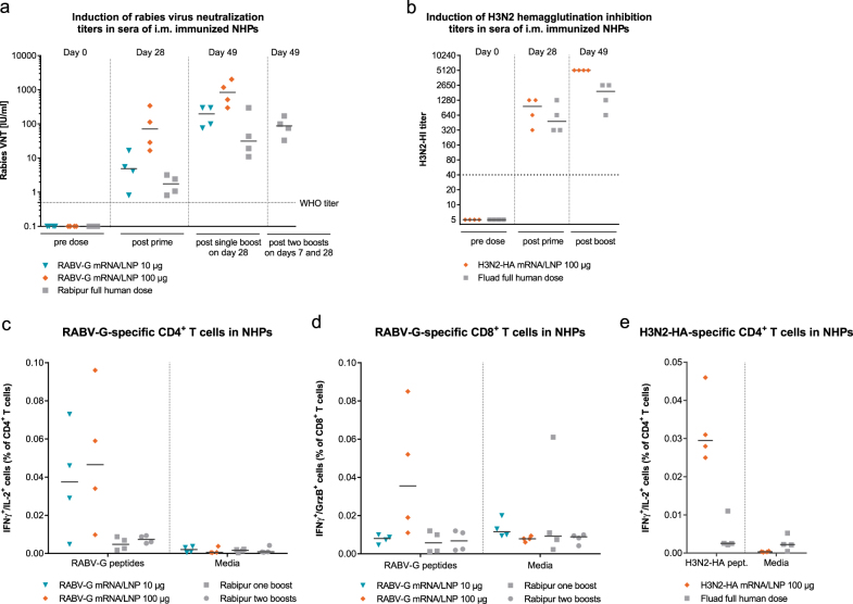
- Compared with licensed vaccines, RABV-G mRNA/LNP after one shot gave median VNTs of 4.9 IU/ml (10 µg) and 71.2 IU/ml (100 µg) versus 1.8 IU/ml for Rabipur®; after boost, 100 µg mRNA reached median 842 IU/ml versus 31.3 IU/ml for Rabipur®. H3N2-HA mRNA/LNP was comparable to or higher than Fluad®, and mRNA/LNP induced antigen-specific CD4+ and CD8+ T cell responses that were absent in Rabipur®- or Fluad®-vaccinated animals.
- LNP-formulated mRNA induced transient local pro-inflammatory cytokines and chemokines, with TNF and IL-6 peaking at 14 h; more than 90% of T and B cells in draining lymph nodes expressed CD69 at 24 h, and CD11b+ Gr1+ cells increased 10-fold within 4 h. Injection-site reactions were limited to slight erythema and/or edema resolving in 1–6 days, with no pain on palpation.
Methods
- Mice (BALB/c, 7–9 weeks) were vaccinated intramuscularly into the tibialis anterior with 25 µl at days 0 and 21; cynomolgus monkeys were vaccinated intramuscularly into the biceps femoris with 500 µl at days 0 and 28, with NHP groups of n = 2 male and 2 female per group.
- Vaccines used sequence-optimized, chemically unmodified mRNA in LNPs composed of an ionizable amino lipid, phospholipid, cholesterol, and PEGylated lipid, encoding RABV-G, H1N1-HA, H3N2-HA, luciferase, or non-translatable fluorescent mRNA; licensed comparators were Rabipur® and Fluad®.
- Analyses included in vivo luciferase imaging, cytokine/chemokine measurement by cytometric bead array or Luminex, flow cytometry of draining lymph nodes, rabies VNT by FAVN, influenza HI assays, intracellular cytokine staining, safety observations, and Mann–Whitney statistical tests.
Limitations
- The paper states that further investigations are required to determine whether monocytes and granulocytes contribute to antigen transport to draining lymph nodes. It also notes that the vaccinated non-human primates were naïve to the influenza strains used, unlike most adult humans who have prior exposure or vaccination, so human responses may differ.
- Evident from the text: NHP immunogenicity groups were small (n = 2 male, 2 female per group), and protection in NHPs was inferred from antibody correlates rather than live-virus challenge. The paper also states that the technology warrants clinical testing, so no human efficacy data are reported.
Model-written summary. Check it against the paper before relying on it.
Integrity signals
Registry
Cites
Statistics
Figures
Verification
Claims the paper makes
60 Own result · 22 Own interpretation · 7 Partly supported · 7 No full text of the sources · 4 Supported · 2 Supported so far · 1 No basis given · accuracy not yet measured
Every source the paper cites for this claim was checked and supports it.
In the paper
“Importantly, protective efficacy against Zika virus challenge infections was also demonstrated in non-human primates (NHPs) after i.d. vaccination.14”
§ Unmodified mRNA in LNPs constitutes a competitive technology for prophylactic vaccines > Introduction
What the sources say
- Supportedcites [14] Pardi, N. et al. Zika virus protection by a single low-dose nucleoside-modified mRNA vaccination. Nature https://doi.org/10.1038/nature21428 (2017). source
The paper reports that rhesus macaques were immunized intradermally and then challenged with Zika virus at week 5. In the passage labeled Abstract, four of five vaccinated macaques had no detectable viremia, while all six controls became infected; the fifth vaccinated animal had only a low, transient viral signal. (LLM, conf=0.94)
boosteddirect- Asserted: passes — The authors put it forward as true.
- Checkable: passes — It can be checked against evidence.
- Load-bearing: passes — The paper’s conclusions depend on it.
- Based: passes — The paper gives a reason why it holds.
- Supported
The cited sources support only part of this claim — read each source’s verdict below.
In the paper
“In recent publications chemically modified nucleosides were used to reach sufficient antigen expression,12–15 which is in contrast to our proprietary mRNA technology, which employs sequence optimization and selected untranslated regions (UTRs) to achieve high antigen expression.16”
§ Unmodified mRNA in LNPs constitutes a competitive technology for prophylactic vaccines > Introduction
What the sources say
- Supportedcites [12] Richner, J. M. et al. Modified mRNA vaccines protect against zika virus infection. Cell. https://doi.org/10.1016/j.cell.2017.02.017 (2017). source
The Introduction says the vaccine uses a nucleoside modification to limit innate immune activation, which can otherwise inhibit translation and reduce antigen expression. The Results (‘An mRNA vaccine platform for ZIKV’) also describe modified mRNA designed to induce high protein expression in vivo. (LLM, conf=0.92)
- Partly supportedcites [13] Richner, J. M. et al. Vaccine mediated protection against zika virus-induced congenital disease. Cell 170, 273–283.e12 (2017). sourceExperimental — accuracy not yet measured
The Method Details section (page unavailable) confirms that the vaccine mRNA used a chemically modified nucleoside, substituting 1-methylpseudoUTP for UTP. It attributes increased translation efficiency to the cap structure, while the Discussion (page unavailable) says modified nucleosides were used to minimize innate immune activation; the paper does not establish that the nucleoside modification was used to reach sufficient antigen expression. (LLM, conf=0.85)
- Supportedcites [14] Pardi, N. et al. Zika virus protection by a single low-dose nucleoside-modified mRNA vaccination. Nature https://doi.org/10.1038/nature21428 (2017). source
The Abstract states that the vaccine mRNA contains 1-methylpseudouridine, a modified nucleoside that increases mRNA translation in vivo. It also reports production of the encoded ZIKV E antigen in transfected cells, supporting the claim that modified nucleosides were used to achieve antigen expression. (LLM, conf=0.91)
- Supportedcites [15] Bahl, K. et al. Preclinical and clinical demonstration of immunogenicity by mRNA vaccines against H10N8 and H7N9 Influenza Viruses. Mol. Ther. J. Am. Soc. Gene Ther. 25, 1316–1327 (2017). source
The Introduction says that replacing uridine with modified nucleosides can overcome immune activation that inhibits mRNA translation and reduces antigen expression. The Discussion says the paper’s modified-mRNA vaccine technology balances immune stimulation with antigen expression and produces high transient protein levels. (LLM, conf=0.91)
plaindirect- Asserted: passes — The authors put it forward as true.
- Checkable: passes — It can be checked against evidence.
- Load-bearing: passes — The paper’s conclusions depend on it.
- Based: passes — The paper gives a reason why it holds.
- Supported
Every source the paper cites for this claim was checked and supports it.
In the paper
“In these studies, mice were vaccinated i.m. with LNP-formulated mRNA vaccines inducing protection against Zika virus challenge infections,12 and protection against placental damage and fetal demise in challenged pregnant mice.13”
§ Unmodified mRNA in LNPs constitutes a competitive technology for prophylactic vaccines > Introduction
What the sources say
- Supportedcites [13] Richner, J. M. et al. Vaccine mediated protection against zika virus-induced congenital disease. Cell 170, 273–283.e12 (2017). source
The Results section, “Activity of a prM-E mRNA LNP vaccine in pregnancy,” describes intramuscular vaccination before mating and Zika virus challenge during pregnancy. The Results section, “Vaccine protection against placental and fetal injury,” reports less placental damage and survival of all fetuses from prM-E-vaccinated dams, compared with fetal demise in placebo-vaccinated dams. Page numbers are not provided in our copy. (LLM, conf=0.94)
plaindirect- Asserted: passes — The authors put it forward as true.
- Checkable: passes — It can be checked against evidence.
- Load-bearing: unclear — It is unclear whether the paper’s conclusions depend on it.
- Based: passes — The paper gives a reason why it holds.
- Supported
Every source the paper cites for this claim was checked and supports it.
In the paper
“The study by Pardi et al. demonstrates that non-replicating mRNA is able to induce antibody titers in NHPs.”
§ Unmodified mRNA in LNPs constitutes a competitive technology for prophylactic vaccines > Introduction
What the sources say
- Supportedcites [14] Pardi, N. et al. Zika virus protection by a single low-dose nucleoside-modified mRNA vaccination. Nature https://doi.org/10.1038/nature21428 (2017). source
The mRNA production section describes a vaccine encoding ZIKV prM-E glycoproteins, consistent with non-replicating mRNA. The macaque findings (section labeled Abstract; page unavailable) report that vaccination induced E-specific IgG and neutralizing antibody titers in rhesus macaques, a non-human primate species. (LLM, conf=0.91)
boosteddirect- Asserted: passes — The authors put it forward as true.
- Checkable: passes — It can be checked against evidence.
- Load-bearing: passes — The paper’s conclusions depend on it.
- Based: passes — The paper gives a reason why it holds.
- Supported
The cited sources support only part of this claim — read each source’s verdict below.
In the paper
“In recent publications chemically modified nucleosides were used to reach sufficient antigen expression,12–15 which is in contrast to our proprietary mRNA technology, which employs sequence optimization and selected untranslated regions (UTRs) to achieve high antigen expression.16”
§ Unmodified mRNA in LNPs constitutes a competitive technology for prophylactic vaccines > Introduction
What the sources say
- Partly supportedcites [16] Thess, A. et al. Sequence-engineered mRNA without chemical nucleoside modifications enables an effective protein therapy in large animals. Mol. Ther. J. Am. Soc. Gene Ther. 23, 1456–1464 (2015). sourceExperimental — accuracy not yet measured
The Materials and Methods describe codon optimization and selection of 5′ and 3′ UTRs, and the Results report high expression from sequence-engineered mRNA. However, the expression experiments described in the Results use luciferase and erythropoietin, not an antigen; the source does not establish high antigen expression specifically. Page numbers were not provided in our copy. (LLM, conf=0.85)
plaindirect- Asserted: passes — The authors put it forward as true.
- Checkable: passes — It can be checked against evidence.
- Load-bearing: passes — The paper’s conclusions depend on it.
- Based: passes — The paper gives a reason why it holds.
- Partly supported
Every source the paper cites for this claim was checked and supports it.
In the paper
“In these studies, mice were vaccinated i.m. with LNP-formulated mRNA vaccines inducing protection against Zika virus challenge infections,12 and protection against placental damage and fetal demise in challenged pregnant mice.13”
§ Unmodified mRNA in LNPs constitutes a competitive technology for prophylactic vaccines > Introduction
What the sources say
- Supportedcites [12] Richner, J. M. et al. Modified mRNA vaccines protect against zika virus infection. Cell. https://doi.org/10.1016/j.cell.2017.02.017 (2017). source
The Results section (“Vaccine efficacy in C57BL/6 mice”) reports intramuscular vaccination with an mRNA LNP vaccine and protection against lethal ZIKV challenge. The separate claim about pregnant mice is attached to another citation and is not part of this verdict. (LLM, conf=0.94)
plaindirect- Asserted: passes — The authors put it forward as true.
- Checkable: passes — It can be checked against evidence.
- Load-bearing: passes — The paper’s conclusions depend on it.
- Based: passes — The paper gives a reason why it holds.
- Supported
A result the paper reports from its own data — it cites no other source for it, so there is none to check. The quote shows it is in the paper.
In the paper
“Activation of the innate immune system was reflected by a transient induction of pro-inflammatory cytokines and chemokines and activation of the majority of immune cells in the dLNs.”
§ Unmodified mRNA in LNPs constitutes a competitive technology for prophylactic vaccines > Abstract
plaindirect- Asserted: passes — The authors put it forward as true.
- Checkable: passes — It can be checked against evidence.
- Load-bearing: passes — The paper’s conclusions depend on it.
- Based: passes — The paper gives a reason why it holds.
A result the paper reports from its own data — it cites no other source for it, so there is none to check. The quote shows it is in the paper.
In the paper
“Single intramuscular vaccination of NHPs with LNP-formulated mRNAs encoding rabies or influenza antigens induced protective antibody titers, which could be boosted and remained stable during an observation period of up to 1 year.”
§ Unmodified mRNA in LNPs constitutes a competitive technology for prophylactic vaccines > Abstract
hedgeddirect- Asserted: passes — The authors put it forward as true.
- Checkable: passes — It can be checked against evidence.
- Load-bearing: passes — The paper’s conclusions depend on it.
- Based: passes — The paper gives a reason why it holds.
A result the paper reports from its own data — it cites no other source for it, so there is none to check. The quote shows it is in the paper.
In the paper
“Here, we demonstrate that vaccines based on sequence optimized, chemically unmodified mRNA formulated in optimized lipid nanoparticles (LNPs) are highly immunogenic and well tolerated in non-human primates (NHPs).”
§ Unmodified mRNA in LNPs constitutes a competitive technology for prophylactic vaccines > Abstract
boosted- Asserted: passes — The authors put it forward as true.
- Checkable: passes — It can be checked against evidence.
- Load-bearing: passes — The paper’s conclusions depend on it.
- Based: passes — The paper gives a reason why it holds.
A result the paper reports from its own data — it cites no other source for it, so there is none to check. The quote shows it is in the paper.
In the paper
“Single intramuscular vaccination of NHPs with LNP-formulated mRNAs encoding rabies or influenza antigens induced protective antibody titers, which could be boosted and remained stable during an observation period of up to 1 year.”
§ Unmodified mRNA in LNPs constitutes a competitive technology for prophylactic vaccines > Abstract
hedgeddirect- Asserted: passes — The authors put it forward as true.
- Checkable: passes — It can be checked against evidence.
- Load-bearing: passes — The paper’s conclusions depend on it.
- Based: passes — The paper gives a reason why it holds.
A result the paper reports from its own data — it cites no other source for it, so there is none to check. The quote shows it is in the paper.
In the paper
“Here, we demonstrate that vaccines based on sequence optimized, chemically unmodified mRNA formulated in optimized lipid nanoparticles (LNPs) are highly immunogenic and well tolerated in non-human primates (NHPs).”
§ Unmodified mRNA in LNPs constitutes a competitive technology for prophylactic vaccines > Abstract
boosteddirect- Asserted: passes — The authors put it forward as true.
- Checkable: passes — It can be checked against evidence.
- Load-bearing: passes — The paper’s conclusions depend on it.
- Based: passes — The paper gives a reason why it holds.
A result the paper reports from its own data — it cites no other source for it, so there is none to check. The quote shows it is in the paper.
In the paper
“First mechanistic insights into the mode of action of the LNP-formulated mRNA vaccines demonstrated a strong activation of the innate immune response at the injection site and in the draining lymph nodes (dLNs).”
§ Unmodified mRNA in LNPs constitutes a competitive technology for prophylactic vaccines > Abstract
boosteddirect- Asserted: passes — The authors put it forward as true.
- Checkable: passes — It can be checked against evidence.
- Load-bearing: passes — The paper’s conclusions depend on it.
- Based: passes — The paper gives a reason why it holds.
A result the paper reports from its own data — it cites no other source for it, so there is none to check. The quote shows it is in the paper.
In the paper
“Single intramuscular vaccination of NHPs with LNP-formulated mRNAs encoding rabies or influenza antigens induced protective antibody titers, which could be boosted and remained stable during an observation period of up to 1 year.”
§ Unmodified mRNA in LNPs constitutes a competitive technology for prophylactic vaccines > Abstract
plaindirect- Asserted: passes — The authors put it forward as true.
- Checkable: passes — It can be checked against evidence.
- Load-bearing: passes — The paper’s conclusions depend on it.
- Based: passes — The paper gives a reason why it holds.
A result the paper reports from its own data — it cites no other source for it, so there is none to check. The quote shows it is in the paper.
In the paper
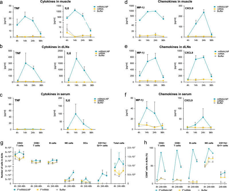
“In contrast to buffer or non-formulated mRNA, LNP-formulated mRNA induced a pronounced but transient release of the pro-inflammatory cytokines tumor necrosis factor (TNF) and IL-6 (Fig. 2a, b) with peak concentrations at 14 h after injection.”
§ Unmodified mRNA in LNPs constitutes a competitive technology for prophylactic vaccines > Results > Unmodified mRNA in LNPs supports high antigen expression in muscle cells and induces a pro-inflammatory environment
plaindirect- Asserted: passes — The authors put it forward as true.
- Checkable: passes — It can be checked against evidence.
- Load-bearing: passes — The paper’s conclusions depend on it.
- Based: passes — The paper gives a reason why it holds.
A result the paper reports from its own data — it cites no other source for it, so there is none to check. The quote shows it is in the paper.
In the paper
“Intramuscular injection of the LNP-formulated F*mRNA induced a strong increase in cellularity, which was absent after injection of unformulated F*mRNA (Fig. 2g).”
§ Unmodified mRNA in LNPs constitutes a competitive technology for prophylactic vaccines > Results > Unmodified mRNA in LNPs supports high antigen expression in muscle cells and induces a pro-inflammatory environment
boosteddirect- Asserted: passes — The authors put it forward as true.
- Checkable: passes — It can be checked against evidence.
- Load-bearing: passes — The paper’s conclusions depend on it.
- Based: passes — The paper gives a reason why it holds.
A result the paper reports from its own data — it cites no other source for it, so there is none to check. The quote shows it is in the paper.
In the paper
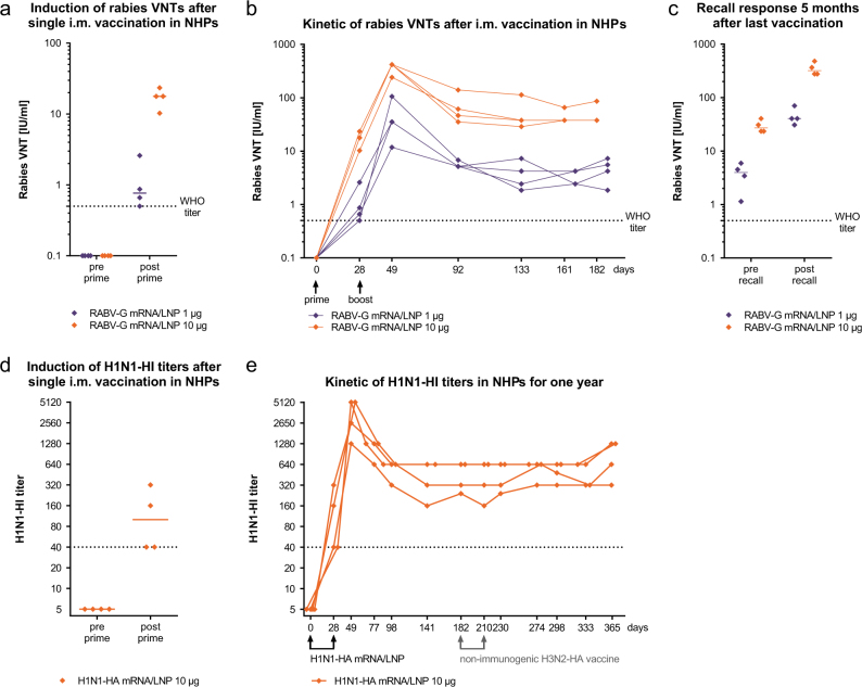
“The observed immunogenicity was dose dependent with a 10-fold higher mRNA dose yielding 10-fold higher VNTs.”
§ Unmodified mRNA in LNPs constitutes a competitive technology for prophylactic vaccines > Results > Intramuscular vaccination with LNP-formulated mRNA leads to long-lived humoral responses in NHPs
plaindirect- Asserted: passes — The authors put it forward as true.
- Checkable: passes — It can be checked against evidence.
- Load-bearing: unclear — It is unclear whether the paper’s conclusions depend on it.
- Based: passes — The paper gives a reason why it holds.
A result the paper reports from its own data — it cites no other source for it, so there is none to check. The quote shows it is in the paper.
In the paper
“A single i.m. immunization with 1 µg LNP-formulated RABV-G mRNA already induced robust VNTs at or above the WHO titer of 0.5 IU/ml in all animals at day 28 after prime vaccination (Fig. 4a).”
§ Unmodified mRNA in LNPs constitutes a competitive technology for prophylactic vaccines > Results > Intramuscular vaccination with LNP-formulated mRNA leads to long-lived humoral responses in NHPs
plaindirect- Asserted: passes — The authors put it forward as true.
- Checkable: passes — It can be checked against evidence.
- Load-bearing: passes — The paper’s conclusions depend on it.
- Based: passes — The paper gives a reason why it holds.
A result the paper reports from its own data — it cites no other source for it, so there is none to check. The quote shows it is in the paper.
In the paper
“Within 4 h after injection of LNP-formulated mRNA, we observed a 10-fold increase in CD11b+ Gr1+ cells compared to mice treated with non-formulated mRNA.”
§ Unmodified mRNA in LNPs constitutes a competitive technology for prophylactic vaccines > Results > Unmodified mRNA in LNPs supports high antigen expression in muscle cells and induces a pro-inflammatory environment
plaindirect- Asserted: passes — The authors put it forward as true.
- Checkable: passes — It can be checked against evidence.
- Load-bearing: passes — The paper’s conclusions depend on it.
- Based: passes — The paper gives a reason why it holds.
A result the paper reports from its own data — it cites no other source for it, so there is none to check. The quote shows it is in the paper.
In the paper
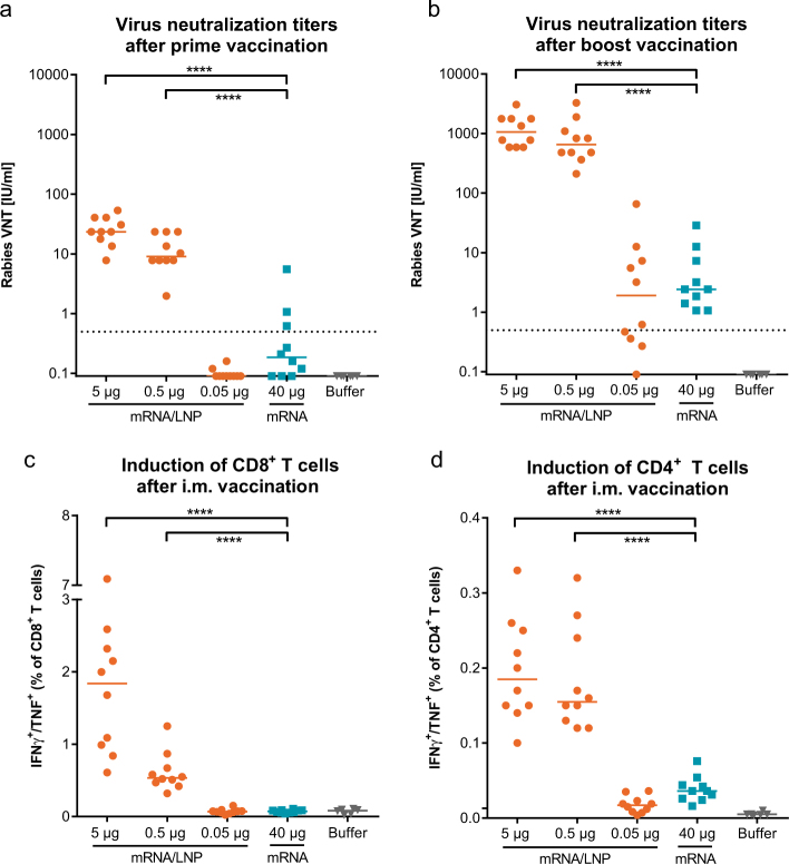
“These neutralizing antibody titers increased more than 50-fold after a second vaccination (Fig. 3b).”
§ Unmodified mRNA in LNPs constitutes a competitive technology for prophylactic vaccines > Results > LNP-formulated mRNA induces strong humoral and cellular responses in mice
plaindirect- Asserted: passes — The authors put it forward as true.
- Checkable: passes — It can be checked against evidence.
- Load-bearing: passes — The paper’s conclusions depend on it.
- Based: passes — The paper gives a reason why it holds.
A result the paper reports from its own data — it cites no other source for it, so there is none to check. The quote shows it is in the paper.
In the paper
“We show for the first time that a single intramuscular injection of non-replicating mRNA vaccines induces functional antibody titers in NHPs comparable to or even higher than vaccination with a full human dose of licensed vaccines based on inactivated virus.”
§ Unmodified mRNA in LNPs constitutes a competitive technology for prophylactic vaccines > Introduction
boosteddirect- Asserted: passes — The authors put it forward as true.
- Checkable: passes — It can be checked against evidence.
- Load-bearing: passes — The paper’s conclusions depend on it.
- Based: passes — The paper gives a reason why it holds.
A result the paper reports from its own data — it cites no other source for it, so there is none to check. The quote shows it is in the paper.
In the paper
“CD11b+ Gr1+ cells, consisting mainly of monocytes and granulocytes, accounted for the largest increase in leukocytes.”
§ Unmodified mRNA in LNPs constitutes a competitive technology for prophylactic vaccines > Results > Unmodified mRNA in LNPs supports high antigen expression in muscle cells and induces a pro-inflammatory environment
plaincorrelational- Asserted: passes — The authors put it forward as true.
- Checkable: passes — It can be checked against evidence.
- Load-bearing: unclear — It is unclear whether the paper’s conclusions depend on it.
- Based: passes — The paper gives a reason why it holds.
A result the paper reports from its own data — it cites no other source for it, so there is none to check. The quote shows it is in the paper.
In the paper
“The VNTs induced by 0.5 µg of LNP-formulated mRNA were more than 250-fold higher (median 650 IU) compared to titers induced by 40 µg of non-formulated mRNA (median 2.4 IU).”
§ Unmodified mRNA in LNPs constitutes a competitive technology for prophylactic vaccines > Results > LNP-formulated mRNA induces strong humoral and cellular responses in mice
plaindirect- Asserted: passes — The authors put it forward as true.
- Checkable: passes — It can be checked against evidence.
- Load-bearing: passes — The paper’s conclusions depend on it.
- Based: passes — The paper gives a reason why it holds.
A result the paper reports from its own data — it cites no other source for it, so there is none to check. The quote shows it is in the paper.
In the paper
“We also observed a transient increase in serum concentrations of the chemokines described above, but to a much lower extent compared to those detected at the injection site or in the dLNs (Fig. 2f).”
§ Unmodified mRNA in LNPs constitutes a competitive technology for prophylactic vaccines > Results > Unmodified mRNA in LNPs supports high antigen expression in muscle cells and induces a pro-inflammatory environment
plaincorrelational- Asserted: passes — The authors put it forward as true.
- Checkable: passes — It can be checked against evidence.
- Load-bearing: unclear — It is unclear whether the paper’s conclusions depend on it.
- Based: passes — The paper gives a reason why it holds.
A result the paper reports from its own data — it cites no other source for it, so there is none to check. The quote shows it is in the paper.
In the paper
“Among the strongly upregulated chemokines were MIP-1β, which plays a pivotal role in the chemotaxis of macrophages, monocytes and NK cells, and CXCL-9, which recruits T cells, NK cells and NKT cells to the site of inflammation.20”
§ Unmodified mRNA in LNPs constitutes a competitive technology for prophylactic vaccines > Results > Unmodified mRNA in LNPs supports high antigen expression in muscle cells and induces a pro-inflammatory environment
boosted- Asserted: passes — The authors put it forward as true.
- Checkable: passes — It can be checked against evidence.
- Load-bearing: unclear — It is unclear whether the paper’s conclusions depend on it.
- Based: passes — The paper gives a reason why it holds.
In the paper
“A prime vaccination with a dose of 0.5 µg LNP-formulated RABV-G mRNA already induced virus neutralization titers (VNTs) above the WHO reference titer of 0.5 IU/ml, which is used as correlate of protection in humans21 (Fig. 3a).”
§ Unmodified mRNA in LNPs constitutes a competitive technology for prophylactic vaccines > Results > LNP-formulated mRNA induces strong humoral and cellular responses in mice
What the sources say
- Not checked — no full text of the sourcecites [21] Plotkin, S. A. Vaccines: correlates of vaccine-induced immunity. Clin. Infect. Dis. 47, 401–409 (2008). source
plaindirect- Asserted: passes — The authors put it forward as true.
- Checkable: passes — It can be checked against evidence.
- Load-bearing: passes — The paper’s conclusions depend on it.
- Based: passes — The paper gives a reason why it holds.
A result the paper reports from its own data — it cites no other source for it, so there is none to check. The quote shows it is in the paper.
In the paper
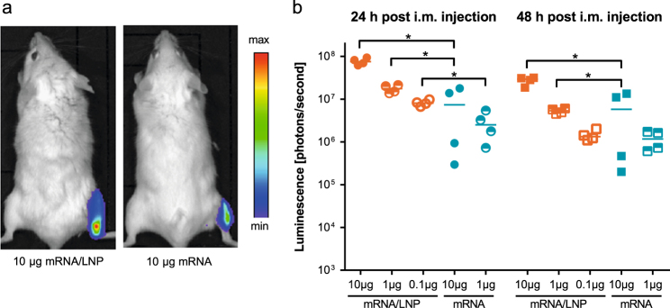
“Mice injected with PpLuc mRNA/LNP showed at 24 and 48 h significantly higher intramuscular luciferase expression than animals that had received non-formulated PpLuc mRNA (Fig. 1).”
§ Unmodified mRNA in LNPs constitutes a competitive technology for prophylactic vaccines > Results > Unmodified mRNA in LNPs supports high antigen expression in muscle cells and induces a pro-inflammatory environment
plaindirect- Asserted: passes — The authors put it forward as true.
- Checkable: passes — It can be checked against evidence.
- Load-bearing: passes — The paper’s conclusions depend on it.
- Based: passes — The paper gives a reason why it holds.
A result the paper reports from its own data — it cites no other source for it, so there is none to check. The quote shows it is in the paper.
In the paper
“Moreover, there was a transient elevation in the concentrations of MCP-1, MIP-1α, and CXCL1, which attract a variety of immune cells such as monocytes, macrophages, dendritic cells and neutrophils (data not shown).”
§ Unmodified mRNA in LNPs constitutes a competitive technology for prophylactic vaccines > Results > Unmodified mRNA in LNPs supports high antigen expression in muscle cells and induces a pro-inflammatory environment
plaindirect- Asserted: passes — The authors put it forward as true.
- Checkable: passes — It can be checked against evidence.
- Load-bearing: unclear — It is unclear whether the paper’s conclusions depend on it.
- Based: passes — The paper gives a reason why it holds.
A result the paper reports from its own data — it cites no other source for it, so there is none to check. The quote shows it is in the paper.
In the paper
“Additionally, LNP-formulated mRNA led to stronger cellular immune responses, which was reflected by significantly higher frequencies of antigen-specific multifunctional CD4+ and CD8+ T cells (Fig. 3c, d) compared to non-formulated mRNA.”
§ Unmodified mRNA in LNPs constitutes a competitive technology for prophylactic vaccines > Results > LNP-formulated mRNA induces strong humoral and cellular responses in mice
boosteddirect- Asserted: passes — The authors put it forward as true.
- Checkable: passes — It can be checked against evidence.
- Load-bearing: passes — The paper’s conclusions depend on it.
- Based: passes — The paper gives a reason why it holds.
A result the paper reports from its own data — it cites no other source for it, so there is none to check. The quote shows it is in the paper.
In the paper
“In the influenza-vaccinated animals H3N2-HA-specific IFN-γ+/IL-2+ CD4+ T cells were observed at day 35 only for the mRNA/LNP vaccine but not for Fluad®”
§ Unmodified mRNA in LNPs constitutes a competitive technology for prophylactic vaccines > Results > LNP-formulated mRNA vaccines are capable of competing with licensed vaccines in NHPs
plain- Asserted: passes — The authors put it forward as true.
- Checkable: passes — It can be checked against evidence.
- Load-bearing: passes — The paper’s conclusions depend on it.
- Based: passes — The paper gives a reason why it holds.
A result the paper reports from its own data — it cites no other source for it, so there is none to check. The quote shows it is in the paper.
In the paper
“The H1N1-HI titers were not changed by the non-immunogenic H3N2-HA vaccination.”
§ Unmodified mRNA in LNPs constitutes a competitive technology for prophylactic vaccines > Results > Intramuscular vaccination with LNP-formulated mRNA leads to long-lived humoral responses in NHPs
plain- Asserted: passes — The authors put it forward as true.
- Checkable: passes — It can be checked against evidence.
- Load-bearing: unclear — It is unclear whether the paper’s conclusions depend on it.
- Based: passes — The paper gives a reason why it holds.
A result the paper reports from its own data — it cites no other source for it, so there is none to check. The quote shows it is in the paper.
In the paper
“Four weeks after a single shot vaccination median VNTs measured for mRNA vaccines were 4.9 IU/ml for the 10 µg dose and 71.2 IU/ml for the 100 µg dose, compared to 1.8 IU/ml for Rabipur®.”
§ Unmodified mRNA in LNPs constitutes a competitive technology for prophylactic vaccines > Results > LNP-formulated mRNA vaccines are capable of competing with licensed vaccines in NHPs
plain- Asserted: passes — The authors put it forward as true.
- Checkable: passes — It can be checked against evidence.
- Load-bearing: passes — The paper’s conclusions depend on it.
- Based: passes — The paper gives a reason why it holds.
A result the paper reports from its own data — it cites no other source for it, so there is none to check. The quote shows it is in the paper.
In the paper
“H3N2-HA-specific CD8+ T cell responses were absent in both cases.”
§ Unmodified mRNA in LNPs constitutes a competitive technology for prophylactic vaccines > Results > LNP-formulated mRNA vaccines are capable of competing with licensed vaccines in NHPs
plain- Asserted: passes — The authors put it forward as true.
- Checkable: passes — It can be checked against evidence.
- Load-bearing: unclear — It is unclear whether the paper’s conclusions depend on it.
- Based: passes — The paper gives a reason why it holds.
A result the paper reports from its own data — it cites no other source for it, so there is none to check. The quote shows it is in the paper.
In the paper
“RABV-G-specific IFN-γ+/GrzB+ CD8+ T cells, which are dedicated cytolytic cells,24 were detected only in animals receiving the 100 µg dose”
§ Unmodified mRNA in LNPs constitutes a competitive technology for prophylactic vaccines > Results > LNP-formulated mRNA vaccines are capable of competing with licensed vaccines in NHPs
plain- Asserted: passes — The authors put it forward as true.
- Checkable: passes — It can be checked against evidence.
- Load-bearing: unclear — It is unclear whether the paper’s conclusions depend on it.
- Based: passes — The paper gives a reason why it holds.
A result the paper reports from its own data — it cites no other source for it, so there is none to check. The quote shows it is in the paper.
In the paper
“Vaccination with the 100 µg dose induced at 24 h in all animals a low increase in IL-6 concentrations.”
§ Unmodified mRNA in LNPs constitutes a competitive technology for prophylactic vaccines > Results > mRNA vaccines exhibit a favorable safety profile
plaindirect- Asserted: passes — The authors put it forward as true.
- Checkable: passes — It can be checked against evidence.
- Load-bearing: passes — The paper’s conclusions depend on it.
- Based: passes — The paper gives a reason why it holds.
A result the paper reports from its own data — it cites no other source for it, so there is none to check. The quote shows it is in the paper.
In the paper
“Body temperatures, which varied considerably among animals and studies, did not display obvious differences between NHPs that had received the mRNA/LNP vaccines or the licensed vaccines”
§ Unmodified mRNA in LNPs constitutes a competitive technology for prophylactic vaccines > Results > mRNA vaccines exhibit a favorable safety profile
plain- Asserted: passes — The authors put it forward as true.
- Checkable: passes — It can be checked against evidence.
- Load-bearing: passes — The paper’s conclusions depend on it.
- Based: passes — The paper gives a reason why it holds.
In the paper
“After just a single vaccination, we observed H1N1-HI titers at or above 1:40, which is used as correlate of protection in humans21 (Fig. 4d).”
§ Unmodified mRNA in LNPs constitutes a competitive technology for prophylactic vaccines > Results > Intramuscular vaccination with LNP-formulated mRNA leads to long-lived humoral responses in NHPs
What the sources say
- Not checked — no full text of the sourcecites [21] Plotkin, S. A. Vaccines: correlates of vaccine-induced immunity. Clin. Infect. Dis. 47, 401–409 (2008). source
plaindirect- Asserted: passes — The authors put it forward as true.
- Checkable: passes — It can be checked against evidence.
- Load-bearing: passes — The paper’s conclusions depend on it.
- Based: passes — The paper gives a reason why it holds.
A result the paper reports from its own data — it cites no other source for it, so there is none to check. The quote shows it is in the paper.
In the paper
“Intramuscular vaccination of NHPs with mRNA/LNP vaccines encoding RABV-G as well as influenza HA of the strains H1N1 and H3N2 induced long lasting antibody responses, which were monitored for up to 1 year and were maintained during this period well above the titers that serve as correlate of protection.”
- Also stated in: § Discussion · plain
“induced long-lived functional antibody responses that correlated with protection for rabies and influenza virus.”
§ Unmodified mRNA in LNPs constitutes a competitive technology for prophylactic vaccines > Discussion
plaindirect- Asserted: passes — The authors put it forward as true.
- Checkable: passes — It can be checked against evidence.
- Load-bearing: passes — The paper’s conclusions depend on it.
- Based: passes — The paper gives a reason why it holds.
- Rests on an assumption: The antibody titers measured serve as correlates of protection for rabies and influenza.
- Also stated in: § Discussion · plain
A result the paper reports from its own data — it cites no other source for it, so there is none to check. The quote shows it is in the paper.
In the paper
“Injection sites showed only slight erythema and/or edema in some of the animals, which resolved 1 to 6 days after injection (Table 1).”
§ Unmodified mRNA in LNPs constitutes a competitive technology for prophylactic vaccines > Results > mRNA vaccines exhibit a favorable safety profile
plain- Asserted: passes — The authors put it forward as true.
- Checkable: passes — It can be checked against evidence.
- Load-bearing: passes — The paper’s conclusions depend on it.
- Based: passes — The paper gives a reason why it holds.
A result the paper reports from its own data — it cites no other source for it, so there is none to check. The quote shows it is in the paper.
In the paper
“However, the additional vaccination with Rabipur® on day 7 did also not result in higher VNTs at day 49 than after two mRNA vaccine administrations.”
§ Unmodified mRNA in LNPs constitutes a competitive technology for prophylactic vaccines > Results > LNP-formulated mRNA vaccines are capable of competing with licensed vaccines in NHPs
plain- Asserted: passes — The authors put it forward as true.
- Checkable: passes — It can be checked against evidence.
- Load-bearing: passes — The paper’s conclusions depend on it.
- Based: passes — The paper gives a reason why it holds.
A result the paper reports from its own data — it cites no other source for it, so there is none to check. The quote shows it is in the paper.
In the paper
“Importantly, the threshold titers were already achieved after a single vaccination”
§ Unmodified mRNA in LNPs constitutes a competitive technology for prophylactic vaccines > Discussion
plaindirect- Asserted: passes — The authors put it forward as true.
- Checkable: passes — It can be checked against evidence.
- Load-bearing: passes — The paper’s conclusions depend on it.
- Based: passes — The paper gives a reason why it holds.
A result the paper reports from its own data — it cites no other source for it, so there is none to check. The quote shows it is in the paper.
In the paper
“An intracellular cytokine staining (ICS) analysis of PBMCs of the rabies-vaccinated animals at day 35, i.e., seven days after the last vaccination, detected specific cellular responses after mRNA/LNP vaccination but not in Rabipur®-vaccinated animals.”
§ Unmodified mRNA in LNPs constitutes a competitive technology for prophylactic vaccines > Results > LNP-formulated mRNA vaccines are capable of competing with licensed vaccines in NHPs
plaindirect- Asserted: passes — The authors put it forward as true.
- Checkable: passes — It can be checked against evidence.
- Load-bearing: passes — The paper’s conclusions depend on it.
- Based: passes — The paper gives a reason why it holds.
A result the paper reports from its own data — it cites no other source for it, so there is none to check. The quote shows it is in the paper.
In the paper
“To evaluate whether the primary responses could be boosted a second vaccination with the RABV-G mRNA/LNP vaccine was performed at day 28 resulting in a 20-fold increase in VNTs.”
§ Unmodified mRNA in LNPs constitutes a competitive technology for prophylactic vaccines > Results > Intramuscular vaccination with LNP-formulated mRNA leads to long-lived humoral responses in NHPs
plaindirect- Asserted: passes — The authors put it forward as true.
- Checkable: passes — It can be checked against evidence.
- Load-bearing: passes — The paper’s conclusions depend on it.
- Based: passes — The paper gives a reason why it holds.
A result the paper reports from its own data — it cites no other source for it, so there is none to check. The quote shows it is in the paper.
In the paper
“No pain on palpation was observed.”
§ Unmodified mRNA in LNPs constitutes a competitive technology for prophylactic vaccines > Results > mRNA vaccines exhibit a favorable safety profile
plain- Asserted: passes — The authors put it forward as true.
- Checkable: passes — It can be checked against evidence.
- Load-bearing: passes — The paper’s conclusions depend on it.
- Based: passes — The paper gives a reason why it holds.
A result the paper reports from its own data — it cites no other source for it, so there is none to check. The quote shows it is in the paper.
In the paper
“Similarly, no changes in food consumption or body weight were associated with any dose (data not shown).”
§ Unmodified mRNA in LNPs constitutes a competitive technology for prophylactic vaccines > Results > mRNA vaccines exhibit a favorable safety profile
plaincorrelational- Asserted: passes — The authors put it forward as true.
- Checkable: passes — It can be checked against evidence.
- Load-bearing: passes — The paper’s conclusions depend on it.
- Based: passes — The paper gives a reason why it holds.
A result the paper reports from its own data — it cites no other source for it, so there is none to check. The quote shows it is in the paper.
In the paper
“IL-6 was absent in almost all animals that received the 1 or 10 µg dose.”
§ Unmodified mRNA in LNPs constitutes a competitive technology for prophylactic vaccines > Results > mRNA vaccines exhibit a favorable safety profile
plain- Asserted: passes — The authors put it forward as true.
- Checkable: passes — It can be checked against evidence.
- Load-bearing: passes — The paper’s conclusions depend on it.
- Based: passes — The paper gives a reason why it holds.
A result the paper reports from its own data — it cites no other source for it, so there is none to check. The quote shows it is in the paper.
In the paper
“A continued follow-up of the H1N1-HI titers demonstrated stable H1N1-HI titers induced by the primary H1N1-HA mRNA/LNP vaccination until the end of the observation period of 1 year.”
§ Unmodified mRNA in LNPs constitutes a competitive technology for prophylactic vaccines > Results > Intramuscular vaccination with LNP-formulated mRNA leads to long-lived humoral responses in NHPs
boosteddirect- Asserted: passes — The authors put it forward as true.
- Checkable: passes — It can be checked against evidence.
- Load-bearing: passes — The paper’s conclusions depend on it.
- Based: passes — The paper gives a reason why it holds.
The paper states this without a source or data behind it here, so there is nothing to check it against. The quote shows it is in the paper.
In the paper
“It should be noted that the animals were naïve regarding the vaccinated influenza strains, indicating induction of de novo immune responses after mRNA vaccination.”
§ Unmodified mRNA in LNPs constitutes a competitive technology for prophylactic vaccines > Discussion
hedgeddirect- Asserted: passes — The authors put it forward as true.
- Checkable: passes — It can be checked against evidence.
- Load-bearing: unclear — It is unclear whether the paper’s conclusions depend on it.
- Based: passes — The paper gives a reason why it holds.
A result the paper reports from its own data — it cites no other source for it, so there is none to check. The quote shows it is in the paper.
In the paper
“RABV-G-specific multifunctional IFN-γ+/IL-2+ CD4+ T cells, which are associated with strong anti-viral responses,23 were observed for both mRNA vaccine doses”
§ Unmodified mRNA in LNPs constitutes a competitive technology for prophylactic vaccines > Results > LNP-formulated mRNA vaccines are capable of competing with licensed vaccines in NHPs
plain- Asserted: passes — The authors put it forward as true.
- Checkable: passes — It can be checked against evidence.
- Load-bearing: passes — The paper’s conclusions depend on it.
- Based: passes — The paper gives a reason why it holds.
A result the paper reports from its own data — it cites no other source for it, so there is none to check. The quote shows it is in the paper.
In the paper
“To demonstrate the existence of B cell memory we vaccinated the same animals 5 months after completed vaccination a third time with RABV-G mRNA/LNPs (recall vaccination) and measured antibody titers 5 days later. In both dose groups we observed a very rapid 10-fold increase in VNTs, demonstrating the induction of a strong recall-response by the mRNA vaccine (Fig. 4c).”
§ Unmodified mRNA in LNPs constitutes a competitive technology for prophylactic vaccines > Results > Intramuscular vaccination with LNP-formulated mRNA leads to long-lived humoral responses in NHPs
boosteddirect- Asserted: passes — The authors put it forward as true.
- Checkable: passes — It can be checked against evidence.
- Load-bearing: passes — The paper’s conclusions depend on it.
- Based: passes — The paper gives a reason why it holds.
A result the paper reports from its own data — it cites no other source for it, so there is none to check. The quote shows it is in the paper.
In the paper
“Similarly important is the observation that LNPs, which were originally developed to deliver siRNA27 and have already demonstrated their safety for intravenous administration in clinical trials,30 are also well tolerated in NHPs after i.m. administration in combination with unmodified mRNA.”
§ Unmodified mRNA in LNPs constitutes a competitive technology for prophylactic vaccines > Discussion
boosted- Asserted: passes — The authors put it forward as true.
- Checkable: passes — It can be checked against evidence.
- Load-bearing: passes — The paper’s conclusions depend on it.
- Based: passes — The paper gives a reason why it holds.
A result the paper reports from its own data — it cites no other source for it, so there is none to check. The quote shows it is in the paper.
In the paper
“The animals presented in Fig. 4 were vaccinated first with the H1N1-HA mRNA/LNP vaccine and three weeks after boost with the RABV-G mRNA/LNP vaccine. Nevertheless, they mounted immune responses against both antigens.”
§ Unmodified mRNA in LNPs constitutes a competitive technology for prophylactic vaccines > Discussion
plaindirect- Asserted: passes — The authors put it forward as true.
- Checkable: passes — It can be checked against evidence.
- Load-bearing: passes — The paper’s conclusions depend on it.
- Based: passes — The paper gives a reason why it holds.
A conclusion the paper draws from its own results — it cites no other source for it, so there is none to check. The quote shows it is in the paper.
In the paper
“These results provide strong evidence that the mRNA technology is a valid approach for the development of effective prophylactic vaccines to prevent infectious diseases.”
§ Unmodified mRNA in LNPs constitutes a competitive technology for prophylactic vaccines > Abstract
boosted- Asserted: passes — The authors put it forward as true.
- Checkable: passes — It can be checked against evidence.
- Load-bearing: passes — The paper’s conclusions depend on it.
- Based: passes — The paper gives a reason why it holds.
- Rests on an assumption: Immunogenicity and protection data in non-human primates predict efficacy of prophylactic vaccines in humans.
A conclusion the paper draws from its own results — it cites no other source for it, so there is none to check. The quote shows it is in the paper.
In the paper
“Importantly, we show that the developed LNP-formulated mRNA vaccines can be used as a vaccination platform allowing multiple, sequential vaccinations against different pathogens.”
§ Unmodified mRNA in LNPs constitutes a competitive technology for prophylactic vaccines > Abstract
plaindirect- Asserted: passes — The authors put it forward as true.
- Checkable: passes — It can be checked against evidence.
- Load-bearing: passes — The paper’s conclusions depend on it.
- Based: passes — The paper gives a reason why it holds.
A conclusion the paper draws from its own results — it cites no other source for it, so there is none to check. The quote shows it is in the paper.
In the paper
“Moreover, we demonstrated the potential of mRNA vaccines as a platform approach by successfully vaccinating the same animals with multiple vaccines based on the same technology.”
§ Unmodified mRNA in LNPs constitutes a competitive technology for prophylactic vaccines > Introduction
boosteddirect- Asserted: passes — The authors put it forward as true.
- Checkable: passes — It can be checked against evidence.
- Load-bearing: passes — The paper’s conclusions depend on it.
- Based: passes — The paper gives a reason why it holds.
A conclusion the paper draws from its own results — it cites no other source for it, so there is none to check. The quote shows it is in the paper.
In the paper
“In the present study, we compare mRNA vaccines to licensed vaccines based on inactivated virus and demonstrate that unmodified mRNA, formulated in optimized LNPs, fulfills all the key requirements to be a viable vaccine platform for human prophylaxis.”
§ Unmodified mRNA in LNPs constitutes a competitive technology for prophylactic vaccines > Introduction
boosted- Asserted: passes — The authors put it forward as true.
- Checkable: passes — It can be checked against evidence.
- Load-bearing: passes — The paper’s conclusions depend on it.
- Based: passes — The paper gives a reason why it holds.
- Rests on an assumption: Immunogenicity and protection data in non-human primates predict efficacy of prophylactic vaccines in humans.
A result the paper reports from its own data — it cites no other source for it, so there is none to check. The quote shows it is in the paper.
In the paper
“These data reveal a gap between self-amplifying and non-replicating mRNA, which cannot be closed only by formulation.”
§ Unmodified mRNA in LNPs constitutes a competitive technology for prophylactic vaccines > Introduction
plain- Asserted: passes — The authors put it forward as true.
- Checkable: passes — It can be checked against evidence.
- Load-bearing: passes — The paper’s conclusions depend on it.
- Based: passes — The paper gives a reason why it holds.
In the paper
“In contrast, non-replicating mRNA did not induce any detectable antibody titers even when formulated with CNE.”
§ Unmodified mRNA in LNPs constitutes a competitive technology for prophylactic vaccines > Introduction
What the sources say
- Not checked — no full text of the sourcecites [11] Brito, L. A. et al. A cationic nanoemulsion for the delivery of next-generation RNA vaccines. Mol. Ther. J. Am. Soc. Gene Ther. 22, 2118–2129 (2014). source
plain- Asserted: passes — The authors put it forward as true.
- Checkable: passes — It can be checked against evidence.
- Load-bearing: passes — The paper’s conclusions depend on it.
- Based: passes — The paper gives a reason why it holds.
A conclusion the paper draws from its own results — it cites no other source for it, so there is none to check. The quote shows it is in the paper.
In the paper
“The higher concentrations at the injection site and in the dLNs strongly suggest a local production of these cytokines.”
§ Unmodified mRNA in LNPs constitutes a competitive technology for prophylactic vaccines > Results > Unmodified mRNA in LNPs supports high antigen expression in muscle cells and induces a pro-inflammatory environment
hedgedconditional- Asserted: passes — The authors put it forward as true.
- Checkable: passes — It can be checked against evidence.
- Load-bearing: unclear — It is unclear whether the paper’s conclusions depend on it.
- Based: passes — The paper gives a reason why it holds.
A conclusion the paper draws from its own results — it cites no other source for it, so there is none to check. The quote shows it is in the paper.
In the paper
“The observed increase in cellularity and the strong activation of immune cells in the dLNs even when a non-translatable mRNA was used suggest that the observed effect is independent of the encoded antigen.”
§ Unmodified mRNA in LNPs constitutes a competitive technology for prophylactic vaccines > Discussion
hedged- Asserted: passes — The authors put it forward as true.
- Checkable: passes — It can be checked against evidence.
- Load-bearing: passes — The paper’s conclusions depend on it.
- Based: passes — The paper gives a reason why it holds.
A conclusion the paper draws from its own results — it cites no other source for it, so there is none to check. The quote shows it is in the paper.
In the paper
“For the 100 µg dose of LNP-formulated RABV-G mRNA, which exceeded the WHO titer of 0.5 IU/ml by 20 to 200-fold at day 28, even a single administration may already be sufficient to induce protective and sustained antibody titers.”
§ Unmodified mRNA in LNPs constitutes a competitive technology for prophylactic vaccines > Results > LNP-formulated mRNA vaccines are capable of competing with licensed vaccines in NHPs
hedgedconditional- Asserted: passes — The authors put it forward as true.
- Checkable: passes — It can be checked against evidence.
- Load-bearing: passes — The paper’s conclusions depend on it.
- Based: passes — The paper gives a reason why it holds.
- Rests on an assumption: Immunogenicity and protection data in non-human primates predict efficacy of prophylactic vaccines in humans.
A conclusion the paper draws from its own results — it cites no other source for it, so there is none to check. The quote shows it is in the paper.
In the paper
“By analyzing the injection site and the dLNs, we revealed that LNP formulation additionally led to the activation of innate immune responses, which support the induction of cellular and humoral responses.”
§ Unmodified mRNA in LNPs constitutes a competitive technology for prophylactic vaccines > Discussion
plaindirect- Asserted: passes — The authors put it forward as true.
- Checkable: passes — It can be checked against evidence.
- Load-bearing: passes — The paper’s conclusions depend on it.
- Based: passes — The paper gives a reason why it holds.
A conclusion the paper draws from its own results — it cites no other source for it, so there is none to check. The quote shows it is in the paper.
In the paper
“suggesting that the vaccines could be used as single shot vaccines, which is especially important for vaccination campaigns in low resource settings.”
§ Unmodified mRNA in LNPs constitutes a competitive technology for prophylactic vaccines > Discussion
hedged- Asserted: passes — The authors put it forward as true.
- Checkable: passes — It can be checked against evidence.
- Load-bearing: passes — The paper’s conclusions depend on it.
- Based: passes — The paper gives a reason why it holds.
A conclusion the paper draws from its own results — it cites no other source for it, so there is none to check. The quote shows it is in the paper.
In the paper
“and confirm the observations in mice, where systemic release of TNF or IL-6 was absent or low, respectively.”
§ Unmodified mRNA in LNPs constitutes a competitive technology for prophylactic vaccines > Results > mRNA vaccines exhibit a favorable safety profile
plain- Asserted: passes — The authors put it forward as true.
- Checkable: passes — It can be checked against evidence.
- Load-bearing: passes — The paper’s conclusions depend on it.
- Based: passes — The paper gives a reason why it holds.
A conclusion the paper draws from its own results — it cites no other source for it, so there is none to check. The quote shows it is in the paper.
In the paper
“These data suggest that the vaccination schedule with two injections of the LNP-formulated mRNA vaccine is sufficient to induce protection against rabies infections.”
§ Unmodified mRNA in LNPs constitutes a competitive technology for prophylactic vaccines > Results > LNP-formulated mRNA vaccines are capable of competing with licensed vaccines in NHPs
hedgeddirect- Asserted: passes — The authors put it forward as true.
- Checkable: passes — It can be checked against evidence.
- Load-bearing: passes — The paper’s conclusions depend on it.
- Based: passes — The paper gives a reason why it holds.
- Rests on an assumption: Immunogenicity and protection data in non-human primates predict efficacy of prophylactic vaccines in humans.
A conclusion the paper draws from its own results — it cites no other source for it, so there is none to check. The quote shows it is in the paper.
In the paper
“Data presented in the current work demonstrate that such a combination is feasible.”
§ Unmodified mRNA in LNPs constitutes a competitive technology for prophylactic vaccines > Discussion
boosted- Asserted: passes — The authors put it forward as true.
- Checkable: passes — It can be checked against evidence.
- Load-bearing: passes — The paper’s conclusions depend on it.
- Based: passes — The paper gives a reason why it holds.
A conclusion the paper draws from its own results — it cites no other source for it, so there is none to check. The quote shows it is in the paper.
In the paper
“These data demonstrate a good tolerability of the mRNA/LNP vaccines in NHPs”
- Also stated in: § Discussion · boosted
“The vaccines were well tolerated in NHPs”
§ Unmodified mRNA in LNPs constitutes a competitive technology for prophylactic vaccines > Results > mRNA vaccines exhibit a favorable safety profile
boosted- Asserted: passes — The authors put it forward as true.
- Checkable: passes — It can be checked against evidence.
- Load-bearing: passes — The paper’s conclusions depend on it.
- Based: passes — The paper gives a reason why it holds.
- Also stated in: § Discussion · boosted
In the paper
“In general, in vitro transcribed mRNA has the risk of triggering various pattern recognition receptors in the endosome (e.g., TLR7/8) or the cytoplasm (e.g., RIG-I), which lead to an anti-viral response, followed by a shutdown of translation that can lower expression of the mRNA-encoded protein.25”
§ Unmodified mRNA in LNPs constitutes a competitive technology for prophylactic vaccines > Discussion
What the sources say
- Not checked — no full text of the sourcecites [25] Karikó, K., Buckstein, M., Ni, H. & Weissman, D. Suppression of RNA recognition by Toll-like receptors: the impact of nucleoside modification and the evolutionary origin of RNA. Immunity 23, 165–175 (2005). source
plaindirect- Asserted: passes — The authors put it forward as true.
- Checkable: passes — It can be checked against evidence.
- Load-bearing: passes — The paper’s conclusions depend on it.
- Based: passes — The paper gives a reason why it holds.
A conclusion the paper draws from its own results — it cites no other source for it, so there is none to check. The quote shows it is in the paper.
In the paper
“In conclusion, the mRNA technology evaluated in the present study fulfills all requirements of a viable vaccine platform”
§ Unmodified mRNA in LNPs constitutes a competitive technology for prophylactic vaccines > Discussion
plain- Asserted: passes — The authors put it forward as true.
- Checkable: passes — It can be checked against evidence.
- Load-bearing: passes — The paper’s conclusions depend on it.
- Based: passes — The paper gives a reason why it holds.
- Rests on an assumption: Nonhuman primates mimic the human situation most closely.
- Rests on an assumption: The antibody titers measured serve as correlates of protection for rabies and influenza.
In the paper
“and have, to our best knowledge, for prophylactic mRNA vaccines only been detected in NHPs after vaccination with self-amplifying mRNA31 but not with non-amplifying mRNA.”
§ Unmodified mRNA in LNPs constitutes a competitive technology for prophylactic vaccines > Discussion
What the sources say
- Not checked — no full text of the sourcecites [31] Bogers, W. M. et al. Potent immune responses in rhesus macaques induced by nonviral delivery of a self-amplifying rna vaccine expressing hiv type 1 envelope with a cationic nanoemulsion. J. Infect. Dis. 211, 947–955 (2015). source
hedged- Asserted: passes — The authors put it forward as true.
- Checkable: passes — It can be checked against evidence.
- Load-bearing: passes — The paper’s conclusions depend on it.
- Based: passes — The paper gives a reason why it holds.
A conclusion the paper draws from its own results — it cites no other source for it, so there is none to check. The quote shows it is in the paper.
In the paper
“Instead, it allows multiple vaccinations targeting different pathogens.”
§ Unmodified mRNA in LNPs constitutes a competitive technology for prophylactic vaccines > Discussion
plaindirect- Asserted: passes — The authors put it forward as true.
- Checkable: passes — It can be checked against evidence.
- Load-bearing: passes — The paper’s conclusions depend on it.
- Based: passes — The paper gives a reason why it holds.
- It may state two separate claims as one. 32
- Probably not a substantial claim. 1
The cited sources support only part of this claim — read each source’s verdict below.
In the paper
“Vaccines based on mRNA could meet these requirements because they offer multifaceted advantages including flexible antigen design, a cost-effective manufacturing process allowing for parallel production of multiple mRNA vaccines1–5 and rapid manufacturing, which could be of crucial importance during pandemics.6”
§ Unmodified mRNA in LNPs constitutes a competitive technology for prophylactic vaccines > Introduction
What the sources say
- Not checked — no full text of the sourcecites [1] Hoerr, I., Obst, R., Rammensee, H. G. & Jung, G. In vivo application of RNA leads to induction of specific cytotoxic T lymphocytes and antibodies. Eur. J. Immunol. 30, 1–7 (2000). source
- Partly supportedcites [2] Kallen, K.-J. et al. A novel, disruptive vaccination technology: self-adjuvanted RNActive(®) vaccines. Hum. Vaccines Immunother. 9, 2263–2276 (2013). sourceExperimental — accuracy not yet measured
The Introduction describes nucleotide-based vaccines as a simple, inexpensive platform compatible with protein engineering, and the Conclusion describes RNA production as flexible and scalable with modest investment (page numbers unavailable). The paper does not establish parallel production of multiple mRNA vaccines. Rapid manufacturing is attached to a separate citation in the citing sentence, not specifically to this source. (LLM, conf=0.84)
- Partly supportedcites [3] Sullenger, B. A. & Nair, S. From the RNA world to the clinic. Science 352, 1417–1420 (2016). sourceExperimental — accuracy not yet measured
The “Therapeutic coding RNAs: mRNA-based immunotherapy” section describes mRNA vaccines encoding different tumor or viral antigens and says direct-injection approaches could replace more complicated and costly cell-based therapies. It does not establish a cost-effective manufacturing process or parallel production of multiple vaccines. Rapid manufacturing is attached to a separate citation in the citing sentence. (LLM, conf=0.83)
- Not checked — no full text of the sourcecites [4] Ulmer, J. B. & Geall, A. J. Recent innovations in mRNA vaccines. Curr. Opin. Immunol. 41, 18–22 (2016). source
- Could not verifycites [5] Nabel, G. J. Designing tomorrow’s vaccines. N. Engl. J. Med 368, 551–560 (2013). sourceExperimental — accuracy not yet measured
The opening section says older vaccine technologies were ill-suited to a rapid response to the 2009 H1N1 pandemic, but it does not say mRNA vaccines can be manufactured rapidly. The passages on manufacturing and gene-based delivery likewise do not address mRNA vaccine production, its cost, or parallel production (searched: mRNA vaccines, messenger RNA, rapid manufacturing). (LLM, conf=0.76)
- Could not verifycites [6] Koff, W. C. et al. Accelerating next-generation vaccine development for global disease prevention. Science 340, 1232910 (2013). sourceExperimental — accuracy not yet measured
The Introduction and Antigen Discovery Technologies sections discuss advances in vaccine design generally, but do not address the claimed advantages of mRNA vaccines. Searches for mRNA, manufacturing, and parallel production found no passage in our copy that substantiates those advantages; the citing sentence assigns rapid manufacturing to a separate citation. (LLM, conf=0.75)
hedgeddirect- Asserted: passes — The authors put it forward as true.
- Checkable: passes — It can be checked against evidence.
- Load-bearing: passes — The paper’s conclusions depend on it.
- Based: passes — The paper gives a reason why it holds.
It may state two separate claims as one.
The cited sources support only part of this claim — read each source’s verdict below.
In the paper
“This approach was specifically optimized for i.d. administration and showed lower efficacy when given by the intramuscular route (i.m.) as preferred route for prophylactic vaccination, highlighting the important role of formulation in vaccine design.”
§ Unmodified mRNA in LNPs constitutes a competitive technology for prophylactic vaccines > Introduction
What the sources say
- Partly supportedcites [7] Schnee, M. et al. An mRNA vaccine encoding rabies virus glycoprotein induces protection against lethal infection in mice and correlates of protection in adult and newborn pigs. PLoS Negl. Trop. Dis. 10, e0004746 (2016). sourceExperimental — accuracy not yet measured
Materials and Methods (mRNA- and protein-based vaccines; page unavailable) says the vaccine contained no chemically modified nucleotides. The Immunizations procedure (page unavailable) says the mRNA vaccine was given intradermally, but describes no intramuscular mRNA group. The copy describes optimization of the mRNA and its formulation, not optimization specifically for intradermal delivery, and does not establish lower efficacy by the intramuscular route. (LLM, conf=0.84)
- Not checked — no full text of the sourcecites [8] Petsch, B. et al. Protective efficacy of in vitro synthesized, specific mRNA vaccines against influenza A virus infection. Nat. Biotechnol. 30, 1210–1216 (2012). source
plaindirect- Asserted: passes — The authors put it forward as true.
- Checkable: passes — It can be checked against evidence.
- Load-bearing: passes — The paper’s conclusions depend on it.
- Based: passes — The paper gives a reason why it holds.
It may state two separate claims as one.
- Partly supported
The cited sources support only part of this claim — read each source’s verdict below.
In the paper
“Meanwhile, induction of protective immune responses against two different influenza strains could be demonstrated also after i.m. administration in NHPs and humans, again using LNP-formulated mRNA vaccines based on chemically modified nucleosides.15”
§ Unmodified mRNA in LNPs constitutes a competitive technology for prophylactic vaccines > Introduction
What the sources say
- Partly supportedcites [15] Bahl, K. et al. Preclinical and clinical demonstration of immunogenicity by mRNA vaccines against H10N8 and H7N9 Influenza Viruses. Mol. Ther. J. Am. Soc. Gene Ther. 25, 1316–1327 (2017). sourceExperimental — accuracy not yet measured
Split into 2 atoms — 0 confirmed, 2 partially supported, 0 disputed, 0 misattributed, 0 unverifiable. Expand the claim to see per-atom verdicts.
hedgeddirect- Asserted: passes — The authors put it forward as true.
- Checkable: passes — It can be checked against evidence.
- Load-bearing: fails — The paper’s conclusions may not depend on it.
- Based: passes — The paper gives a reason why it holds.
Probably not a substantial claim. The paper’s conclusions may not depend on it.
- Partly supported
The sources with a verdict support this claim; the rest weren’t checked yet, have no full text we could read, or their verdicts aren’t rated yet.
In the paper
“Studies with self-amplifying mRNA vaccines investigated the effect of different formulations such as lipid-nanoparticles (LNP)10 or cationic nanoemulsion (CNE)11 on the immunogenicity of mRNA vaccines. Self-amplifying vaccines benefited from formulation with LNP or CNE and were able to induce protective antibody titers.”
§ Unmodified mRNA in LNPs constitutes a competitive technology for prophylactic vaccines > Introduction
What the sources say
- Supportedcites [10] Geall, A. J. et al. Nonviral delivery of self-amplifying RNA vaccines. Proc. Natl. Acad. Sci. USA 109, 14604–14609 (2012). source
The paper is cited for the LNP formulation; the citing sentence assigns CNE to a separate citation. In “Protection from Virus Challenge” (page unavailable), LNP-formulated self-amplifying RNA raised RSV-neutralizing antibody titers tenfold over naked RNA, above the threshold correlated with protection in cotton rats. (LLM, conf=0.94)
- Not checked — no full text of the sourcecites [11] Brito, L. A. et al. A cationic nanoemulsion for the delivery of next-generation RNA vaccines. Mol. Ther. J. Am. Soc. Gene Ther. 22, 2118–2129 (2014). source
plaindirect- Asserted: passes — The authors put it forward as true.
- Checkable: passes — It can be checked against evidence.
- Load-bearing: passes — The paper’s conclusions depend on it.
- Based: passes — The paper gives a reason why it holds.
It may state two separate claims as one.
- Supported
The cited sources support only part of this claim — read each source’s verdict below.
In the paper
“Using exclusively unmodified nucleosides, we have demonstrated that non-replicating mRNA vaccines are immunogenic and capable of inducing protection against lethal rabies and influenza virus infections after intradermal vaccination (i.d.) in mice, rats, ferrets, and pigs.7,8”
§ Unmodified mRNA in LNPs constitutes a competitive technology for prophylactic vaccines > Introduction
What the sources say
- Partly supportedcites [7] Schnee, M. et al. An mRNA vaccine encoding rabies virus glycoprotein induces protection against lethal infection in mice and correlates of protection in adult and newborn pigs. PLoS Negl. Trop. Dis. 10, e0004746 (2016). sourceExperimental — accuracy not yet measured
Materials and Methods states that the rabies mRNA contained no chemically modified nucleotides and that mice and pigs received intradermal injections. The Abstract reports protection against lethal rabies challenge in mice, while the Results report protective-correlate antibody titers—not challenge-proven protection—in pigs. This paper does not establish the sentence’s influenza or rat and ferret claims. (LLM, conf=0.90)
- Not checked — no full text of the sourcecites [8] Petsch, B. et al. Protective efficacy of in vitro synthesized, specific mRNA vaccines against influenza A virus infection. Nat. Biotechnol. 30, 1210–1216 (2012). source
boosteddirect- Asserted: passes — The authors put it forward as true.
- Checkable: passes — It can be checked against evidence.
- Load-bearing: passes — The paper’s conclusions depend on it.
- Based: passes — The paper gives a reason why it holds.
It may state two separate claims as one.
- Partly supported
The cited sources support only part of this claim — read each source’s verdict below.
In the paper
“Recent studies using chemically modified mRNA revealed protective efficacy of LNP-formulated mRNA vaccines for Zika virus after i.m. administration in mice or i.d. administration in NHPs, as well as immunogenicity for influenza HA of the strains H10N8 and H7N9 after i.m. administration in NHPs and humans.12–15”
§ Unmodified mRNA in LNPs constitutes a competitive technology for prophylactic vaccines > Discussion
What the sources say
- Partly supportedcites [12] Richner, J. M. et al. Modified mRNA vaccines protect against zika virus infection. Cell. https://doi.org/10.1016/j.cell.2017.02.017 (2017). sourceExperimental — accuracy not yet measured
Split into 2 atoms — 1 confirmed, 1 partially supported, 0 disputed, 0 misattributed, 0 unverifiable. Expand the claim to see per-atom verdicts.
- Partly supportedcites [13] Richner, J. M. et al. Vaccine mediated protection against zika virus-induced congenital disease. Cell 170, 273–283.e12 (2017). sourceExperimental — accuracy not yet measured
Split into 2 atoms — 1 confirmed, 0 partially supported, 0 disputed, 0 misattributed, 1 unverifiable. Expand the claim to see per-atom verdicts.
- Partly supportedcites [14] Pardi, N. et al. Zika virus protection by a single low-dose nucleoside-modified mRNA vaccination. Nature https://doi.org/10.1038/nature21428 (2017). sourceExperimental — accuracy not yet measured
Split into 2 atoms — 1 confirmed, 0 partially supported, 0 disputed, 0 misattributed, 1 unverifiable. Expand the claim to see per-atom verdicts.
- Partly supportedcites [15] Bahl, K. et al. Preclinical and clinical demonstration of immunogenicity by mRNA vaccines against H10N8 and H7N9 Influenza Viruses. Mol. Ther. J. Am. Soc. Gene Ther. 25, 1316–1327 (2017). sourceExperimental — accuracy not yet measured
Split into 2 atoms — 1 confirmed, 1 partially supported, 0 disputed, 0 misattributed, 0 unverifiable. Expand the claim to see per-atom verdicts.
plaindirect- Asserted: passes — The authors put it forward as true.
- Checkable: passes — It can be checked against evidence.
- Load-bearing: passes — The paper’s conclusions depend on it.
- Based: passes — The paper gives a reason why it holds.
It may state two separate claims as one.
- Partly supported
The sources with a verdict support this claim; the rest weren’t checked yet, have no full text we could read, or their verdicts aren’t rated yet.
In the paper
“We have previously demonstrated that i.d. administration of protamine-formulated prophylactic mRNA vaccines based on unmodified nucleosides can protect mice against influenza or rabies challenge infections and induces also in ferrets and pigs strong humoral responses.7,8”
§ Unmodified mRNA in LNPs constitutes a competitive technology for prophylactic vaccines > Discussion
What the sources say
- Supportedcites [7] Schnee, M. et al. An mRNA vaccine encoding rabies virus glycoprotein induces protection against lethal infection in mice and correlates of protection in adult and newborn pigs. PLoS Negl. Trop. Dis. 10, e0004746 (2016). source
The Methods describe intradermal administration of protamine-complexed mRNA without chemically modified nucleotides. The Introduction reports protection against rabies challenge in mice and virus-neutralizing antibody responses in adult and newborn pigs. This rabies paper supports those portions of the sentence; the sentence also cites another source for the influenza and ferret portions. (LLM, conf=0.91)
- Not checked — no full text of the sourcecites [8] Petsch, B. et al. Protective efficacy of in vitro synthesized, specific mRNA vaccines against influenza A virus infection. Nat. Biotechnol. 30, 1210–1216 (2012). source
boosteddirect- Asserted: passes — The authors put it forward as true.
- Checkable: passes — It can be checked against evidence.
- Load-bearing: passes — The paper’s conclusions depend on it.
- Based: passes — The paper gives a reason why it holds.
It may state two separate claims as one.
- Supported
A result the paper reports from its own data — it cites no other source for it, so there is none to check. The quote shows it is in the paper.
In the paper
“The strongest elevation in cell numbers was observed 24 h after injection, except for NK cells which increased over time.”
§ Unmodified mRNA in LNPs constitutes a competitive technology for prophylactic vaccines > Results > Unmodified mRNA in LNPs supports high antigen expression in muscle cells and induces a pro-inflammatory environment
plaindirect- Asserted: passes — The authors put it forward as true.
- Checkable: passes — It can be checked against evidence.
- Load-bearing: unclear — It is unclear whether the paper’s conclusions depend on it.
- Based: passes — The paper gives a reason why it holds.
It may state two separate claims as one.
A result the paper reports from its own data — it cites no other source for it, so there is none to check. The quote shows it is in the paper.
In the paper
“The increase in cellularity in dLNs was accompanied by a strong activation of both adaptive and innate immune cells, which peaked at 24 h after injection, when more than 90% of the T and B cells expressed the activation marker CD69 (Fig. 2h).”
§ Unmodified mRNA in LNPs constitutes a competitive technology for prophylactic vaccines > Results > Unmodified mRNA in LNPs supports high antigen expression in muscle cells and induces a pro-inflammatory environment
plaincorrelational- Asserted: passes — The authors put it forward as true.
- Checkable: passes — It can be checked against evidence.
- Load-bearing: passes — The paper’s conclusions depend on it.
- Based: passes — The paper gives a reason why it holds.
It may state two separate claims as one.
A result the paper reports from its own data — it cites no other source for it, so there is none to check. The quote shows it is in the paper.
In the paper
“The IL-6 concentrations remained 10-fold lower in the serum compared to the injection site and returned to baseline at 96 h after injection (Fig. 2c).”
§ Unmodified mRNA in LNPs constitutes a competitive technology for prophylactic vaccines > Results > Unmodified mRNA in LNPs supports high antigen expression in muscle cells and induces a pro-inflammatory environment
plain- Asserted: passes — The authors put it forward as true.
- Checkable: passes — It can be checked against evidence.
- Load-bearing: passes — The paper’s conclusions depend on it.
- Based: passes — The paper gives a reason why it holds.
It may state two separate claims as one.
A result the paper reports from its own data — it cites no other source for it, so there is none to check. The quote shows it is in the paper.
In the paper
“Importantly, the increase in cytokine concentrations was predominantly local with systemic TNF concentrations below detection limit and only a transient increase in IL-6 concentrations.”
§ Unmodified mRNA in LNPs constitutes a competitive technology for prophylactic vaccines > Results > Unmodified mRNA in LNPs supports high antigen expression in muscle cells and induces a pro-inflammatory environment
plaindirect- Asserted: passes — The authors put it forward as true.
- Checkable: passes — It can be checked against evidence.
- Load-bearing: passes — The paper’s conclusions depend on it.
- Based: passes — The paper gives a reason why it holds.
It may state two separate claims as one.
A result the paper reports from its own data — it cites no other source for it, so there is none to check. The quote shows it is in the paper.
In the paper
“The immune responses could be boosted and stayed stable during an observation period of up to 1 year.”
§ Unmodified mRNA in LNPs constitutes a competitive technology for prophylactic vaccines > Introduction
hedgeddirect- Asserted: passes — The authors put it forward as true.
- Checkable: passes — It can be checked against evidence.
- Load-bearing: passes — The paper’s conclusions depend on it.
- Based: passes — The paper gives a reason why it holds.
It may state two separate claims as one.
A result the paper reports from its own data — it cites no other source for it, so there is none to check. The quote shows it is in the paper.
In the paper
“In contrast to the LNP formulation, the non-LNP formulation did not induce antigen-specific humoral responses and no H3N2-HI titers could be detected after prime or boost vaccination (Supplementary Fig. 1).”
§ Unmodified mRNA in LNPs constitutes a competitive technology for prophylactic vaccines > Results > Intramuscular vaccination with LNP-formulated mRNA leads to long-lived humoral responses in NHPs
plain- Asserted: passes — The authors put it forward as true.
- Checkable: passes — It can be checked against evidence.
- Load-bearing: passes — The paper’s conclusions depend on it.
- Based: passes — The paper gives a reason why it holds.
It may state two separate claims as one.
A result the paper reports from its own data — it cites no other source for it, so there is none to check. The quote shows it is in the paper.
In the paper
“Antibody titers of all groups benefited from a second vaccination given at day 28, and VNTs (measured on day 49) induced by 100 µg of mRNA vaccine reached a median of 842 IU/ml, outperforming Rabipur® more than 20-fold (median 31.3 IU/ml).”
§ Unmodified mRNA in LNPs constitutes a competitive technology for prophylactic vaccines > Results > LNP-formulated mRNA vaccines are capable of competing with licensed vaccines in NHPs
plaindirect- Asserted: passes — The authors put it forward as true.
- Checkable: passes — It can be checked against evidence.
- Load-bearing: passes — The paper’s conclusions depend on it.
- Based: passes — The paper gives a reason why it holds.
It may state two separate claims as one.
A result the paper reports from its own data — it cites no other source for it, so there is none to check. The quote shows it is in the paper.
In the paper
“A second dose of both vaccines further increased the titers, to yet higher titers for the mRNA/LNP vaccine.”
§ Unmodified mRNA in LNPs constitutes a competitive technology for prophylactic vaccines > Results > LNP-formulated mRNA vaccines are capable of competing with licensed vaccines in NHPs
plaindirect- Asserted: passes — The authors put it forward as true.
- Checkable: passes — It can be checked against evidence.
- Load-bearing: passes — The paper’s conclusions depend on it.
- Based: passes — The paper gives a reason why it holds.
It may state two separate claims as one.
A result the paper reports from its own data — it cites no other source for it, so there is none to check. The quote shows it is in the paper.
In the paper
“The RABV-G mRNA/LNP vaccine induced after a single vaccination neutralizing antibody titers above 0.5 IU/ml, which were comparable (for the 10 µg mRNA dose) or higher (for the 100 µg mRNA dose) than the VNTs induced by a full human dose of Rabipur® (Fig. 5a).”
§ Unmodified mRNA in LNPs constitutes a competitive technology for prophylactic vaccines > Results > LNP-formulated mRNA vaccines are capable of competing with licensed vaccines in NHPs
plaindirect- Asserted: passes — The authors put it forward as true.
- Checkable: passes — It can be checked against evidence.
- Load-bearing: passes — The paper’s conclusions depend on it.
- Based: passes — The paper gives a reason why it holds.
It may state two separate claims as one.
A result the paper reports from its own data — it cites no other source for it, so there is none to check. The quote shows it is in the paper.
In the paper
“We observed that i.m. application of unmodified mRNA in optimized LNPs led to high expression of the encoded protein in mice, which translated into strong humoral and cellular immune responses.”
§ Unmodified mRNA in LNPs constitutes a competitive technology for prophylactic vaccines > Discussion
boosteddirect- Asserted: passes — The authors put it forward as true.
- Checkable: passes — It can be checked against evidence.
- Load-bearing: passes — The paper’s conclusions depend on it.
- Based: passes — The paper gives a reason why it holds.
It may state two separate claims as one.
A result the paper reports from its own data — it cites no other source for it, so there is none to check. The quote shows it is in the paper.
In the paper
“Importantly, all vaccinated animals maintained H1N1-HI titers clearly above the protective limit for 5 months starting after prime vaccination pointing to longevity of the mRNA-mediated humoral response.”
§ Unmodified mRNA in LNPs constitutes a competitive technology for prophylactic vaccines > Results > Intramuscular vaccination with LNP-formulated mRNA leads to long-lived humoral responses in NHPs
boosteddirect- Asserted: passes — The authors put it forward as true.
- Checkable: passes — It can be checked against evidence.
- Load-bearing: passes — The paper’s conclusions depend on it.
- Based: passes — The paper gives a reason why it holds.
It may state two separate claims as one.
A result the paper reports from its own data — it cites no other source for it, so there is none to check. The quote shows it is in the paper.
In the paper
“Systemic cytokine concentrations after vaccination stayed for most animals and time points below the detection limit (IL-1β, IL-4, IL-5, IFN-γ and TNF) or did not increase significantly above pre-dose concentrations (IL-2, IL-8 and G-CSF)”
§ Unmodified mRNA in LNPs constitutes a competitive technology for prophylactic vaccines > Results > mRNA vaccines exhibit a favorable safety profile
plain- Asserted: passes — The authors put it forward as true.
- Checkable: passes — It can be checked against evidence.
- Load-bearing: passes — The paper’s conclusions depend on it.
- Based: passes — The paper gives a reason why it holds.
It may state two separate claims as one.
A result the paper reports from its own data — it cites no other source for it, so there is none to check. The quote shows it is in the paper.
In the paper
“Monitoring the antibody titers for 6 months we could demonstrate that after an initial decline neutralizing titers stabilized at a level of about 40 IU/ml for the 10 µg mRNA dose and about 4 IU/ml for the 1 µg mRNA dose (Fig. 4b).”
§ Unmodified mRNA in LNPs constitutes a competitive technology for prophylactic vaccines > Results > Intramuscular vaccination with LNP-formulated mRNA leads to long-lived humoral responses in NHPs
boosted- Asserted: passes — The authors put it forward as true.
- Checkable: passes — It can be checked against evidence.
- Load-bearing: passes — The paper’s conclusions depend on it.
- Based: passes — The paper gives a reason why it holds.
It may state two separate claims as one.
A result the paper reports from its own data — it cites no other source for it, so there is none to check. The quote shows it is in the paper.
In the paper
“A single dose of the H3N2-HA mRNA/LNP vaccine was sufficient to induce H3N2-HI titers, which were above the titer of 1:40 and comparable to titers induced by a full human dose of Fluad®.”
§ Unmodified mRNA in LNPs constitutes a competitive technology for prophylactic vaccines > Results > LNP-formulated mRNA vaccines are capable of competing with licensed vaccines in NHPs
plaindirect- Asserted: passes — The authors put it forward as true.
- Checkable: passes — It can be checked against evidence.
- Load-bearing: passes — The paper’s conclusions depend on it.
- Based: passes — The paper gives a reason why it holds.
It may state two separate claims as one.
A result the paper reports from its own data — it cites no other source for it, so there is none to check. The quote shows it is in the paper.
In the paper
“A second dose of the H1N1-HA mRNA/LNP vaccine strongly increased the H1N1-HI titers, which again after an initial decline stabilized at a titer of about 640 (Fig. 4e).”
§ Unmodified mRNA in LNPs constitutes a competitive technology for prophylactic vaccines > Results > Intramuscular vaccination with LNP-formulated mRNA leads to long-lived humoral responses in NHPs
boosteddirect- Asserted: passes — The authors put it forward as true.
- Checkable: passes — It can be checked against evidence.
- Load-bearing: passes — The paper’s conclusions depend on it.
- Based: passes — The paper gives a reason why it holds.
It may state two separate claims as one.
A result the paper reports from its own data — it cites no other source for it, so there is none to check. The quote shows it is in the paper.
In the paper
“We observed only limited reactogenicity at the injection site and minor changes in systemic cytokine and chemokine concentrations.”
§ Unmodified mRNA in LNPs constitutes a competitive technology for prophylactic vaccines > Discussion
plain- Asserted: passes — The authors put it forward as true.
- Checkable: passes — It can be checked against evidence.
- Load-bearing: passes — The paper’s conclusions depend on it.
- Based: passes — The paper gives a reason why it holds.
It may state two separate claims as one.
A result the paper reports from its own data — it cites no other source for it, so there is none to check. The quote shows it is in the paper.
In the paper
“The mRNA/LNP vaccines induced even CD4+ and CD8+ T cell responses against RABV-G and CD4+ T cell responses against H3N2-HA, which were absent in NHPs that had received the licensed vaccines Rabipur® or Fluad®”
§ Unmodified mRNA in LNPs constitutes a competitive technology for prophylactic vaccines > Discussion
plaindirect- Asserted: passes — The authors put it forward as true.
- Checkable: passes — It can be checked against evidence.
- Load-bearing: passes — The paper’s conclusions depend on it.
- Based: passes — The paper gives a reason why it holds.
It may state two separate claims as one.
A conclusion the paper draws from its own results — it cites no other source for it, so there is none to check. The quote shows it is in the paper.
In the paper
“Notably, our data demonstrate that mRNA vaccines can compete with licensed vaccines based on inactivated virus or are even superior in respect of functional antibody and T cell responses.”
§ Unmodified mRNA in LNPs constitutes a competitive technology for prophylactic vaccines > Abstract
boosteddirect- Asserted: passes — The authors put it forward as true.
- Checkable: passes — It can be checked against evidence.
- Load-bearing: passes — The paper’s conclusions depend on it.
- Based: passes — The paper gives a reason why it holds.
It may state two separate claims as one.
A conclusion the paper draws from its own results — it cites no other source for it, so there is none to check. The quote shows it is in the paper.
In the paper
“Taken together, these results suggest that i.m. injection of LNP-formulated mRNA vaccines induces a broad but transient local immunostimulatory milieu, which is relevant for the induction of strong adaptive immune responses.”
§ Unmodified mRNA in LNPs constitutes a competitive technology for prophylactic vaccines > Results > Unmodified mRNA in LNPs supports high antigen expression in muscle cells and induces a pro-inflammatory environment
hedgeddirect- Asserted: passes — The authors put it forward as true.
- Checkable: passes — It can be checked against evidence.
- Load-bearing: passes — The paper’s conclusions depend on it.
- Based: passes — The paper gives a reason why it holds.
It may state two separate claims as one.
In the paper
“the Zika studies used mRNA harboring 1-methylpseudouridine (m1Ψ) which has been demonstrated to reduce immunostimulation and increase protein expression.26”
§ Unmodified mRNA in LNPs constitutes a competitive technology for prophylactic vaccines > Discussion
What the sources say
- Not checked — no full text of the sourcecites [26] Andries, O. et al. N(1)-methylpseudouridine-incorporated mRNA outperforms pseudouridine-incorporated mRNA by providing enhanced protein expression and reduced immunogenicity in mammalian cell lines and mice. J. Control. Release 217, 337–344 (2015). source
boosteddirect- Asserted: passes — The authors put it forward as true.
- Checkable: passes — It can be checked against evidence.
- Load-bearing: passes — The paper’s conclusions depend on it.
- Based: passes — The paper gives a reason why it holds.
It may state two separate claims as one.
A conclusion the paper draws from its own results — it cites no other source for it, so there is none to check. The quote shows it is in the paper.
In the paper
“The specific approach we describe here yields strong immunogenicity in NHPs, which is even superior to licensed vaccines based on inactivated virus.”
§ Unmodified mRNA in LNPs constitutes a competitive technology for prophylactic vaccines > Discussion
boosteddirect- Asserted: passes — The authors put it forward as true.
- Checkable: passes — It can be checked against evidence.
- Load-bearing: passes — The paper’s conclusions depend on it.
- Based: passes — The paper gives a reason why it holds.
It may state two separate claims as one.
In the paper
“An important finding of our study is that LNP-formulated mRNA can be used as a platform technology, which does not induce anti-vector immunity that can be observed with viral vectors such as adenoviruses.29”
§ Unmodified mRNA in LNPs constitutes a competitive technology for prophylactic vaccines > Discussion
What the sources say
- Not checked — no full text of the sourcecites [29] Lopez-Gordo, E., Podgorski, I. I., Downes, N. & Alemany, R. Circumventing antivector immunity: potential use of nonhuman adenoviral vectors. Hum. Gene Ther. 25, 285–300 (2014). source
plain- Asserted: passes — The authors put it forward as true.
- Checkable: passes — It can be checked against evidence.
- Load-bearing: passes — The paper’s conclusions depend on it.
- Based: passes — The paper gives a reason why it holds.
It may state two separate claims as one.
A conclusion the paper draws from its own results — it cites no other source for it, so there is none to check. The quote shows it is in the paper.
In the paper
“In summary, we demonstrated that intramuscular application of LNP-formulated prophylactic mRNA vaccines based on sequence-optimized, unmodified nucleosides leads to a strong induction of local innate immune responses and systemic adaptive immune responses.”
§ Unmodified mRNA in LNPs constitutes a competitive technology for prophylactic vaccines > Discussion
boosteddirect- Asserted: passes — The authors put it forward as true.
- Checkable: passes — It can be checked against evidence.
- Load-bearing: passes — The paper’s conclusions depend on it.
- Based: passes — The paper gives a reason why it holds.
It may state two separate claims as one.
A conclusion the paper draws from its own results — it cites no other source for it, so there is none to check. The quote shows it is in the paper.
In the paper
“Here, we demonstrate that mRNA vaccines can compete with or even outperform licensed vaccines.”
§ Unmodified mRNA in LNPs constitutes a competitive technology for prophylactic vaccines > Discussion
boosteddirect- Asserted: passes — The authors put it forward as true.
- Checkable: passes — It can be checked against evidence.
- Load-bearing: passes — The paper’s conclusions depend on it.
- Based: passes — The paper gives a reason why it holds.
It may state two separate claims as one.
A conclusion the paper draws from its own results — it cites no other source for it, so there is none to check. The quote shows it is in the paper.
In the paper
“Notably, the humoral and cellular immune responses in NHP induced by mRNA/LNP vaccines against rabies and influenza H3N2 were superior to the licensed vaccines Rabipur® and Fluad®, respectively.”
§ Unmodified mRNA in LNPs constitutes a competitive technology for prophylactic vaccines > Discussion
plaindirect- Asserted: passes — The authors put it forward as true.
- Checkable: passes — It can be checked against evidence.
- Load-bearing: passes — The paper’s conclusions depend on it.
- Based: passes — The paper gives a reason why it holds.
It may state two separate claims as one.
A conclusion the paper draws from its own results — it cites no other source for it, so there is none to check. The quote shows it is in the paper.
In the paper
“The maintenance of the H1N1-HI titers during the RABV-G mRNA vaccinations and the induction of high titers of rabies neutralizing antibodies in H1N1-HA-vaccinated animals using the same vaccine format suggest that LNP-formulated mRNA enables multiple prophylactic vaccinations.”
§ Unmodified mRNA in LNPs constitutes a competitive technology for prophylactic vaccines > Discussion
hedgeddirect- Asserted: passes — The authors put it forward as true.
- Checkable: passes — It can be checked against evidence.
- Load-bearing: passes — The paper’s conclusions depend on it.
- Based: passes — The paper gives a reason why it holds.
It may state two separate claims as one.
The paper relies on this without support.
Muscle tissue has low infiltration by immune cells and lacks costimulatory molecules and optimal antigen presentation on muscle cells.
“The induction of strong immune responses after intramuscular injection of mRNA represents a very high hurdle due to a low infiltration of muscle tissue by immune cells and the lack of costimulatory molecules and optimal antigen presentation on muscle cells.”
The paper relies on this without support.
Potent intramuscular mRNA vaccines must allow high antigen expression and presentation and induce strong immunostimulatory signals to improve immune cell infiltration.
“Thus, potent intramuscular mRNA vaccines must allow high antigen expression and presentation and induce strong immunostimulatory signals to improve immune cell infiltration.”
The paper relies on this without support.
Immunogenicity and protection data in non-human primates predict efficacy of prophylactic vaccines in humans.
“In the present study, we compare mRNA vaccines to licensed vaccines based on inactivated virus and demonstrate that unmodified mRNA, formulated in optimized LNPs, fulfills all the key requirements to be a viable vaccine platform for human prophylaxis.”
The paper relies on this without support.
Fluad® contains the surface antigens HA and neuraminidase of the influenza strains H1N1, H3N2 and B/Brisbane, as well as the adjuvant MF59C.1.
“Fluad® contains the surface antigens HA and neuraminidase of the influenza strains H1N1, H3N2 and B/Brisbane, as well as the adjuvant MF59C.1.”
The paper relies on this without support.
Nonhuman primates mimic the human situation most closely.
“we tested whether this also translates to NHPs, which mimic the human situation most closely.”
The paper relies on this without support.
The antibody titers measured serve as correlates of protection for rabies and influenza.
“were maintained during this period well above the titers that serve as correlate of protection.”
Claims other papers make about this one
Each is checked against this paper’s own text: does it say what the citing paper says it does?
The source backs part of the claim, not all of it — or backs it for a different population, condition or comparison.
In the citing paper
“Due to their intrinsic immunostimulatory capacities, LNP-formulated mRNAs with unmodified nucleotides are a potent modality to deliver antigens for presentation on HLA class I and II molecules.”
From Preclinical development of an mRNA-based multiepitope immunotherapeutic for glioblastoma (2025) · cites this paper as [22]
Experimental — accuracy not yet measuredThe source backs part of the claim, not all of it — or backs it for a different population, condition or comparison.
In the citing paper
“They primarily induce CD8+ T-cell responses [22], which can exceed those induced by LNP-formulated mRNAs with modified nucleotides [34, 35], as well as CD4+ T-cell responses.”
From Preclinical development of an mRNA-based multiepitope immunotherapeutic for glioblastoma (2025) · cites this paper as [22]
Experimental — accuracy not yet measuredThe source backs part of the claim, not all of it — or backs it for a different population, condition or comparison.
In the citing paper
“Both CVnCoV and CV2CoV are based on RNActive technology4–7 and consist of non-chemically modified sequence-engineered mRNA without pseudouridine6–12.”
From Optimization of non-coding regions for a non-modified mRNA COVID-19 vaccine (2022) · cites this paper as [8]
Experimental — accuracy not yet measuredThe source backs part of the claim, not all of it — or backs it for a different population, condition or comparison.
In the citing paper
“preclinical studies showed that protecting the mRNA by lipid encapsulation significantly enhanced the response to the CV7201 [9], [24].”
From Proof-of-concept of a low-dose unmodified mRNA-based rabies vaccine formulated with lipid nanoparticles in human volunteers: A phase 1 trial (2021) · cites this paper as [9]
Experimental — accuracy not yet measuredThe cited source says what the claim attributes to it.
In the citing paper
“Further preclinical research in animal models has found that formulation of the mRNA in lipid nanoparticles (LNP) protects the mRNA and enhances the immune response [9].”
From Proof-of-concept of a low-dose unmodified mRNA-based rabies vaccine formulated with lipid nanoparticles in human volunteers: A phase 1 trial (2021) · cites this paper as [9]
The cited source says what the claim attributes to it.
In the citing paper
“Intracellular delivery of oligonucleotides can be improved by encapsulating them in lipid nanoparticles (LNP) [9], [23]”
From Proof-of-concept of a low-dose unmodified mRNA-based rabies vaccine formulated with lipid nanoparticles in human volunteers: A phase 1 trial (2021) · cites this paper as [9]
In the citing paper
“In non-human primates, two 100 µg doses spaced 28 days apart were well tolerated and provoked 20-fold higher antibody titres than a commercially licensed rabies vaccine189.”
From mRNA vaccines for infectious diseases: principles, delivery and clinical translation (2021) · cites this paper as [189]
In the citing paper
“in phase I trials of CV7202, a 5 μg dose had unacceptable reactogenicity, and 1 μg was the highest dose that was well tolerated.”
From mRNA vaccines for infectious diseases: principles, delivery and clinical translation (2021) · cites this paper as [189]
In the citing paper
“in phase I trials of CV7202, a 5 μg dose had unacceptable reactogenicity, and 1 μg was the highest dose that was well tolerated.”
From mRNA vaccines for infectious diseases: principles, delivery and clinical translation (2021) · cites this paper as [189]
In the citing paper
“prompting CureVac to adopt LNPs as their preferred delivery platform for their subsequent rabies candidate CV7202 (ref.189).”
From mRNA vaccines for infectious diseases: principles, delivery and clinical translation (2021) · cites this paper as [189]
In the citing paper
“In their final COVID-19 vaccines, both BioNTech and Moderna have made use of a highly-purified and N1-methyl-pseudouridine (1mΨ) modified mRNA. In contrast, CureVac has applied its “unmodified” mRNA vaccine technology”
From The dawn of mRNA vaccines: the COVID-19 case (2021) · cites this paper as [11]
In the citing paper
“the CVnCoV vaccine candidate contains an “unmodified” mRNA, which employs sequence engineering (e.g., reduction in uridine content), selected UTRs, and a stringent purification protocol to remove dsRNA fragments [11]”
From The dawn of mRNA vaccines: the COVID-19 case (2021) · cites this paper as [11]
In the citing paper
“Several preclinical studies have indicated that an unmodified mRNA platform induces a more pronounced type-I IFN-polarized innate immune response [11,62]”
From The dawn of mRNA vaccines: the COVID-19 case (2021) · cites this paper as [11]
In the citing paper
“Similar to the more classical vaccine approaches, the COVID-19 mRNA vaccines are injected in the muscle, where they trigger localized and transient inflammation that recruits different immune cells to the injection site (Fig. 3 ) [11,41]”
From The dawn of mRNA vaccines: the COVID-19 case (2021) · cites this paper as [11]
In the citing paper
“owing to their relatively small size (~100 nm), neutral surface charge and diffusible PEG lipid coating, mRNA LNPs might also enter the lymphatics to directly target LN-resident APCs and B cells [11,58]”
From The dawn of mRNA vaccines: the COVID-19 case (2021) · cites this paper as [11]
Citations
Cites
Karikó, K., Buckstein, M., Ni, H., & Weissman, D. (2005) · Immunity · doi:10.1016/j.immuni.2005.06.008 · Cited by 2,728
T-cell quality in memory and protection: implications for vaccine design
Seder, R. A., Darrah, P. A., & Roederer, M. (2008) · Nature reviews. Immunology · doi:10.1038/nri2274 · Cited by 1,565
Maximizing the potency of siRNA lipid nanoparticles for hepatic gene silencing in vivo
Jayaraman, M., Ansell, S. M., Mui, B. L., Tam, Y. K., Chen, J., Du, X., Butler, D., Eltepu, L., Matsuda, S., Narayanannair, J. K., et al. (2012) · Angewandte Chemie International Edition · doi:10.1002/anie.201203263 · Cited by 1,267 · cited 2×
“Several mechanisms have been described how LNPs containing ionizable amino lipids could enhance cellular delivery of nucleic acids.(27) Firstly, they self-assemble with the polyanionic mRNA to form particles, which provide protection of the mRNA from RNases and improve uptake of the mRNA by endocytosis.”
BackgroundMethodSelf-citationPardi, N., Tuyishime, S., Muramatsu, H., Kariko, K., Mui, B. L., Tam, Y. K., Madden, T. D., Hope, M. J., & Weissman, D. (2015) · Journal of Controlled Release · doi:10.1016/j.jconrel.2015.08.007 · Cited by 1,014
Self-citationCXCR3 ligands: redundant, collaborative and antagonistic functions
Groom, J. R., & Luster, A. D. (2011) · Immunology and Cell Biology · doi:10.1038/icb.2010.158 · Cited by 1,006
Safety and efficacy of RNAi therapy for transthyretin amyloidosis
Coelho, T., Adams, D., Silva, A., Lozeron, P., Hawkins, P. N., Mant, T., Perez, J., Chiesa, J., Warrington, S., Tranter, E., et al. (2013) · New England Journal of Medicine · doi:10.1056/nejmoa1208760 · Cited by 971
Modified mRNA vaccines protect against zika virus infection
Richner, J. M., Himansu, S., Dowd, K. A., Butler, S. L., Salazar, V., Fox, J. M., Julander, J. G., Tang, W. W., Shresta, S., Pierson, T. C., et al. (2017) · Cell · doi:10.1016/j.cell.2017.02.017 · Cited by 909
Zika virus protection by a single low-dose nucleoside-modified mRNA vaccination
Pardi, N., Hogan, M. J., Pelc, R. S., Muramatsu, H., Andersen, H., DeMaso, C. R., Dowd, K. A., Sutherland, L. L., Scearce, R. M., Parks, R., et al. (2017) · Nature · doi:10.1038/nature21428 · Cited by 884 · cited 1×
“vaccination.(14) Immunogenicity data in NHPs are absolutely desirable taking into account the experiences with early DNA vaccines, for which efficacy could not be translated from mice to NHPs.”
BackgroundSelf-citationVaccines: Correlates of Vaccine‐Induced Immunity
Plotkin, S. A. (2008) · Clinical Infectious Diseases · doi:10.1086/589862 · Cited by 829
The who's who of T ‐cell differentiation: Human memory T ‐cell subsets
Mahnke, Y. D., Brodie, T. M., Sallusto, F., Roederer, M., & Lugli, E. (2013) · European Journal of Immunology · doi:10.1002/eji.201343751 · Cited by 817
Cited by
Sorted by citations
mRNA vaccines for infectious diseases: principles, delivery and clinical translation
Chaudhary, N., Weissman, D., & Whitehead, K. A. (2021) · Nature Reviews Drug Discovery · doi:10.1038/s41573-021-00283-5 · Cited by 1,555
Immunological considerations for COVID-19 vaccine strategies
Jeyanathan, M., Afkhami, S., Smaill, F., Miller, M. S., Lichty, B. D., & Xing, Z. (2020) · Nature Reviews Immunology · doi:10.1038/s41577-020-00434-6 · Cited by 1,015
Optimization of lipid nanoparticles for intramuscular administration of mRNA vaccines
Hassett, K. J., Benenato, K. E., Jacquinet, E., Lee, A., Woods, A., Yuzhakov, O., Himansu, S., Deterling, J., Geilich, B. M., Ketova, T., et al. (2019) · Molecular Therapy — Nucleic Acids · doi:10.1016/j.omtn.2019.01.013 · Cited by 927
The clinical progress of mRNA vaccines and immunotherapies
Barbier, A. J., Jiang, A. Y., Zhang, P., Wooster, R., & Anderson, D. G. (2022) · Nature Biotechnology · doi:10.1038/s41587-022-01294-2 · Cited by 823
mRNA-based therapeutics: powerful and versatile tools to combat diseases
Qin, S., Tang, X., Chen, Y., Chen, K., Fan, N., Xiao, W., Zheng, Q., Li, G., Teng, Y., Wu, M., et al. (2022) · Signal Transduction and Targeted Therapy · doi:10.1038/s41392-022-01007-w · Cited by 824
“The optimized LNP includes an ionizable amino lipid, a PEG-modified lipid, phospholipid, and cholesterol.(350) CV7202 showed good tolerance in a clinical trial (NCT03713086).”
Background
Metadata
- Authors
- Lutz, J., Lazzaro, S., Habbeddine, M., Schmidt, K. E., Baumhof, P., Mui, B. L., Tam, Y. K., Madden, T. D., Hope, M. J., Heidenreich, R., & Fotin-Mleczek, M.
- Affiliations
- CureVac (Germany) (DE)
- Acuitas Therapeutics (Canada) (CA)
- Venue
- npj Vaccines
- Published
- 2017-10-19
- Volume
- 2
- Issue
- 1
- Pages
- 29-29
- ISSN
- 2059-0105 (ISSN-L)
- PMC
- PMC5648897
- OpenAlex
- W2761745911
- Cited by
- 257
- References
- 36
- Citations by year
- 2026: 13 · 2025: 25 · 2024: 39 · 2023: 39 · 2022: 41 · 2021: 51 · 2020: 26 · 2019: 14 · 2018: 9
- FWCI
- 4.77
- Type
- journal article
- Language
- en
- Open access
- gold
- doi.org
- Locations
- npj Vaccines (publishedVersion, cc-by)
- PubMed (publishedVersion)
- Europe PMC (PubMed Central) (submittedVersion, cc-by)
- PubMed Central (submittedVersion, cc-by)
- Topics
- Life Sciences › Biochemistry, Genetics and Molecular Biology › Molecular Biology › RNA Interference and Gene Delivery
- Life Sciences › Immunology and Microbiology › Immunology › Immunotherapy and Immune Responses
- Life Sciences › Immunology and Microbiology › Immunology › Immune responses and vaccinations
- Keywords
- mRNA vaccines, lipid nanoparticles, rabies vaccine, influenza vaccination, prophylactic vaccination, non-human primates, intramuscular vaccination, antibody response, T cell response, innate immune activation, draining lymph nodes, cytokines and chemokines, sequential vaccination, vaccine platforms
About these checks
Registry, retraction, Cites and statistics results are computed, not judged, with one exception: a printed reference that no index lists is read once by a model before it counts as a citation error. Claims are extracted by a model and kept only when their quote appears verbatim in the text; each cited source is then checked, with a model’s help, for whether it says what the claim relies on. Those verdicts can be wrong — read the claim’s quote and the cited source before relying on one.
A language model drew the claims from the paper on 2026-10-11, under version 3 of the claim definition (model deepseek-v4.1-flash, prompt extract-v3).
A second model labelled each claim on 2026-10-11 (model jev-1.13-20260917, definition revision gold-guideline-v0.2).
Cited sources are checked with a language model’s help (gpt-6-sol).
Statistics and arithmetic are recomputed, not judged.
Confidence bands are shown only for fields calibrated on our annotated test set. None is calibrated yet, so no band is shown.
What counts as a claim ›How we measure confidence ›How we check this ›
Are you an author, or do you see a problem with this page? Request a review
