

The DOI matches its Crossref record; 45 of the 45 references it cites were found in a registry or an index; 13 of the 25 sources its claims cite were checked for support.
MedMNIST v2 - A large-scale lightweight benchmark for 2D and 3D biomedical image classification
Open accessAbstract
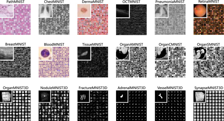
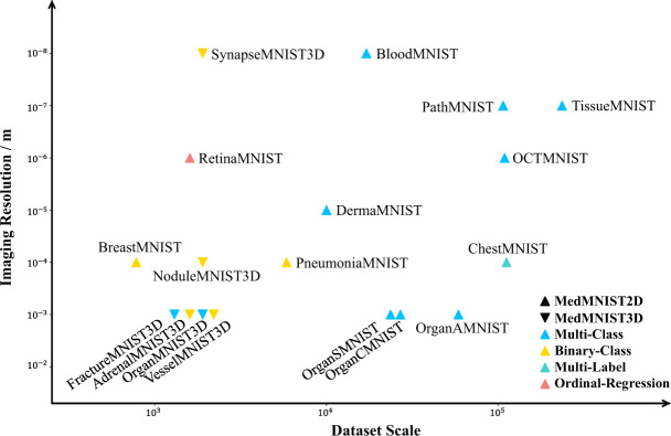
We introduce MedMNIST v2 , a large-scale MNIST-like dataset collection of standardized biomedical images, including 12 datasets for 2D and 6 datasets for 3D. All images are pre-processed into a small size of 28 × 28 (2D) or 28 × 28 × 28 (3D) with the corresponding classification labels so that no background knowledge is required for users. Covering primary data modalities in biomedical images, MedMNIST v2 is designed to perform classification on lightweight 2D and 3D images with various dataset scales (from 100 to 100,000) and diverse tasks (binary/multi-class, ordinal regression, and multi-label). The resulting dataset, consisting of 708,069 2D images and 9,998 3D images in total, could support numerous research/educational purposes in biomedical image analysis, computer vision, and machine learning. We benchmark several baseline methods on MedMNIST v2, including 2D/3D neural networks and open-source/commercial AutoML tools. The data and code are publicly available at https://medmnist.com/ .
Overview
AI snapshot
Written from the paper’s full text.
TL;DR
MedMNIST v2 is a standardized MNIST-like benchmark of 12 2D datasets (708,069 images) and 6 3D datasets (9,998 images) for biomedical image classification at 28×28 or 28×28×28. It benchmarks ResNets and AutoML tools, finding ResNets competitive and 3D/ACS convolutions generally better than 2.5D on 3D datasets.
Key findings
- MedMNIST v2 contains 12 2D datasets totaling 708,069 images and 6 3D datasets totaling 9,998 images, all preprocessed to 28×28 (2D) or 28×28×28 (3D), covering binary/multi-class, ordinal regression, and multi-label tasks.
- On MedMNIST2D, Google AutoML Vision achieved the highest average AUC (0.927), close to ResNet-18 (224) (0.925) and ResNet-50 (224) (0.923); ResNets had higher average ACC (0.821 for ResNet-18/50 at 224) than Google AutoML Vision (0.809). auto-sklearn was lowest (average AUC 0.878, ACC 0.722).
- On MedMNIST3D, ResNet-50 + 3D convolution achieved the best average AUC (0.863) and ACC (0.780); ResNet-18 + 3D was 0.849/0.767, auto-sklearn 0.815/0.765, and AutoKeras 0.763/0.737. 2.5D models were inferior to 3D and ACS models.
- Under the same ResNet backbone, 224 resolution generally outperformed 28 on 2D; at 28, ResNet-18 often outperformed ResNet-50. For 3D, 3D and ACS models were comparable, and ResNet-50 with 3D convolution surpassed ResNet-18.
Methods
- Dataset construction: 12 2D and 6 3D datasets from source datasets, preprocessed to MNIST-like sizes via cubic spline interpolation; official splits used where available, otherwise 9:1 train-validation from training or 7:1:2 patient-level random split; labels from source datasets, sometimes simplified.
- Benchmark: ResNet-18/50 with early stopping for 2D (28 or 224 input) and ResNet-18/50 with 2.5D/3D/ACS convolutions for 3D; auto-sklearn, AutoKeras, and Google AutoML Vision (2D only); training used cross-entropy, Adam, batch size 128 for 2D and 32 for 3D, 100 epochs, and learning-rate decay.
- Evaluation: AUC and ACC per dataset and average over MedMNIST2D/MedMNIST3D; each benchmark was repeated at least 3 times.
Limitations
- Stated: the dataset is NOT intended for clinical use; substantially reducing resolution may yield images insufficient to represent and capture different disease pathologies.
- Stated: labels could be simplified (merged or deleted classes) if classification on small images is too difficult.
- Evident from the text: the benchmark focuses on the machine-learning part rather than end-to-end systems; 3D datasets are much smaller (9,998 total) than 2D datasets (708,069), and AdrenalMNIST3D source CT scans are not provided due to patient privacy.
Model-written summary. Check it against the paper before relying on it.
Integrity signals
Registry
Cites
Statistics
Figures
Verification
Claims the paper makes
27 No basis given · 16 Own result · 7 Own interpretation · 6 Supported · 4 Own method · 3 Partly supported · 3 Sources not found · 3 No full text of the sources · 1 Supported so far · 1 Couldn’t confirm · accuracy not yet measured
The sources with a verdict support this claim; the rest weren’t checked yet, have no full text we could read, or their verdicts aren’t rated yet.
In the paper
“The BloodMNIST is based on a dataset26,27 of individual normal cells, captured from individuals without infection, hematologic or oncologic disease and free of any pharmacologic treatment at the moment of blood collection. It contains a total of 17,092 images and is organized into 8 classes.”
§ MedMNIST v2 - A large-scale lightweight benchmark for 2D and 3D biomedical image classification > Methods > Details for MedMNIST2D
What the sources say
- Supportedcites [26] Acevedo, A. et al. A dataset of microscopic peripheral blood cell images for development of automatic recognition systems. Data in Brief 30, 105474, https://doi.org/10.1016/j.dib.2020.105474 (2020). source
The Abstract states that the dataset contains 17,092 images of individual normal cells, organized into eight groups, and describes the donors’ health and treatment criteria. The Data section confirms the image count, eight groups, and pixel dimensions of the images. This paper documents the source dataset identified in the citing sentence. (LLM, conf=0.92)
- Not checked — no full text of the sourcecites [27] Acevedo, A. et al. A dataset for microscopic peripheral blood cell images for development of automatic recognition systems. Mendeley Data https://doi.org/10.17632/snkd93bnjr.1 (2020). source
plainn = 17,092- Asserted: passes — The authors put it forward as true.
- Checkable: passes — It can be checked against evidence.
- Load-bearing: unclear — It is unclear whether the paper’s conclusions depend on it.
- Based: passes — The paper gives a reason why it holds.
- Supported
The paper states this without a source or data behind it here, so there is nothing to check it against. The quote shows it is in the paper.
In the paper
“Furthermore, we provide standard train-validation-test splits for all datasets in MedMNIST v2, therefore algorithms could be easily compared.”
§ MedMNIST v2 - A large-scale lightweight benchmark for 2D and 3D biomedical image classification > Background & Summary
plain- Asserted: passes — The authors put it forward as true.
- Checkable: passes — It can be checked against evidence.
- Load-bearing: passes — The paper’s conclusions depend on it.
- Based: passes — The paper gives a reason why it holds.
The paper states this without a source or data behind it here, so there is nothing to check it against. The quote shows it is in the paper.
In the paper
“All images are pre-processed into a small size of 28 × 28 (2D) or 28 × 28 × 28 (3D) with the corresponding classification labels so that no background knowledge is required for users.”
- Also stated in: § Design principles · plain
“All images are pre-processed into a MNIST-like format, i.e., 28 × 28 (2D) or 28 × 28 × 28 (3D), with cubic spline interpolation operation for image resizing.”
§ MedMNIST v2 - A large-scale lightweight benchmark for 2D and 3D biomedical image classification > Abstract
plain- Asserted: passes — The authors put it forward as true.
- Checkable: passes — It can be checked against evidence.
- Load-bearing: passes — The paper’s conclusions depend on it.
- Based: passes — The paper gives a reason why it holds.
- Rests on an assumption: Resizing biomedical images to 28 × 28 (2D) or 28 × 28 × 28 (3D) preserves sufficient information for meaningful classification.
- Also stated in: § Design principles · plain
The paper states this without a source or data behind it here, so there is nothing to check it against. The quote shows it is in the paper.
In the paper
“Covering primary data modalities in biomedical images, MedMNIST v2 is designed to perform classification on lightweight 2D and 3D images with various dataset scales (from 100 to 100,000) and diverse tasks (binary/multi-class, ordinal regression, and multi-label).”
§ MedMNIST v2 - A large-scale lightweight benchmark for 2D and 3D biomedical image classification > Abstract
plain- Asserted: passes — The authors put it forward as true.
- Checkable: passes — It can be checked against evidence.
- Load-bearing: passes — The paper’s conclusions depend on it.
- Based: passes — The paper gives a reason why it holds.
The paper states this without a source or data behind it here, so there is nothing to check it against. The quote shows it is in the paper.
In the paper
“Considering the popularity of 3D imaging in biomedical area, we carefully develop 6 3D datasets following the same design principle as 2D ones.”
§ MedMNIST v2 - A large-scale lightweight benchmark for 2D and 3D biomedical image classification > Background & Summary
plain- Asserted: passes — The authors put it forward as true.
- Checkable: passes — It can be checked against evidence.
- Load-bearing: passes — The paper’s conclusions depend on it.
- Based: passes — The paper gives a reason why it holds.
The paper states this without a source or data behind it here, so there is nothing to check it against. The quote shows it is in the paper.
In the paper
“As MedMNIST v1 is more medical-oriented, we additionally provide 2 2D bioimage datasets.”
§ MedMNIST v2 - A large-scale lightweight benchmark for 2D and 3D biomedical image classification > Background & Summary
plain- Asserted: passes — The authors put it forward as true.
- Checkable: passes — It can be checked against evidence.
- Load-bearing: unclear — It is unclear whether the paper’s conclusions depend on it.
- Based: passes — The paper gives a reason why it holds.
In the paper
“As an MNIST-like8 dataset collection to perform classification tasks on small images, it primarily focuses on the machine learning part rather than the end-to-end system.”
§ MedMNIST v2 - A large-scale lightweight benchmark for 2D and 3D biomedical image classification > Background & Summary
What the sources say
- Not matchedcites [8] LeCun, Y., Cortes, C. & Burges, C. Mnist handwritten digit database. http://yann.lecun.com/exdb/mnist/ (2010).
Couldn’t be matched to a paper in Crossref, so it can’t be checked.
plain- Asserted: passes — The authors put it forward as true.
- Checkable: passes — It can be checked against evidence.
- Load-bearing: unclear — It is unclear whether the paper’s conclusions depend on it.
- Based: passes — The paper gives a reason why it holds.
- Not matched
A result the paper reports from its own data — it cites no other source for it, so there is none to check. The quote shows it is in the paper.
In the paper
“The resulting dataset, consisting of 708,069 2D images and 9,998 3D images in total, could support numerous research/educational purposes in biomedical image analysis, computer vision, and machine learning.”
§ MedMNIST v2 - A large-scale lightweight benchmark for 2D and 3D biomedical image classification > Abstract
plain- Asserted: passes — The authors put it forward as true.
- Checkable: passes — It can be checked against evidence.
- Load-bearing: passes — The paper’s conclusions depend on it.
- Based: passes — The paper gives a reason why it holds.
A result the paper reports from its own data — it cites no other source for it, so there is none to check. The quote shows it is in the paper.
In the paper
“2.5D models have poorer performance compared with 3D and ACS models”
§ MedMNIST v2 - A large-scale lightweight benchmark for 2D and 3D biomedical image classification > Technical Validation > Benchmark on each dataset
plain- Asserted: passes — The authors put it forward as true.
- Checkable: passes — It can be checked against evidence.
- Load-bearing: unclear — It is unclear whether the paper’s conclusions depend on it.
- Based: passes — The paper gives a reason why it holds.
A result the paper reports from its own data — it cites no other source for it, so there is none to check. The quote shows it is in the paper.
In the paper
“Auto-sklearn is superior to ResNet-18 + 2.5D and ResNet-50 + 2.5D in general”
§ MedMNIST v2 - A large-scale lightweight benchmark for 2D and 3D biomedical image classification > Technical Validation > Benchmark on each dataset
plaindirect- Asserted: passes — The authors put it forward as true.
- Checkable: passes — It can be checked against evidence.
- Load-bearing: unclear — It is unclear whether the paper’s conclusions depend on it.
- Based: passes — The paper gives a reason why it holds.
- It may state two separate claims as one. 31
- Probably not a substantial claim. 17
Every source the paper cites for this claim was checked and supports it.
In the paper
“MedMNIST v2 is extended from our preliminary version, MedMNIST v19, with 10 2D datasets for medical image classification.”
§ MedMNIST v2 - A large-scale lightweight benchmark for 2D and 3D biomedical image classification > Background & Summary
What the sources say
- Supportedcites [9] Yang, J., Shi, R. & Ni, B. Medmnist classification decathlon: A lightweight automl benchmark for medical image analysis. In International Symposium on Biomedical Imaging, 191–195 (2021). source
The Introduction (p. 1) presents MedMNIST as a collection of 10 datasets for classification on 28 × 28 images, and the Dataset section (p. 2) describes those images as 2D. The citation identifies the preliminary MedMNIST collection; this earlier paper need not describe the later v2 extension. (LLM, conf=0.88)
plaindirect- Asserted: passes — The authors put it forward as true.
- Checkable: passes — It can be checked against evidence.
- Load-bearing: unclear — It is unclear whether the paper’s conclusions depend on it.
- Based: passes — The paper gives a reason why it holds.
It may state two separate claims as one.
- Supported
Every source the paper cites for this claim was checked and supports it.
In the paper
“Each subset is saved in NumPy39 npz format, named as <data> mnist.npz for MedMNIST2D and <data> mnist3d.npz for MedMNIST3D, and is comprised of 6 keys (“train_images”, “train_labels”, “val_images”, “val_labels”, “test_images”, “test_labels”).”
§ MedMNIST v2 - A large-scale lightweight benchmark for 2D and 3D biomedical image classification > Data Records
What the sources say
- Supportedcites [39] Harris, C. R. et al. Array programming with numpy. Nature 585, 357–362 (2020). source
The citation is attached to “NumPy,” identifying the software rather than sourcing the MedMNIST file names or six keys. The Main section describes NumPy as an open-source library providing a multidimensional Python array object, which supports that identification. (LLM, conf=0.91)
plain- Asserted: passes — The authors put it forward as true.
- Checkable: passes — It can be checked against evidence.
- Load-bearing: fails — The paper’s conclusions may not depend on it.
- Based: passes — The paper gives a reason why it holds.
Probably not a substantial claim. The paper’s conclusions may not depend on it. It may state two separate claims as one.
- Supported
Every source the paper cites for this claim was checked and supports it.
In the paper
“The VesselMNIST3D is based on an open-access 3D intracranial aneurysm dataset, IntrA34, containing 103 3D models (meshes) of entire brain vessels collected by reconstructing MRA images. 1,694 healthy vessel segments and 215 aneurysm segments are generated automatically from the complete models.”
§ MedMNIST v2 - A large-scale lightweight benchmark for 2D and 3D biomedical image classification > Methods > Details for MedMNIST3D
What the sources say
- Supportedcites [34] Yang, X., Xia, D., Kin, T. & Igarashi, T. Intra: 3d intracranial aneurysm dataset for deep learning. In Conference on Computer Vision and Pattern Recognition (2020). source
Section 3.1 (Data) identifies IntrA as a dataset of 103 complete 3D brain-vessel models reconstructed from MRA images. It also reports 1,694 healthy vessel segments and 215 aneurysm segments generated automatically from those models. (LLM, conf=0.94)
plain- Asserted: passes — The authors put it forward as true.
- Checkable: passes — It can be checked against evidence.
- Load-bearing: unclear — It is unclear whether the paper’s conclusions depend on it.
- Based: passes — The paper gives a reason why it holds.
It may state two separate claims as one.
- Supported
The cited sources support only part of this claim — read each source’s verdict below.
In the paper
“The SynapseMNIST3D is a new 3D volume dataset to classify whether a synapse is excitatory or inhibitory. It uses a 3D image volume of an adult rat acquired by a multi-beam scanning electron microscope.”
§ MedMNIST v2 - A large-scale lightweight benchmark for 2D and 3D biomedical image classification > Methods > Details for MedMNIST3D
What the sources say
- Partly supportedcites [37] Wei, D. et al. Mitoem dataset: Large-scale 3d mitochondria instance segmentation from em images. In Conference on Medical Image Computing and Computer Assisted Intervention, 66–76 (Springer, 2020). sourceExperimental — accuracy not yet measured
The Dataset Acquisition section (page unavailable) says the adult-rat tissue used for MitoEM-R was imaged with a multi-beam scanning electron microscope. But the Introduction identifies MitoEM as a 3D mitochondria instance-segmentation dataset; our copy does not identify SynapseMNIST3D or describe excitatory-versus-inhibitory synapse classification. (LLM, conf=0.88)
plainSingle case- Asserted: passes — The authors put it forward as true.
- Checkable: passes — It can be checked against evidence.
- Load-bearing: passes — The paper’s conclusions depend on it.
- Based: passes — The paper gives a reason why it holds.
It may state two separate claims as one.
- Partly supported
The cited sources support only part of this claim — read each source’s verdict below.
In the paper
“We use the BBBC05128, available from the Broad Bioimage Benchmark Collection29. The dataset contains 236,386 human kidney cortex cells, segmented from 3 reference tissue specimens and organized into 8 categories.”
§ MedMNIST v2 - A large-scale lightweight benchmark for 2D and 3D biomedical image classification > Methods > Details for MedMNIST2D
What the sources say
- Partly supportedcites [28] Woloshuk, A. et al. In situ classification of cell types in human kidney tissue using 3d nuclear staining. Cytometry Part A (2020). sourceExperimental — accuracy not yet measured
The Data organization section describes approximately 230,000 nuclei from three reference tissue specimens, and the Results describe eight classification categories and derived 2D datasets. The copy does not give the exact count of 236,386 or identify the dataset as TissueMNIST or BBBC051; it says the dataset would be made available through an online repository such as the Broad Bioimage Benchmark Collection. (LLM, conf=0.86)
- Supportedcites [29] Ljosa, V., Sokolnicki, K. L. & Carpenter, A. E. Annotated high-throughput microscopy image sets for validation. Nature methods 9, 637–637 (2012). source
Citation 29 is attached to the Broad Bioimage Benchmark Collection, not to the BBBC051 dataset details. The paper describes the BBBC as a publicly available collection of microscopy image sets and gives its access URL. The dataset-specific figures in the citing sentence are not attributed to this citation. (LLM, conf=0.93)
plainn = 3- Asserted: passes — The authors put it forward as true.
- Checkable: passes — It can be checked against evidence.
- Load-bearing: unclear — It is unclear whether the paper’s conclusions depend on it.
- Based: passes — The paper gives a reason why it holds.
It may state two separate claims as one.
- Partly supported
The cited sources support only part of this claim — read each source’s verdict below.
In the paper
“The DermaMNIST is based on the HAM1000019–21, a large collection of multi-source dermatoscopic images of common pigmented skin lesions. The dataset consists of 10,015 dermatoscopic images categorized as 7 different diseases, formulized as a multi-class classification task.”
§ MedMNIST v2 - A large-scale lightweight benchmark for 2D and 3D biomedical image classification > Methods > Details for MedMNIST2D
What the sources say
- Partly supportedcites [19] Tschandl, P., Rosendahl, C. & Kittler, H. The ham10000 dataset, a large collection of multi-source dermatoscopic images of common pigmented skin lesions. Scientific data 5, 180161 (2018). sourceExperimental — accuracy not yet measured
The Abstract reports 10,015 dermatoscopic images, and Methods (“Unifying pathologic diagnoses”) says HAM10000 has seven diagnostic classes intended as a benchmark for classification. The paper does not identify DermaMNIST or establish that DermaMNIST is based on HAM10000, so it supports the underlying dataset description but not that relationship. (LLM, conf=0.84)
- Not checked — no full text of the sourcecites [20] Tschandl, P. The HAM10000 dataset, a large collection of multi-source dermatoscopic images of common pigmented skin lesions. Harvard Dataverse https://doi.org/10.7910/DVN/DBW86T (2018). source
- Partly supportedcites [21] Codella, N. et al. Skin lesion analysis toward melanoma detection 2018: A challenge hosted by the international skin imaging collaboration (isic). Preprint at https://arxiv.org/abs/1902.03368v2 (2019). sourceExperimental — accuracy not yet measured
Section 2.3 (p. 4) reports 10,015 dermoscopic training images with seven classification labels and cites the HAM10000 paper, identified in the References (p. 8). It does not identify DermaMNIST, establish that DermaMNIST is based on HAM10000, or describe it as a 2D dataset; the 10,015 figure is stated for the challenge’s training set. (LLM, conf=0.88)
plain- Asserted: passes — The authors put it forward as true.
- Checkable: passes — It can be checked against evidence.
- Load-bearing: unclear — It is unclear whether the paper’s conclusions depend on it.
- Based: passes — The paper gives a reason why it holds.
It may state two separate claims as one.
- Partly supported
Every source the paper cites for this claim was checked and supports it.
In the paper
“The data files of MedMNIST v2 dataset can be accessed at Zenodo38. It contains 12 pre-processed 2D datasets (MedMNIST2D) and 6 pre-processed 3D datasets (MedMNIST3D).”
§ MedMNIST v2 - A large-scale lightweight benchmark for 2D and 3D biomedical image classification > Data Records
What the sources say
- Supportedcites [38] Yang, J. et al. Medmnist v2: A large-scale lightweight benchmark for 2d and 3d biomedical image classification. Zenodo https://doi.org/10.5281/zenodo.5208230 (2021). source
The Abstract states that MedMNIST v2 includes 12 2D and 6 3D datasets, with all images pre-processed. The Zenodo record’s Files section lists the downloadable dataset files, supporting the access claim. (LLM, conf=0.95)
plain- Asserted: passes — The authors put it forward as true.
- Checkable: passes — It can be checked against evidence.
- Load-bearing: unclear — It is unclear whether the paper’s conclusions depend on it.
- Based: passes — The paper gives a reason why it holds.
It may state two separate claims as one.
- Supported
Every source the paper cites for this claim was checked and supports it.
In the paper
“The BreastMNIST is based on a dataset25 of 780 breast ultrasound images. It is categorized into 3 classes: normal, benign, and malignant. As we use low-resolution images, we simplify the task into binary classification by combining normal and benign as positive and classifying them against malignant as negative.”
§ MedMNIST v2 - A large-scale lightweight benchmark for 2D and 3D biomedical image classification > Methods > Details for MedMNIST2D
What the sources say
- Supportedcites [25] Al-Dhabyani, W., Gomaa, M., Khaled, H. & Fahmy, A. Dataset of breast ultrasound images. Data in Brief 28, 104863, https://doi.org/10.1016/j.dib.2019.104863 (2020). source
The Data section reports 780 breast ultrasound images categorized as normal, benign, and malignant, supporting the dataset description for which this source is cited. The binary grouping is described as the citing authors’ own simplification, not as a feature attributed to this source. (LLM, conf=0.91)
plain- Asserted: passes — The authors put it forward as true.
- Checkable: passes — It can be checked against evidence.
- Load-bearing: fails — The paper’s conclusions may not depend on it.
- Based: passes — The paper gives a reason why it holds.
Probably not a substantial claim. The paper’s conclusions may not depend on it. It may state two separate claims as one.
- Supported
The cited sources that could be checked couldn’t confirm or dispute this claim.
In the paper
“The source of the OrganMNIST3D is the same as that of the Organ{A,C,S}MNIST. Instead of 2D images, we directly use the 3D bounding boxes and process the images into 28 × 28 × 28 to perform multi-class classification of 11 body organs.”
§ MedMNIST v2 - A large-scale lightweight benchmark for 2D and 3D biomedical image classification > Methods > Details for MedMNIST3D
What the sources say
- Could not verifycites [30] Bilic, P. et al. The liver tumor segmentation benchmark (lits). Medical Image Analysis 84,102680 (2023). sourceExperimental — accuracy not yet measured
The Methods > Dataset passage describes LiTS as a collection of abdominal CT scans, but does not identify it as the source of OrganMNIST3D or Organ{A,C,S}MNIST. Searches for those dataset names and for 3D bounding boxes, 28 × 28 × 28 images, and classification of 11 organs found no supporting passage in our copy. (LLM, conf=0.75)
- Not checked — no full text of the sourcecites [31] Xu, X. et al. Efficient multiple organ localization in ct image using 3d region proposal network. IEEE Transactions on Medical Imaging 38, 1885–1898 (2019). source
plain- Asserted: passes — The authors put it forward as true.
- Checkable: passes — It can be checked against evidence.
- Load-bearing: unclear — It is unclear whether the paper’s conclusions depend on it.
- Based: passes — The paper gives a reason why it holds.
It may state two separate claims as one.
- Could not verify
Every source the paper cites for this claim was checked and supports it.
In the paper
“The NoduleMNIST3D is based on the LIDC-IDRI32, a large public lung nodule dataset, containing images from thoracic CT scans. The dataset is designed for both lung nodule segmentation and 5-level malignancy classification task. To perform binary classification, we categorize cases with malignancy level 1/2 into negative class and 4/5 into positive class, ignoring the cases with malignancy level 3.”
§ MedMNIST v2 - A large-scale lightweight benchmark for 2D and 3D biomedical image classification > Methods > Details for MedMNIST3D
What the sources say
- Supportedcites [32] Armato, S. G. III et al. The lung image database consortium (lidc) and image database resource initiative (idri): A completed reference database of lung nodules on ct scans. Medical Physics 38, 915–931, https://doi.org/10.1118/1.3528204 (2011). source
The citation is attached to the identification of LIDC-IDRI as a large, public dataset of lung nodules from thoracic CT scans. The paper reports 1018 thoracic CT scans (II.A), describes its nodule annotations (Results), and says the database is publicly available (II.E). The later binary-classification rule is presented as the citing authors’ own procedure, not as a finding attributed to this source. (LLM, conf=0.91)
plain- Asserted: passes — The authors put it forward as true.
- Checkable: passes — It can be checked against evidence.
- Load-bearing: unclear — It is unclear whether the paper’s conclusions depend on it.
- Based: passes — The paper gives a reason why it holds.
It may state two separate claims as one.
- Supported
The paper states this without a source or data behind it here, so there is nothing to check it against. The quote shows it is in the paper.
In the paper
“The MedMNIST v2 dataset consists of 12 2D and 6 3D standardized datasets from carefully selected sources covering primary data modalities (e.g., X-ray, OCT, ultrasound, CT, electron microscope), diverse classification tasks (binary/multi-class, ordinal regression, and multi-label) and dataset scales (from 100 to 100,000).”
§ MedMNIST v2 - A large-scale lightweight benchmark for 2D and 3D biomedical image classification > Methods > Design principles
plain- Asserted: passes — The authors put it forward as true.
- Checkable: passes — It can be checked against evidence.
- Load-bearing: passes — The paper’s conclusions depend on it.
- Based: passes — The paper gives a reason why it holds.
It may state two separate claims as one.
The paper states this without a source or data behind it here, so there is nothing to check it against. The quote shows it is in the paper.
In the paper
“The MedMNIST uses the classification labels from the source datasets directly in most cases, but the labels could be simplified (merged or deleted classes) if the classification tasks on the small images are too difficult.”
§ MedMNIST v2 - A large-scale lightweight benchmark for 2D and 3D biomedical image classification > Methods > Design principles
hedgedconditional- Asserted: passes — The authors put it forward as true.
- Checkable: passes — It can be checked against evidence.
- Load-bearing: fails — The paper’s conclusions may not depend on it.
- Based: passes — The paper gives a reason why it holds.
Probably not a substantial claim. The paper’s conclusions may not depend on it. It may state two separate claims as one.
- Rests on an assumption: The classification labels from the source datasets are accurate and suitable for the benchmark tasks.
A result the paper reports from its own data — it cites no other source for it, so there is none to check. The quote shows it is in the paper.
In the paper
“We introduce MedMNIST v2, a large-scale MNIST-like dataset collection of standardized biomedical images, including 12 datasets for 2D and 6 datasets for 3D.”
- Also stated in: § Background & Summary · plain
“MedMNIST v2 is a large-scale benchmark for 2D and 3D biomedical image classification, covering 12 2D datasets with 708,069 images and 6 3D datasets with 9,998 images.”
§ MedMNIST v2 - A large-scale lightweight benchmark for 2D and 3D biomedical image classification > Abstract
plain- Asserted: passes — The authors put it forward as true.
- Checkable: passes — It can be checked against evidence.
- Load-bearing: passes — The paper’s conclusions depend on it.
- Based: passes — The paper gives a reason why it holds.
It may state two separate claims as one.
- Also stated in: § Background & Summary · plain
The paper states this without a source or data behind it here, so there is nothing to check it against. The quote shows it is in the paper.
In the paper
“All source datasets are either associated with the Creative Commons (CC) Licenses or developed by us, which allows us to develop derivative datasets based on them.”
§ MedMNIST v2 - A large-scale lightweight benchmark for 2D and 3D biomedical image classification > Methods > Design principles
plaindirect- Asserted: passes — The authors put it forward as true.
- Checkable: passes — It can be checked against evidence.
- Load-bearing: passes — The paper’s conclusions depend on it.
- Based: passes — The paper gives a reason why it holds.
It may state two separate claims as one.
The paper states this without a source or data behind it here, so there is nothing to check it against. The quote shows it is in the paper.
In the paper
“Covering primary data modalities in biomedical images, MedMNIST v2 is designed to perform classification on lightweight 2D and 3D images with various dataset scales (from 100 to 100,000) and diverse tasks (binary/multi-class, ordinal regression, and multi-label).”
§ MedMNIST v2 - A large-scale lightweight benchmark for 2D and 3D biomedical image classification > Abstract
plain- Asserted: passes — The authors put it forward as true.
- Checkable: passes — It can be checked against evidence.
- Load-bearing: passes — The paper’s conclusions depend on it.
- Based: passes — The paper gives a reason why it holds.
It may state two separate claims as one.
The paper states this without a source or data behind it here, so there is nothing to check it against. The quote shows it is in the paper.
In the paper
“It is designed to be:Diverse: It covers diverse data modalities, dataset scales (from 100 to 100,000), and tasks (binary/multi-class, multi-label, and ordinal regression).”
§ MedMNIST v2 - A large-scale lightweight benchmark for 2D and 3D biomedical image classification > Background & Summary
plain- Asserted: passes — The authors put it forward as true.
- Checkable: passes — It can be checked against evidence.
- Load-bearing: passes — The paper’s conclusions depend on it.
- Based: passes — The paper gives a reason why it holds.
It may state two separate claims as one.
The paper states this without a source or data behind it here, so there is nothing to check it against. The quote shows it is in the paper.
In the paper
“Standardized: Each sub-dataset is pre-processed into the same format (see details in Methods), which requires no background knowledge for users.”
§ MedMNIST v2 - A large-scale lightweight benchmark for 2D and 3D biomedical image classification > Background & Summary
plaindirect- Asserted: passes — The authors put it forward as true.
- Checkable: passes — It can be checked against evidence.
- Load-bearing: passes — The paper’s conclusions depend on it.
- Based: passes — The paper gives a reason why it holds.
It may state two separate claims as one.
The paper states this without a source or data behind it here, so there is nothing to check it against. The quote shows it is in the paper.
In the paper
“Some datasets are under CC-BY-NC license; we have contacted the authors and obtained the permission to re-distribute the datasets.”
§ MedMNIST v2 - A large-scale lightweight benchmark for 2D and 3D biomedical image classification > Methods > Design principles
plain- Asserted: passes — The authors put it forward as true.
- Checkable: passes — It can be checked against evidence.
- Load-bearing: unclear — It is unclear whether the paper’s conclusions depend on it.
- Based: passes — The paper gives a reason why it holds.
It may state two separate claims as one.
A result the paper reports from its own data — it cites no other source for it, so there is none to check. The quote shows it is in the paper.
In the paper
““{train,val,test}_labels”: an array containing ground-truth labels, with a shape of N × 1 for multi-class/binary-class/ordinal regression datasets, of N × L for multi-lable binary-class datasets. N denotes the number of samples in training/validation/test set and L denotes the number of task labels in the multi-label dataset (i.e., 14 for the ChestMNIST).”
§ MedMNIST v2 - A large-scale lightweight benchmark for 2D and 3D biomedical image classification > Data Records
plain- Asserted: passes — The authors put it forward as true.
- Checkable: passes — It can be checked against evidence.
- Load-bearing: fails — The paper’s conclusions may not depend on it.
- Based: passes — The paper gives a reason why it holds.
Probably not a substantial claim. The paper’s conclusions may not depend on it. It may state two separate claims as one.
A result the paper reports from its own data — it cites no other source for it, so there is none to check. The quote shows it is in the paper.
In the paper
““{train,val,test}_images”: an array containing images, with a shape of N × 28 × 28 for 2D gray-scale datasets, of N × 28 × 28 × 3 for 2D RGB datasets, of N × 28 × 28 × 28 for 3D datasets.”
§ MedMNIST v2 - A large-scale lightweight benchmark for 2D and 3D biomedical image classification > Data Records
plain- Asserted: passes — The authors put it forward as true.
- Checkable: passes — It can be checked against evidence.
- Load-bearing: fails — The paper’s conclusions may not depend on it.
- Based: passes — The paper gives a reason why it holds.
Probably not a substantial claim. The paper’s conclusions may not depend on it. It may state two separate claims as one.
In the paper
“The RetinaMNIST is based on the DeepDRiD24 challenge, which provides a dataset of 1,600 retina fundus images. The task is ordinal regression for 5-level grading of diabetic retinopathy severity.”
§ MedMNIST v2 - A large-scale lightweight benchmark for 2D and 3D biomedical image classification > Methods > Details for MedMNIST2D
What the sources say
- Not matchedcites [24] DeepDRiD. The 2nd diabetic retinopathy–grading and image quality estimation challenge. https://isbi.deepdr.org/data.html (2020).
Couldn’t be matched to a paper in Crossref, so it can’t be checked.
plain- Asserted: passes — The authors put it forward as true.
- Checkable: passes — It can be checked against evidence.
- Load-bearing: unclear — It is unclear whether the paper’s conclusions depend on it.
- Based: passes — The paper gives a reason why it holds.
It may state two separate claims as one.
- Not matched
The paper states this without a source or data behind it here, so there is nothing to check it against. The quote shows it is in the paper.
In the paper
“The data type of the dataset is uint8.”
§ MedMNIST v2 - A large-scale lightweight benchmark for 2D and 3D biomedical image classification > Data Records
plain- Asserted: passes — The authors put it forward as true.
- Checkable: passes — It can be checked against evidence.
- Load-bearing: fails — The paper’s conclusions may not depend on it.
- Based: passes — The paper gives a reason why it holds.
Probably not a substantial claim. The paper’s conclusions may not depend on it.
A result the paper reports from its own data — it cites no other source for it, so there is none to check. The quote shows it is in the paper.
In the paper
“For 2D datasets, Google AutoML Vision is well-performing in general, however it could not always win, even compared with the baseline ResNet-18 and ResNet-50.”
§ MedMNIST v2 - A large-scale lightweight benchmark for 2D and 3D biomedical image classification > Technical Validation > Benchmark on each dataset
hedged- Asserted: passes — The authors put it forward as true.
- Checkable: passes — It can be checked against evidence.
- Load-bearing: unclear — It is unclear whether the paper’s conclusions depend on it.
- Based: passes — The paper gives a reason why it holds.
It may state two separate claims as one.
A claim is checked only against the cited paper’s full text, never its abstract, and none is available for its sources.
In the paper
“The OCTMNIST is based on a prior dataset22,23 of 109,309 valid optical coherence tomography (OCT) images for retinal diseases. The dataset is comprised of 4 diagnosis categories, leading to a multi-class classification task.”
§ MedMNIST v2 - A large-scale lightweight benchmark for 2D and 3D biomedical image classification > Methods > Details for MedMNIST2D
What the sources say
- Not checked — no full text of the sourcecites [22] Kermany, D. S. et al. Identifying medical diagnoses and treatable diseases by image-based deep learning. Cell 172, 1122–1131.e9, https://doi.org/10.1016/j.cell.2018.02.010 (2018). source
- Not checked — no full text of the sourcecites [23] Kermany, D. S., Zhang, K. & Goldbaum, M. Large dataset of labeled optical coherence tomography (oct) and chest x-ray images https://doi.org/10.17632/rscbjbr9sj.3 (2018). source
plaindirect- Asserted: passes — The authors put it forward as true.
- Checkable: passes — It can be checked against evidence.
- Load-bearing: unclear — It is unclear whether the paper’s conclusions depend on it.
- Based: passes — The paper gives a reason why it holds.
It may state two separate claims as one.
A result the paper reports from its own data — it cites no other source for it, so there is none to check. The quote shows it is in the paper.
In the paper
“For MedMNIST2D, Google AutoML Vision outperforms all the other methods in average AUC, however, it is very close to the performance of baseline ResNets.”
§ MedMNIST v2 - A large-scale lightweight benchmark for 2D and 3D biomedical image classification > Technical Validation > Benchmark on each dataset
plaincorrelational- Asserted: passes — The authors put it forward as true.
- Checkable: passes — It can be checked against evidence.
- Load-bearing: unclear — It is unclear whether the paper’s conclusions depend on it.
- Based: passes — The paper gives a reason why it holds.
It may state two separate claims as one.
The paper states this without a source or data behind it here, so there is nothing to check it against. The quote shows it is in the paper.
In the paper
“AutoKeras performs well on datasets with large scales, however relatively worse on datasets with small scale.”
§ MedMNIST v2 - A large-scale lightweight benchmark for 2D and 3D biomedical image classification > Technical Validation > Benchmark on each dataset
plaincorrelational- Asserted: passes — The authors put it forward as true.
- Checkable: passes — It can be checked against evidence.
- Load-bearing: unclear — It is unclear whether the paper’s conclusions depend on it.
- Based: passes — The paper gives a reason why it holds.
It may state two separate claims as one.
A claim is checked only against the cited paper’s full text, never its abstract, and none is available for its sources.
In the paper
“The PneumoniaMNIST is based on a prior dataset22,23 of 5,856 pediatric chest X-Ray images. The task is binary-class classification of pneumonia against normal.”
§ MedMNIST v2 - A large-scale lightweight benchmark for 2D and 3D biomedical image classification > Methods > Details for MedMNIST2D
What the sources say
- Not checked — no full text of the sourcecites [22] Kermany, D. S. et al. Identifying medical diagnoses and treatable diseases by image-based deep learning. Cell 172, 1122–1131.e9, https://doi.org/10.1016/j.cell.2018.02.010 (2018). source
- Not checked — no full text of the sourcecites [23] Kermany, D. S., Zhang, K. & Goldbaum, M. Large dataset of labeled optical coherence tomography (oct) and chest x-ray images https://doi.org/10.17632/rscbjbr9sj.3 (2018). source
plain- Asserted: passes — The authors put it forward as true.
- Checkable: passes — It can be checked against evidence.
- Load-bearing: unclear — It is unclear whether the paper’s conclusions depend on it.
- Based: passes — The paper gives a reason why it holds.
It may state two separate claims as one.
In the paper
“The ChestMNIST is based on the NIH-ChestXray14 dataset18, a dataset comprising 112,120 frontal-view X-Ray images of 30,805 unique patients with the text-mined 14 disease labels, which could be formulized as a multi-label binary-class classification task.”
§ MedMNIST v2 - A large-scale lightweight benchmark for 2D and 3D biomedical image classification > Methods > Details for MedMNIST2D
What the sources say
- Not checked — no full text of the sourcecites [18] Wang, X. et al. Chestx-ray8: Hospital-scale chest x-ray database and benchmarks on weakly-supervised classification and localization of common thorax diseases. In Conference on Computer Vision and Pattern Recognition, 3462–3471 (2017). source
hedgedn = 30,805- Asserted: passes — The authors put it forward as true.
- Checkable: passes — It can be checked against evidence.
- Load-bearing: unclear — It is unclear whether the paper’s conclusions depend on it.
- Based: passes — The paper gives a reason why it holds.
It may state two separate claims as one.
A result the paper reports from its own data — it cites no other source for it, so there is none to check. The quote shows it is in the paper.
In the paper
“Under the same ResNet backbone, 2.5D models are inferior to 3D and ACS models and perform worse than auto-sklearn and AutoKeras.”
§ MedMNIST v2 - A large-scale lightweight benchmark for 2D and 3D biomedical image classification > Technical Validation > Benchmark on each dataset
plain- Asserted: passes — The authors put it forward as true.
- Checkable: passes — It can be checked against evidence.
- Load-bearing: unclear — It is unclear whether the paper’s conclusions depend on it.
- Based: passes — The paper gives a reason why it holds.
It may state two separate claims as one.
The paper states this without a source or data behind it here, so there is nothing to check it against. The quote shows it is in the paper.
In the paper
“They are sliced before being resized into 1 × 28 × 28.”
§ MedMNIST v2 - A large-scale lightweight benchmark for 2D and 3D biomedical image classification > Technical Validation > Benchmark on each dataset
plain- Asserted: passes — The authors put it forward as true.
- Checkable: passes — It can be checked against evidence.
- Load-bearing: passes — The paper’s conclusions depend on it.
- Based: passes — The paper gives a reason why it holds.
Probably not a substantial claim.
The paper states this without a source or data behind it here, so there is nothing to check it against. The quote shows it is in the paper.
In the paper
“Since there is no severe class imbalance on our datasets”
§ MedMNIST v2 - A large-scale lightweight benchmark for 2D and 3D biomedical image classification > Technical Validation > Evaluation
plain- Asserted: passes — The authors put it forward as true.
- Checkable: passes — It can be checked against evidence.
- Load-bearing: passes — The paper’s conclusions depend on it.
- Based: fails — The paper may give no reason why it holds.
Probably not a substantial claim. The paper may give no reason why it holds.
The paper states this without a source or data behind it here, so there is nothing to check it against. The quote shows it is in the paper.
In the paper
“Organ{A,C,S}MNIST and OrganMNIST3D are generated from the same source dataset, and share the same task and the same data split.”
§ MedMNIST v2 - A large-scale lightweight benchmark for 2D and 3D biomedical image classification > Technical Validation > Benchmark on each dataset
plaincorrelational- Asserted: passes — The authors put it forward as true.
- Checkable: passes — It can be checked against evidence.
- Load-bearing: unclear — It is unclear whether the paper’s conclusions depend on it.
- Based: passes — The paper gives a reason why it holds.
It may state two separate claims as one.
The paper states this without a source or data behind it here, so there is nothing to check it against. The quote shows it is in the paper.
In the paper
“On the other hand, OrganMNIST3D is resized into 28 × 28 × 28 directly.”
§ MedMNIST v2 - A large-scale lightweight benchmark for 2D and 3D biomedical image classification > Technical Validation > Benchmark on each dataset
plain- Asserted: passes — The authors put it forward as true.
- Checkable: passes — It can be checked against evidence.
- Load-bearing: passes — The paper’s conclusions depend on it.
- Based: passes — The paper gives a reason why it holds.
Probably not a substantial claim.
The paper states this without a source or data behind it here, so there is nothing to check it against. The quote shows it is in the paper.
In the paper
“Organ{A,C,S}MNIST are sampled slices of 3D bounding boxes of 3D CT images in axial/coronal/sagittal views (planes), respectively.”
§ MedMNIST v2 - A large-scale lightweight benchmark for 2D and 3D biomedical image classification > Technical Validation > Benchmark on each dataset
plain- Asserted: passes — The authors put it forward as true.
- Checkable: passes — It can be checked against evidence.
- Load-bearing: passes — The paper’s conclusions depend on it.
- Based: passes — The paper gives a reason why it holds.
Probably not a substantial claim.
A result the paper reports from its own data — it cites no other source for it, so there is none to check. The quote shows it is in the paper.
In the paper
“The ResNets surpass auto-sklearn and AutoKeras, and outperform Google AutoML Vision in average ACC.”
§ MedMNIST v2 - A large-scale lightweight benchmark for 2D and 3D biomedical image classification > Technical Validation > Benchmark on each dataset
plaindirect- Asserted: passes — The authors put it forward as true.
- Checkable: passes — It can be checked against evidence.
- Load-bearing: passes — The paper’s conclusions depend on it.
- Based: passes — The paper gives a reason why it holds.
It may state two separate claims as one.
What the paper did — it cites no source for this, so there is none to check. The quote shows it is in the paper.
In the paper
“All benchmark experiments are repeated at least 3 times for more stable results than in MedMNIST v1.”
§ MedMNIST v2 - A large-scale lightweight benchmark for 2D and 3D biomedical image classification > Background & Summary
plaindirect- Asserted: passes — The authors put it forward as true.
- Checkable: passes — It can be checked against evidence.
- Load-bearing: fails — The paper’s conclusions may not depend on it.
- Based: passes — The paper gives a reason why it holds.
Probably not a substantial claim. The paper’s conclusions may not depend on it.
None of the sources cited for this claim could be matched to a paper in Crossref, so there is nothing to check it against.
In the paper
“It is as diverse as the VDD4 and MSD5 to fairly evaluate the generalizable performance of machine learning algorithms in different settings, but both 2D and 3D biomedical images are provided.”
§ MedMNIST v2 - A large-scale lightweight benchmark for 2D and 3D biomedical image classification > Background & Summary
What the sources say
- Not matchedcites VDD4
Couldn’t be matched to a paper in Crossref, so it can’t be checked.
- Not matchedcites MSD5
Couldn’t be matched to a paper in Crossref, so it can’t be checked.
plain- Asserted: passes — The authors put it forward as true.
- Checkable: passes — It can be checked against evidence.
- Load-bearing: passes — The paper’s conclusions depend on it.
- Based: passes — The paper gives a reason why it holds.
It may state two separate claims as one.
- Not matched
A conclusion the paper draws from its own results — it cites no other source for it, so there is none to check. The quote shows it is in the paper.
In the paper
“The resulting dataset, consisting of 708,069 2D images and 9,998 3D images in total, could support numerous research/educational purposes in biomedical image analysis, computer vision, and machine learning.”
§ MedMNIST v2 - A large-scale lightweight benchmark for 2D and 3D biomedical image classification > Abstract
hedged- Asserted: passes — The authors put it forward as true.
- Checkable: fails — It may be too vague to check against evidence.
- Load-bearing: passes — The paper’s conclusions depend on it.
- Based: passes — The paper gives a reason why it holds.
Probably not a substantial claim. It may be too vague to check against evidence.
- Rests on an assumption: Resizing biomedical images to 28 × 28 (2D) or 28 × 28 × 28 (3D) preserves sufficient information for meaningful classification.
What the paper did — it cites no source for this, so there is none to check. The quote shows it is in the paper.
In the paper
“For the dataset without an official split, we split the dataset randomly at the patient level with a ratio of 7:1:2 into training-validation-test.”
§ MedMNIST v2 - A large-scale lightweight benchmark for 2D and 3D biomedical image classification > Methods > Design principles
plain- Asserted: passes — The authors put it forward as true.
- Checkable: passes — It can be checked against evidence.
- Load-bearing: passes — The paper’s conclusions depend on it.
- Based: passes — The paper gives a reason why it holds.
Probably not a substantial claim.
- Rests on an assumption: Patient-level random splitting avoids data leakage for datasets without official splits.
A conclusion the paper draws from its own results — it cites no other source for it, so there is none to check. The quote shows it is in the paper.
In the paper
“The diverse dataset design could lead to diverse task difficulty, which is desirable as a biomedical image classification benchmark.”
§ MedMNIST v2 - A large-scale lightweight benchmark for 2D and 3D biomedical image classification > Methods > Design principles
hedgedconditional- Asserted: passes — The authors put it forward as true.
- Checkable: fails — It may be too vague to check against evidence.
- Load-bearing: passes — The paper’s conclusions depend on it.
- Based: passes — The paper gives a reason why it holds.
Probably not a substantial claim. It may be too vague to check against evidence.
What the paper did — it cites no source for this, so there is none to check. The quote shows it is in the paper.
In the paper
“We use the official data split from source dataset (if provided) to avoid data leakage.”
§ MedMNIST v2 - A large-scale lightweight benchmark for 2D and 3D biomedical image classification > Methods > Design principles
plaindirect- Asserted: passes — The authors put it forward as true.
- Checkable: passes — It can be checked against evidence.
- Load-bearing: passes — The paper’s conclusions depend on it.
- Based: passes — The paper gives a reason why it holds.
Probably not a substantial claim.
- Rests on an assumption: The official data splits from source datasets avoid data leakage.
A conclusion the paper draws from its own results — it cites no other source for it, so there is none to check. The quote shows it is in the paper.
In the paper
“ACC could also serve as a good metric”
§ MedMNIST v2 - A large-scale lightweight benchmark for 2D and 3D biomedical image classification > Technical Validation > Evaluation
hedged- Asserted: passes — The authors put it forward as true.
- Checkable: fails — It may be too vague to check against evidence.
- Load-bearing: fails — The paper’s conclusions may not depend on it.
- Based: passes — The paper gives a reason why it holds.
Probably not a substantial claim. It may be too vague to check against evidence. The paper’s conclusions may not depend on it.
What the paper did — it cites no source for this, so there is none to check. The quote shows it is in the paper.
In the paper
“We calculate the mean value of at least 3 trials for each method on each dataset.”
§ MedMNIST v2 - A large-scale lightweight benchmark for 2D and 3D biomedical image classification > Technical Validation > Benchmark on each dataset
plain- Asserted: passes — The authors put it forward as true.
- Checkable: passes — It can be checked against evidence.
- Load-bearing: passes — The paper’s conclusions depend on it.
- Based: passes — The paper gives a reason why it holds.
Probably not a substantial claim.
The paper relies on this without support.
The classification labels from the source datasets are accurate and suitable for the benchmark tasks.
“The MedMNIST uses the classification labels from the source datasets directly in most cases, but the labels could be simplified (merged or deleted classes) if the classification tasks on the small images are too difficult.”
The paper relies on this without support.
Resizing biomedical images to 28 × 28 (2D) or 28 × 28 × 28 (3D) preserves sufficient information for meaningful classification.
“All images are pre-processed into a MNIST-like format, i.e., 28 × 28 (2D) or 28 × 28 × 28 (3D), with cubic spline interpolation operation for image resizing.”
The paper relies on this without support.
The official data splits from source datasets avoid data leakage.
“We use the official data split from source dataset (if provided) to avoid data leakage.”
The paper relies on this without support.
Patient-level random splitting avoids data leakage for datasets without official splits.
“For the dataset without an official split, we split the dataset randomly at the patient level with a ratio of 7:1:2 into training-validation-test.”
The paper relies on this without support.
The selected 18 datasets are representative of primary data modalities and tasks in biomedical image analysis.
“The MedMNIST v2 dataset consists of 12 2D and 6 3D standardized datasets from carefully selected sources covering primary data modalities (e.g., X-ray, OCT, ultrasound, CT, electron microscope), diverse classification tasks (binary/multi-class, ordinal regression, and multi-label) and dataset scales (from 100 to 100,000).”
Citations
Cites
Deep Residual Learning for Image Recognition
He, K., Zhang, X., Ren, S., & Sun, J. (2016) · 2016 IEEE Conference on Computer Vision and Pattern Recognition (CVPR) · doi:10.1109/cvpr.2016.90 · Cited by 235,474
Adam: A Method for Stochastic Optimization
Kingma, D. P., & Ba, J. (2014) · arXiv · doi:10.48550/arxiv.1412.6980 · Cited by 98,946
Harris, C. R., Millman, K. J., van der Walt, S. J., Gommers, R., Virtanen, P., Cournapeau, D., Wieser, E., Taylor, J., Berg, S., Smith, N. J., et al. (2020) · Nature · doi:10.1038/s41586-020-2649-2 · Cited by 23,826
Advances in neural information processing systems 7
Cios, K. J., & Shields, M. E. (1997) · Neurocomputing · doi:10.1016/s0925-2312(97)90018-7 · Cited by 22,093
Harris, C., Millman, K., van der Walt, S., Gommers, R., Virtanen, P., Cournapeau, D., Wieser, E., Taylor, J., Berg, S., Smith, N., et al. (2020) · RePEc: Research Papers in Economics · Cited by 17,844
A survey on deep learning in medical image analysis
Litjens, G., Kooi, T., Bejnordi, B. E., Setio, A. A. A., Ciompi, F., Ghafoorian, M., van der Laak, J. A. W. M., van Ginneken, B., & Sánchez, C. I. (2017) · Medical Image Analysis · doi:10.1016/j.media.2017.07.005 · Cited by 15,332 · cited 7×
“AdrenalMNIST3D New Shape from Abdominal CT BC (2) 1,584 1,188/98/298”
BackgroundInfluentialnnU-Net: a self-configuring method for deep learning-based biomedical image segmentation
Isensee, F., Jaeger, P. F., Kohl, S. A. A., Petersen, J., & Maier-Hein, K. H. (2021) · Nature Methods · doi:10.1038/s41592-020-01008-z · Cited by 10,477 · cited 1×
“21 Dermatoscope MC (7) 10,015 7,007/1,003/2,005”
The use of the area under the ROC curve in the evaluation of machine learning algorithms
Bradley, A. P. (1997) · Pattern Recognition · doi:10.1016/s0031-3203(96)00142-2 · Cited by 7,217
Deep learning in medical image analysis
Shen, D., Wu, G., & Suk, H.-I. (2017) · Annual Review of Biomedical Engineering · doi:10.1146/annurev-bioeng-071516-044442 · Cited by 4,968
Identifying Medical Diagnoses and Treatable Diseases by Image-Based Deep Learning
Kermany, D. S., Goldbaum, M., Cai, W., Valentim, C. C. S., Liang, H., Baxter, S. L., McKeown, A., Yang, G., Wu, X., Yan, F., et al. (2018) · Cell · doi:10.1016/j.cell.2018.02.010 · Cited by 4,842
Research works printed but not linked: 1
- Pedregosa, F. et al. Scikit-learn: Machine learning in python. the Journal of machine Learning research 12, 2825–2830 (2011).
Cited by
Sorted by citations
Items 201–300 of 1,054
MedVK: Efficient medical image classification via decoupled vision Kolmogorov-Arnold Networks
Pan, W., & Wang, X. (2026) · Pattern Recognition Letters · doi:10.1016/j.patrec.2026.03.004 · Cited by 8
Data free knowledge distillation with feature synthesis and spatial consistency for image analysis
Liang, P., Chen, J., Wu, Y., Pu, B., Huang, H., Chang, Q., & Ran, G. (2024) · Scientific Reports · doi:10.1038/s41598-024-78757-w · Cited by 8
A Faster Privacy-Preserving Medical Image Diagnosis Scheme with Machine Learning
Ran, J., & Li, D. (2025) · Journal of Imaging Informatics in Medicine · doi:10.1007/s10278-024-01384-4 · Cited by 9
Deep Possibilistic C -means Clustering Algorithm on Medical Datasets
Gu, Y., Ni, T., & Jiang, Y. (2022) · Computational and Mathematical Methods in Medicine · doi:10.1155/2022/3469979 · Cited by 8
RetractedDRIP: deep regularizers for inverse problems
Eliasof, M., Haber, E., & Treister, E. (2023) · Inverse Problems · doi:10.1088/1361-6420/ad0f3b · Cited by 8
Understanding the Effects of Data Encoding on Quantum-Classical Convolutional Neural Networks
Monnet, M., Chaabani, N., Drăgan, T.-A., Schachtner, B. M., & Lorenz, J. M. (2024) · IEEE International Conference on Quantum Computing and Engineering (QCE) · doi:10.1109/qce60285.2024.00170 · Cited by 7
Dmixnet: a dendritic multi-layered perceptron architecture for image recognition
Xu, W., Song, Y., Gupta, S., Jia, D., Tang, J., Lei, Z., & Gao, S. (2025) · Artificial Intelligence Review · doi:10.1007/s10462-025-11123-y · Cited by 10
“The experiments evaluated the performance of DMixNet against other state-of-the-art architectures, including AS-MLP, ResMLP, VGG19 (Simonyan and Zisserman 2014), ConvNeXt V2 (Woo et al. 2023), ViT, and ConvFormer (Yu et al. 2024), on the CIFAR-10, CIFAR-100, Tiny-ImageNet datasets, COIL-100, and PathMNIST.”
InfluentialCoherence Awareness in Diffractive Neural Networks
Kleiner, M., Michaeli, L., & Michaeli, T. (2025) · Laser & Photonics Review · doi:10.1002/lpor.202401299 · Cited by 8
Ramadan, F., Shomron, G., & Gabbay, F. (2025) · IEEE Access · doi:10.1109/access.2025.3548966 · Cited by 7
Feng, W., Zhou, S., Jiang, Y., Tang, F., & Ge, Z. (2025) · IEEE Journal of Biomedical and Health Informatics · doi:10.1109/jbhi.2025.3556984 · Cited by 7
“TheOrganAMNIST[36]datasetisbasedonabdominalcom-putedtomography(CT)imagesfromtheLiverTumorSegmen-tationBenchmark(LiTS)[38].”
Enhanced Hybrid Quantum Neural Network for Breast Cancer Detection
Rifat, R. C., Tonmoy, M. T. U. I., Aziz, R. T., & Qayum, M. A. (2024) · International Conference on Computer and Information Technology (ICCIT) · doi:10.1109/iccit64611.2024.11021989 · Cited by 7
“Considering ResNet50’s excellent accuracy in classifying the MedMNIST dataset shown in [20], we choose ResNet50 as a feature extractor.”
Cheng, M., Guo, Y., Zhao, H., Zhang, H., Liang, P., & Gao, J. (2025) · European Journal of Radiology Open · doi:10.1016/j.ejro.2025.100667 · Cited by 7
“The ResNet model was pre-trained on the large-scale annotated biomedical ImageNet database [34].”
Medical Knowledge Intervention Prompt Tuning for Medical Image Classification
Du, Y., Yu, N., & Wang, S. (2025) · IEEE Transactions on Medical Imaging · doi:10.1109/tmi.2025.3584841 · Cited by 9
“Specifically, we conduct experiments on the dermatoscope modality using the Der-maMNIST [66], Derm7pt [67], and ISIC 2018 [68] datasets.”
From Colors to Classes: Emergence of Concepts in Vision Transformers
Dorszewski, T., Tětková, L., Jenssen, R., Hansen, L. K., & Wickstrøm, K. (2025) · Communications in computer and information science · doi:10.1007/978-3-032-08317-3_2 · Cited by 9
“To investigate the effect of finetuning, we also analyze sup-ViT, DINOv2, CLIP and ResNet50 finetuned to Caltech-UCSD Birds-200-2011 Dataset (CUB) [32] and a dataset of the MedMNIST collection [34], the bloodMNIST dataset [1].”
Cheng, P., Zhang, K., Lyu, M., Liu, H., Sun, L., & Wu, Y. (2025) · Journal of Biomedical Informatics · doi:10.1016/j.jbi.2025.104946 · Cited by 14
Huang, B., Liu, Y., Tang, B., & Fang, G. (2026) · Neural Processing Letters · doi:10.1007/s11063-025-11823-0 · Cited by 5
MedMambaLite: Hardware-Aware Mamba for Medical Image Classification
Aalishah, R., Navardi, M., & Mohsenin, T. (2025) · IEEE Biomedical Circuits and Systems Conference (BioCAS) · doi:10.1109/biocas67066.2025.00057 · Cited by 8
“This study evaluates model performance using 10 datasets from MedMNIST v2 [37] and four medical imaging datasets [38]–[41], covering diverse domains.”
fastMONAI: A low-code deep learning library for medical image analysis
Kaliyugarasan, S., & Lundervold, A. S. (2023) · Software Impacts · doi:10.1016/j.simpa.2023.100583 · Cited by 8
Haque, M. M., Akter, S., & Ashrafi, A. F. (2024) · doi:10.1109/iceeict62016.2024.10534544 · Cited by 7
Weight-Based Privacy-Preserving Asynchronous SplitFed for Multimedia Healthcare Data
Stephanie, V., Khalil, I., & Atiquzzaman, M. (2024) · ACM Transactions on Multimedia Computing Communications and Applications · doi:10.1145/3695876 · Cited by 9
Universal adversarial perturbations for multiple classification tasks with quantum classifiers
Qiu, Y. (2023) · Machine Learning Science and Technology · doi:10.1088/2632-2153/acffa3 · Cited by 11
RR-CP: Reliable-Region-Based Conformal Prediction for Trustworthy Medical Image Classification
Zhang, Y., Wang, S., Zhang, Y., & Chen, D. Z. (2023) · Lecture notes in computer science · doi:10.1007/978-3-031-44336-7_2 · Cited by 8
EvalAttAI: A Holistic Approach to Evaluating Attribution Maps in Robust and Non-Robust Models
Nielsen, I. E., Ramachandran, R. P., Bouaynaya, N. C., Fathallah‐Shaykh, H. M., & Rasool, G. (2023) · IEEE Access · doi:10.1109/access.2023.3300242 · Cited by 9
Support vector machine with discriminative low‐rank embedding
Liang, G., Lai, Z., & Kong, H. (2024) · CAAI Transactions on Intelligence Technology · doi:10.1049/cit2.12329 · Cited by 7
Sparse Bayesian Networks: Efficient Uncertainty Quantification in Medical Image Analysis
Abboud, Z., Lombaert, H., & Kadoury, S. (2024) · Lecture notes in computer science · doi:10.1007/978-3-031-72117-5_63 · Cited by 6
Jiang, S., Liu, Y., Wang, Z., Li, Y., Chi, H., & Du, X. (2025) · IEEE Transactions on Dependable and Secure Computing · doi:10.1109/tdsc.2025.3565196 · Cited by 6
“Datasets: We use MedMNIST [31].”
Label-Semantic-Based Prompt Tuning for Vision Transformer Adaptation in Medical Image Analysis
Bai, Y., Bai, L., Yang, X., & Liang, J. (2025) · IEEE Transactions on Circuits and Systems for Video Technology · doi:10.1109/tcsvt.2025.3578111 · Cited by 7
“[49], which encompass various medical imaging modalities such as CT, ultrasound, and OCT.”
CED-HDC: Lightweight Concurrent Error Detection for Reliable Hyperdimensional Computing
Roodsari, M. S., Meyers, V., & Tahoori, M. B. (2025) · IEEE VLSI Test Symposium (VTS) · doi:10.1109/vts65138.2025.11022900 · Cited by 7
Sheng, H., Zhang, Y., Cong, R., Wang, S., Yang, D., Cui, Z., Huang, X., Chen, R., Liu, J., Ke, W., et al. (2025) · iScience · doi:10.1016/j.isci.2025.112987 · Cited by 9
“17 The availability of large-scale medical image datasets 18,19 allows deep neural network to develop full potential and play an important role in medical field such as medical image interpretation, 20 medical image segmentation, 21 medical image quality assessment, 22 medical image-guided therapy,…”
An Open-Source Clinical Case Dataset for Medical Image Classification and Multimodal AI Applications
Offidani, M. A. N., Roffet, F., Galtier, M. C. G., Massiris, M., & Delrieux, C. A. (2025) · Data · doi:10.3390/data10080123 · Cited by 7
TET Loss: A Temperature-Entropy Calibrated Transfer Loss for Reliable Medical Image Classification
Pan, W. (2026) · Journal of Imaging Informatics in Medicine · doi:10.1007/s10278-025-01816-9 · Cited by 9
Applications, image analysis, and interpretation of computer vision in medical imaging
Matsuzaka, Y., & Iyoda, M. (2026) · Frontiers in Radiology · doi:10.3389/fradi.2025.1733003 · Cited by 12
Benchmarking MedMNIST dataset on real quantum hardware
Singh, G., Jin, H., & Merz, K. M. (2026) · Scientific Reports · doi:10.1038/s41598-026-35605-3 · Cited by 15
“Recently, machine learning (ML) has demonstrated remarkable potential in this domain, excelling in tasks such as medical image classification [1, 2, 3], segmentation [4, 5], and reconstruction [6, 7].”
InfluentialLearning Large Margin Sparse Embeddings for Open Set Medical Diagnosis
Liu, M., Xu, L., & Zhang, J. (2023) · Lecture notes in computer science · doi:10.1007/978-3-031-43993-3_53 · Cited by 7
Feature Importance Estimation Using Gradient Based Method for Multimodal Fused Neural Networks
Azmat, M., & Alessio, A. M. (2022) · IEEE Nuclear Science Symposium and Medical Imaging Conference (NSS/MIC) · doi:10.1109/nss/mic44845.2022.10398969 · Cited by 6
Multi-task learning for medical foundation models
Yang, J. (2024) · Nature Computational Science · doi:10.1038/s43588-024-00658-9 · Cited by 6
Self-citationFeature-Based Dataset Fingerprinting for Clustered Federated Learning on Medical Image Data
Scheliga, D., Mäder, P., & Seeland, M. (2024) · Applied Artificial Intelligence · doi:10.1080/08839514.2024.2394756 · Cited by 6
Low-Rank Knowledge Decomposition for Medical Foundation Models
Zhou, Y., Li, H., Du, S., Yao, J., Zhang, Y., & Wang, Y. (2024) · IEEE Computer Society Conference on Computer Vision and Pattern Recognition/Proceedings - IEEE Computer Society Conference on Computer Vision and Pattern Recognition/Proceedings · doi:10.1109/cvpr52733.2024.01103 · Cited by 6
Data-to-Model Distillation: Data-Efficient Learning Framework
Sajedi, A., Khaki, S., Liu, L., Amjadian, E., Lawryshyn, Y., & Plataniotis, K. N. (2024) · Lecture notes in computer science · doi:10.1007/978-3-031-72775-7_25 · Cited by 6
Oehri, S., Ebert, N., Abdullah, A., Stricker, D., & Wasenmüller, O. (2024) · Lecture notes in computer science · doi:10.1007/978-3-031-78166-7_12 · Cited by 8
Kuş, Z., Aydın, M., Kıraz, B., & Kıraz, A. (2025) · Artificial Intelligence in Medicine · doi:10.1016/j.artmed.2024.103064 · Cited by 6
Panzade, P. P., Asl, J. R., Takabi, D., & Cai, Z. (2025) · IEEE Internet of Things Journal · doi:10.1109/jiot.2025.3552321 · Cited by 6
“3) DermaMNIST From MedMNIST: The DermaMNIST dataset [33] is a subset of MedMNIST [34].”
FLAN: feature-wise latent additive neural models for biological applications
Nguyen, A.-P., Vasilaki, S., & Martínez, M. R. (2023) · Briefings in Bioinformatics · doi:10.1093/bib/bbad056 · Cited by 8
Cao, D., Wang, C., Sun, H., Cao, C., Kang, M., Zheng, H., Zhou, S., Guan, X., Cao, Y., & Tong, Q. (2023) · IEEE International Conference on e-Health Networking, Applications and Services (Healthcom) · doi:10.1109/healthcom56612.2023.10472358 · Cited by 7
Walke, S. M., Mandake, M. B., & Naniwadekar, M. (2024) · ChemistrySelect · doi:10.1002/slct.202304082 · Cited by 6
Image Distillation for Safe Data Sharing in Histopathology
Li, Z., & Kainz, B. (2024) · Lecture notes in computer science · doi:10.1007/978-3-031-72117-5_43 · Cited by 6
Wide-field imaging and recognition through cascaded complex scattering media
Zhang, H., Wang, L., Xiao, Q., et al. (2024) · Optics Express · doi:10.1364/oe.534163 · Cited by 5
Rethinking deep clustering paradigms: Self-supervision is all you need
Shaheen, A., Mrabah, N., Ksantini, R., et al. (2024) · Neural Networks · doi:10.1016/j.neunet.2024.106773 · Cited by 5
Boosting Federated Learning with Diffusion Models for Non-IID and Imbalanced Data
Hoefler, M. A., Mazouka, T., Mueller, K., & Samek, W. (2024) · IEEE International Conference on Big Data (BigData) · doi:10.1109/bigdata62323.2024.10825355 · Cited by 7
DFNet: A Differential Feature-Incorporated Residual Network for Image Recognition
Cai, P., Zhang, Y., He, H., Lei, Z., & Gao, S. (2025) · Journal of Bionic Engineering · doi:10.1007/s42235-025-00654-3 · Cited by 7
Tailored self-supervised pretraining improves brain MRI diagnostic models
Huang, X., Wang, Z. W., Zhou, W., Yang, K., Wen, K., Liu, H., Huang, S., & Lyu, M. (2025) · Computerized Medical Imaging and Graphics · doi:10.1016/j.compmedimag.2025.102560 · Cited by 8
An Input Recovery Side-Channel Attack on Dnn Accelerator with Three-Dimensional Power Surface
Wu, L., Wu, L., Ba, Z., & Zhang, X. (2025) · IEEE International Symposium on Hardware Oriented Security and Trust (HOST) · doi:10.1109/host64725.2025.11050042 · Cited by 6
“The ChestMNIST dataset contains multiple frontal chest X-ray images, encompassing 14 disease labels pertinent to the medical domain [34].”
Federated Learning for Medical Image Analysis: Privacy-Preserving Paradigms and Clinical Challenges
Hu, J., Yang, Z., Wang, P., Zhao, G., Huang, H., Zong, Z., & Wu, D. O. (2025) · Transactions on Artificial Intelligence · doi:10.53941/tai.2025.100010 · Cited by 7
UA-PDFL: A personalized approach for decentralized federated learning
Zhu, H., Fan, Y., & Xie, Z. (2025) · Neurocomputing · doi:10.1016/j.neucom.2025.131579 · Cited by 8
DSA mamba: A model for advanced medical image classification
Wang, Z., Yin, H., Yu, J., Gong, M., Chen, Q., Chen, Q., He, Z., & Wang, D. (2025) · Expert Systems with Applications · doi:10.1016/j.eswa.2025.130064 · Cited by 5
Ultrastructural reconstruction of the endodermal nerve net of Hydra vulgaris
Zhang, S., Ofer, N., Dupré, C., Wang, X., Schalek, R. L., Wu, Y., Lin, M., Roth, M. D., Adhinarta, J. K., Gohain, A., et al. (2025) · Current Biology · doi:10.1016/j.cub.2025.10.001 · Cited by 5
Self-citationZhao, Z., Pang, S., Wang, L., Qiao, S., Zhao, Y., Wang, S., & Lv, Z. (2025) · Expert Systems with Applications · doi:10.1016/j.eswa.2025.130332 · Cited by 5
Telçeken, M. (2025) · Biomedical Signal Processing and Control · doi:10.1016/j.bspc.2025.109381 · Cited by 5
Federated Learning for Histopathology Image Classification: A Systematic Review
Touhami, M., Fauzi, M. F. A., Rehman, Z. U., & Mansor, S. B. (2026) · Diagnostics · doi:10.3390/diagnostics16010137 · Cited by 9
Passive all-optical nonlinear neuron activation via PPLN nanophotonic waveguides
Fu, W., Shi, X., Mohanraj, S. S., Shi, L., Gao, Y., Wang, Z., Wang, J., Xu, C., Qi, L. L. L., Aashna, P., et al. (2026) · eLight · doi:10.1186/s43593-026-00125-0 · Cited by 8
Pathology Public Datasets for Artificial Intelligence: A Systematic Review
Chinnachinnanagari, A. R., Debsarkar, S. S., & Prasath, V. B. S. (2026) · Journal of Imaging Informatics in Medicine · doi:10.1007/s10278-026-01899-y · Cited by 7
CT-Agent: a multimodal-LLM agent for 3D CT radiology question answering
Mao, Y., Xu, W., Qin, Y., & Gao, Y. (2026) · Science China Information Sciences · doi:10.1007/s11432-025-4818-7 · Cited by 14
Trained to Leak: Hiding Trojan Side-Channels in Neural Network Weights
Meyers, V., Hefenbrock, M., Gnad, D. R. E., & Tahoori, M. B. (2024) · IEEE International Symposium on Hardware Oriented Security and Trust (HOST) · doi:10.1109/host55342.2024.10545350 · Cited by 6
Evolution-aware VAriance (EVA) Coreset Selection for Medical Image Classification
Hong, Y., Zhang, X., Zhang, X., & Zhou, J. T. (2024) · ACM International Conference on Multimedia (ACM MM) · doi:10.1145/3664647.3681592 · Cited by 6
Elbatel, M., Kamnitsas, K., & Li, X. (2024) · Lecture notes in computer science · doi:10.1007/978-3-031-72378-0_61 · Cited by 8
Wolf, D., Payer, T., Lisson, C. S., Lisson, C. G., Beer, M., GOETZ, M. A., & Ropinski, T. (2024) · Computers in Biology and Medicine · doi:10.1016/j.compbiomed.2024.109242 · Cited by 5
Akbacak, E., & Muzoğlu, N. (2024) · Computational Intelligence · doi:10.1111/coin.70000 · Cited by 6
Jeong, I., Cho, N., Ahn, S.-J., Lee�, H., & Gil, H. (2024) · The Korean Journal of Internal Medicine · doi:10.3904/kjim.2024.098 · Cited by 6
DPFedSAM-Meas: Comparison Of Differential Privacy Federated Learning In Medical Image Classification
Jiang, S., Wang, Z., He, Z., Li, Y., Li, X., Chi, H., & Du, X. (2024) · Globecom · doi:10.1109/globecom52923.2024.10901140 · Cited by 5
“Three real-world datasets [22] are used in our experiments.”
FedGP: Genetic Programming for Evolutionary Aggregation in Federated Learning with Non-IID Data
Pacioni, E., de Vega, F. F., & Calvaresi, D. (2025) · Lecture notes in computer science · doi:10.1007/978-3-031-90062-4_26 · Cited by 5
Model Conversion via Differentially Private Data-Free Distillation
Liu, B., Wang, P., Li, S., Zeng, D., & Ge, S. (2023) · International Joint Conference on Artificial Intelligence · doi:10.24963/ijcai.2023/243 · Cited by 7
Naik, N. K., Sethy, P. K., Devi, A. G., et al. (2023) · Journal of Agriculture and Food Research · doi:10.1016/j.jafr.2023.100929 · Cited by 5
SFHarmony: Source Free Domain Adaptation for Distributed Neuroimaging Analysis
Dinsdale, N. K., Jenkinson, M., & Namburete, A. I. L. (2023) · IEEE International Conference on Computer Vision · doi:10.1109/iccv51070.2023.01056 · Cited by 4
He, X. J., & Chu, X. (2023) · IEEE International Conference on Medical Artificial Intelligence (MedAI) · doi:10.1109/medai59581.2023.00053 · Cited by 5
Evolutionary Neural Architecture Search for 2D and 3D Medical Image Classification
Ali, M. J., Moalic, L., Essaid, M., et al. (2024) · Lecture notes in computer science · doi:10.1007/978-3-031-63751-3_9 · Cited by 5
Federated Learning with a Single Shared Image
Soni, S., Saeed, A., & Asano, Y. M. (2024) · IEEE/CVF Conference on Computer Vision and Pattern Recognition Workshops (CVPRW) · doi:10.1109/cvprw63382.2024.00774 · Cited by 5
Few-shot learning for inference in medical imaging with subspace feature representations
Liu, J., Fan, K., Cai, X., & Niranjan, M. (2024) · PLoS ONE · doi:10.1371/journal.pone.0309368 · Cited by 9
On the Impact of Model Compression for Bayesian Federated Learning: An Analysis on Healthcare Data
Barbieri, L., Savazzi, S., & Nicoli, M. (2024) · IEEE Signal Processing Letters · doi:10.1109/lsp.2024.3495586 · Cited by 6
DM-GAN: A Data Augmentation-Based Approach for Imbalanced Medical Image Classification
Ding, H., Zhang, K., & Huang, N. (2024) · IEEE International Conference on Bioinformatics and Biomedicine (BIBM) · doi:10.1109/bibm62325.2024.10821792 · Cited by 5
Monkeypox data enhancement and diagnosis using improved DCGAN
Tulasiram, J., Banothu, B., & Nickolas, S. (2025) · Soft Computing · doi:10.1007/s00500-025-10509-y · Cited by 5
“The MedMNIST datasets are carefully selected and organized to facilitate the advancement and assessment of CNN algorithms specifically designed for the interpretation of biomedical images.”
Dataset distillation in medical imaging: a feasibility study
Li, M., Cui, C., Liu, Q., Deng, R., Yao, T., Lionts, M., & Huo, Y. (2025) · SPIE Medical Imaging: Image Perception, Observer Performance, and Technology Assessment · doi:10.1117/12.3047345 · Cited by 9
Khader, A., & Alquran, H. (2023) · Bioengineering · doi:10.3390/bioengineering10070764 · Cited by 5
Primed: Private Federated Training and Encrypted Inference on Medical Images in HealthcarePreprint
Gopalakrishnan, A., Kulkarni, N. P., Raghavendra, C. B., Manjappa, R., Honnavalli, P. B., & Eswaran, S. (2022) · SSRN Electronic Journal · doi:10.2139/ssrn.4196696 · Cited by 5
An UltraMNIST classification benchmark to train CNNs for very large images
Gupta, D. K., Bamba, U., Thakur, A., et al. (2024) · Scientific Data · doi:10.1038/s41597-024-03587-4 · Cited by 5
A Deep Feature Ensemble Methodology for 2D Biomedical Image Classification
Rajoria, R., Kanodia, B., Saha, D., et al. (2025) · Lecture notes in networks and systems · doi:10.1007/978-981-97-8090-7_7 · Cited by 4
MTTM: Memory-Augmented with Mamba for 3D Medical Images Analysis
Wei, H., Yang, Y., Sun, S., et al. (2025) · IEEE International Conference on Acoustics Speech and Signal Processing · doi:10.1109/icassp49660.2025.10888361 · Cited by 4
“• We demonstrate the superior performance of MTTM compared to existing methods on four 3D medical image classification datasets of the MedMNIST v2 [26], showcasing its effectiveness in various tasks.”
Zuo, L., & Mak, M. (2025) · IEEE Journal of Biomedical and Health Informatics · doi:10.1109/jbhi.2025.3566767 · Cited by 4
“Sincethereis noofficialdatasetdivision,weadoptedthetestsetdivision outlinedin[26],andreserved20%ofthetrainingdatafor validation. c) BreastMNIST: The MedMNIST benchmark [46] offers a range of medical imaging datasets designed for rapid evaluation of machine learning methods in healthcare.”
Kang, M., Chikontwe, P., Kim, S., Jin, K. H., Adeli, E., Pohl, K. M., & Park, S. H. (2025) · Medical Image Analysis · doi:10.1016/j.media.2025.103714 · Cited by 4
“We used pre-processed samples from MedMNIST (Yang et al., 2023a), where the original images were resized to 28 × 28.”
InfluentialLiu, J., Ssewamala, F. M., An, R., & Ji, M. (2025) · JMIR AI · doi:10.2196/68260 · Cited by 4
“AutoML reduces the need for machine learning expertise, enabling clinicians to use advanced technologies without programming skills and integrating them into research and clinical practice [52-55].”
Elvitigala, C., Khalil, I., & Atiquzzaman, M. (2025) · IEEE Transactions on Consumer Electronics · doi:10.1109/tce.2025.3595845 · Cited by 5
Bhatia, A. S., Kais, S., & Alam, M. A. (2025) · IEEE Journal of Biomedical and Health Informatics · doi:10.1109/jbhi.2025.3596156 · Cited by 19
A Lightweight Dual-Output Vision Transformer for Enhanced Lung Nodule Classification Using CT Images
Mahmoud, M. A., Wen, Y., Liufu, Y., et al. (2025) · Technology in Cancer Research & Treatment · doi:10.1177/15330338251370439 · Cited by 4
“25 The model architecture employs a pretrained Ef fi cientFormerV2-S2 as the backbone in a multitask transformer con fi guration for binary classi fi cation.”
Yoon, T., Hwang, M., & Yang, E. (2025) · IEEE Transactions on Pattern Analysis and Machine Intelligence · doi:10.1109/tpami.2025.3602282 · Cited by 4
“OrganAMNIST [43] is a subset of a large set of datasets called MedMNIST, which contains abdominal computerized tomography (CT) images of size 32 × 32.”
Quantum splitting convolutional neural network-based distributed quantum disease detection model
Li, Y., Qi, Z., Li, Y., Yang, H., Shang, R., & Jiao, L. (2025) · Neurocomputing · doi:10.1016/j.neucom.2025.131410 · Cited by 7
Multiplex network-based representation of vision transformers for visual explainability
Marchetti, M., Traini, D., Domenico Ursino, A., & Virgili, L. (2025) · Neural Computing and Applications · doi:10.1007/s00521-025-11591-x · Cited by 4
From Slices to Volumes: Multi-scale Fusion of 2D and 3D Features for CT Scan Report Generation
Hosseini, A., Ibrahim, A., & Serag, A. (2025) · Lecture notes in computer science · doi:10.1007/978-3-032-04978-0_26 · Cited by 4
Multi-omics approaches for image classification in disease diagnosis
Lin, Y., Chen, S., Che, J., Sun, M., & Wang, Y. (2025) · Frontiers in Cellular and Infection Microbiology · doi:10.3389/fcimb.2025.1616189 · Cited by 4
Federated Generative Models in Medical Imaging: Current Advances, Challenges, and Future Directions
Mahmood, H., Alamgir, Z., Javed, S. T., Karim, S., & Awais, M. (2026) · IEEE Access · doi:10.1109/access.2026.3650810 · Cited by 4
Calibration-informed metrics for instance-level predictive reliability in medical AI
Cabitza, F. (2026) · Artificial Intelligence in Medicine · doi:10.1016/j.artmed.2026.103366 · Cited by 5
Wang, S., Safari, M., Li, Q., Chang, C., Qiu, R. L. J., Roper, J. R., Yu, D. S., & Yang, X. (2026) · Medical Image Analysis · doi:10.1016/j.media.2026.103992 · Cited by 15
Metadata
- Authors
- Yang, J., Shi, R., Wei, D., Liu, Z., Zhao, L., Ke, B., Pfister, H., & Ni, B.
- Affiliations
- Shanghai Jiao Tong University (CN)
- Boston College (US)
- RWTH Aachen University (DE)
- Fudan University (CN)
- Harvard University (US)
- Corresponding author
- Ni, Bingbing
- Venue
- Scientific Data
- Published
- 2023-01-19
- Volume
- 10
- Issue
- 1
- Pages
- 41-41
- ISSN
- 2052-4463 (ISSN-L)
- PMC
- PMC9852451
- OpenAlex
- W4317436377
- Semantic Scholar
- a66606a0bb11a1d5a7a14ec09df8a3481121ad6c
- Cited by
- 1,043
- References
- 43
- Citations by year
- 2026: 355 · 2025: 373 · 2024: 203 · 2023: 101
- Type
- data paper
- Language
- en
- Open access
- gold
- doi.org
- Locations
- Scientific Data (publishedVersion, cc-by)
- PubMed (publishedVersion)
- PubMed Central (submittedVersion, cc-by-nc)
- DOAJ (DOAJ: Directory of Open Access Journals) (submittedVersion, cc-by-sa)
- Funders
- National Natural Science Foundation of China (U20B200011, 61976137)
- Shanghai Jiao Tong University (YG2021ZD18)
- Topics
- Physical Sciences › Computer Science › Computer Vision and Pattern Recognition › Advanced Neural Network Applications
- Physical Sciences › Computer Science › Artificial Intelligence › Domain Adaptation and Few-Shot Learning
- Life Sciences › Biochemistry, Genetics and Molecular Biology › Biophysics › Cell Image Analysis Techniques
- Keywords
- biomedical image classification, 3D medical images, benchmark dataset, ordinal regression, multi-label classification, automated machine learning, 3D neural networks
- MeSH terms
- Machine Learning, Image Processing, Computer-Assisted, Neural Networks, Computer, Benchmarking, Imaging, Three-Dimensional
About these checks
Registry, retraction, Cites and statistics results are computed, not judged, with one exception: a printed reference that no index lists is read once by a model before it counts as a citation error. Claims are extracted by a model and kept only when their quote appears verbatim in the text; each cited source is then checked, with a model’s help, for whether it says what the claim relies on. Those verdicts can be wrong — read the claim’s quote and the cited source before relying on one.
A language model drew the claims from the paper on 2026-10-21, under version 3 of the claim definition (model deepseek-v4.1-flash, prompt extract-v3).
A second model labelled each claim on 2026-10-21 (model jev-1.13-20260917, definition revision gold-guideline-v0.2).
Cited sources are checked with a language model’s help (gpt-6-sol).
Statistics and arithmetic are recomputed, not judged.
Confidence bands are shown only for fields calibrated on our annotated test set. None is calibrated yet, so no band is shown.
What counts as a claim ›How we measure confidence ›How we check this ›
Are you an author, or do you see a problem with this page? Request a review
