

The DOI matches its Crossref record; 7 of the 7 references it cites were found in a registry or an index.
In vivo degeneration and the fate of inorganic nanoparticles
Abstract
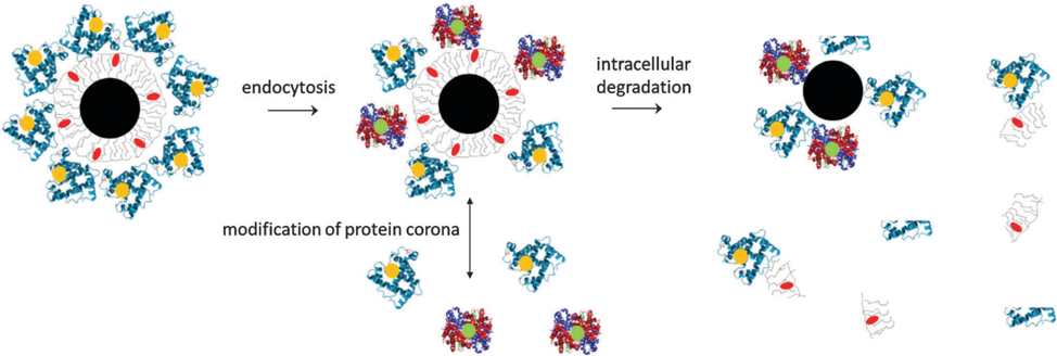
Nanoparticles comprise of a core, a surface coating, and a corona of adsorbed biomolecules, of which all parts can have a different fate.
Overview
The claims and statistics of this paper haven’t been checked yet.
Run the check — free, no account needed. It reads the open-access text and checks the sources it cites.
Integrity signals
Registry
Cites
Statistics
Figures
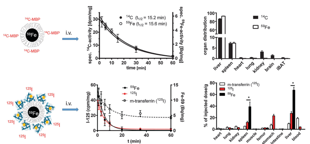
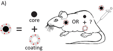
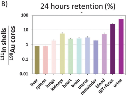
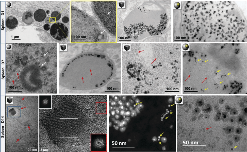
Verification
Claims the paper makes
The claims and statistics of this paper haven’t been checked yet.
Run the check — free, no account needed. It reads the open-access text and checks the sources it cites.
Claims other papers make about this one
Each is checked against this paper’s own text: does it say what the citing paper says it does?
In the citing paper
“Empirical evidence shows that all three components of NPs (core, shell and corona) may degrade individually in vivo and can drastically modify the life cycle and biodistribution of the whole heterostructure, such that the biodistribution and fate of each sub-component would need to be analyzed individually for regulatory and nanomedical approval purposes [136].”
From Particle toxicology and health - where are we? (2019) · cites this paper as [136]
Self-check
Percentages, sums and reported differences re-computed from the paper’s own numbers.
The self-check needs the paper’s full text.
Citations
Cites
In vivo tumor targeting and spectroscopic detection with surface-enhanced Raman nanoparticle tags
Qian, X., Peng, X.-H., Ansari, D. O., Yin-Goen, Q., Chen, G. Z., Shin, D. M., Yang, L., Young, A. N., Wang, M. D., & Nie, S. (2008) · Nature Biotechnology · doi:10.1038/nbt1377 · Cited by 2,201
Monopoli, M. P., Walczyk, D., Campbell, A., Elia, G., Lynch, I., Baldelli Bombelli, F., & Dawson, K. A. (2011) · Journal of the American Chemical Society · doi:10.1021/ja107583h · Cited by 1,835
Walkey, C. D., & Chan, W. C. W. (2012) · Chem. Soc. Rev. · doi:10.1039/c1cs15233e · Cited by 1,758
Time Evolution of the Nanoparticle Protein Corona
Casals, E., Pfaller, T., Duschl, A., Oostingh, G. J., & Puntes, V. (2010) · ACS Nano · doi:10.1021/nn901372t · Cited by 1,211
Kittler, S., Greulich, C., Diendorf, J., Köller, M., & Epple, M. (2010) · Chemistry of Materials · doi:10.1021/cm100023p · Cited by 1,126
Multifunctional magnetic nanoparticles for targeted imaging and therapy
MCCARTHY, J., & WEISSLEDER, R. (2008) · Advanced Drug Delivery Reviews · doi:10.1016/j.addr.2008.03.014 · Cited by 892
The “Sweet” Side of the Protein Corona: Effects of Glycosylation on Nanoparticle–Cell Interactions
Wan, S., Kelly, P. M., Mahon, E., Stöckmann, H., Rudd, P. M., Caruso, F., Dawson, K. A., Yan, Y., & Monopoli, M. P. (2015) · ACS Nano · doi:10.1021/nn506060q · Cited by 223
Research works printed but not linked: 170
- Webster, 2013, Nanomedicine, 8, 525, 10.2217/nnm.13.37 · DOI
- Reese, 2013, Health Matrix Clevel, 23, 537
- Docter, 2015, Nanomedicine, 10, 503, 10.2217/nnm.14.184 · DOI
- Setyawati, 2015, Chem. Soc. Rev., 44, 8174, 10.1039/C5CS00499C · DOI
- Docter, 2015, Chem. Soc. Rev., 44, 6094, 10.1039/C5CS00217F · DOI
- Pautler, 2010, Int. J. Nanomed., 5, 803
- McNeil, 2009, Wiley Interdiscip. Rev.: Nanomed. Nanobiotechnol., 1, 264
- Riehemann, 2009, Angew. Chem., Int. Ed., 48, 872, 10.1002/anie.200802585 · DOI
- Wahajuddin, 2012, Int. J. Nanomed., 7, 3445, 10.2147/IJN.S30320 · DOI
- Rizzo, 2013, Curr. Opin. Biotechnol., 24, 1159, 10.1016/j.copbio.2013.02.020 · DOI
- Seeney, 2012, Nanomedicine, 7, 289, 10.2217/nnm.11.183 · DOI
- Prabhu, 2012, J. Biomed. Nanotechnol., 8, 859, 10.1166/jbn.2012.1459 · DOI
- Murday, 2009, Nanomedicine, 5, 251, 10.1016/j.nano.2009.06.001 · DOI
- Ferrari, 2009, Clin. Pharmacol. Ther., 85, 466, 10.1038/clpt.2008.276 · DOI
- Ventola, 2012, P T., 37, 582
- Hou, 2014, J. Biomed. Mater. Res., Part A, 102, 1726, 10.1002/jbm.a.34839 · DOI
- Pino, 2014, J. Biomed. Opt., 19, 101507, 10.1117/1.JBO.19.10.101507 · DOI
- Colombo, 2012, Chem. Soc. Rev., 41, 4306, 10.1039/c2cs15337h · DOI
- Helou, 2013, Lab Chip, 13, 1035, 10.1039/c3lc41310a · DOI
- Ding, 2014, Biomaterials, 35, 8703, 10.1016/j.biomaterials.2014.06.033 · DOI
- Dias, 2013, Angew. Chem., Int. Ed., 52, 11526, 10.1002/anie.201305835 · DOI
- del Pino, 2010, Nano Lett., 10, 3914, 10.1021/nl102485v · DOI
- Child, 2011, ACS Nano, 5, 7910, 10.1021/nn202163v · DOI
- Sperling, 2008, Chem. Soc. Rev., 37, 1896, 10.1039/b712170a · DOI
- Perez-Hernandez, 2015, ACS Nano, 9, 52, 10.1021/nn505468v · DOI
- Bao, 2013, Small, 9, 68, 10.1002/smll.201201779 · DOI
- Polo, 2013, Chem. Commun., 49, 3676, 10.1039/c3cc39112d · DOI
- Ambrosone, 2014, Nanomedicine, 9, 1913, 10.2217/nnm.14.100 · DOI
- Parak, 2005, Nanotechnology, 16, R5, 10.1088/0957-4484/16/2/R01 · DOI
- Kim, 2004, Nat. Biotechnol., 22, 93, 10.1038/nbt920 · DOI
- Bao, 2014, Nanomedicine, 9, 2353, 10.2217/nnm.14.155 · DOI
- Nystrom, 2012, J. Controlled Release, 161, 403, 10.1016/j.jconrel.2012.01.027 · DOI
- Bawa, 2011, Curr. Drug Delivery, 8, 227, 10.2174/156720111795256156 · DOI
- Baun, 2008, Nanomedicine, 3, 605, 10.2217/17435889.3.5.605 · DOI
- Rahman, 2012, Curr. Drug Discovery Technol., 9, 319, 10.2174/157016312803305898 · DOI
- Ventola, 2012, P T., 37, 631
- Rivera Gil, 2013, Acc. Chem. Res., 46, 743, 10.1021/ar300039j · DOI
- Pelaz, 2013, Small, 9, 1573, 10.1002/smll.201201229 · DOI
- Parak, 2003, Nanotechnology, 14, R15, 10.1088/0957-4484/14/7/201 · DOI
- Fadeel, 2013, Wiley Interdiscip. Rev.: Nanomed. Nanobiotechnol., 5, 111
- Lin, 2009, ACS Nano, 3, 395, 10.1021/nn800632j · DOI
- Shang, 2011, Small, 7, 2614, 10.1002/smll.201100746 · DOI
- Clarke, 2010, Nano Lett., 10, 2147, 10.1021/nl100825n · DOI
- Gerion, 2001, J. Phys. Chem. B, 105, 8861, 10.1021/jp0105488 · DOI
- Selvan, 2007, Angew. Chem., Int. Ed., 46, 2448, 10.1002/anie.200604245 · DOI
- Zhang, 2011, Small, 7, 3113, 10.1002/smll.201100608 · DOI
- Klapper, 2015, Nanoscale, 7, 9980, 10.1039/C5NR01694K · DOI
- Dubertret, 2002, Science, 298, 1759, 10.1126/science.1077194 · DOI · Paper page
- Kolosnjaj-Tabi, 2015, ACS Nano, 9, 7925, 10.1021/acsnano.5b00042 · DOI
- Levy, 2010, Nanotechnology, 21, 395103, 10.1088/0957-4484/21/39/395103 · DOI
- Yang, 2012, ACS Nano, 6, 1795, 10.1021/nn2048526 · DOI
- Zhao, 2015, J. Am. Chem. Soc., 137, 675, 10.1021/ja511843w · DOI
- Branca, 2015, J. Magn. Magn. Mater., 377, 348, 10.1016/j.jmmm.2014.10.086 · DOI
- Wang, 2015, Adv. Mater., 27, 576, 10.1002/adma.201401124 · DOI
- Zeng, 2014, Adv. Mater., 26, 2694, 10.1002/adma.201304744 · DOI
- Montenegro, 2013, Adv. Drug Delivery Rev., 65, 677, 10.1016/j.addr.2012.12.003 · DOI
- Zhang, 2010, ChemPhysChem, 11, 730, 10.1002/cphc.200900849 · DOI
- Carrillo-Carrion, 2014, Curr. Opin. Chem. Eng., 4, 88, 10.1016/j.coche.2013.11.006 · DOI
- Cedervall, 2007, Proc. Natl. Acad. Sci. U. S. A., 104, 2050, 10.1073/pnas.0608582104 · DOI · Paper page
- Nel, 2009, Nat. Mater., 8, 543, 10.1038/nmat2442 · DOI · Paper page
- Monopoli, 2012, Nat. Nanotechnol., 7, 779, 10.1038/nnano.2012.207 · DOI · Paper page
- Tenzer, 2013, Nat. Nanotechnol., 8, 772, 10.1038/nnano.2013.181 · DOI · Paper page
- Xia, 2010, Nat. Nanotechnol., 5, 671, 10.1038/nnano.2010.164 · DOI
- Moore, 2015, Chem. Soc. Rev., 44, 6287, 10.1039/C4CS00487F · DOI
- Lartigue, 2012, ACS Nano, 6, 2665, 10.1021/nn300060u · DOI
- Howes, 2014, Science, 346, 53, 10.1126/science.1247390 · DOI
- Jedlovszky-Hajdu, 2012, Langmuir, 28, 14983, 10.1021/la302446h · DOI
- Monopoli, 2011, Nat. Nanotechnol., 6, 11, 10.1038/nnano.2010.267 · DOI
- Kelly, 2015, Nat. Nanotechnol., 10, 472, 10.1038/nnano.2015.47 · DOI · Paper page
- Lundqvist, 2008, Proc. Natl. Acad. Sci. U. S. A., 105, 14265, 10.1073/pnas.0805135105 · DOI · Paper page
- Milani, 2012, ACS Nano, 6, 2532, 10.1021/nn204951s · DOI
- Walczyk, 2010, J. Am. Chem. Soc., 132, 5761, 10.1021/ja910675v · DOI
- Lynch, 2007, Adv. Colloid Interface Sci., 134–135, 167, 10.1016/j.cis.2007.04.021 · DOI
- Cedervall, 2007, Angew. Chem., Int. Ed., 46, 5754, 10.1002/anie.200700465 · DOI
- Bantz, 2014, Beilstein J. Nanotechnol., 5, 1774, 10.3762/bjnano.5.188 · DOI
- Bargheer, 2015, Beilstein J. Nanotechnol., 6, 36, 10.3762/bjnano.6.5 · DOI
- Treuel, 2015, Beilstein J. Nanotechnol., 6, 857, 10.3762/bjnano.6.88 · DOI
- Gebauer, 2012, Langmuir, 28, 9673, 10.1021/la301104a · DOI
- Hillaireu, 2009, Cell. Mol. Life Sci., 66, 2873, 10.1007/s00018-009-0053-z · DOI
- Nazarenus, 2014, Beilstein J. Nanotechnol., 5, 1477, 10.3762/bjnano.5.161 · DOI
- Jones, 2013, J. Clin. Invest., 123, 3061, 10.1172/JCI66895 · DOI
- Freund, 2012, ACS Nano, 6, 7318, 10.1021/nn3024267 · DOI
- Moore, 2000, Radiology, 214, 568, 10.1148/radiology.214.2.r00fe19568 · DOI
- Bargheer, 2015, Beilstein J. Nanotechnol., 6, 111, 10.3762/bjnano.6.11 · DOI
- Carambia, 2015, J. Hepatol., 62, 1349, 10.1016/j.jhep.2015.01.006 · DOI
- Rehberg, 2012, ACS Nano, 6, 1370, 10.1021/nn204187c · DOI
- Kolosnjaj-Tabi, 2014, ACS Nano, 8, 4268, 10.1021/nn405356r · DOI
- Jung, 2014, Circ. Cardiovasc. Imaging, 7, 303, 10.1161/CIRCIMAGING.113.000607 · DOI
- Lipka, 2010, Biomaterials, 31, 6574, 10.1016/j.biomaterials.2010.05.009 · DOI
- Nohynek, 2007, Crit. Rev. Toxicol., 37, 251, 10.1080/10408440601177780 · DOI · Paper page
- Schleh, 2012, Nanotoxicology, 6, 36, 10.3109/17435390.2011.552811 · DOI · Paper page
- Frey, 1997, Bioconjugate Chem., 8, 424, 10.1021/bc970036p · DOI
- Rancan, 2012, ACS Nano, 6, 6829, 10.1021/nn301622h · DOI
- Vogt, 2014, Beilstein J. Nanotechnol., 5, 2363, 10.3762/bjnano.5.245 · DOI
- Semmler-Behnke, 2008, Small, 4, 2108, 10.1002/smll.200800922 · DOI
- Sadauskas, 2009, Chem. Cent. J., 3, 16, 10.1186/1752-153X-3-16 · DOI
- Sinnecker, 2014, Beilstein J. Nanotechnol., 5, 2092, 10.3762/bjnano.5.218 · DOI
- Levy, 2011, Nanoscale, 3, 4402, 10.1039/c1nr10778j · DOI
- Di Corato, 2014, Biomaterials, 35, 6400, 10.1016/j.biomaterials.2014.04.036 · DOI
- Levy, 2012, Contrast Media Mol. Imaging, 7, 373, 10.1002/cmmi.504 · DOI
- Soenen, 2015, Chem. Rev., 115, 2109, 10.1021/cr400714j · DOI
- Caballero-Díaz, 2013, Part. Part. Syst. Charact., 30, 1079, 10.1002/ppsc.201300215 · DOI
- Loza, 2014, J. Mater. Chem. B, 2, 1634, 10.1039/c3tb21569e · DOI
- Lévy, 2010, Nanotechnology, 21, 395103, 10.1088/0957-4484/21/39/395103 · DOI
- Levy, 2011, Biomaterials, 32, 3988, 10.1016/j.biomaterials.2011.02.031 · DOI
- Kirchner, 2005, Nano Lett., 5, 331, 10.1021/nl047996m · DOI · Paper page
- Mahon, 2012, Chem. Commun., 48, 7970, 10.1039/c2cc34023b · DOI
- Kagan, 2010, Nat. Nanotechnol., 5, 354, 10.1038/nnano.2010.44 · DOI
- Yu, 2000, Science, 287, 637, 10.1126/science.287.5453.637 · DOI
- Sée, 2009, ACS Nano, 3, 2461, 10.1021/nn9006994 · DOI
- Lunov, 2010, Biomaterials, 31, 9015, 10.1016/j.biomaterials.2010.08.003 · DOI
- Bose, 2014, Part. Part. Syst. Charact., 31, 439, 10.1002/ppsc.201300308 · DOI
- Chanana, 2013, Angew. Chem., Int. Ed., 52, 4179, 10.1002/anie.201208019 · DOI
- Ali, 2011, Anal. Chem., 83, 2877, 10.1021/ac103261y · DOI
- Kreyling, 2014, ACS Nano, 8, 222, 10.1021/nn403256v · DOI · Paper page
- Harisinghani, 2001, Clin. Radiol., 56, 714, 10.1053/crad.2001.0764 · DOI
- Salvati, 2011, Nanomedicine, 7, 818, 10.1016/j.nano.2011.03.005 · DOI
- Kreyling, 2015, Nat. Nanotechnol., 10, 619, 10.1038/nnano.2015.111 · DOI
- Walkey, 2014, ACS Nano, 8, 2439, 10.1021/nn406018q · DOI
- Adiseshaiah, 2010, Wiley Interdiscip. Rev.: Nanomed. Nanobiotechnol., 2, 99
- Leszczynski, 2010, Nat. Nanotechnol., 5, 633, 10.1038/nnano.2010.182 · DOI
- Dobrovolskaia, 2014, Nanomedicine, 10, 1453, 10.1016/j.nano.2014.01.009 · DOI
- Lesniak, 2012, ACS Nano, 6, 5845, 10.1021/nn300223w · DOI · Paper page
- Fleischer, 2014, Acc. Chem. Res., 47, 2651, 10.1021/ar500190q · DOI
- Tay, 2014, Adv. Funct. Mater., 24, 5936, 10.1002/adfm.201401664 · DOI
- Sakulkhu, 2014, Sci. Rep., 4, 5020, 10.1038/srep05020 · DOI
- Sacchetti, 2013, ACS Nano, 7, 1974, 10.1021/nn400409h · DOI
- Nel, 2013, Acc. Chem. Res., 46, 605, 10.1021/ar400005v · DOI
- Wang, 2013, Nanomedicine, 9, 1159, 10.1016/j.nano.2013.04.010 · DOI
- Caracciolo, 2011, Langmuir, 27, 15048, 10.1021/la202912f · DOI
- Lundqvist, 2011, ACS Nano, 5, 7503, 10.1021/nn202458g · DOI · Paper page
- Syed, 2015, Cancer Treat. Res., 166, 227, 10.1007/978-3-319-16555-4_10 · DOI
- Liu, 2015, Nanoscale, 7, 9664, 10.1039/C5NR01537E · DOI
- Lin, 2014, ACS Nano, 8, 4450, 10.1021/nn5012754 · DOI
- Li, 2014, ACS Nano, 8, 1771, 10.1021/nn406166n · DOI
- Konduru, 2014, Part. Fibre Toxicol., 11, 55, 10.1186/s12989-014-0055-3 · DOI · Paper page
- Gilbert, 2012, ACS Nano, 6, 4921, 10.1021/nn300425a · DOI
- Wang, 2014, Small, 10, 385, 10.1002/smll.201301597 · DOI
- Zhang, 2014, J. Am. Chem. Soc., 136, 6406, 10.1021/ja501699e · DOI
- Bertoli, 2014, Small, 10, 3307, 10.1002/smll.201303841 · DOI
- Mahmoudi, 2013, ACS Nano, 7, 6555, 10.1021/nn305337c · DOI
- Murthy, 2013, J. Am. Chem. Soc., 135, 7799, 10.1021/ja400701c · DOI
- Natte, 2013, Colloids Surf., B, 104, 213, 10.1016/j.colsurfb.2012.11.019 · DOI
- Pozzi, 2014, Nanoscale, 6, 2782, 10.1039/c3nr05559k · DOI · Paper page
- Pelaz, 2015, ACS Nano, 9, 6996, 10.1021/acsnano.5b01326 · DOI
- del_Pino, 2014, Mater. Horiz., 1, 301, 10.1039/C3MH00106G · DOI
- Röcker, 2009, Nat. Nanotechnol., 4, 577, 10.1038/nnano.2009.195 · DOI
- Hühn, 2013, ACS Nano, 7, 3253, 10.1021/nn3059295 · DOI
- Alvarez-Puebla, 2011, Proc. Natl. Acad. Sci. U. S. A., 108, 8157, 10.1073/pnas.1016530108 · DOI
- Guerrini, 2015, ACS Appl. Mater. Interfaces, 7, 9420, 10.1021/acsami.5b01056 · DOI
- Guerrini, 2013, J. Am. Chem. Soc., 135, 10314, 10.1021/ja405120x · DOI
- Richter, 1942, Biochem. J., 36, 746, 10.1042/bj0360746 · DOI
- Paulsson, 2009, Nano Lett., 9, 117, 10.1021/nl802643h · DOI
- Kruger, 2003, Angew. Chem., Int. Ed., 42, 2251, 10.1002/anie.200351000 · DOI
- Mejias, 2013, J. Controlled Release, 171, 225, 10.1016/j.jconrel.2013.07.019 · DOI
- Lartigue, 2013, ACS Nano, 7, 3939, 10.1021/nn305719y · DOI · Paper page
- Weissleder, 1989, Am. J. Roentgenol., 152, 167, 10.2214/ajr.152.1.167 · DOI
- Majumdar, 1990, Invest. Radiol., 25, 771, 10.1097/00004424-199007000-00004 · DOI
- Heine, 2014, Beilstein J. Nanotechnol., 5, 1432, 10.3762/bjnano.5.155 · DOI
- Beaumont, 2009, Semin. Hematol., 46, 328, 10.1053/j.seminhematol.2009.06.004 · DOI
- Arbab, 2005, NMR Biomed., 18, 383, 10.1002/nbm.970 · DOI
- Javed, 2014, Small, 10, 3325, 10.1002/smll.201400281 · DOI
- Luciani, 2010, Biomaterials, 31, 7061, 10.1016/j.biomaterials.2010.05.062 · DOI
- Silva, 2012, Pharm. Res., 29, 1392, 10.1007/s11095-012-0680-1 · DOI
- Silva, 2013, Nanoscale, 5, 11374, 10.1039/c3nr01541f · DOI
- Marangon, 2012, Nano Lett., 12, 4830, 10.1021/nl302273p · DOI
- de Jonge, 2009, Proc. Natl. Acad. Sci. U. S. A., 106, 2159, 10.1073/pnas.0809567106 · DOI
- Pohlmann, 2015, Nano Lett., 15, 2329, 10.1021/nl504481k · DOI
- Kagan, 2014, ACS Nano, 8, 5610, 10.1021/nn406484b · DOI
- Elgrabli, 2015, ACS Nano, 9, 10113, 10.1021/acsnano.5b03708 · DOI
Cited by
Sorted by citations
Diverse Applications of Nanomedicine
Pelaz, B., Alexiou, C., Alvarez-Puebla, R. A., Alves, F., Andrews, A. M., Ashraf, S., Balogh, L. P., Ballerini, L., Bestetti, A., Brendel, C., et al. (2017) · ACS Nano · doi:10.1021/acsnano.6b06040 · Cited by 1,350
Self-citationWu, H., Li, F., Shao, W., Gao, J., & Ling, D. (2019) · ACS Central Science · doi:10.1021/acscentsci.8b00850 · Cited by 338
Wuttke, S., Lismont, M., Escudero, A., Rungtaweevoranit, B., & Parak, W. J. (2017) · Biomaterials · doi:10.1016/j.biomaterials.2017.01.025 · Cited by 286
Self-citationParticle toxicology and health - where are we?
Riediker, M., Zink, D., Kreyling, W., Oberdörster, G., Elder, A., Graham, U., Lynch, I., Duschl, A., Ichihara, G., Ichihara, S., et al. (2019) · Particle and Fibre Toxicology · doi:10.1186/s12989-019-0302-8 · Cited by 237
Beldman, T. J., Senders, M. L., Alaarg, A., Pérez-Medina, C., Tang, J., Zhao, Y., Fay, F., Deichmöller, J., Born, B., Desclos, E., et al. (2017) · ACS Nano · doi:10.1021/acsnano.7b01385 · Cited by 196
Metadata
- Authors
- Feliu, N., Docter, D., Heine, M., del Pino, P., Ashraf, S., Kolosnjaj-Tabi, J., Macchiarini, P., Nielsen, P., Alloyeau, D., Gazeau, F., Stauber, R. H., & Parak, W. J.
- Venue
- Chemical Society Reviews
- Publisher
- Royal Society of Chemistry (RSC)
- Published
- 2016
- Volume
- 45
- Issue
- 9
- Pages
- 2440-2457
- ISSN
- 0306-0012
- 1460-4744
- Cited by
- 423
- References
- 7
- Language
- en
- Funders
- Bundesministerium für Bildung und Forschung (MRCyte/NanoBEL)
- European Commission (FutureNanoNeeds)
- Alexander von Humboldt-Stiftung
About these checks
Registry, retraction, Cites and statistics results are computed, not judged, with one exception: a printed reference that no index lists is read once by a model before it counts as a citation error. Claims are extracted by a model and kept only when their quote appears verbatim in the text; each cited source is then checked, with a model’s help, for whether it says what the claim relies on. Those verdicts can be wrong — read the claim’s quote and the cited source before relying on one.
Statistics and arithmetic are recomputed, not judged.
Confidence bands are shown only for fields calibrated on our annotated test set. None is calibrated yet, so no band is shown.
What counts as a claim ›How we measure confidence ›How we check this ›
Are you an author, or do you see a problem with this page? Request a review
How to cite
Cite CoChat for the checks on this page, and the paper for its own findings.
- This page
- CoChat. (2026). Citation check: In vivo degeneration and the fate of inorganic nanoparticles. https://app.cochat.ai/papers/10.1039/c5cs00699f
- The paper
- Feliu, N., Docter, D., Heine, M., del Pino, P., Ashraf, S., Kolosnjaj-Tabi, J., Macchiarini, P., Nielsen, P., Alloyeau, D., Gazeau, F., Stauber, R. H., & Parak, W. J. (2016). In vivo degeneration and the fate of inorganic nanoparticles. Chemical Society Reviews. https://doi.org/10.1039/c5cs00699f
