

The DOI matches its Crossref record; 62 of the 62 references it cites were found in a registry or an index.
Cell survival, DNA damage, and oncogenic transformation after a transient and reversible apoptotic response
Open accessAbstract
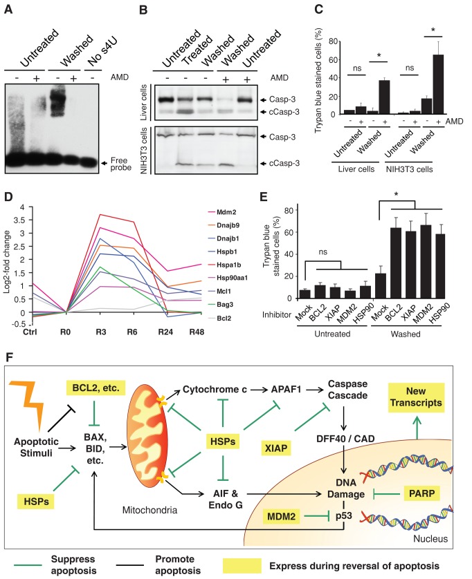
Apoptosis serves as a protective mechanism by eliminating damaged cells through programmed cell death. After apoptotic cells pass critical checkpoints, including mitochondrial fragmentation, executioner caspase activation, and DNA damage, it is assumed that cell death inevitably follows. However, this assumption has not been tested directly. Here we report an unexpected reversal of late-stage apoptosis in primary liver and heart cells, macrophages, NIH 3T3 fibroblasts, cervical cancer HeLa cells, and brain cells. After exposure to an inducer of apoptosis, cells exhibited multiple morphological and biochemical hallmarks of late-stage apoptosis, including mitochondrial fragmentation, caspase-3 activation, and DNA damage. Surprisingly, the vast majority of dying cells arrested the apoptotic process and recovered when the inducer was washed away. Of importance, some cells acquired permanent genetic changes and underwent oncogenic transformation at a higher frequency than controls. Global gene expression analysis identified a molecular signature of the reversal process. We propose that reversal of apoptosis is an unanticipated mechanism to rescue cells from crisis and propose to name this mechanism “anastasis” (Greek for “rising to life”). Whereas carcinogenesis represents a harmful side effect, potential benefits of anastasis could include preservation of cells that are difficult to replace and stress-induced genetic diversity.
Overview
The claims and statistics of this paper haven’t been checked yet.
Run the check — free, no account needed. It reads the open-access text and checks the sources it cites.
Integrity signals
Registry
Cites
Statistics
Figures
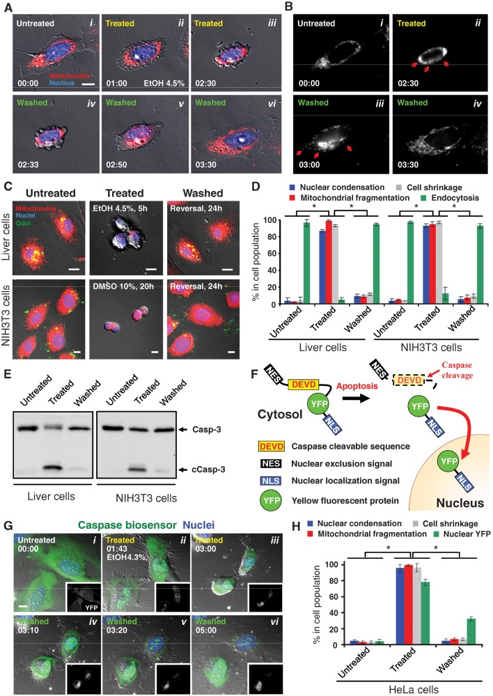
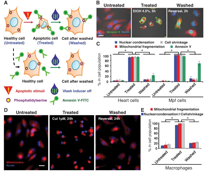
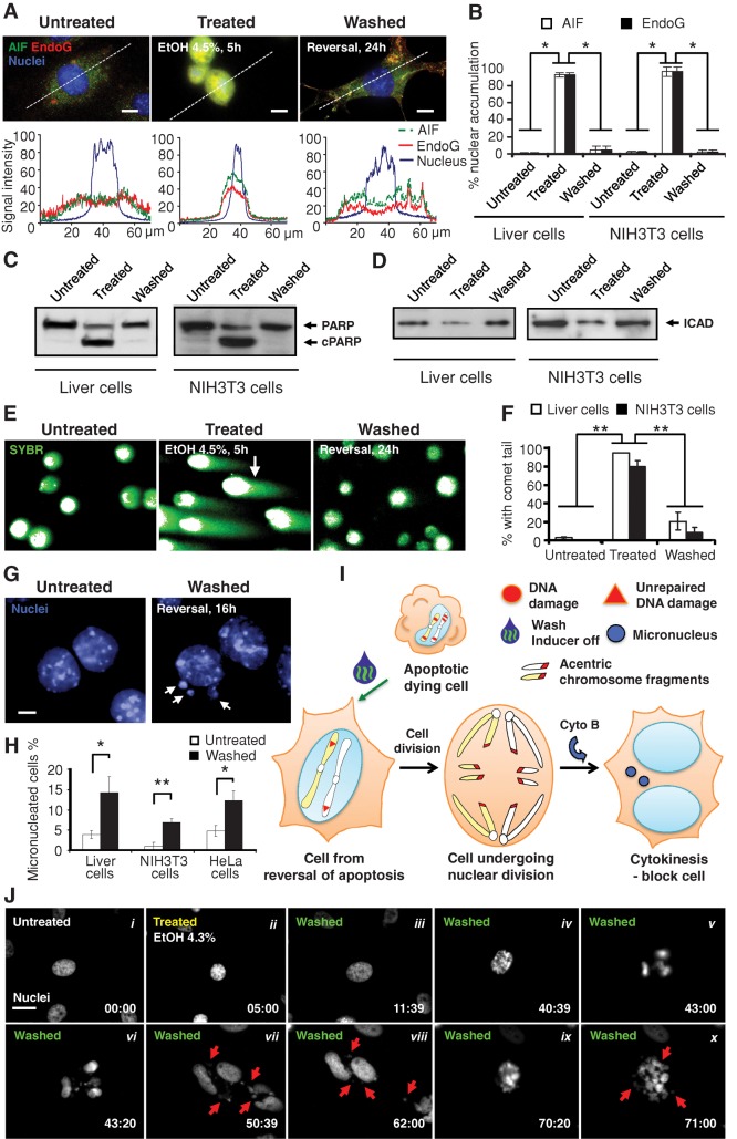
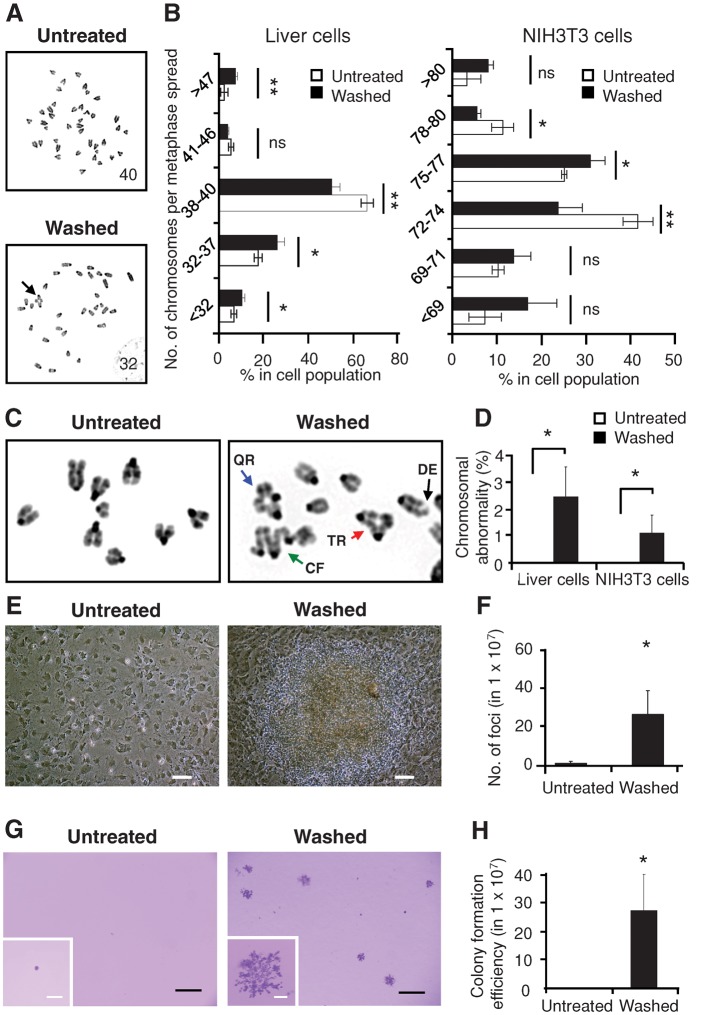
Verification
Claims the paper makes
The claims and statistics of this paper haven’t been checked yet.
Run the check — free, no account needed. It reads the open-access text and checks the sources it cites.
Claims other papers make about this one
Each is checked against this paper’s own text: does it say what the citing paper says it does?
In the citing paper
“Finally, at least some cells exposed to transient apoptotic stimuli appear to survive MOMP affecting a limited number of mitochondria and the partial activation of executioner caspases by a hitherto poorly characterized process called anastasis (most likely constituting a robust adaptative response upstream of the boundary between cellular life and death) [226–228].”
From Molecular mechanisms of cell death: recommendations of the nomenclature committee on cell death 2018. (2018) · cites this paper as [228]
Self-check
Percentages, sums and reported differences re-computed from the paper’s own numbers.
The self-check needs the paper’s full text.
Citations
Cites
Apoptosis: A Basic Biological Phenomenon with Wideranging Implications in Tissue Kinetics
Kerr, J. F. R., Wyllie, A. H., & Currie, A. R. (1972) · British Journal of Cancer · doi:10.1038/bjc.1972.33 · Cited by 15,673
Molecular characterization of mitochondrial apoptosis-inducing factor
Susin, S. A., Lorenzo, H. K., Zamzami, N., Marzo, I., Snow, B. E., Brothers, G. M., Mangion, J., Jacotot, E., Costantini, P., Loeffler, M., et al. (1999) · Nature · doi:10.1038/17135 · Cited by 4,060
Stratton, M. R., Campbell, P. J., & Futreal, P. A. (2009) · Nature · doi:10.1038/nature07943 · Cited by 3,625
The Pathophysiology of Mitochondrial Cell Death
Green, D. R., & Kroemer, G. (2004) · Science · doi:10.1126/science.1099320 · Cited by 3,487
Classification of cell death: recommendations of the Nomenclature Committee on Cell Death 2009
Kroemer, G., Galluzzi, L., Vandenabeele, P., Abrams, J., Alnemri, E. S., Baehrecke, E. H., Blagosklonny, M. V., El-Deiry, W. S., Golstein, P., Green, D. R., et al. (2009) · Cell Death & Differentiation · doi:10.1038/cdd.2008.150 · Cited by 3,399
A caspase-activated DNase that degrades DNA during apoptosis, and its inhibitor ICAD
Enari, M., Sakahira, H., Yokoyama, H., Okawa, K., Iwamatsu, A., & Nagata, S. (1998) · Nature · doi:10.1038/34112 · Cited by 3,239
Apoptosis: controlled demolition at the cellular level
Taylor, R. C., Cullen, S. P., & Martin, S. J. (2007) · Nature Reviews Molecular Cell Biology · doi:10.1038/nrm2312 · Cited by 2,708
Programmed Cell Death in Animal Development
Jacobson, M. D., Weil, M., & RAFF, M. C. (1997) · Cell · doi:10.1016/s0092-8674(00)81873-5 · Cited by 2,685
The Significance of Responses of the Genome to Challenge
McClintock, B. (1984) · Science · doi:10.1126/science.15739260 · Cited by 2,638
Cleavage of poly(ADP-ribose) polymerase by a proteinase with properties like ICE
Lazebnik, Y. A., Kaufmann, S. H., Desnoyers, S., Poirier, G. G., & Earnshaw, W. C. (1994) · Nature · doi:10.1038/371346a0 · Cited by 2,563
Research works printed but not linked: 1
- Gordon WC, 2002, Invest Ophthalmol Visual Sci, 43, 3511
Cited by
Sorted by citations
Galluzzi, L., Vitale, I., Aaronson, S. A., Abrams, J. M., Adam, D., Agostinis, P., Alnemri, E. S., Altucci, L., Amelio, I., Andrews, D. W., et al. (2018) · Cell Death & Differentiation · doi:10.1038/s41418-017-0012-4 · Cited by 6,680
Mitochondria as multifaceted regulators of cell death
Bock, F. J., & Tait, S. W. G. (2019) · Nature Reviews Molecular Cell Biology · doi:10.1038/s41580-019-0173-8 · Cited by 2,531
Regulation of apoptosis in health and disease: the balancing act of BCL-2 family proteins
Singh, R., Letai, A., & Sarosiek, K. (2019) · Nature Reviews Molecular Cell Biology · doi:10.1038/s41580-018-0089-8 · Cited by 2,111
Essential versus accessory aspects of cell death: recommendations of the NCCD 2015
Galluzzi, L., Bravo-San Pedro, J. M., Vitale, I., Aaronson, S. A., Abrams, J. M., Adam, D., Alnemri, E. S., Altucci, L., Andrews, D., Annicchiarico-Petruzzelli, M., et al. (2015) · Cell Death & Differentiation · doi:10.1038/cdd.2014.137 · Cited by 1,002
Self-citationMOMP, cell suicide as a BCL-2 family business
Kalkavan, H., & Green, D. R. (2017) · Cell Death and Differentiation · doi:10.1038/cdd.2017.179 · Cited by 707
Live to die another way: modes of programmed cell death and the signals emanating from dying cells
Fuchs, Y., & Steller, H. (2015) · Nature Reviews Molecular Cell Biology · doi:10.1038/nrm3999 · Cited by 662
Paradoxical roles of caspase-3 in regulating cell survival, proliferation, and tumorigenesis
Eskandari, E., & Eaves, C. J. (2022) · The Journal of Cell Biology · doi:10.1083/jcb.202201159 · Cited by 504
Mechanisms and Consequences of Double‐Strand DNA Break Formation in Chromatin
Cannan, W. J., & Pederson, D. S. (2016) · Journal of Cellular Physiology · doi:10.1002/jcp.25048 · Cited by 499
Ichim, G., Lopez, J., Ahmed, S. U., Muthalagu, N., Giampazolias, E., Delgado, M. E., Haller, M., Riley, J. S., Mason, S. M., Athineos, D., et al. (2015) · Molecular Cell · doi:10.1016/j.molcel.2015.01.018 · Cited by 479
Human topoisomerases and their roles in genome stability and organization
Pommier, Y. G., Nussenzweig, A., Takeda, S., & Austin, C. A. (2022) · Nature Reviews Molecular Cell Biology · doi:10.1038/s41580-022-00452-3 · Cited by 493
Glover, H. L., Schreiner, A., Dewson, G., & Tait, S. W. G. (2024) · Nature Cell Biology · doi:10.1038/s41556-024-01429-4 · Cited by 515
Melatonin, a Full Service Anti-Cancer Agent: Inhibition of Initiation, Progression and Metastasis
Reiter, R. J., Rosales‐Corral, S., Tan, D., Acuña-Castroviejo, D., Qin, L., Yang, S., & Xu, K. (2017) · International Journal of Molecular Sciences · doi:10.3390/ijms18040843 · Cited by 453
Cell death in the gut epithelium and implications for chronic inflammation
Patankar, J. V., & Becker, C. (2020) · Nature Reviews Gastroenterology & Hepatology · doi:10.1038/s41575-020-0326-4 · Cited by 429
DNA Damage/Repair Management in Cancers
Alhmoud, J. F., Woolley, J. F., Moustafa, A. A., & Malki, M. I. (2020) · Cancers · doi:10.3390/cancers12041050 · Cited by 410
Mitochondria—Judges and Executioners of Cell Death Sentences
Bhola, P. D., & Letai, A. (2016) · Molecular Cell · doi:10.1016/j.molcel.2016.02.019 · Cited by 326
Holland, A. J., & Cleveland, D. W. (2012) · Nature Medicine · doi:10.1038/nm.2988 · Cited by 315
Do TUNEL and Other Apoptosis Assays Detect Cell Death in Preclinical Studies?
Mirzayans, R., & Murray, D. L. (2020) · International Journal of Molecular Sciences · doi:10.3390/ijms21239090 · Cited by 231
Rethink of EGFR in Cancer With Its Kinase Independent Function on Board
Thomas, R., & Weihua, Z. (2019) · Frontiers in Oncology · doi:10.3389/fonc.2019.00800 · Cited by 215
Flipping the dogma – phosphatidylserine in non-apoptotic cell death
Shlomovitz, I., Speir, M., & Gerlic, M. (2019) · Cell Communication and Signaling · doi:10.1186/s12964-019-0437-0 · Cited by 189
Caspase-dependent non-apoptotic processes in development
Nakajima, Y.-I., & Kuranaga, E. (2017) · Cell Death and Differentiation · doi:10.1038/cdd.2017.36 · Cited by 184
Stress-Induced Mutagenesis: Implications in Cancer and Drug Resistance
Fitzgerald, D. M., Hastings, P. J., & Rosenberg, S. M. (2016) · Annual Review of Cancer Biology · doi:10.1146/annurev-cancerbio-050216-121919 · Cited by 178
Apoptosis: A mammalian cell bioprocessing perspective
Grilo, A. L., & Mantalaris, A. (2019) · Biotechnology Advances · doi:10.1016/j.biotechadv.2019.02.012 · Cited by 175
Mitochondria, cholesterol and cancer cell metabolism
Ribas, V., García‐Ruiz, C., & Fernández-Checa, J. C. (2016) · Clinical and Translational Medicine · doi:10.1186/s40169-016-0106-5 · Cited by 166
A molecular signature for anastasis, recovery from the brink of apoptotic cell death
Sun, G., Guzman, E., Balasanyan, V., Conner, C. M., Wong, K., Zhou, H. R., Kosik, K. S., & Montell, D. J. (2017) · Journal of Cell Biology · doi:10.1083/jcb.201706134 · Cited by 151
Self-citationGudipaty, S. A., Conner, C. M., Rosenblatt, J., & Montell, D. J. (2018) · Annual Review of Cell and Developmental Biology · doi:10.1146/annurev-cellbio-100616-060748 · Cited by 150
Self-citationCardiac regeneration based on mechanisms of cardiomyocyte proliferation and differentiation
Senyo, S. E., Lee, R. T., & Kühn, B. (2014) · Stem Cell Research · doi:10.1016/j.scr.2014.09.003 · Cited by 136
HEI-OC1 cells as a model for investigating drug cytotoxicity
Kalinec, G. M., Thein, P., Park, C., & Kalinec, F. (2016) · Hearing Research · doi:10.1016/j.heares.2016.02.019 · Cited by 139
Ding, X., Sun, G., Argaw, Y. G., Wong, J. O., Easwaran, S., & Montell, D. J. (2016) · eLife · doi:10.7554/elife.10936 · Cited by 128
Self-citationSystematic Quantification of Population Cell Death Kinetics in Mammalian Cells
Forcina, G. C., Conlon, M., Wells, A. C., Cao, J. Y., & Dixon, S. J. (2017) · Cell Systems · doi:10.1016/j.cels.2017.05.002 · Cited by 122
In vivo CaspaseTracker biosensor system for detecting anastasis and non-apoptotic caspase activity
Tang, H. L., Tang, H. M., Fung, M. C., & Hardwick, J. M. (2015) · Scientific Reports · doi:10.1038/srep09015 · Cited by 115
Self-citationIhenacho, U. K., Nolden, K. A., Harwig, M. C., Widlansky, M. E., & Hill, R. B. (2021) · Frontiers in Endocrinology · doi:10.3389/fendo.2021.660095 · Cited by 117
Anastasis: recovery from the brink of cell death
Tang, H. M., & Tang, H. L. (2018) · Royal Society Open Science · doi:10.1098/rsos.180442 · Cited by 117
Self-citationRole of the nucleus in apoptosis: signaling and execution
Prokhorova, E. A., Zamaraev, A. V., Kopeina, G. S., Zhivotovsky, B., & Lavrik, I. N. (2015) · Cellular and Molecular Life Sciences · doi:10.1007/s00018-015-2031-y · Cited by 116
Rab5 and Alsin regulate stress-activated cytoprotective signaling on mitochondria
Hsu, F., Spannl, S., Ferguson, C., Hyman, A. A., Parton, R. G., & Zerial, M. (2018) · eLife · doi:10.7554/elife.32282 · Cited by 110
To the edge of cell death and back
Gong, Y., Crawford, J. C., Heckmann, B. L., & Green, D. R. (2018) · FEBS Journal · doi:10.1111/febs.14714 · Cited by 110
Self-inflicted DNA double-strand breaks sustain tumorigenicity and stemness of cancer cells
Liu, X., Li, F., Huang, Q., Zhang, Z., Zhou, L., Deng, Y., Zhou, M., Fleenor, D. E., He, W., Kastan, M. B., et al. (2017) · Cell Research · doi:10.1038/cr.2017.41 · Cited by 99
Biological events and molecular signaling following MLKL activation during necroptosis
Gong, Y., Guy, C. S., Crawford, J. C., & Green, D. R. (2017) · Cell Cycle · doi:10.1080/15384101.2017.1371889 · Cited by 93
DNA Degradation and Its DefectsPreprint
Kawane, K., Motani, K., & Nagata, S. (2014) · Cold Spring Harbor Perspectives in Biology · doi:10.1101/cshperspect.a016394 · Cited by 93
A matter of life and death: stem cell survival in tissue regeneration and tumour formation
Soteriou, D., & Fuchs, Y. (2018) · Nature Reviews Cancer · doi:10.1038/nrc.2017.122 · Cited by 87
New and revisited approaches to preserving the reperfused myocardium
Kloner, R. A., Brown, D. A., CSETE, M. E., Dai, W., Downey, J. M., Gottlieb, R. A., Hale, S. L., & Shi, J. (2017) · Nature Reviews Cardiology · doi:10.1038/nrcardio.2017.102 · Cited by 86
Cell recovery by reversal of ferroptosis
Tang, H. M., & Tang, H. L. (2019) · Biology Open · doi:10.1242/bio.043182 · Cited by 83
Self-citationDo cytotoxicity and cell death cause false positive results in the in vitro comet assay?
Azqueta, A., Stopper, H., Zegura, B., Dusinska, M., & Møller, P. (2022) · Mutation Research/Genetic Toxicology and Environmental Mutagenesis · doi:10.1016/j.mrgentox.2022.503520 · Cited by 80
Apoptosis – Fueling the oncogenic fire
Ferrer, C. C., Berthenet, K., & Ichim, G. (2020) · FEBS Journal · doi:10.1111/febs.15624 · Cited by 82
Mirzayans, R., & Murray, D. L. (2020) · International Journal of Molecular Sciences · doi:10.3390/ijms21041308 · Cited by 76
Regulating cell morphogenesis: The drosophila jun N‐terminal kinase pathway
Ríos‐Barrera, L. D., & Riesgo‐Escovar, J. R. (2012) · genesis · doi:10.1002/dvg.22354 · Cited by 75
On the Complexity of Mechanisms and Consequences of Chromothripsis: An Update
Koltsova, A. S., Pendina, A. A., Efimova, O. A., Chiryaeva, O. G., Kuznetzova, T. V., & Баранов, В. С. (2019) · Frontiers in Genetics · doi:10.3389/fgene.2019.00393 · Cited by 72
Time-dependent replicative senescence vs. disturbed flow-induced pre-mature aging in atherosclerosis
Dominic, A., Banerjee, P., Hamilton, D. J., Le, N., & Abe, J. (2020) · Redox Biology · doi:10.1016/j.redox.2020.101614 · Cited by 72
Smirnova, L. (2015) · ALTEX · doi:10.14573/altex.1509271 · Cited by 71
Apoptosis and Cancer: Force Awakens, Phantom Menace, or Both?
Cao, K., & Tait, S. W. G. (2018) · International review of cytology · doi:10.1016/bs.ircmb.2017.12.003 · Cited by 74
Cosolo, A., Jaiswal, J., Csordás, G., Grass, I., Uhlířová, M., & Classen, A.-K. (2019) · eLife · doi:10.7554/elife.41036 · Cited by 72
Optogenetic activators of apoptosis, necroptosis, and pyroptosis
Shkarina, K., Hasel, E., Santos, J. C., Ramos, S., Leptin, M., & Brož, P. (2022) · The Journal of Cell Biology · doi:10.1083/jcb.202109038 · Cited by 69
Thermotolerance and plasticity of camel somatic cells exposed to acute and chronic heat stress
Saadeldin, I. M., Swelum, A. A., Elsafadi, M. A., Mahmood, A., Osama, A., Shikshaky, H., Alfayez, M. A., Alowaimer, A. N., & Magdeldin, S. (2019) · Journal of Advanced Research · doi:10.1016/j.jare.2019.11.009 · Cited by 68
Chromothripsis: potential origin in gametogenesis and preimplantation cell divisions. A review
Pellestor, F., Gâtinois, V., Puechberty, J., Geneviève, D., & Lefort, G. (2014) · Fertility and Sterility · doi:10.1016/j.fertnstert.2014.09.006 · Cited by 68
Sun, G., Ding, X., Argaw, Y. G., Guo, X., & Montell, D. J. (2020) · Nature Communications · doi:10.1038/s41467-020-19068-2 · Cited by 64
Self-citationZhao, R., Kaakati, R. N., Lee, A. K., Liu, X., Li, F., & Li, C. (2018) · Cancer and Metastasis Reviews · doi:10.1007/s10555-018-9736-y · Cited by 63
When dying is not the end: Apoptotic caspases as drivers of proliferation
Pérez-Garijo, A. (2017) · Seminars in Cell and Developmental Biology · doi:10.1016/j.semcdb.2017.11.036 · Cited by 59
Hamed, M., Osman, A. G. M., Badrey, A. E. A., Soliman, H. A. M., & Sayed, A. E. H. (2021) · Frontiers in Physiology · doi:10.3389/fphys.2021.742922 · Cited by 59
Li, X., Guo, S., Min, L., Guo, Q., & Zhang, S. (2019) · International Journal of Molecular Medicine · doi:10.3892/ijmm.2019.4258 · Cited by 57
RetractedCell survival following direct executioner-caspase activation
Nano, M., Mondo, J. A., Harwood, J., Balasanyan, V., & Montell, D. J. (2023) · Proceedings of the National Academy of Sciences · doi:10.1073/pnas.2216531120 · Cited by 58
Self-citationSilver, B. B., & Nelson, C. M. (2018) · Frontiers in Cell and Developmental Biology · doi:10.3389/fcell.2018.00021 · Cited by 56
Scrambling the genome in cancer: causes and consequences of complex chromosome rearrangements
Krupina, K., Goginashvili, A., & Cleveland, D. W. (2023) · Nature Reviews Genetics · doi:10.1038/s41576-023-00663-0 · Cited by 63
Manalo, R. V. M., & Medina, P. M. B. (2018) · Frontiers in Neuroscience · doi:10.3389/fnins.2018.00137 · Cited by 55
Senescence-Associated Secretory Phenotype as a Hinge Between Cardiovascular Diseases and Cancer
Banerjee, P., Kotla, S., Velatooru, L. R., Abe, R. J., Olmsted‐Davis, E. A., Cooke, J. P., Schadler, K., Deswal, A. M., Herrmann, J., Lin, S. H., et al. (2021) · Frontiers in Cardiovascular Medicine · doi:10.3389/fcvm.2021.763930 · Cited by 55
Apoptosis Reversal Promotes Cancer Stem Cell-Like Cell Formation
Xu, Y., So, C., Lam, H., Fung, M.-C., & Tsang, S. Y. (2018) · Neoplasia · doi:10.1016/j.neo.2018.01.005 · Cited by 54
Self-citationChoe, S. C., Hamacher‐Brady, A., & Brady, N. R. (2015) · Cell Communication and Signaling · doi:10.1186/s12964-015-0115-9 · Cited by 55
Hornik, T. C., Vilalta, A., & Brown, G. C. (2015) · Journal of Cell Science · doi:10.1242/jcs.174631 · Cited by 57
Molecular signature of anastasis for reversal of apoptosis
Tang, H. M., Talbot, C. C., Fung, M. C., & Tang, H. L. (2017) · F1000Research · doi:10.12688/f1000research.10568.2 · Cited by 71
Self-citationEssential roles of Caspase-3 in facilitating Myc-induced genetic instability and carcinogenesis
Cartwright, I. M., Liu, X., Zhou, M., Li, F., & Li, C. (2017) · eLife · doi:10.7554/elife.26371 · Cited by 54
Mnich, K., Carleton, L. A., Kavanagh, E. T., Doyle, K., Afshin Samali, A., & Gorman, A. M. (2014) · Cell Death and Disease · doi:10.1038/cddis.2014.173 · Cited by 52
Suzuki, J., Kawasaki, Y., Nozawa, R., Isome, M., Suzuki, S., Takahashi, A., & Suzuki, H. (2003) · PubMed · Cited by 49
Luchetti, F., Carloni, S., Nasoni, M. G., Reiter, R. J., & Balduini, W. (2022) · Journal of Pineal Research · doi:10.1111/jpi.12800 · Cited by 46
Molecular profiling of anastatic cancer cells: potential role of the nuclear export pathway
Seervi, M., Sumi, S., Chandrasekharan, A., Sharma, A. K., & Santhoshkumar, T. R. (2019) · Cellular Oncology · doi:10.1007/s13402-019-00451-1 · Cited by 45
Chakraborty, S., Mir, K. B., Seligson, N. D., Nayak, D., Kumar, R., & Goswami, A. (2020) · Cancer and Metastasis Reviews · doi:10.1007/s10555-020-09866-x · Cited by 45
Antiaging Effects of Urolithin A on Replicative Senescent Human Skin Fibroblasts
Liu, C.-F., Li, X., Zhang, Z., Qiu, L., Ding, S.-X., Xue, J., Zhao, G., & Li, J. (2018) · Rejuvenation Research · doi:10.1089/rej.2018.2066 · Cited by 46
Non-lethal outcomes of engaging regulated cell death pathways in cancer
Kalkavan, H., Rühl, S., Shaw, J. J. P., & Green, D. R. (2023) · Nature Cancer · doi:10.1038/s43018-023-00571-6 · Cited by 48
Q&A: Cellular near death experiences—what is anastasis?
Sun, G., & Montell, D. J. (2017) · BMC Biology · doi:10.1186/s12915-017-0441-z · Cited by 44
Self-citationThe impact of non-genetic heterogeneity on cancer cell death
Inde, Z., & Dixon, S. J. (2017) · Critical Reviews in Biochemistry and Molecular Biology · doi:10.1080/10409238.2017.1412395 · Cited by 41
Overview of BCL-2 Family Proteins and Therapeutic Potentials
Huska, J. D., Lamb, H. M., & Hardwick, J. M. (2018) · Methods in molecular biology · doi:10.1007/978-1-4939-8861-7_1 · Cited by 44
Death and survival from executioner caspase activation
Sun, G. (2023) · Seminars in Cell and Developmental Biology · doi:10.1016/j.semcdb.2023.07.005 · Cited by 46
Valon, L., & Levayer, R. (2019) · Biology of the Cell · doi:10.1111/boc.201800075 · Cited by 43
Anastasis: Return Journey from Cell Death
Zaitceva, V., Kopeina, G. S., & Zhivotovsky, B. (2021) · Cancers · doi:10.3390/cancers13153671 · Cited by 41
Amino Acid Derivatives of Ligustrazine-Oleanolic Acid as New Cytotoxic Agents
Chu, F., Xu, X., Li, G., Gu, S., Xu, K., Gong, Y., Xu, B., Wang, M., Zhang, H., Zhang, Y., et al. (2014) · Molecules · doi:10.3390/molecules191118215 · Cited by 41
Eldeeb, M. A., Fahlman, R. P., Esmaili, M., & Ragheb, M. A. (2018) · International Journal of Molecular Sciences · doi:10.3390/ijms19113414 · Cited by 41
Rapid coupling between gravitational forces and the transcriptome in human myelomonocytic U937 cells
Thiel, C. S., Tauber, S., Christoffel, S., Huge, A., Lauber, B. A., Polzer, J., Paulsen, K., Lier, H., Engelmann, F., Schmitz, B., et al. (2018) · Scientific Reports · doi:10.1038/s41598-018-31596-y · Cited by 40
DNA Damage/Repair Management in Cancers
Alhmoud, J. F., Woolley, J. F., Moustafa, A. A., & Mallei, M. I. (2021) · doi:10.1201/9781003180449-12 · Cited by 40
Apoptotic signaling: Beyond cell death
Nano, M., & Montell, D. J. (2023) · Seminars in Cell and Developmental Biology · doi:10.1016/j.semcdb.2023.11.002 · Cited by 40
Self-citationStrategies for Tracking Anastasis, A Cell Survival Phenomenon that Reverses Apoptosis
Tang, H. L., Tang, H. M., Hardwick, J. M., & Fung, M. C. (2015) · Journal of Visualized Experiments · doi:10.3791/51964 · Cited by 47
Self-citationCurcumin protection activities against γ-Rays-induced molecular and biochemical lesions
Tawfik, S. S., Abouelella, A. M., & Shahein, Y. E. (2013) · BMC Research Notes · doi:10.1186/1756-0500-6-375 · Cited by 37
Conod, A., Silvano, M., & Altaba, A. R. I. (2022) · Cell Reports · doi:10.1016/j.celrep.2022.110490 · Cited by 34
Sublethal engagement of apoptotic pathways in residual cancer
Killarney, S. T., Tait, S. W. G., Green, D. R., & Wood, K. C. (2023) · Trends in Cell Biology · doi:10.1016/j.tcb.2023.07.005 · Cited by 35
Xu, Y., Surman, D. R., Diggs, L. P., Xi, S., Gao, S., Gurusamy, D., McLoughlin, K. C., Drake, J. A., Feingold, P. L., Brown, K., et al. (2019) · Oncogene · doi:10.1038/s41388-019-1029-6 · Cited by 35
Non-apoptotic roles of apoptotic proteases: new tricks for an old dog
Su, T. T. (2020) · Open Biology · doi:10.1098/rsob.200130 · Cited by 32
Lethal and Non-Lethal Functions of Caspases in the DNA Damage Response
Lopez, K. E., & Bouchier‐Hayes, L. (2022) · Cells · doi:10.3390/cells11121887 · Cited by 38
Chromothripsis—Explosion in Genetic Science
Shorokhova, M., Nikolsky, N. N., & Гринчук, Т. М. (2021) · Cells · doi:10.3390/cells10051102 · Cited by 35
BAX and SMAC regulate bistable properties of the apoptotic caspase system
McKenna, S., García-Gutiérrez, L., Matallanas, D., & Fey, D. (2021) · Scientific Reports · doi:10.1038/s41598-021-82215-2 · Cited by 33
Anastasis confers ovarian cancer cells increased malignancy through elevated p38 MAPK activation
Sun, L., Yao, C., Li, X., Wang, Y., Wang, R., Wang, Μ., Liu, Q., Montell, D. J., Shao, C., Gong, Y., et al. (2022) · Cell Death and Differentiation · doi:10.1038/s41418-022-01081-1 · Cited by 34
Self-citationCell death: Revisiting the roads to ruin
Green, D. R. (2024) · Developmental Cell · doi:10.1016/j.devcel.2024.08.008 · Cited by 33
Simovic-Lorenz, M., & Ernst, A. (2024) · Nature reviews. Cancer · doi:10.1038/s41568-024-00769-5 · Cited by 33
Drug-tolerant persister cells in cancer: bridging the gaps between bench and bedside
Wang, Z., Wang, M., Dong, B., Wang, Y., Ding, Z., & Shen, S. (2025) · Nature Communications · doi:10.1038/s41467-025-66376-6 · Cited by 46
Pandya, V., Githaka, J. M., Patel, N., Veldhoen, R. A., Hugh, J. C., Damaraju, S., McMullen, T. P. W., Mackey, J. R., & Goping, I. S. (2020) · Cell Death and Disease · doi:10.1038/s41419-020-2654-2 · Cited by 32
Metadata
- Authors
- Tang, H. L., Tang, H. M., Mak, K. H., Hu, S., Wang, S. S., Wong, K. M., Wong, C. S. T., Wu, H. Y., Law, H. T., Liu, K., Talbot, C. C., Lau, W. K., Montell, D. J., & Fung, M. C.
- Affiliations
- Johns Hopkins University (US)
- Chinese University of Hong Kong (HK)
- School of Life Sciences, Chinese University of Hong Kong, Shatin, Hong Kong
- Corresponding author
- Tang, Ho Lam
- Montell, Denise J.
- Fung, Ming Chiu
- Venue
- Molecular Biology of the Cell
- Published
- 2012-06-15
- Volume
- 23
- Issue
- 12
- Pages
- 2240-2252
- ISSN
- 1059-1524 (ISSN-L)
- 1939-4586
- PMC
- PMC3374744
- OpenAlex
- W2166525237
- Cited by
- 293
- References
- 62
- Citations by year
- 2026: 18 · 2025: 22 · 2024: 31 · 2023: 25 · 2022: 26 · 2021: 16 · 2020: 24 · 2019: 28 · 2018: 27 · 2017: 20 · 2016: 14 · 2015: 20 · 2014: 13 · 2013: 3 · 2012: 5
- FWCI
- 5.28
- Type
- journal article
- Language
- en
- Open access
- green
- www.ncbi.nlm.nih.gov
- Licence
- cc-by-nc-sa
- Locations
- Molecular Biology of the Cell (publishedVersion)
- Europe PMC (PubMed Central) (publishedVersion, cc-by-nc-sa)
- PubMed Central (publishedVersion, cc-by-nc-sa)
- PubMed (publishedVersion)
- Funders
- Johns Hopkins University
- National Institute of General Medical Sciences (R01GM46425, R01GM046425)
- National Center for Research Resources (S10RR024550)
- Topics
- Life Sciences › Biochemistry, Genetics and Molecular Biology › Molecular Biology › Cell death mechanisms and regulation
- Life Sciences › Biochemistry, Genetics and Molecular Biology › Cell Biology › Autophagy in Disease and Therapy
- Health Sciences › Medicine › Oncology › Cancer-related Molecular Pathways
- Keywords
- anastasis, oncogenic transformation, DNA damage, caspase-3 activation, mitochondrial fragmentation, cell survival, carcinogenesis, hepatocytes, cardiomyocytes, macrophages, fibroblasts, HeLa cells, brain cells
- MeSH terms
- Ethanol, Animals, Animals, Newborn, Anti-Infective Agents, Local, Antineoplastic Agents, Cell Survival, Cell Transformation, Neoplastic, Cells, Cultured, DNA Damage, HeLa Cells, Humans, Liver, Mice, Inbred BALB C, Microscopy, Fluorescence, Rats, Sprague-Dawley, Apoptosis, Reverse Transcriptase Polymerase Chain Reaction, Oligonucleotide Array Sequence Analysis, Gene Expression Profiling, Myocytes, Cardiac, NIH 3T3 Cells, Depsipeptides, Mice, Rats, Transcriptome
About these checks
Registry, retraction, Cites and statistics results are computed, not judged, with one exception: a printed reference that no index lists is read once by a model before it counts as a citation error. Claims are extracted by a model and kept only when their quote appears verbatim in the text; each cited source is then checked, with a model’s help, for whether it says what the claim relies on. Those verdicts can be wrong — read the claim’s quote and the cited source before relying on one.
Statistics and arithmetic are recomputed, not judged.
Confidence bands are shown only for fields calibrated on our annotated test set. None is calibrated yet, so no band is shown.
What counts as a claim ›How we measure confidence ›How we check this ›
Are you an author, or do you see a problem with this page? Request a review
