

The DOI matches its Crossref record; 7 of the 7 references it cites were found in a registry or an index.
PCSK9 and LDLR degradation: regulatory mechanisms in circulation and in cells
Open accessAbstract
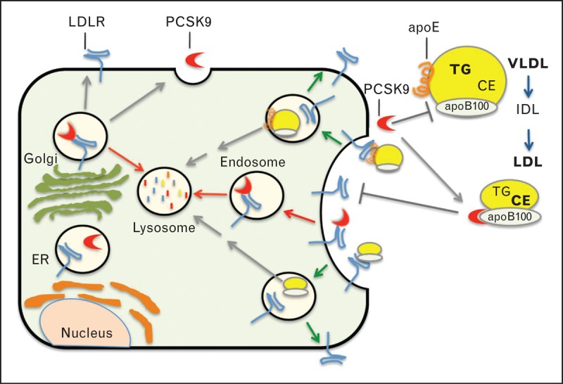
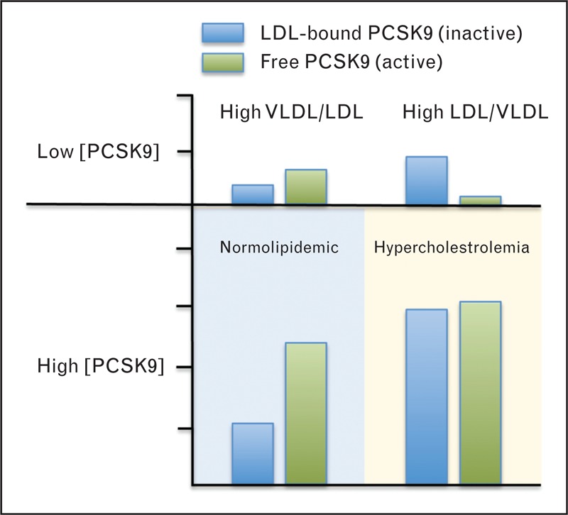
PURPOSE OF REVIEW: Proprotein convertase subtilisin/kexin type-9 (PCSK9) binds to LDL receptor (LDLR) and targets it for lysosomal degradation in cells. Decreased hepatic clearance of plasma LDL-cholesterol is the primary gauge of PCSK9 activity in humans; however, PCSK9's evolutionary role may extend to other lipoprotein classes and processes. This review highlights studies that are providing novel insights into physiological regulation of PCSK9 transcription and plasma PCSK9 activity. RECENT FINDINGS: Recent studies indicate that circulating PCSK9 binds to apolipoprotein B100 on LDL particles, which in turn inhibits PCSK9's ability to bind to cell surface LDLRs. Negative feedback of secreted PCSK9 activity by LDL could serve to increase plasma excursion of triglyceride-rich lipoproteins and monitor lipoprotein remodeling. Recent findings have identified hepatocyte nuclear factor-1α as a key transcriptional regulator that cooperates with sterol regulatory element-binding protein-2 to control PCSK9 expression in hepatocytes in response to nutritional and hormonal inputs, as well as acute inflammation. SUMMARY: PCSK9 is an established target for cholesterol-lowering therapies. Further study of PCSK9 regulatory mechanisms may identify additional control points for pharmacological inhibition of PCSK9-mediated LDLR degradation. PCSK9 function could reflect ancient roles in the fasting-feeding cycle and in linking lipoprotein metabolism with innate immunity.
Overview
The claims and statistics of this paper haven’t been checked yet.
Run the check — free, no account needed. It reads the open-access text and checks the sources it cites.
Integrity signals
Registry
Cites
Statistics
Figures
Verification
Claims the paper makes
The claims and statistics of this paper haven’t been checked yet.
Run the check — free, no account needed. It reads the open-access text and checks the sources it cites.
Claims other papers make about this one
Each is checked against this paper’s own text: does it say what the citing paper says it does?
The source backs part of the claim, not all of it — or backs it for a different population, condition or comparison.
In the citing paper
“In the MK-0616 higher dose group (18 to 20 mg), LDL-C closely tracked with ApoB, supporting a shared mechanism of particle clearance through PCSK9 inhibition, consistent with its known effect on LDL receptor recycling [18].”
From Oral PCSK9 inhibitors for elevated LDL-C: A systematic review and meta-analysis of randomized trials (2026) · cites this paper as [18]
Experimental — accuracy not yet measuredThe cited source says what the claim attributes to it.
In the citing paper
“By inhibiting this interaction, PCSK9 targeted therapy preserves LDL receptor recycling, increases hepatic receptor abundance, and enhances clearance of circulating ApoB-containing lipoproteins [18].”
From Oral PCSK9 inhibitors for elevated LDL-C: A systematic review and meta-analysis of randomized trials (2026) · cites this paper as [18]
The cited source says what the claim attributes to it.
In the citing paper
“Mechanistically, PCSK9 binds hepatic LDL receptors and targets them for lysosomal degradation, thereby limiting receptor recycling and reducing hepatocyte surface receptor availability.”
From Oral PCSK9 inhibitors for elevated LDL-C: A systematic review and meta-analysis of randomized trials (2026) · cites this paper as [18]
The cited source says what the claim attributes to it.
In the citing paper
“By remaining bound to the LDLR, PCSK9 prevents the conformational change of the LDLR that is required for its recycling back to the plasma membrane, therefore, inhibiting this recycling [38]. Instead, the PCSK9–LDLR complex is directed to the lysosome where both proteins are degraded (Figure 1C), thereby reducing the amount of LDLR in the cell [39].”
From Post-translational regulation of the low-density lipoprotein receptor provides new targets for cholesterol regulation (2024) · cites this paper as [39]
The cited source says what the claim attributes to it.
In the citing paper
“Proprotein convertase subtilisin/kexin type 9 and the asialoglycoprotein receptor 1 both independently interact with the LDLR and direct it towards the lysosome for degradation.”
From Post-translational regulation of the low-density lipoprotein receptor provides new targets for cholesterol regulation (2024) · cites this paper as [39]
The cited source says what the claim attributes to it.
In the citing paper
“As PCSK9 reroutes internalized LDLR for intracellular degradation (Lagace, 2014)”
From γ-secretase inhibition lowers plasma triglyceride-rich lipoproteins by stabilizing the LDL receptor (2018) · cites this paper as Lagace, 2014
The cited source says what the claim attributes to it.
In the citing paper
“PCSK9 plays an important role in cholesterol homeostasis as a result of its ability to reduce cell-surface LDLR levels7.”
From Triciribine increases LDLR expression and LDL uptake through stabilization of LDLR mRNA (2018) · cites this paper as [7]
The cited source says what the claim attributes to it.
In the citing paper
“Post-translational regulation of LDLR is primarily governed by PCSK9 that upon binding to cell-surface LDLR mediates its degradation7.”
From Triciribine increases LDLR expression and LDL uptake through stabilization of LDLR mRNA (2018) · cites this paper as [7]
The cited source says what the claim attributes to it.
In the citing paper
“PCSK9 is another potent LDLR degrader aside from IDOL (28, 29).”
From SUMOylation of the ubiquitin ligase IDOL decreases LDL receptor levels and is reversed by SENP1 (2021) · cites this paper as [29]
Self-check
Percentages, sums and reported differences re-computed from the paper’s own numbers.
The self-check needs the paper’s full text.
Citations
Cites
SREBPs: activators of the complete program of cholesterol and fatty acid synthesis in the liver
Horton, J. D., Goldstein, J. L., & Brown, M. S. (2002) · Journal of Clinical Investigation · doi:10.1172/jci0215593 · Cited by 4,092
Sequence Variations in PCSK9, Low LDL, and Protection against Coronary Heart Disease
Cohen, J. C., Boerwinkle, E., Mosley, T. H., & Hobbs, H. H. (2006) · New England Journal of Medicine · doi:10.1056/nejmoa054013 · Cited by 3,181
Cohen, J., Pertsemlidis, A., Kotowski, I. K., Graham, R., Garcia, C. K., & Hobbs, H. H. (2005) · Nature Genetics · doi:10.1038/ng1509 · Cited by 1,381
Li, S., Brown, M. S., & Goldstein, J. L. (2010) · Proceedings of the National Academy of Sciences · doi:10.1073/pnas.0914798107 · Cited by 733
A 52-Week Placebo-Controlled Trial of Evolocumab in Hyperlipidemia
Blom, D. J., Hala, T., Bolognese, M., Lillestol, M. J., Toth, P. D., Burgess, L., Ceska, R., Roth, E., Koren, M. J., Ballantyne, C. M., et al. (2014) · New England Journal of Medicine · doi:10.1056/nejmoa1316222 · Cited by 720
Kosenko, T., Golder, M., Leblond, G., Weng, W., & Lagace, T. A. (2013) · Journal of Biological Chemistry · doi:10.1074/jbc.m112.421370 · Cited by 181
Self-citationPostmus, I., Trompet, S., de Craen, A. J. M., Buckley, B. M., Ford, I., Stott, D. J., Sattar, N., Slagboom, P. E., Westendorp, R. G. J., & Jukema, J. W. (2013) · Journal of Lipid Research · doi:10.1194/jlr.m033969 · Cited by 67
Research works printed but not linked: 57
- Abifadel, 2009, Hum Mutat, 30, 520, 10.1002/humu.20882 · DOI
- Abifadel, 2003, Nat Genet, 34, 154, 10.1038/ng1161 · DOI · Paper page
- Lagace, 2006, J Clin Invest, 116, 2995, 10.1172/JCI29383 · DOI · Paper page
- Zhang, 2007, J Biol Chem, 282, 18602, 10.1074/jbc.M702027200 · DOI · Paper page
- Benjannet, 2004, J Biol Chem, 279, 48865, 10.1074/jbc.M409699200 · DOI · Paper page
- Kwon, 2008, Proc Natl Acad Sci U S A, 105, 1820, 10.1073/pnas.0712064105 · DOI · Paper page
- Chan, 2009, Proc Natl Acad Sci U S A, 106, 9820, 10.1073/pnas.0903849106 · DOI · Paper page
- Poirier, 2013, Drug Des Devel Ther, 7, 1135
- Stein, 2013, Curr Atheroscler Rep, 15, 310, 10.1007/s11883-013-0310-3 · DOI
- Seidah, 2003, Proc Natl Acad Sci U S A, 100, 928, 10.1073/pnas.0335507100 · DOI · Paper page
- Seidah, 2013, J Biol Chem, 288, 21473, 10.1074/jbc.R113.481549 · DOI
- Cunningham, 2007, Nat Struct Mol Biol, 14, 413, 10.1038/nsmb1235 · DOI · Paper page
- McNutt, 2007, J Biol Chem, 282, 20799, 10.1074/jbc.C700095200 · DOI · Paper page
- Grefhorst, 2008, J Lipid Res, 49, 1303, 10.1194/jlr.M800027-JLR200 · DOI · Paper page
- Poirier, 2009, J Biol Chem, 284, 28856, 10.1074/jbc.M109.037085 · DOI · Paper page
- Zhang, 2008, Proc Natl Acad Sci U S A, 105, 13045, 10.1073/pnas.0806312105 · DOI · Paper page
- Lo Surdo, 2011, EMBO Rep, 12, 1300, 10.1038/embor.2011.205 · DOI · Paper page
- Tavori, 2013, Circulation, 127, 2403, 10.1161/CIRCULATIONAHA.113.001592 · DOI · Paper page
- Dubuc, 2004, Arterioscler Thromb Vasc Biol, 24, 1454, 10.1161/01.ATV.0000134621.14315.43 · DOI · Paper page
- JeongJeong, 2008, J Lipid Res, 49, 399, 10.1194/jlr.M700443-JLR200 · DOI · Paper page
- Brown, 1997, Cell, 89, 331, 10.1016/S0092-8674(00)80213-5 · DOI · Paper page
- Browning, 2010, J Lipid Res, 51, 3359, 10.1194/jlr.P009860 · DOI
- Persson, 2010, Arterioscler Thromb Vasc Biol, 30, 2666, 10.1161/ATVBAHA.110.214130 · DOI · Paper page
- Horton, 2007, Trends Biochem Sci, 2, 71, 10.1016/j.tibs.2006.12.008 · DOI · Paper page
- Twisk, 2000, J Clin Invest, 105, 521, 10.1172/JCI8623 · DOI
- Blasiole, 2008, J Biol Chem, 283, 11374, 10.1074/jbc.M710457200 · DOI · Paper page
- Lambert, 2006, Endocrinology, 147, 4985, 10.1210/en.2006-0098 · DOI
- Herbert, 2010, Arterioscler Thromb Vasc Biol, 30, 1333, 10.1161/ATVBAHA.110.204040 · DOI
- Sun, 2012, Arterioscler Thromb Vasc Biol, 32, 1585, 10.1161/ATVBAHA.112.250043 · DOI
- Li, 2009, J Biol Chem, 284, 28885, 10.1074/jbc.M109.052407 · DOI
- Armendariz, 2009, Curr Opin Lipidol, 20, 106, 10.1097/MOL.0b013e3283295ee9 · DOI
- Dong, 2010, J Lipid Res, 51, 1486, 10.1194/jlr.M003566 · DOI
- Ai, 2012, J Clin Invest, 122, 1262, 10.1172/JCI61919 · DOI
- Shimomura, 2000, Mol Cell, 6, 77, 10.1016/S1097-2765(00)00009-5 · DOI
- Laplante, 2012, Cell, 149, 274, 10.1016/j.cell.2012.03.017 · DOI · Paper page
- Lakoski, 2009, J Clin Endocrinol Metab, 94, 2537, 10.1210/jc.2009-0141 · DOI · Paper page
- Kwakernaak, 2014, Clin Biochem, 47, 679, 10.1016/j.clinbiochem.2014.03.008 · DOI
- Benjannet, 2010, J Biol Chem, 285, 40965, 10.1074/jbc.M110.154815 · DOI
- Holla, 2011, Biochem Biophys Res Commun, 406, 234, 10.1016/j.bbrc.2011.02.023 · DOI
- Benjannet, 2006, J Biol Chem, 281, 30561, 10.1074/jbc.M606495200 · DOI · Paper page
- Han, 2014, J Lipid Res, 55, 1505, 10.1194/jlr.M049346 · DOI
- Essalmani, 2011, J Biol Chem, 286, 4257, 10.1074/jbc.M110.192104 · DOI · Paper page
- Azzam, 2012, Trends Endocrinol Metab, 23, 169, 10.1016/j.tem.2012.02.001 · DOI · Paper page
- Vreugdenhil, 2001, J Clin Invest, 107, 225, 10.1172/JCI10832 · DOI
- Feingold, 2008, Biochem Biophys Res Commun, 374, 341, 10.1016/j.bbrc.2008.07.023 · DOI
- Xiao, 2012, Pharmacol Rep, 64, 889, 10.1016/S1734-1140(12)70883-6 · DOI
- Barter, 2003, Arterioscler Thromb Vasc Biol, 23, 160, 10.1161/01.ATV.0000054658.91146.64 · DOI
- Annema, 2012, Nutr Metab (Lond), 9, 25, 10.1186/1743-7075-9-25 · DOI
- Moore, 2013, Nat Rev Immunol, 13, 709, 10.1038/nri3520 · DOI · Paper page
- Le May, 2013, Arterioscler Thromb Vasc Biol, 33, 1484, 10.1161/ATVBAHA.112.300263 · DOI
- Van der Veen, 2009, J Biol Chem, 284, 19211, 10.1074/jbc.M109.014860 · DOI
- Brown, 1997, Nature, 388, 629, 10.1038/41672 · DOI · Paper page
- Brown, 1981, N Engl J Med, 305, 515, 10.1056/NEJM198108273050909 · DOI
- Raal, 2013, J Am Heart Assoc, 2, e000028, 10.1161/JAHA.112.000028 · DOI
- Richard, 2012, Br J Nutr, 107, 705, 10.1017/S0007114511003436 · DOI
- Raal, 2014, J Am Coll Cardiol, 63, 1278, 10.1016/j.jacc.2014.01.006 · DOI
- Koren, 2014, J Am Coll Cardiol, 63, 2531, 10.1016/j.jacc.2014.03.018 · DOI · Paper page
Cited by
Sorted by citations
Mechanisms and regulation of cholesterol homeostasis
Luo, J., Yang, H., & Song, B.-L. (2020) · Nature Reviews Molecular Cell Biology · doi:10.1038/s41580-019-0190-7 · Cited by 1,980
Metabolism of triglyceride-rich lipoproteins in health and dyslipidaemia
Borén, J., Taskinen, M.-R., Björnson, E., & Packard, C. J. (2022) · Nature Reviews Cardiology · doi:10.1038/s41569-022-00676-y · Cited by 315
Langsted, A., Nordestgaard, B. G., Benn, M., Tybjærg-Hansen, A., & Kamstrup, P. R. (2016) · The Journal of Clinical Endocrinology & Metabolism · doi:10.1210/jc.2016-1206 · Cited by 136
Dijk, W., Ruppert, P. M. M., Oost, L. J., & Kersten, S. (2018) · Journal of Biological Chemistry · doi:10.1074/jbc.ra118.002426 · Cited by 72
PCSK9: A Multi-Faceted Protein That Is Involved in Cardiovascular Biology
Sundararaman, S. S., Döring, Y., & van der Vorst, E. P. C. (2021) · Biomedicines · doi:10.3390/biomedicines9070793 · Cited by 67
Metadata
- Authors
- Lagace, T. A.
- Affiliations
- Ottawa Heart Institute (CA)
- Corresponding author
- Lagace, Thomas A.
- ORCID
- Lagace, Thomas A.
- Venue
- Current Opinion in Lipidology
- Published
- 2014-10
- Volume
- 25
- Issue
- 5
- Pages
- 387-393
- ISSN
- 0957-9672 (ISSN-L)
- PMC
- PMC4166010
- OpenAlex
- W2036304437
- Cited by
- 372
- References
- 7
- Citations by year
- 2026: 28 · 2025: 41 · 2024: 55 · 2023: 37 · 2022: 38 · 2021: 36 · 2020: 36 · 2019: 25 · 2018: 25 · 2017: 20 · 2016: 18 · 2015: 13
- FWCI
- 3.21
- Type
- journal article
- Language
- en
- Open access
- hybrid
- doi.org
- Licence
- cc-by-nc-nd
- Locations
- Current Opinion in Lipidology (publishedVersion, cc-by-nc-nd)
- PubMed Central (publishedVersion, cc-by-nc-nd)
- PubMed (publishedVersion)
- Funders
- Heart and Stroke Foundation of Canada
- Canadian Institutes of Health Research (106462_1)
- Topics
- Health Sciences › Medicine › Cardiology and Cardiovascular Medicine › Lipoproteins and Cardiovascular Health
- Life Sciences › Biochemistry, Genetics and Molecular Biology › Biochemistry › Cholesterol and Lipid Metabolism
- Health Sciences › Medicine › Cardiology and Cardiovascular Medicine › Lipid metabolism and disorders
- Keywords
- PCSK9, LDL receptor, LDLR degradation, lysosomal degradation, apolipoprotein B-100, triglyceride-rich lipoproteins, lipoprotein remodeling, HNF1A, SREBP2, PCSK9 inhibitors, innate immunity, lipoprotein metabolism
- MeSH terms
- Proprotein Convertase 9, Apolipoproteins B, Gene Expression Regulation, Humans, Immunity, Innate, Inflammation, Lipoproteins, LDL, Lysosomes, Protein Binding, Receptors, LDL, Serine Endopeptidases, Hepatocytes, Proprotein Convertases, Proteolysis
About these checks
Registry, retraction, Cites and statistics results are computed, not judged, with one exception: a printed reference that no index lists is read once by a model before it counts as a citation error. Claims are extracted by a model and kept only when their quote appears verbatim in the text; each cited source is then checked, with a model’s help, for whether it says what the claim relies on. Those verdicts can be wrong — read the claim’s quote and the cited source before relying on one.
Statistics and arithmetic are recomputed, not judged.
Confidence bands are shown only for fields calibrated on our annotated test set. None is calibrated yet, so no band is shown.
What counts as a claim ›How we measure confidence ›How we check this ›
Are you an author, or do you see a problem with this page? Request a review
