

The DOI matches its Crossref record; 1 of the 1 references it cites were found in a registry or an index.
IgE antibodies increase honeybee venom responsiveness and detoxification efficiency of mast cells
Abstract
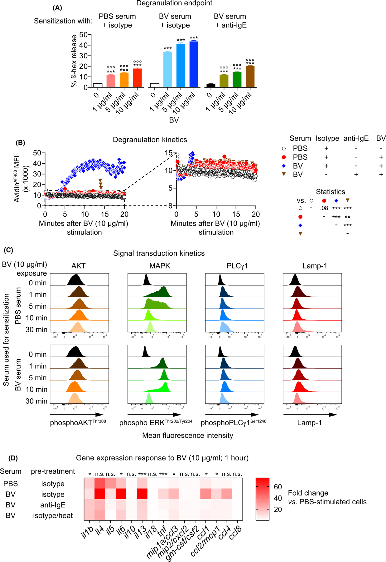
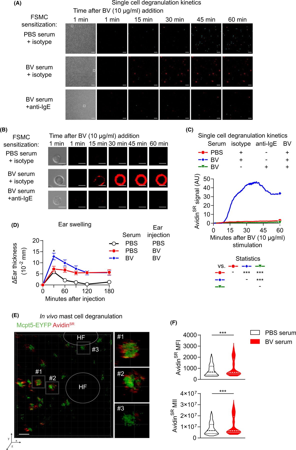
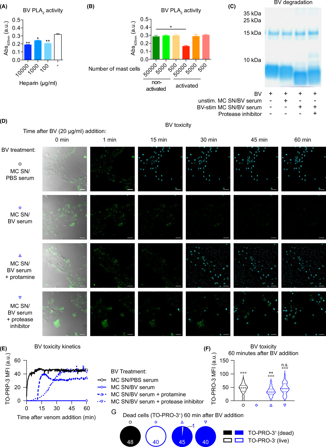
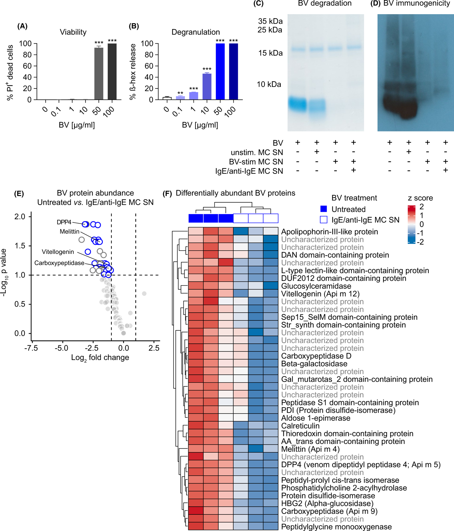
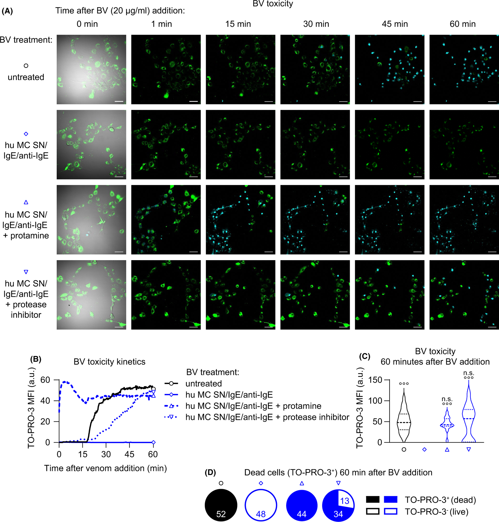
Background: In contrast to their clearly defined roles in allergic diseases, the physiologic functions of Immunoglobulin E antibodies (IgEs) and mast cells (MCs) remain enigmatic. Recent research supports the toxin hypothesis, showing that MCs and IgE‐related type 2 immune responses can enhance host defense against certain noxious substances, including honeybee venom (BV). However, the mechanisms by which MCs can interfere with BV toxicity are unknown. In this study, we assessed the role of IgE and certain MC products in MC‐mediated BV detoxification. Methods: We applied in vitro and in vivo fluorescence microscopyimaging, and flow cytometry, fibroblast‐based toxicity assays and mass spectrometry to investigate IgE‐mediated detoxification of BV cytotoxicity by mouse and human MCs in vitro. Pharmacologic strategies to interfere with MC‐derived heparin and proteases helped to define the importance of specific detoxification mechanisms. Results: Venom‐specific IgE increased the degranulation and cytokine responses of MCs to BV in vitro. Passive serum sensitization enhanced MC degranulation in vivo. IgE‐activated mouse or human MCs exhibited enhanced potential for detoxifying BV by both proteolytic degradation and heparin‐related interference with toxicity. Mediators released by IgE‐activated human MCs efficiently degraded multiple BV toxins. Conclusions: Our results both reveal that IgE sensitization enhances the MC's ability to detoxify BV and also assign efficient toxin‐neutralizing activity to MC‐derived heparin and proteases. Our study thus highlights the potential importance of IgE, MCs, and particular MC products in defense against BV.
Overview
The claims and statistics of this paper haven’t been checked yet.
Run the check — free, no account needed. It reads the open-access text and checks the sources it cites.
Integrity signals
Registry
Cites
Statistics
Figures
Verification
Claims the paper makes
The claims and statistics of this paper haven’t been checked yet.
Run the check — free, no account needed. It reads the open-access text and checks the sources it cites.
Claims other papers make about this one
Each is checked against this paper’s own text: does it say what the citing paper says it does?
In the citing paper
“They also mediate protection from venoms produced by a range of animals, including snakes and bees. 5 , 6 , 7”
From Mast cell ontogeny: From fetal development to life‐long health and disease (2023) · cites this paper as [7]
In the citing paper
“In adults, mast cells have important functions in protective immunity from certain bacteria, 1 helminths 2 , 3 , 4 and animal venoms. 6 , 7”
From Mast cell ontogeny: From fetal development to life‐long health and disease (2023) · cites this paper as [7]
In the citing paper
“On the other hand, mast cells can also be protective, for example against Staphylococcus, honeybee, and snake venom. 1 , 5 , 6 , 7”
From Mast cell ontogeny: From fetal development to life‐long health and disease (2023) · cites this paper as [7]
Self-check
Percentages, sums and reported differences re-computed from the paper’s own numbers.
The self-check needs the paper’s full text.
Citations
Cites
IgE and mast cells in allergic disease
Galli, S. J., & Tsai, M. (2012) · Nature Medicine · doi:10.1038/nm.2755 · Cited by 1,799
Self-citation
Research works printed but not linked: 37
- 10.1038/nri3690 · DOI · Paper page
- 10.1016/j.jaci.2017.06.003 · DOI
- 10.1038/nri3427 · DOI · Paper page
- 10.1007/s00281-016-0565-1 · DOI
- 10.1016/j.jaci.2018.10.054 · DOI
- 10.1146/annurev.genom.9.081307.164356 · DOI
- 10.1038/nature11047 · DOI · Paper page
- 10.1086/417049 · DOI · Paper page
- 10.1353/pbm.1974.0027 · DOI · Paper page
- 10.3389/fimmu.2019.02090 · DOI
- 10.1111/all.13262 · DOI
- 10.1126/science.177.4046.314 · DOI · Paper page
- 10.1126/science.1128877 · DOI · Paper page
- 10.1084/jem.20071262 · DOI · Paper page
- 10.1172/JCI46139 · DOI · Paper page
- 10.1016/j.jaci.2015.08.005 · DOI · Paper page
- 10.1016/j.immuni.2013.10.005 · DOI · Paper page
- 10.1016/j.immuni.2013.10.006 · DOI · Paper page
- 10.1016/0041-0101(95)00011-A · DOI
- 10.1038/ncomms7174 · DOI
- 10.1002/eji.201444546 · DOI
- 10.1172/JCI85538 · DOI
- 10.1111/imr.12634 · DOI
- 10.1111/imr.12629 · DOI
- 10.4049/jimmunol.106.1.233 · DOI · Paper page
- 10.1038/nature03085 · DOI · Paper page
- Jaques LB, 1973, Protamine–antagonist to heparin, Can Med Assoc J, 108, 1291
- 10.3389/fimmu.2014.00077 · DOI
- 10.1016/j.jaci.2013.10.060 · DOI
- 10.1038/nri2273 · DOI
- 10.1038/nri2782 · DOI · Paper page
- 10.1038/nri3476 · DOI
- 10.1038/nri3632 · DOI
- 10.1126/sciimmunol.aat1604 · DOI
- 10.1007/s00103-012-1652-7 · DOI
- 10.1111/all.13071 · DOI
- 10.1097/01.all.0000173783.42906.95 · DOI · Paper page
Cited by
Sorted by citations
Mast cell ontogeny: From fetal development to life‐long health and disease
Chia, S. L., Kapoor, S., Carvalho, C., Bajénoff, M., & Gentek, R. (2023) · Immunological Reviews · doi:10.1111/imr.13191 · Cited by 60
Metadata
- Authors
- Starkl, P., Gaudenzio, N., Marichal, T., Reber, L. L., Sibilano, R., Watzenboeck, M. L., Fontaine, F., Mueller, A. C., Tsai, M., Knapp, S., & Galli, S. J.
- Affiliations
- Laboratory of Infection Biology Department of Medicine I Medical University of Vienna Vienna Austria
- Department of Pathology Stanford University School of Medicine Stanford CA USA
- Department of Biomedical Imaging and Image‐guided Therapy Medical University of Vienna Vienna Austria
- CeMM ‐ Research Center for Molecular Medicine of the Austrian Academy of Sciences Vienna Austria
- Venue
- Allergy
- Publisher
- Wiley
- Published
- 2021-05-06
- Volume
- 77
- Issue
- 2
- Pages
- 499-512
- ISSN
- 0105-4538
- 1398-9995
- PMC
- PMC8502784
- Cited by
- 33
- References
- 1
- Language
- en
- Funders
- H2020 Marie Skłodowska-Curie Actions (655153)
- Austrian Science Fund (J3399‐B21, P31113‐B30)
- Fondation Acteria
- Fonds De La Recherche Scientifique - FNRS (F.4508.18)
- Institut National de la Santé et de la Recherche Médicale
- National Institute of Allergy and Infectious Diseases (R01 AI23990, R01 AI070813, R01 AI132494, R01 AR067145)
- Agence Nationale de la Recherche (ANR‐18‐CE18‐0023)
About these checks
Registry, retraction, Cites and statistics results are computed, not judged, with one exception: a printed reference that no index lists is read once by a model before it counts as a citation error. Claims are extracted by a model and kept only when their quote appears verbatim in the text; each cited source is then checked, with a model’s help, for whether it says what the claim relies on. Those verdicts can be wrong — read the claim’s quote and the cited source before relying on one.
Statistics and arithmetic are recomputed, not judged.
Confidence bands are shown only for fields calibrated on our annotated test set. None is calibrated yet, so no band is shown.
What counts as a claim ›How we measure confidence ›How we check this ›
Are you an author, or do you see a problem with this page? Request a review
How to cite
Cite CoChat for the checks on this page, and the paper for its own findings.
- This page
- CoChat. (2026). Citation check: IgE antibodies increase honeybee venom responsiveness and detoxification efficiency of mast cells. https://app.cochat.ai/papers/10.1111/all.14852
- The paper
- Starkl, P., Gaudenzio, N., Marichal, T., Reber, L. L., Sibilano, R., Watzenboeck, M. L., Fontaine, F., Mueller, A. C., Tsai, M., Knapp, S., & Galli, S. J. (2022). IgE antibodies increase honeybee venom responsiveness and detoxification efficiency of mast cells. Allergy. https://doi.org/10.1111/all.14852
