

The DOI matches its Crossref record; 21 of the 21 references it cites were found in a registry or an index.
Mapping the developing human immune system across organs
Published version of a preprint
Abstract
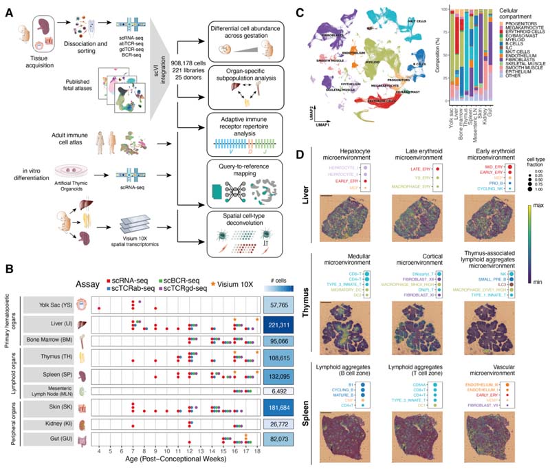
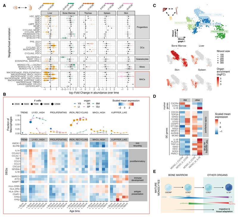
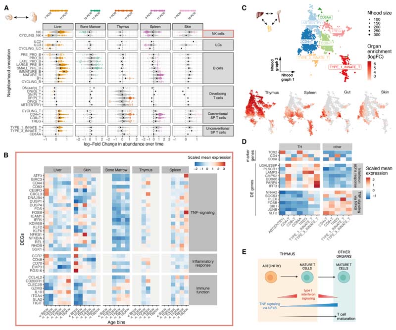
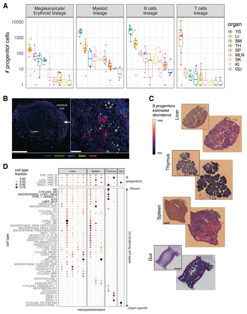
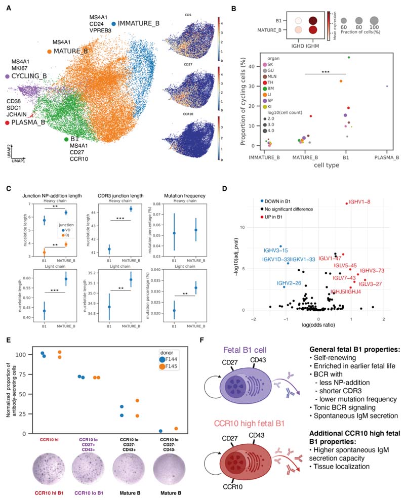
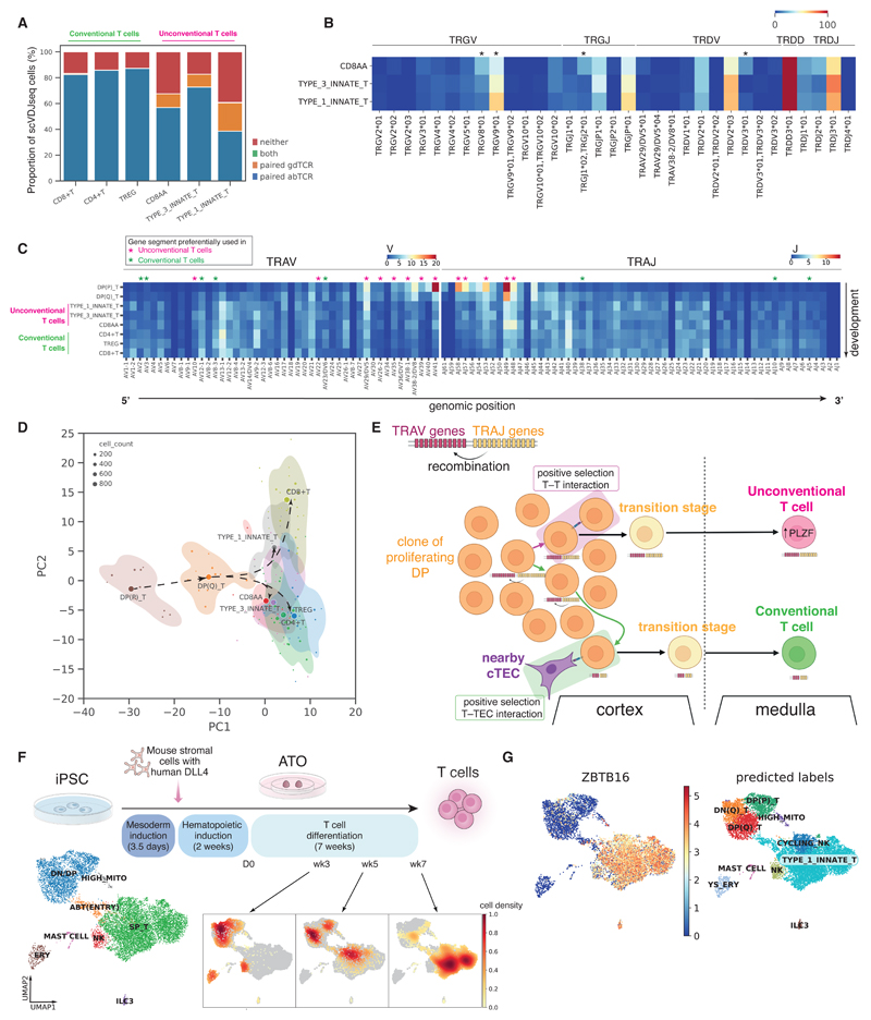
Single-cell genomics studies have decoded the immune cell composition of several human prenatal organs but were limited in describing the developing immune system as a distributed network across tissues. We profiled nine prenatal tissues combining single-cell RNA sequencing, antigen-receptor sequencing, and spatial transcriptomics to reconstruct the developing human immune system. This revealed the late acquisition of immune-effector functions by myeloid and lymphoid cell subsets and the maturation of monocytes and T cells before peripheral tissue seeding. Moreover, we uncovered system-wide blood and immune cell development beyond primary hematopoietic organs, characterized human prenatal B1 cells, and shed light on the origin of unconventional T cells. Our atlas provides both valuable data resources and biological insights that will facilitate cell engineering, regenerative medicine, and disease understanding.
Overview
The claims and statistics of this paper haven’t been checked yet.
Run the check — free, no account needed. It reads the open-access text and checks the sources it cites.
Integrity signals
Registry
Cites
Statistics
Figures
Verification
Claims the paper makes
The claims and statistics of this paper haven’t been checked yet.
Run the check — free, no account needed. It reads the open-access text and checks the sources it cites.
Claims other papers make about this one
Each is checked against this paper’s own text: does it say what the citing paper says it does?
In the citing paper
“These do not yet have granules but express cardinal markers of mast cells such as Kit, as well as several mast cell proteases, at least on transcriptional level. 54 , 59 , 60”
From Mast cell ontogeny: From fetal development to life‐long health and disease (2023) · cites this paper as [60]
In the citing paper
“These transcriptional differences parallel those seen in fetal macrophages. 60”
From Mast cell ontogeny: From fetal development to life‐long health and disease (2023) · cites this paper as [60]
In the citing paper
“In line with this, human fetal mast cells show considerable heterogeneity across developmental stages (7‐17 weeks of gestation) and tissues analyzed, that is, liver, BM, thymus, spleen, and skin. 60”
From Mast cell ontogeny: From fetal development to life‐long health and disease (2023) · cites this paper as [60]
In the citing paper
“Although the idea that mast cells support developmental processes has gained traction, 10 , 11 , 60 , 117 mice that lack mast cells develop normally without obvious defects, and mast cell deficiencies have not been reported in humans. The current consensus therefore is that mast cells are dispensable for development.”
From Mast cell ontogeny: From fetal development to life‐long health and disease (2023) · cites this paper as [60]
In the citing paper
“At least on transcript level, fetal mast cells also express key effectors like proteases. 53 , 54 , 63 This is also true for human fetal mast cells, which express, for example, TPSAB1, the gene encoding tryptase, and carry granules. 59 , 60 , 63”
From Mast cell ontogeny: From fetal development to life‐long health and disease (2023) · cites this paper as [60]
In the citing paper
“Finally, single cell transcriptional profiling recently demonstrated that the earliest mast cells found in human fetuses express transcripts for pro‐inflammatory cytokines and chemokines involved in recruitment of endothelial cells. 60”
From Mast cell ontogeny: From fetal development to life‐long health and disease (2023) · cites this paper as [60]
In the citing paper
“Although there is currently no experimental data showing this directly, human fetal mast cells acquire transcriptional signatures compatible with immune effector functions already from 10 weeks of gestation onwards. 60 , 116”
From Mast cell ontogeny: From fetal development to life‐long health and disease (2023) · cites this paper as [60]
Self-check
Percentages, sums and reported differences re-computed from the paper’s own numbers.
The self-check needs the paper’s full text.
Citations
Cites
SCANPY: large-scale single-cell gene expression data analysis
Wolf, F. A., Angerer, P., & Theis, F. J. (2018) · Genome Biology · doi:10.1186/s13059-017-1382-0 · Cited by 8,881
Enrichr: interactive and collaborative HTML5 gene list enrichment analysis tool
Chen, E. Y., Tan, C. M., Kou, Y., Duan, Q., Wang, Z., Meirelles, G. V., Clark, N. R., & Ma’ayan, A. (2013) · BMC Bioinformatics · doi:10.1186/1471-2105-14-128 · Cited by 7,814
From Louvain to Leiden: guaranteeing well-connected communities
Traag, V. A., Waltman, L., & van Eck, N. J. (2019) · Scientific Reports · doi:10.1038/s41598-019-41695-z · Cited by 5,635
Scrublet: computational identification of cell doublets in single-cell transcriptomic data
Wolock, S. L., Lopez, R., & Klein, A. M. (2018) · doi:10.1101/357368 · Cited by 2,758 · cited 1×
“Low-quality cells were filtered out [minimum number of reads = 2000, minimum number of genes = 500, minimum Scrublet (77) doublet detection score <0.”
BackgroundDeep generative modeling for single-cell transcriptomics
Lopez, R., Regier, J., Cole, M. B., Jordan, M. I., & Yosef, N. (2018) · Nature Methods · doi:10.1038/s41592-018-0229-2 · Cited by 2,530
Single-cell RNA-seq reveals new types of human blood dendritic cells, monocytes, and progenitors
Villani, A.-C., Satija, R., Reynolds, G., Sarkizova, S., Shekhar, K., Fletcher, J., Griesbeck, M., Butler, A., Zheng, S., Lazo, S., et al. (2017) · Science · doi:10.1126/science.aah4573 · Cited by 2,413
Self-citationConfronting false discoveries in single-cell differential expression
Squair, J. W., Gautier, M., Kathe, C., Anderson, M. A., James, N. D., Hutson, T. H., Hudelle, R., Qaiser, T., Matson, K. J. E., Barraud, Q., et al. (2021) · doi:10.1101/2021.03.12.435024 · Cited by 1,086 · cited 1×
“Briefly, single-cell expression profiles were first aggregated into pseudobulk expression profiles x̂ for each cell type c and sample s [as recommended by (85, 86)].”
MethodThe double life of a B-1 cell: self-reactivity selects for protective effector functions
Baumgarth, N. (2011) · Nature Reviews Immunology · doi:10.1038/nri2901 · Cited by 969
Specification of tissue-resident macrophages during organogenesis
Mass, E., Ballesteros, I., Farlik, M., Halbritter, F., Günther, P., Crozet, L., Jacome-Galarza, C. E., Händler, K., Klughammer, J., Kobayashi, Y., et al. (2016) · Science · doi:10.1126/science.aaf4238 · Cited by 891
Characterization of cell fate probabilities in single-cell data with Palantir
Setty, M., Kiseliovas, V., Levine, J., Gayoso, A., Mazutis, L., & Pe’er, D. (2019) · Nature Biotechnology · doi:10.1038/s41587-019-0068-4 · Cited by 833 · cited 1×
“S20, C and D, the Palantir method as implemented in CellRank was used (94, 95).”
Method
Research works printed but not linked: 82
- 10.1126/science.aaz9330 · DOI
- 10.1242/dev.083147 · DOI
- 10.1126/science.aat5031 · DOI
- 10.1038/s41422-019-0228-6 · DOI
- 10.1016/j.immuni.2019.09.008 · DOI
- 10.1126/science.aay3224 · DOI
- 10.1016/j.devcel.2020.11.010 · DOI
- 10.1038/s41586-021-03929-x · DOI
- 10.1038/s41587-021-01139-4 · DOI · Paper page
- 10.1038/s41467-018-06318-7 · DOI · Paper page
- 10.3389/fgene.2021.781272 · DOI
- 10.1126/science.abl5197 · DOI
- 10.1038/s41587-021-01033-z · DOI · Paper page
- 10.3389/fimmu.2019.02700 · DOI
- 10.1002/ijc.21596 · DOI
- F. Hua, Y. Tian, CCL4 promotes the cell proliferation, invasion and migration of endometrial carcinoma by targeting the VEGF-A signal pathway. Int. J. Clin. Exp. Pathol. 10, 11288–11299 (2017). 31966483
- 10.1016/S0065-230X(10)06003-3 · DOI
- 10.1074/jbc.M208231200 · DOI
- 10.1034/j.1600-0463.2002.100501.x · DOI
- 10.1016/j.yexcr.2015.11.022 · DOI
- 10.1016/j.devcel.2017.01.001 · DOI
- 10.1016/j.smim.2008.02.003 · DOI
- 10.1038/nature19814 · DOI
- 10.1038/s41590-018-0294-9 · DOI
- 10.1016/j.cell.2021.04.039 · DOI
- 10.1038/ni.3419 · DOI
- 10.1084/jem.20151604 · DOI
- 10.1007/s10875-021-01059-7 · DOI
- 10.1034/j.1398-9995.2000.00118.x · DOI
- 10.1084/jem.20101499 · DOI
- 10.3389/fimmu.2012.00122 · DOI
- 10.1111/nyas.12137 · DOI
- 10.1002/eji.1830190315 · DOI
- 10.1084/jem.157.1.202 · DOI
- 10.1016/j.immuni.2011.11.017 · DOI
- A. B. Kantor, C. E. Merrill, L. A. Herzenberg, J. L. Hillson, An unbiased analysis of V(H)-D-J(H) sequences from B-1a, B-1b, and conventional B cells. J. Immunol. 158, 1175–1186 (1997). 9013957 · DOI
- 10.1002/j.1460-2075.1995.tb07157.x · DOI
- 10.1084/jem.168.2.811 · DOI
- 10.1038/s41586-021-03852-1 · DOI
- 10.1016/j.immuni.2006.10.010 · DOI
- 10.1016/j.coi.2010.12.016 · DOI
- 10.1073/pnas.1412058112 · DOI
- 10.1126/sciimmunol.abf0125 · DOI
- 10.1038/s41586-021-03578-0 · DOI
- 10.1016/j.celrep.2017.05.045 · DOI
- 10.1084/jem.20091519 · DOI
- 10.1073/pnas.1105118108 · DOI
- 10.1038/s41467-021-22589-z · DOI
- 10.1101/gad.10.8.948 · DOI
- 10.1038/nri913 · DOI
- 10.1016/j.stem.2018.12.011 · DOI
- 10.1016/j.exphem.2021.10.002 · DOI
- 10.1182/blood.2021013450 · DOI
- 10.1084/jem.20200737 · DOI
- 10.2147/JBM.S7224 · DOI
- 10.1038/s41593-021-00880-y · DOI
- 10.1016/j.immuni.2021.09.016 · DOI
- 10.1038/ni1301 · DOI
- 10.1073/pnas.0811632106 · DOI
- 10.1073/pnas.1015841108 · DOI
- 10.1016/j.coi.2018.01.001 · DOI
- 10.1126/science.aau8475 · DOI
- 10.1038/s41592-019-0392-0 · DOI · Paper page
- S. J. Fleming J. C. Marioni M. Babadi CellBender remove-background: a deep generative model for unsupervised removal of background noise from scRNA-seq datasets bioRxiv 791699 [Preprint] (2019); .10.1101/791699 · DOI · Paper page
- 10.1016/j.cels.2018.11.005 · DOI · Paper page
- F. Pedregosa., Scikit-learn: Machine learning in python. j. mach. learn. res. 12, 2825–2830 (2011).
- K. Polański, M. D. Young, Z. Miao, K. B. Meyer, S. A. Teichmann, J. E. Park, BBKNN: Fast batch alignment of single cell transcriptomes. Bioinformatics 36, 964–965 (2020). 31400197 · DOI · Paper page
- 10.1093/bioinformatics/btaa1009 · DOI
- 10.1038/s41467-021-25960-2 · DOI · Paper page
- 10.1093/bioinformatics/btp616 · DOI · Paper page
- 10.1093/bioinformatics/btaa611 · DOI
- 10.1038/s41591-021-01329-2 · DOI · Paper page
- 10.1093/bioinformatics/btv359 · DOI
- 10.1038/s41596-020-0292-x · DOI · Paper page
- 10.1038/s41588-021-00972-2 · DOI
- 10.1038/s41592-021-01346-6 · DOI
- 10.1038/s41592-021-01358-2 · DOI
- I. Virshup S. Rybakov F. J. Theis P. Angerer F. A. Wolf anndata: Annotated data bioRxiv 473007 [Preprint] (2021); 10.1101/2021.12.16.473007 · DOI
- 10.1126/sciimmunol.abf7777 · DOI · Paper page
- 10.1073/pnas.1417683112 · DOI
- 10.1016/S1074-7613(01)00185-6 · DOI
- 10.1038/382635a0 · DOI
Cited by
Sorted by citations
Tissue-specific macrophages: how they develop and choreograph tissue biology
Mass, E., Nimmerjahn, F., Kierdorf, K., & Schlitzer, A. (2023) · Nature Reviews Immunology · doi:10.1038/s41577-023-00848-y · Cited by 621
BANKSY unifies cell typing and tissue domain segmentation for scalable spatial omics data analysis
Singhal, V., Chou, N., Lee, J., Yue, Y., Liu, J., Chock, W. K., Lin, L., Chang, Y.-C., Teo, E. M. L., Aow, J., et al. (2024) · Nature Genetics · doi:10.1038/s41588-024-01664-3 · Cited by 282
Novel antigen-presenting cell imparts Treg-dependent tolerance to gut microbiota
Akagbosu, B., Tayyebi, Z., Shibu, G., Paucar Iza, Y. A., Deep, D., Parisotto, Y. F., Fisher, L., Pasolli, H. A., Thevin, V., Elmentaite, R., et al. (2022) · Nature · doi:10.1038/s41586-022-05309-5 · Cited by 273
Self-citationImpact of the Human Cell Atlas on medicine
Rood, J. E., Maartens, A., Hupalowska, A., Teichmann, S. A., & Regev, A. (2022) · Nature Medicine · doi:10.1038/s41591-022-02104-7 · Cited by 222
Self-citationA prenatal skin atlas reveals immune regulation of human skin morphogenesis
Gopee, N. H., Winheim, E., Olabi, B., Admane, C., Foster, A. R., Huang, N., Botting, R. A., Torabi, F., Sumanaweera, D., Le, A. P., et al. (2024) · Nature · doi:10.1038/s41586-024-08002-x · Cited by 108
Self-citation
Metadata
- Authors
- Suo, C., Dann, E., Goh, I., Jardine, L., Kleshchevnikov, V., Park, J.-E., Botting, R. A., Stephenson, E., Engelbert, J., Tuong, Z. K., Polanski, K., Yayon, N., Xu, C., Suchanek, O., Elmentaite, R., Domínguez Conde, C., He, P., Pritchard, S., Miah, M., Moldovan, C., Steemers, A. S., Mazin, P., Prete, M., Horsfall, D., Marioni, J. C., Clatworthy, M. R., Haniffa, M., & Teichmann, S. A.
- Affiliations
- Wellcome Sanger Institute, Wellcome Genome Campus, Hinxton, Cambridge, UK.
- Biosciences Institute, Newcastle University, Newcastle upon Tyne, UK.
- Molecular Immunity Unit, University of Cambridge Department of Medicine, Cambridge, UK.
- Department of Cellular Pathology, Newcastle upon Tyne Hospitals NHS Foundation Trust, Newcastle upon Tyne, UK.
- ORCID
- Suo, Chenqu
- Dann, Emma
- Goh, Issac
- Jardine, Laura
- Kleshchevnikov, Vitalii
- Park, Jong-Eun
- Botting, Rachel A.
- Stephenson, Emily
- Tuong, Zewen Kelvin
- Polanski, Krzysztof
- Yayon, Nadav
- Xu, Chuan
- Suchanek, Ondrej
- Elmentaite, Rasa
- Domínguez Conde, Cecilia
- He, Peng
- Moldovan, Corina
- Steemers, Alexander S.
- Mazin, Pavel
- Prete, Martin
- Horsfall, Dave
- Marioni, John C.
- Clatworthy, Menna R.
- Haniffa, Muzlifah
- Teichmann, Sarah A.
- Venue
- Science
- Published
- 2022-06-03
- Volume
- 376
- Issue
- 6597
- ISSN
- 0036-8075
- 1095-9203
- PMC
- PMC7612819
- Cited by
- 340
- References
- 21
- Language
- en
About these checks
Registry, retraction, Cites and statistics results are computed, not judged, with one exception: a printed reference that no index lists is read once by a model before it counts as a citation error. Claims are extracted by a model and kept only when their quote appears verbatim in the text; each cited source is then checked, with a model’s help, for whether it says what the claim relies on. Those verdicts can be wrong — read the claim’s quote and the cited source before relying on one.
Statistics and arithmetic are recomputed, not judged.
Confidence bands are shown only for fields calibrated on our annotated test set. None is calibrated yet, so no band is shown.
What counts as a claim ›How we measure confidence ›How we check this ›
Are you an author, or do you see a problem with this page? Request a review
How to cite
Cite CoChat for the checks on this page, and the paper for its own findings.
- This page
- CoChat. (2026). Citation check: Mapping the developing human immune system across organs. https://app.cochat.ai/papers/10.1126/science.abo0510
- The paper
- Suo, C., Dann, E., Goh, I., Jardine, L., Kleshchevnikov, V., Park, J.-E., Botting, R. A., Stephenson, E., Engelbert, J., Tuong, Z. K., Polanski, K., Yayon, N., Xu, C., Suchanek, O., Elmentaite, R., Domínguez Conde, C., He, P., Pritchard, S., Miah, M., . . . Teichmann, S. A. (2022). Mapping the developing human immune system across organs. Science. https://doi.org/10.1126/science.abo0510
