

The DOI matches its Crossref record; 65 of the 65 references it cites were found in a registry or an index.
Hallucinating symmetric protein assemblies
Open accessPublished version of a preprint
Abstract
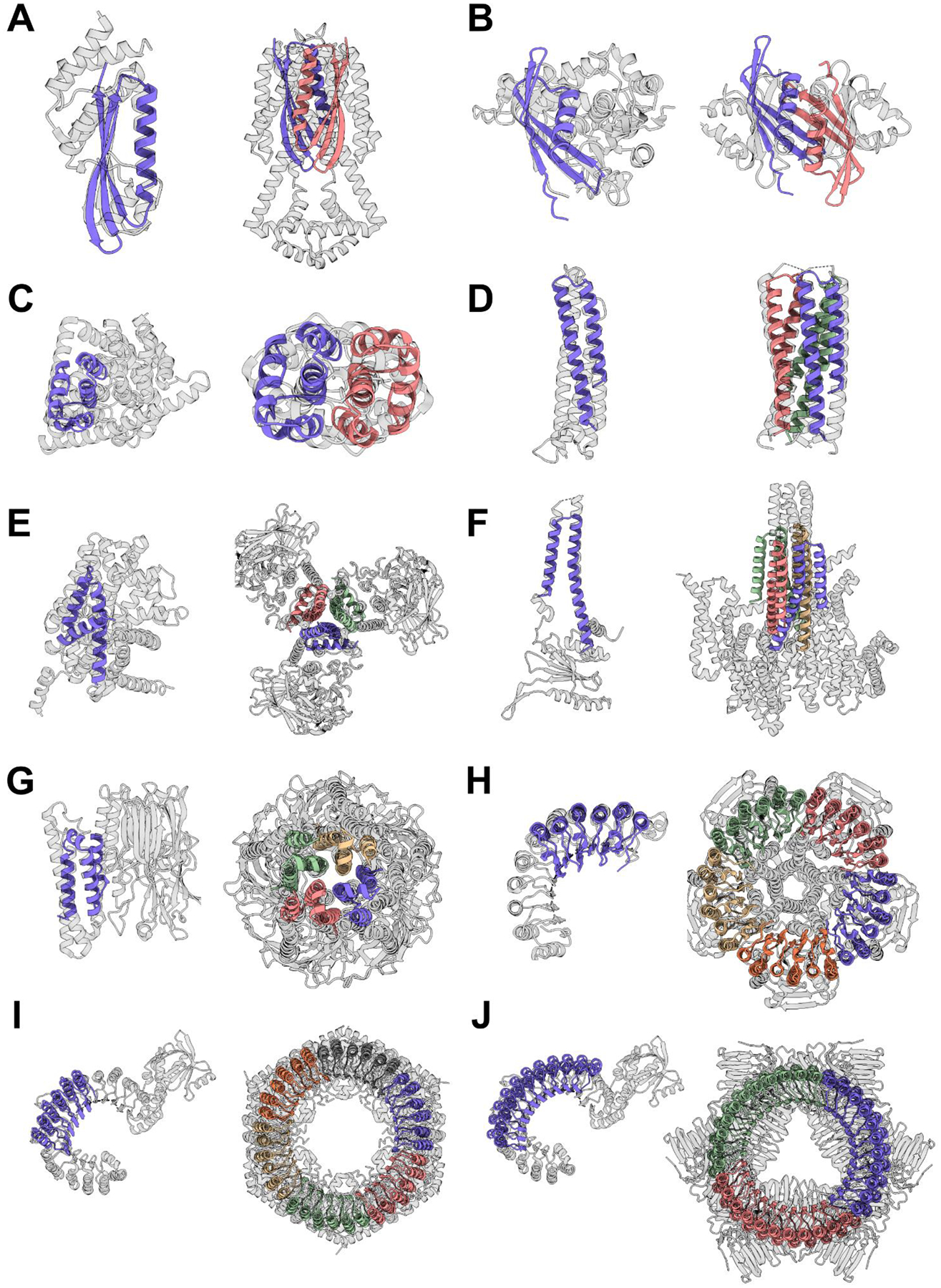
Deep learning generative approaches provide an opportunity to broadly explore protein structure space beyond the sequences and structures of natural proteins. Here, we use deep network hallucination to generate a wide range of symmetric protein homo-oligomers given only a specification of the number of protomers and the protomer length. Crystal structures of seven designs are very similar to the computational models (median root mean square deviation: 0.6 angstroms), as are three cryo–electron microscopy structures of giant 10-nanometer rings with up to 1550 residues and C 33 symmetry; all differ considerably from previously solved structures. Our results highlight the rich diversity of new protein structures that can be generated using deep learning and pave the way for the design of increasingly complex components for nanomachines and biomaterials.
Overview
The claims and statistics of this paper haven’t been checked yet.
Free: it reads the open-access text and checks the first 5 claims that cite a source we can check. Create a free account to check every claim.
Integrity signals
Registry
Cites
Statistics
Figures


Verification
Claims the paper makes
The claims and statistics of this paper haven’t been checked yet.
Free: it reads the open-access text and checks the first 5 claims that cite a source we can check. Create a free account to check every claim.
Claims other papers make about this one
Each is checked against this paper’s own text: does it say what the citing paper says it does?
The source backs part of the claim, not all of it — or backs it for a different population, condition or comparison.
In the citing paper
“This measure of in silico success has been found to correlate with experimental success4,7,26 and is significantly more stringent than template modelling (TM)-score-based metrics used elsewhere5,16,27–29”
From De novo design of protein structure and function with RFdiffusion (2023) · cites this paper as [7]
Experimental — accuracy not yet measuredThe source backs part of the claim, not all of it — or backs it for a different population, condition or comparison.
In the citing paper
“Cyclic oligomers have been designed using structure prediction networks with an adaptation of Hallucination that searches for sequences predicted to fold to the desired cyclic symmetry, but this approach fails for higher-order dihedral, tetrahedral, octahedral and icosahedral symmetries, probably in part because of the much lower representation of such structures in the PDB7.”
From De novo design of protein structure and function with RFdiffusion (2023) · cites this paper as [7]
Experimental — accuracy not yet measured
Self-check
Percentages, sums and reported differences re-computed from the paper’s own numbers.
The self-check needs the paper’s full text.
Citations
Cites
Highly accurate protein structure prediction with AlphaFold
Jumper, J., Evans, R., Pritzel, A., Green, T., Figurnov, M., Ronneberger, O., Tunyasuvunakool, K., Bates, R., Žídek, A., Potapenko, A., et al. (2021) · Nature · doi:10.1038/s41586-021-03819-2 · Cited by 43,764
Coot : model-building tools for molecular graphics
Emsley, P., & Cowtan, K. (2004) · Acta Crystallographica Section D Biological Crystallography · doi:10.1107/s0907444904019158 · Cited by 30,571
PHENIX : a comprehensive Python-based system for macromolecular structure solution
Adams, P. D., Afonine, P. V., Bunkóczi, G., Chen, V. B., Davis, I. W., Echols, N., Headd, J. J., Hung, L.-W., Kapral, G. J., Grosse-Kunstleve, R. W., et al. (2010) · Acta Crystallographica Section D Biological Crystallography · doi:10.1107/s0907444909052925 · Cited by 23,716
Phaser crystallographic software
McCoy, A. J., Grosse-Kunstleve, R. W., Adams, P. D., Winn, M. D., Storoni, L. C., & Read, R. J. (2007) · Journal of Applied Crystallography · doi:10.1107/s0021889807021206 · Cited by 20,612
Kabsch, W. (2010) · Acta Crystallographica Section D Biological Crystallography · doi:10.1107/s0907444909047337 · Cited by 16,463
Kabsch, W., & Sander, C. (1983) · Biopolymers · doi:10.1002/bip.360221211 · Cited by 15,320
Refinement of Macromolecular Structures by the Maximum-Likelihood Method
Murshudov, G. N., Vagin, A. A., & Dodson, E. J. (1997) · Acta Crystallographica Section D Biological Crystallography · doi:10.1107/s0907444996012255 · Cited by 14,742
Overview of the CCP 4 suite and current developments
Winn, M. D., Ballard, C. C., Cowtan, K. D., Dodson, E. J., Emsley, P., Evans, P. R., Keegan, R. M., Krissinel, E. B., Leslie, A. G. W., McCoy, A., et al. (2011) · Acta Crystallographica Section D Biological Crystallography · doi:10.1107/s0907444910045749 · Cited by 12,353
cryoSPARC: algorithms for rapid unsupervised cryo-EM structure determination
Punjani, A., Rubinstein, J. L., Fleet, D. J., & Brubaker, M. A. (2017) · Nature Methods · doi:10.1038/nmeth.4169 · Cited by 11,626
MotionCor2: anisotropic correction of beam-induced motion for improved cryo-electron microscopy
Zheng, S. Q., Palovcak, E., Armache, J.-P., Verba, K. A., Cheng, Y., & Agard, D. A. (2017) · Nature Methods · doi:10.1038/nmeth.4193 · Cited by 7,777
Accurate prediction of protein structures and interactions using a 3-track network
Baek, M., DiMaio, F., Anishchenko, I., Dauparas, J., Ovchinnikov, S., Lee, G. R., Wang, J., Cong, Q., Kinch, L. N., Schaeffer, R. D., et al. (2021) · doi:10.1101/2021.06.14.448402 · Cited by 5,549
Self-citationJones, D. T. (1999) · Journal of Molecular Biology · doi:10.1006/jmbi.1999.3091 · Cited by 5,449
Deep Inside Convolutional Networks: Visualising Image Classification Models and Saliency Maps
Simonyan, K., Vedaldi, A., & Zisserman, A. (2013) · arXiv · doi:10.48550/arxiv.1312.6034 · Cited by 5,370
CTFFIND4: Fast and accurate defocus estimation from electron micrographs
Rohou, A., & Grigorieff, N. (2015) · Journal of Structural Biology · doi:10.1016/j.jsb.2015.08.008 · Cited by 4,913
MolProbity: More and better reference data for improved all‐atom structure validation
Williams, C. J., Headd, J. J., Moriarty, N. W., Prisant, M. G., Videau, L. L., Deis, L. N., Verma, V., Keedy, D. A., Hintze, B. J., Chen, V. B., et al. (2018) · Protein Science · doi:10.1002/pro.3330 · Cited by 4,599
TM-align: a protein structure alignment algorithm based on the TM-score
Zhang, Y. (2005) · Nucleic Acids Research · doi:10.1093/nar/gki524 · Cited by 3,774
Deep Neural Networks are Easily Fooled: High Confidence Predictions for Unrecognizable Images
Nguyen, A., Yosinski, J., & Clune, J. (2014) · arXiv · doi:10.48550/arxiv.1412.1897 · Cited by 3,258
Deep neural networks are easily fooled: High confidence predictions for unrecognizable images
Nguyen, A., Yosinski, J., & Clune, J. (2015) · 2015 IEEE Conference on Computer Vision and Pattern Recognition (CVPR) · doi:10.1109/cvpr.2015.7298640 · Cited by 3,258
Robust deep learning–based protein sequence design using ProteinMPNN
Dauparas, J., Anishchenko, I., Bennett, N., Bai, H., Ragotte, R. J., Milles, L. F., Wicky, B. I. M., Courbet, A., de Haas, R. J., Bethel, N., et al. (2022) · Science · doi:10.1126/science.add2187 · Cited by 2,210
Self-citationRobust deep learning based protein sequence design using ProteinMPNN
Dauparas, J., Anishchenko, I., Bennett, N., Bai, H., Ragotte, R. J., Milles, L. F., Wicky, B. I. M., Courbet, A., de Haas, R. J., Bethel, N., et al. (2022) · doi:10.1101/2022.06.03.494563 · Cited by 2,107
Self-citationThe Rosetta All-Atom Energy Function for Macromolecular Modeling and Design
Alford, R. F., Leaver-Fay, A., Jeliazkov, J. R., O’Meara, M. J., DiMaio, F. P., Park, H., Shapovalov, M. V., Renfrew, P. D., Mulligan, V. K., Kappel, K., et al. (2017) · Journal of Chemical Theory and Computation · doi:10.1021/acs.jctc.7b00125 · Cited by 1,532
Self-citationShape Complementarity at Protein/Protein Interfaces
Lawrence, M. C., & Colman, P. M. (1993) · Journal of Molecular Biology · doi:10.1006/jmbi.1993.1648 · Cited by 1,215
The leucine-rich repeat: a versatile binding motif
Kobe, B., & Deisenhofer, J. (1994) · Trends in Biochemical Sciences · doi:10.1016/0968-0004(94)90090-6 · Cited by 1,202
Protein Data Bank: the single global archive for 3D macromolecular structure data
wwPDB consortium, Burley, S. K., Berman, H. M., Bhikadiya, C., Bi, C., Chen, L., Costanzo, L. D., Christie, C., Duarte, J. M., Dutta, S., et al. (2019) · Nucleic Acids Research · doi:10.1093/nar/gky949 · Cited by 1,163
How significant is a protein structure similarity with TM-score = 0.5?
Xu, J., & Zhang, Y. (2010) · Bioinformatics · doi:10.1093/bioinformatics/btq066 · Cited by 1,142
Mariani, V., Biasini, M., Barbato, A., & Schwede, T. (2013) · Bioinformatics · doi:10.1093/bioinformatics/btt473 · Cited by 1,138
Punjani, A., & Fleet, D. J. (2021) · Journal of Structural Biology · doi:10.1016/j.jsb.2021.107702 · Cited by 977
Structural Symmetry and Protein Function
Goodsell, D. S., & Olson, A. J. (2000) · Annual Review of Biophysics and Biomolecular Structure · doi:10.1146/annurev.biophys.29.1.105 · Cited by 950
cisTEM, user-friendly software for single-particle image processing
Grant, T., Rohou, A., & Grigorieff, N. (2018) · eLife · doi:10.7554/elife.35383 · Cited by 865
De novo protein design by deep network hallucination
Anishchenko, I., Chidyausiku, T. M., Ovchinnikov, S., Pellock, S. J., & Baker, D. (2020) · doi:10.1101/2020.07.22.211482 · Cited by 629
Self-citationDesign of therapeutic proteins with enhanced stability
Chennamsetty, N., Voynov, V., Kayser, V., Helk, B., & Trout, B. L. (2009) · Proceedings of the National Academy of Sciences · doi:10.1073/pnas.0904191106 · Cited by 598
Inceptionism: Going Deeper into Neural Networks
Mordvintsev, A., Olah, C., & Tyka, M. (2015) · Cited by 574
Accurate design of megadalton-scale two-component icosahedral protein complexes
Bale, J. B., Gonen, S., Liu, Y., Sheffler, W., Ellis, D., Thomas, C., Cascio, D., Yeates, T. O., Gonen, T., King, N. P., et al. (2016) · Science · doi:10.1126/science.aaf8818 · Cited by 564
Self-citationDeep learning methods for designing proteins scaffolding functional sites
Wang, J., Lisanza, S., Juergens, D., Tischer, D., Anishchenko, I., Baek, M., Watson, J. L., Chun, J. H., Milles, L. F., Dauparas, J., et al. (2021) · doi:10.1101/2021.11.10.468128 · Cited by 487
Self-citationScaffolding protein functional sites using deep learning
Wang, J., Lisanza, S., Juergens, D., Tischer, D., Watson, J. L., Castro, K. M., Ragotte, R., Saragovi, A., Milles, L. F., Baek, M., et al. (2022) · Science · doi:10.1126/science.abn2100 · Cited by 487
Self-citationHigh-Resolution Protein Design with Backbone Freedom
Harbury, P. B., Plecs, J. J., Tidor, B., Alber, T., & Kim, P. S. (1998) · Science · doi:10.1126/science.282.5393.1462 · Cited by 458
Learning inverse folding from millions of predicted structures
Hsu, C., Verkuil, R., Liu, J., Lin, Z., Hie, B., Sercu, T., Lerer, A., & Rives, A. (2022) · bioRxiv (Cold Spring Harbor Laboratory) · doi:10.1101/2022.04.10.487779 · Cited by 416
Computational design of water-soluble α-helical barrels
Thomson, A. R., Wood, C. W., Burton, A. J., Bartlett, G. J., Sessions, R. B., Brady, R. L., & Woolfson, D. N. (2014) · Science · doi:10.1126/science.1257452 · Cited by 330
Leginon: An Automated System for Acquisition of Images from Vitreous Ice Specimens
Carragher, B., Kisseberth, N., Kriegman, D., Milligan, R. A., Potter, C. S., Pulokas, J., & Reilein, A. (2000) · Journal of Structural Biology · doi:10.1006/jsbi.2000.4314 · Cited by 313
De novo design of protein homo-oligomers with modular hydrogen-bond network–mediated specificity
Boyken, S. E., Chen, Z., Groves, B., Langan, R. A., Oberdorfer, G., Ford, A., Gilmore, J. M., Xu, C., DiMaio, F., Pereira, J. H., et al. (2016) · Science · doi:10.1126/science.aad8865 · Cited by 304
Self-citationHigh thermodynamic stability of parametrically designed helical bundles
Huang, P.-S., Oberdorfer, G., Xu, C., Pei, X. Y., Nannenga, B. L., Rogers, J. M., DiMaio, F., Gonen, T., Luisi, B., & Baker, D. (2014) · Science · doi:10.1126/science.1257481 · Cited by 277
Self-citationProteins evolve on the edge of supramolecular self-assembly
Garcia-Seisdedos, H., Empereur-Mot, C., Elad, N., & Levy, E. D. (2017) · Nature · doi:10.1038/nature23320 · Cited by 253
Principles of assembly reveal a periodic table of protein complexes
Ahnert, S. E., Marsh, J. A., Hernández, H., Robinson, C. V., & Teichmann, S. A. (2015) · Science · doi:10.1126/science.aaa2245 · Cited by 253
De novo design of a four-fold symmetric TIM-barrel protein with atomic-level accuracy
Huang, P.-S., Feldmeier, K., Parmeggiani, F., Fernandez Velasco, D. A., Höcker, B., & Baker, D. (2016) · Nature Chemical Biology · doi:10.1038/nchembio.1966 · Cited by 251
Self-citationMukherjee, S., & Zhang, Y. (2009) · Nucleic Acids Research · doi:10.1093/nar/gkp318 · Cited by 181
Protein sequence design with a learned potential
Anand, N., Eguchi, R., Mathews, I. I., Perez, C. P., Derry, A., Altman, R. B., & Huang, P.-S. (2022) · Nature Communications · doi:10.1038/s41467-022-28313-9 · Cited by 171
De novo design of a Zn2+-binding protein
Handel, T. M., & DeGrado, W. F. (1990) · Journal of the American Chemical Society · doi:10.1021/ja00174a039 · Cited by 154
Computational design of self-assembling cyclic protein homo-oligomers
Fallas, J. A., Ueda, G., Sheffler, W., Nguyen, V., McNamara, D. E., Sankaran, B., Pereira, J. H., Parmeggiani, F., Brunette, T., Cascio, D., et al. (2016) · Nature Chemistry · doi:10.1038/nchem.2673 · Cited by 150
Self-citationProtein sequence design by conformational landscape optimization
Norn, C., Wicky, B. I. M., Juergens, D., Liu, S., Kim, D., Tischer, D., Koepnick, B., Anishchenko, I., Players, F., Baker, D., et al. (2021) · Proceedings of the National Academy of Sciences · doi:10.1073/pnas.2017228118 · Cited by 144
Self-citationSymmetry and simplicity spontaneously emerge from the algorithmic nature of evolution
Johnston, I. G., Dingle, K., Greenbury, S. F., Camargo, C. Q., Doye, J. P. K., Ahnert, S. E., & Louis, A. A. (2022) · Proceedings of the National Academy of Sciences · doi:10.1073/pnas.2113883119 · Cited by 134
Rational design of α-helical tandem repeat proteins with closed architectures
Doyle, L., Hallinan, J. P., Bolduc, J. M., Parmeggiani, F., Baker, D. A., Stoddard, B. L., & Bradley, P. H. (2015) · Nature · doi:10.1038/nature16191 · Cited by 132
Self-citationStructure-based protein design with deep learning
Ovchinnikov, S., & Huang, P.-S. (2021) · Current Opinion in Chemical Biology · doi:10.1016/j.cbpa.2021.08.004 · Cited by 93
Diffusion probabilistic modeling of protein backbones in 3D for the motif-scaffolding problem
Trippe, B. L., Yim, J., Tischer, D., Baker, D., Broderick, T., Barzilay, R., & Jaakkola, T. (2022) · arXiv · doi:10.48550/arxiv.2206.04119 · Cited by 92
Self-citationProtein Structure and Sequence Generation with Equivariant Denoising Diffusion Probabilistic Models
Anand, N., & Achim, T. (2022) · arXiv · doi:10.48550/arxiv.2205.15019 · Cited by 90
Design of multi-scale protein complexes by hierarchical building block fusion
Hsia, Y., Mout, R., Sheffler, W., Edman, N. I., Vulovic, I., Park, Y., Redler, R. L., Bick, M. J., Bera, A. K., Courbet, A., et al. (2021) · Nature Communications · doi:10.1038/s41467-021-22276-z · Cited by 86
Self-citationSNAC-tag for sequence-specific chemical protein cleavage
Dang, B., Mravic, M., Hu, H., Schmidt, N., Mensa, B., & DeGrado, W. F. (2019) · Nature Methods · doi:10.1038/s41592-019-0357-3 · Cited by 77
Reconfigurable asymmetric protein assemblies through implicit negative design
Sahtoe, D. D., Praetorius, F., Courbet, A., Hsia, Y., Wicky, B. I. M., Edman, N. I., Miller, L. M., Timmermans, B. J. R., Decarreau, J. A., Morris, H. M., et al. (2022) · Science · doi:10.1126/science.abj7662 · Cited by 71
Self-citationAlphaDesign: A de novo protein design framework based on AlphaFold
Jendrusch, M., Korbel, J. O., & Sadiq, S. K. (2021) · doi:10.1101/2021.10.11.463937 · Cited by 65
Computational design of mechanically coupled axle-rotor protein assemblies
Courbet, A., Hansen, J. M., Hsia, Y., Bethel, N. P., Park, Y., Xu, C., Moyer, A. P., Boyken, S. E., Ueda, G., Nattermann, U., et al. (2022) · Science · doi:10.1126/science.abm1183 · Cited by 56
Self-citationGeneration of ordered protein assemblies using rigid three-body fusion
Vulovic, I., Yao, Q., Park, Y., Courbet, A., Norris, A. S., Büsch, F., Sahasrabuddhe, A., Merten, H., Sahtoe, D. D., Ueda, G., et al. (2021) · Proceedings of the National Academy of Sciences · doi:10.1073/pnas.2015037118 · Cited by 34
Self-citationUsing AlphaFold for Rapid and Accurate Fixed Backbone Protein Design
Moffat, L., Greener, J. G., & Jones, D. T. (2021) · doi:10.1101/2021.08.24.457549 · Cited by 33
Nannenga, B. L., Iadanza, M. G., Vollmar, B. S., & Gonen, T. (2013) · Current Protocols in Protein Science · doi:10.1002/0471140864.ps1715s72 · Cited by 30
Symmetry disruption commits vault particles to disassembly
Guerra, P., González-Álamos, M., Llauró, A., Casañas, A., Querol‐Audí, J., de Pablo, P. J., & Verdaguer, N. (2022) · Science Advances · doi:10.1126/sciadv.abj7795 · Cited by 23
Engineering and functionalization of large circular tandem repeat protein nanoparticles
Correnti, C., Hallinan, J. P., Doyle, L., Ruff, R. O., Jaeger-Ruckstuhl, C. A., Xu, Y., Shen, B. W., Qu, A., Polkinghorn, C. R., Friend, D. J., et al. (2020) · Nature Structural & Molecular Biology · doi:10.1038/s41594-020-0397-5 · Cited by 19
bwicky/oligomer_hallucination: Initial release
Wicky, B. I. M. (2022) · Zenodo (CERN European Organization for Nuclear Research) · doi:10.5281/zenodo.7112140 · Cited by 1
Self-citation
Cited by
Evolutionary-scale prediction of atomic level protein structure with a language model
Lin, Z., Akin, H., Rao, R., Hie, B., Zhu, Z., Lu, W., Smetanin, N., Verkuil, R., Kabeli, O., Shmueli, Y., et al. (2022) · doi:10.1101/2022.07.20.500902 · Cited by 5,990
Watson, J. L., Juergens, D., Bennett, N. R., Trippe, B. L., Yim, J., Eisenach, H. E., Ahern, W., Borst, A. J., Ragotte, R. J., Milles, L. F., et al. (2022) · doi:10.1101/2022.12.09.519842 · Cited by 2,369
Self-citationDe novo design of protein structure and function with RFdiffusion
Watson, J. L., Juergens, D., Bennett, N. R., Trippe, B. L., Yim, J., Eisenach, H. E., Ahern, W., Borst, A. J., Ragotte, R. J., Milles, L. F., et al. (2023) · Nature · doi:10.1038/s41586-023-06415-8 · Cited by 2,325
Self-citationRobust deep learning–based protein sequence design using ProteinMPNN
Dauparas, J., Anishchenko, I., Bennett, N., Bai, H., Ragotte, R. J., Milles, L. F., Wicky, B. I. M., Courbet, A., de Haas, R. J., Bethel, N., et al. (2022) · Science · doi:10.1126/science.add2187 · Cited by 2,210
Self-citationRobust deep learning based protein sequence design using ProteinMPNN
Dauparas, J., Anishchenko, I., Bennett, N., Bai, H., Ragotte, R. J., Milles, L. F., Wicky, B. I. M., Courbet, A., de Haas, R. J., Bethel, N., et al. (2022) · doi:10.1101/2022.06.03.494563 · Cited by 2,107
Self-citation
Metadata
- Authors
- Wicky, B. I. M., Milles, L. F., Courbet, A., Ragotte, R. J., Dauparas, J., Kinfu, E., Tipps, S., Kibler, R. D., Baek, M., DiMaio, F., Li, X., Carter, L., Kang, A., Nguyen, H., Bera, A. K., & Baker, D.
- Affiliations
- University of Washington (US)
- Howard Hughes Medical Institute (US)
- Corresponding author
- Baker, D.
- ORCID
- Venue
- Science
- Published
- 2022-10-07
- Volume
- 378
- Issue
- 6615
- Pages
- 56-61
- ISSN
- 0036-8075 (ISSN-L)
- 1095-9203
- PMC
- PMC9724707
- OpenAlex
- W4296031103
- Cited by
- 240
- References
- 58
- Citations by year
- 2026: 46 · 2025: 72 · 2024: 62 · 2023: 52 · 2022: 8
- FWCI
- 15.84
- Type
- journal article
- Language
- en
- Open access
- green
- doi.org
- Licence
- cc-by-nc-nd
- Locations
- Science (publishedVersion)
- bioRxiv (Cold Spring Harbor Laboratory) (acceptedVersion, cc-by-nc-nd)
- PubMed (publishedVersion)
- PubMed Central (acceptedVersion, cc-by)
- Funders
- National Science Foundation (P30 GM124165, P30 CA015704, CA015704, DE-AC02–06CH11357, P41-GM103311, 1629214)
- Howard Hughes Medical Institute
- U.S. Department of Energy (DE‐ AC02‐06CH11357, Contract No. DE-AC02-06CH11357., AC02‐06CH11357, P30 GM124165, 06CH11357, No. DE-AC02-06CH11357, DE-AC02-)
- Alfred P. Sloan Foundation (g-2021-16899)
- University of Washington (DE-AC02-06CH11357, P30 CA015704)
- Open Philanthropy Project
- National Institutes of Health (S10 OD032290, DE-AC02–06CH11357, GM124165, CA015704)
- Office of Science (Contract No. DE-AC02-06CH11357, AC02‐06CH11357, DE-AC02, No. DE-AC02-06CH11357., 06CH11357, DE-AC02-06CH11357)
- University of California, San Francisco (NIH P41-GM103311, P41-GM103311)
- National Cancer Institute (P30CA015704)
- National Institute of General Medical Sciences (GM103311, P41GM103311, P41GM103533, DE-AC02-06CH11357, NIH P41-GM103311, 103533, GM124165, P30GM124165)
- Division of Chemistry (1629214, CHE 1629214)
- Argonne National Laboratory (DE- AC02-06-CH11357, DE-AC02, AC02–06CH11357, No. DE-AC02-06CH11357, 06CH11357, Contract No. DE-AC02-06CH11357., P30 GM124165)
- Topics
- Life Sciences › Biochemistry, Genetics and Molecular Biology › Molecular Biology › Protein Structure and Dynamics
- Life Sciences › Biochemistry, Genetics and Molecular Biology › Structural Biology › Enzyme Structure and Function
- Life Sciences › Biochemistry, Genetics and Molecular Biology › Molecular Biology › Machine Learning in Bioinformatics
- Keywords
- protein structure design, homo-oligomers, nanorobots, biomaterials
- MeSH terms
- Deep Learning, Biocompatible Materials, Models, Molecular, Protein Conformation, Protein Engineering, Cryoelectron Microscopy, Protein Subunits
About these checks
Registry, retraction, Cites and statistics results are computed, not judged, with one exception: a printed reference that no index lists is read once by a model before it counts as a citation error. Claims are extracted by a model and kept only when their quote appears verbatim in the text; each cited source is then checked, with a model’s help, for whether it says what the claim relies on. Those verdicts can be wrong — read the claim’s quote and the cited source before relying on one.
Statistics and arithmetic are recomputed, not judged.
Confidence bands are shown only for fields calibrated on our annotated test set. None is calibrated yet, so no band is shown.
What counts as a claim ›How we measure confidence ›How we check this ›
Are you an author, or do you see a problem with this page? Request a review
Data sources
Bibliographic data: Semantic Scholar, OpenAlex, Crossref.
Contains information from the Semantic Scholar Academic Graph Datasets, made available under the ODC Attribution License (ODC-BY 1.0).
Semantic Scholar’s data is described in: Kinney, R., et al. (2023). The Semantic Scholar Open Data Platform. arXiv:2301.10140.
