

The DOI matches its Crossref record; 13 of the 13 references it cites were found in a registry or an index.
AMPK Inhibits ULK1-Dependent Autophagosome Formation and Lysosomal Acidification via Distinct Mechanisms
Overview
The claims and statistics of this paper haven’t been checked yet.
Run the check — free, no account needed. It reads the open-access text and checks the sources it cites.
Integrity signals
Registry
Cites
Statistics
Figures
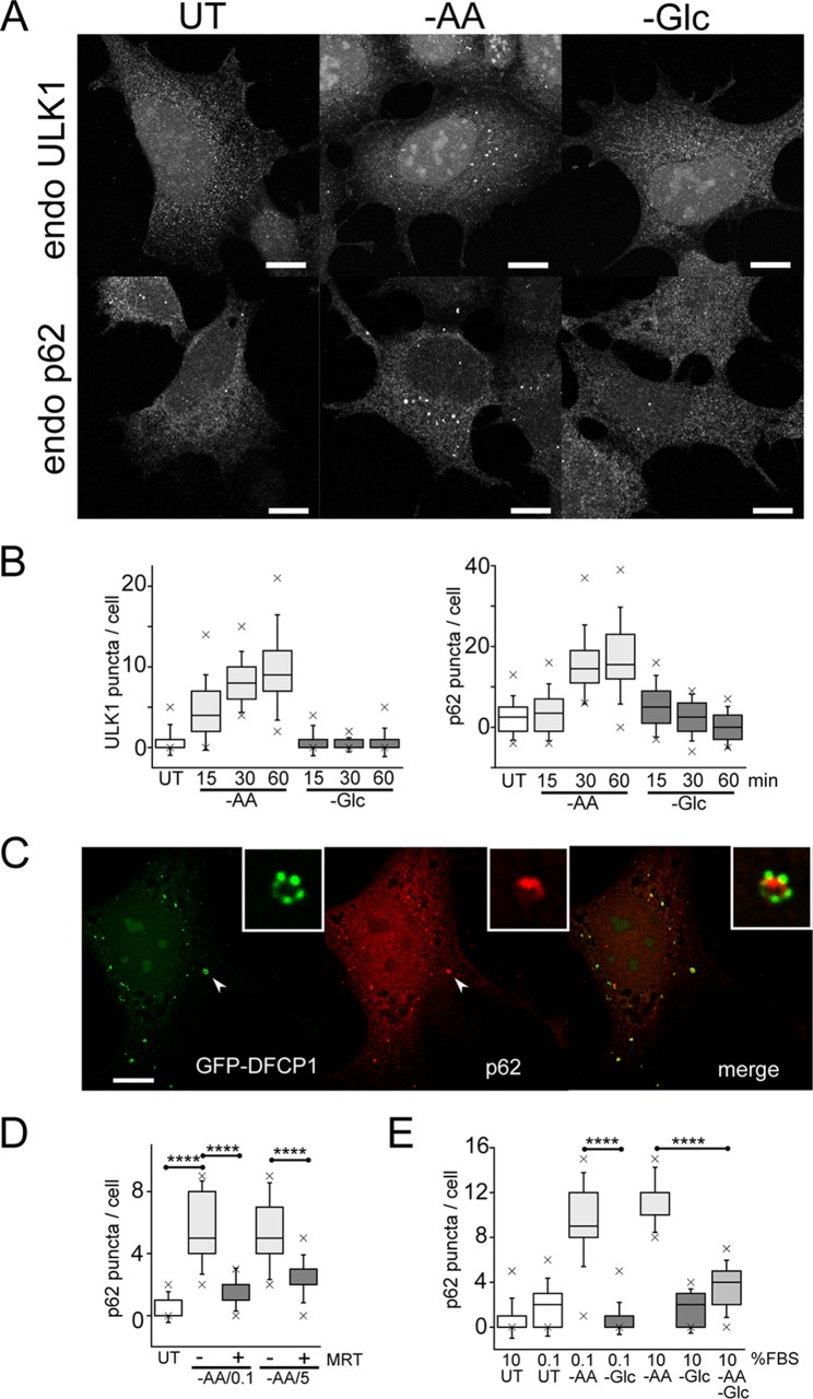
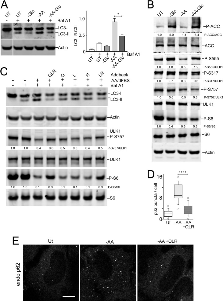
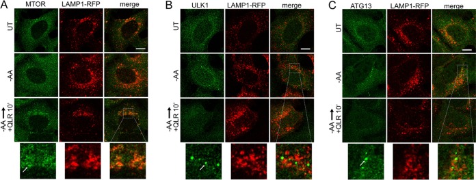
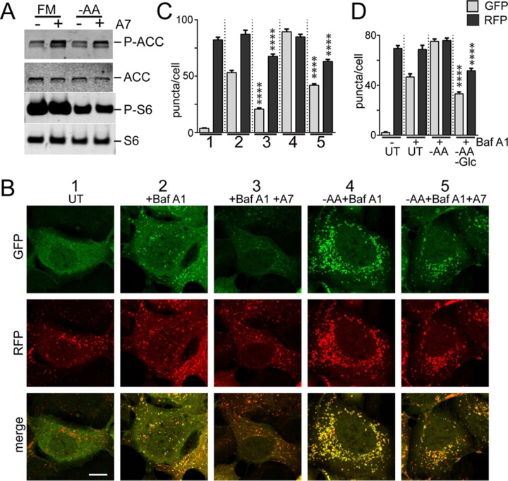
Verification
Claims the paper makes
The claims and statistics of this paper haven’t been checked yet.
Run the check — free, no account needed. It reads the open-access text and checks the sources it cites.
Claims other papers make about this one
Each is checked against this paper’s own text: does it say what the citing paper says it does?
The cited source doesn’t support the claim as written.
In the citing paper
“whereas it appears to stimulate autophagy in many other cell types, including fibroblasts, colon carcinoma cells and skeletal muscle [656-665]”
From Guidelines for the use and interpretation of assays for monitoring autophagy (4th edition) 1 (2021) · cites this paper as [656]
Checked by a reasoning model — may be wrong.
Experimental — accuracy not yet measuredThe source backs part of the claim, not all of it — or backs it for a different population, condition or comparison.
In the citing paper
“In liver cells, AMPK suppresses autophagy at the level of cargo sequestration, as indicated by the rapid sequestration-inhibitory effects of a variety of AMPK activators”
From Guidelines for the use and interpretation of assays for monitoring autophagy (4th edition) 1 (2021) · cites this paper as [656]
Experimental — accuracy not yet measured
Self-check
Percentages, sums and reported differences re-computed from the paper’s own numbers.
The self-check needs the paper’s full text.
Citations
Cites
AMPK and mTOR regulate autophagy through direct phosphorylation of Ulk1
Kim, J., Kundu, M., Viollet, B., & Guan, K.-L. (2011) · Nature Cell Biology · doi:10.1038/ncb2152 · Cited by 7,227
Pankiv, S., Clausen, T. H., Lamark, T., Brech, A., Bruun, J.-A., Outzen, H., Øvervatn, A., Bjørkøy, G., & Johansen, T. (2007) · Journal of Biological Chemistry · doi:10.1074/jbc.m702824200 · Cited by 4,578
The role of autophagy during the early neonatal starvation period
Kuma, A., Hatano, M., Matsui, M., Yamamoto, A., Nakaya, H., Yoshimori, T., Ohsumi, Y., Tokuhisa, T., & Mizushima, N. (2004) · Nature · doi:10.1038/nature03029 · Cited by 2,865
Kimura, S., Noda, T., & Yoshimori, T. (2007) · Autophagy · doi:10.4161/auto.4451 · Cited by 2,278
Zoncu, R., Bar-Peled, L., Efeyan, A., Wang, S., Sancak, Y., & Sabatini, D. M. (2011) · Science · doi:10.1126/science.1207056 · Cited by 1,673
Termination of autophagy and reformation of lysosomes regulated by mTOR
Yu, L., McPhee, C. K., Zheng, L., Mardones, G. A., Rong, Y., Peng, J., Mi, N., Zhao, Y., Liu, Z., Wan, F., et al. (2010) · Nature · doi:10.1038/nature09076 · Cited by 1,544
The role for autophagy in cancer
White, E. (2015) · Journal of Clinical Investigation · doi:10.1172/jci73941 · Cited by 1,332
The CASTOR Proteins Are Arginine Sensors for the mTORC1 Pathway
Chantranupong, L., Scaria, S. M., Saxton, R. A., Gygi, M. P., Shen, K., Wyant, G. A., Wang, T., Harper, J. W., Gygi, S. P., & Sabatini, D. M. (2016) · Cell · doi:10.1016/j.cell.2016.02.035 · Cited by 843
Differential regulation of mTORC1 by leucine and glutamine
Jewell, J. L., Kim, Y. C., Russell, R. C., Yu, F.-X., Park, H. W., Plouffe, S. W., Tagliabracci, V. S., & Guan, K.-L. (2015) · Science · doi:10.1126/science.1259472 · Cited by 757
The ATG conjugation systems are important for degradation of the inner autophagosomal membrane
Tsuboyama, K., Koyama-Honda, I., Sakamaki, Y., Koike, M., Morishita, H., & Mizushima, N. (2016) · Science · doi:10.1126/science.aaf6136 · Cited by 485
PLIC proteins or ubiquilins regulate autophagy‐dependent cell survival during nutrient starvation
N'Diaye, E., Kajihara, K. K., Hsieh, I., Morisaki, H., Debnath, J., & Brown, E. J. (2009) · The EMBO Reports · doi:10.1038/embor.2008.238 · Cited by 323
Regulation of autophagy by coordinated action of mTORC1 and protein phosphatase 2A
Wong, P.-M., Feng, Y., Wang, J., Shi, R., & Jiang, X. (2015) · Nature Communications · doi:10.1038/ncomms9048 · Cited by 239
The GATOR2 Component Wdr24 Regulates TORC1 Activity and Lysosome Function
Cai, W., Wei, Y., Jarnik, M., Reich, J., & Lilly, M. A. (2016) · PLOS Genetics · doi:10.1371/journal.pgen.1006036 · Cited by 61
Research works printed but not linked: 48
- 10.1016/j.cell.2014.11.006 · DOI · Paper page
- 10.1101/gad.283416.116 · DOI
- 10.1083/jcb.150.6.1507 · DOI · Paper page
- 10.1038/ncb2757 · DOI · Paper page
- 10.1016/j.molcel.2015.05.031 · DOI · Paper page
- 10.1016/j.febslet.2015.05.020 · DOI
- 10.1126/science.1196371 · DOI · Paper page
- 10.1073/pnas.1100844108 · DOI · Paper page
- 10.4161/auto.23066 · DOI
- 10.1038/ncomms12420 · DOI · Paper page
- 10.1073/pnas.1107969108 · DOI
- 10.1038/cdd.2015.26 · DOI
- 10.1016/j.molcel.2014.12.007 · DOI
- 10.1038/nsmb.2475 · DOI
- 10.1016/j.molcel.2015.10.037 · DOI
- 10.1016/j.molcel.2013.12.019 · DOI
- 10.1111/j.1432-1033.1987.tb11011.x · DOI
- 10.1074/jbc.273.37.23758 · DOI · Paper page
- 10.1093/hmg/ddg109 · DOI
- 10.1042/BJ20121122 · DOI
- 10.1074/jbc.M113.490581 · DOI
- 10.1016/j.ccr.2004.06.007 · DOI
- 10.1038/nature11745 · DOI
- 10.4161/auto.27344 · DOI · Paper page
- 10.1074/jbc.M703663200 · DOI · Paper page
- 10.1128/MCB.01082-08 · DOI · Paper page
- 10.4161/auto.6.6.12709 · DOI · Paper page
- 10.1083/jcb.201009067 · DOI
- 10.1074/jbc.C114.627778 · DOI
- 10.1083/jcb.201607005 · DOI
- 10.7554/Elife.11058 · DOI · Paper page
- 10.1074/jbc.M706536200 · DOI
- 10.1038/cr.2013.11 · DOI
- 10.1016/j.cell.2015.02.041 · DOI
- 10.1038/nrd3504 · DOI
- 10.1242/jcs.181016 · DOI · Paper page
- 10.1038/s41467-017-00303-2 · DOI · Paper page
- 10.1016/j.molcel.2013.12.011 · DOI
- 10.4161/auto.20586 · DOI
- 10.1016/j.molcel.2011.06.018 · DOI · Paper page
- 10.3390/cells5020024 · DOI
- 10.1074/jbc.M113.525782 · DOI
- 10.1038/nrm3565 · DOI
- 10.1016/j.tibs.2015.08.005 · DOI · Paper page
- 10.1074/jbc.M115.659128 · DOI
- 10.1128/MCB.25.2.575-589.2005 · DOI
- 10.2174/138920312800493142 · DOI
- 10.1083/jcb.200604129 · DOI
Cited by
Sorted by citations
Guidelines for the use and interpretation of assays for monitoring autophagy (4th edition) 1
Klionsky, D. J., Abdel-Aziz, A. K., Abdelfatah, S., Abdellatif, M., Abdoli, A., Abel, S., Abeliovich, H., Abildgaard, M. H., Abudu, Y. P., Acevedo-Arozena, A., et al. (2021) · Autophagy · doi:10.1080/15548627.2020.1797280 · Cited by 2,897
Self-citationPanwar, V., Singh, A., Bhatt, M., Tonk, R. K., Azizov, S., Raza, A. S., Sengupta, S., Kumar, D., & Garg, M. (2023) · Signal Transduction and Targeted Therapy · doi:10.1038/s41392-023-01608-z · Cited by 967
Large neutral amino acid levels tune perinatal neuronal excitability and survival
Knaus, L. S., Basilico, B., Malzl, D., Gerykova Bujalkova, M., Smogavec, M., Schwarz, L. A., Gorkiewicz, S., Amberg, N., Pauler, F. M., Knittl-Frank, C., et al. (2023) · Cell · doi:10.1016/j.cell.2023.02.037 · Cited by 70
Stanciu, G. D., Rusu, R. N., Bild, V., Filipiuc, L. E., Tamba, B.-I., & Ababei, D. C. (2021) · Biomedicines · doi:10.3390/biomedicines9050576 · Cited by 47
Metadata
- Authors
- Nwadike, C., Williamson, L. E., Gallagher, L. E., Guan, J.-L., & Chan, E. Y. W.
- Venue
- Molecular and Cellular Biology
- Publisher
- Informa UK Limited
- Published
- 2018-05-01
- Volume
- 38
- Issue
- 10
- ISSN
- 1098-5549
- PMC
- PMC5954193
- Cited by
- 110
- References
- 13
- Language
- en
- Funders
- National Institutes of Health (NS094144)
- RCUK | Biotechnology and Biological Sciences Research Council
- Tertiary Education Trust Fund
- Cancer Research UK
About these checks
Registry, retraction, Cites and statistics results are computed, not judged, with one exception: a printed reference that no index lists is read once by a model before it counts as a citation error. Claims are extracted by a model and kept only when their quote appears verbatim in the text; each cited source is then checked, with a model’s help, for whether it says what the claim relies on. Those verdicts can be wrong — read the claim’s quote and the cited source before relying on one.
Statistics and arithmetic are recomputed, not judged.
Confidence bands are shown only for fields calibrated on our annotated test set. None is calibrated yet, so no band is shown.
What counts as a claim ›How we measure confidence ›How we check this ›
Are you an author, or do you see a problem with this page? Request a review
