

The DOI matches its Crossref record; 61 of the 61 references it cites were found in a registry or an index.
Pro-inflammatory activation of primary microglia and macrophages increases 18 kDa translocator protein expression in rodents but not humans
Open accessAbstract
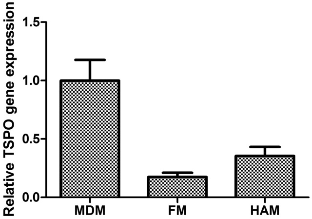
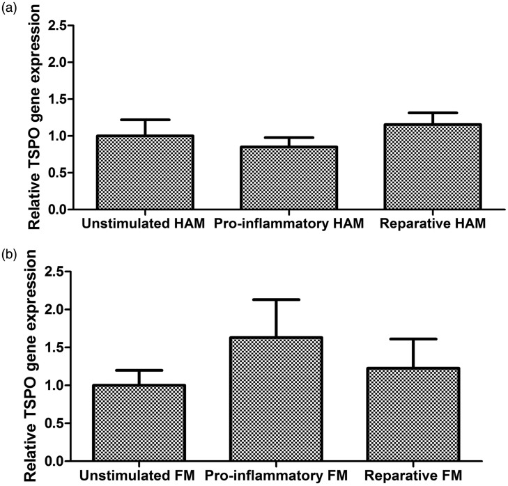
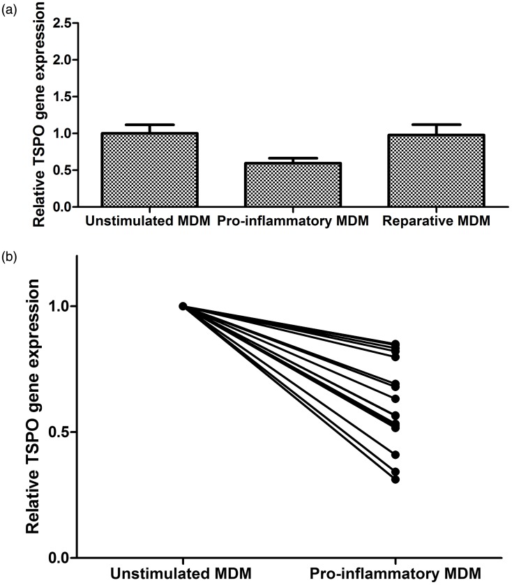
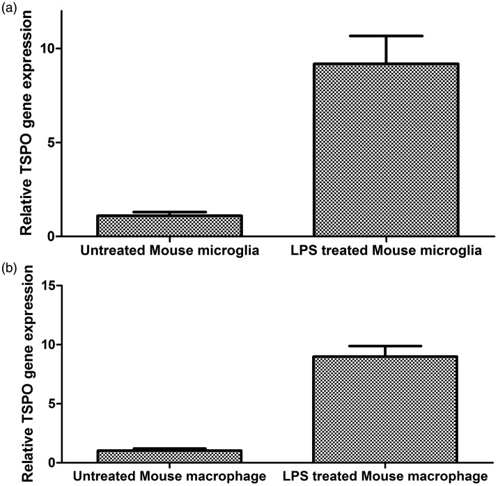
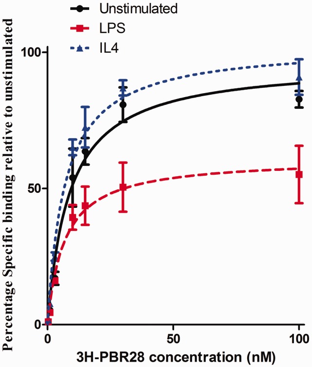
The 18kDa Translocator Protein (TSPO) is the most commonly used tissue-specific marker of inflammation in positron emission tomography (PET) studies. It is expressed in myeloid cells such as microglia and macrophages, and in rodent myeloid cells expression increases with cellular activation. We assessed the effect of myeloid cell activation on TSPO gene expression in both primary human and rodent microglia and macrophages in vitro, and also measured TSPO radioligand binding with 3 H-PBR28 in primary human macrophages. As observed previously, we found that TSPO expression increases (∼9-fold) in rodent-derived macrophages and microglia upon pro-inflammatory stimulation. However, TSPO expression does not increase with classical pro-inflammatory activation in primary human microglia (fold change 0.85 [95% CI 0.58–1.12], p = 0.47). In contrast, pro-inflammatory activation of human monocyte-derived macrophages is associated with a reduction of both TSPO gene expression (fold change 0.60 [95% CI 0.45–0.74], p = 0.02) and TSPO binding site abundance (fold change 0.61 [95% CI 0.49–0.73], p < 0.0001). These findings have important implications for understanding the biology of TSPO in activated macrophages and microglia in humans. They are also clinically relevant for the interpretation of PET studies using TSPO targeting radioligands, as they suggest changes in TSPO expression may reflect microglial and macrophage density rather than activation phenotype.
Overview
The claims and statistics of this paper haven’t been checked yet.
Run the check — free, no account needed. It reads the open-access text and checks the sources it cites.
Integrity signals
Registry
Cites
Statistics
Figures
Verification
Claims the paper makes
The claims and statistics of this paper haven’t been checked yet.
Run the check — free, no account needed. It reads the open-access text and checks the sources it cites.
Claims other papers make about this one
Each is checked against this paper’s own text: does it say what the citing paper says it does?
The source backs part of the claim, not all of it — or backs it for a different population, condition or comparison.
In the citing paper
“Recently, we have shown in vitro that, although TSPO gene and protein expression and TSPO radioligand binding increases in rodent microglia and macrophages with inflammatory stimulation, relative TSPO gene expression in human microglia and TSPO ligand binding in human monocyte-derived macrophages are reduced with pro-inflammatory activation (Owen et al., 2017).”
From A quantitative neuropathological assessment of translocator protein expression in multiple sclerosis (2019) · cites this paper as Owen et al., 2017
Experimental — accuracy not yet measuredThe source backs part of the claim, not all of it — or backs it for a different population, condition or comparison.
In the citing paper
“Recently, we have shown in vitro that, although TSPO gene and protein expression and TSPO radioligand binding increases in rodent microglia and macrophages with inflammatory stimulation, relative TSPO gene expression in human microglia and TSPO ligand binding in human monocyte-derived macrophages are reduced with pro-inflammatory activation (Owen et al., 2017).”
From A quantitative neuropathological assessment of translocator protein expression in multiple sclerosis (2019) · cites this paper as Owen et al., 2017
Experimental — accuracy not yet measuredThe cited source says what the claim attributes to it.
In the citing paper
“However, in humans, TSPO is not upregulated in either pro-inflammatory macrophages (Narayan et al., 2017) or primary microglia (Owen et al., 2017).”
From A quantitative neuropathological assessment of translocator protein expression in multiple sclerosis (2019) · cites this paper as Owen et al., 2017
The cited source says what the claim attributes to it.
In the citing paper
“This new observation thus confirms in vitro data suggesting that the TSPO expression signal does not distinguish between microglial phenotypes in human tissue (Owen et al., 2017).”
From A quantitative neuropathological assessment of translocator protein expression in multiple sclerosis (2019) · cites this paper as Owen et al., 2017
Self-check
Percentages, sums and reported differences re-computed from the paper’s own numbers.
The self-check needs the paper’s full text.
Citations
Cites
Amyloid oligomers exacerbate tau pathology in a mouse model of tauopathy
Selenica, M.-L. B., Brownlow, M., Jimenez, J. P., Lee, D. C., Pena, G., Dickey, C. A., Gordon, M. N., & Morgan, D. (2013) · Neurodegenerative Diseases · doi:10.1159/000337230 · Cited by 10,359
Identification of a unique TGF-β–dependent molecular and functional signature in microglia
Butovsky, O., Jedrychowski, M. P., Moore, C. S., Cialic, R., Lanser, A. J., Gabriely, G., Koeglsperger, T., Dake, B., Wu, P. M., Doykan, C. E., et al. (2014) · Nature Neuroscience · doi:10.1038/nn.3599 · Cited by 2,543 · cited 1×
“Furthermore, many of the markers with differential expression profiles in human microglia relative to human macrophages are not expressed by rodent myeloid cells.(37,38) Our findings add to these differences and suggest that extrapolating TSPO biology from rodent to human myeloid cells should be done with extreme caut…”
BackgroundSelf-citationMitochondrial reactive oxygen species trigger hypoxia-induced transcription
Chandel, N. S., Maltepe, E., Goldwasser, E., Mathieu, C. E., Simon, M. C., & Schumacker, P. T. (1998) · Proceedings of the National Academy of Sciences · doi:10.1073/pnas.95.20.11715 · Cited by 1,926
Microglial physiology: unique stimuli, specialized responses
Ransohoff, R. M., & Perry, V. H. (2009) · Annual Review of Immunology · doi:10.1146/annurev.immunol.021908.132528 · Cited by 1,858 · cited 3×
“Microglia also express receptors for neurotransmitters including g-aminobutyric acid (GABA) and glutamate, providing another pathway for microglial phenotypic regulation.(1,50,51) Caution should therefore be used when extrapolating these findings in vivo and further work will therefore examine directly how TSPO expres…”
BackgroundInnate immune activation in neurodegenerative disease
Heneka, M. T., Kummer, M. P., & Latz, E. (2014) · Nature Reviews Immunology · doi:10.1038/nri3705 · Cited by 1,437 · cited 3×
“Dysregulated or inappropriate microglial activation is thought to contribute to many neurodegenerative diseases.(4) Modulating microglial phenotype has therefore been hypothesised as a potential therapeutic strategy for diseases such as Alzheimer’s and Parkinson’s Disease.”
BackgroundMethodInfluentialMicroglia in physiology and disease
Wolf, S. A., Boddeke, H. W. G. M., & Kettenmann, H. (2017) · Annual Review of Physiology · doi:10.1146/annurev-physiol-022516-034406 · Cited by 1,426 · cited 1×
“Rodent microglia are used extensively given the scarcity of primary human microglia; however, there are several important differences between the two cell types.(34,35) For example, in rodent but not human microglia, transforming growth Figure 2.”
In vivo imaging of microglial activation with [11C](R)-PK11195 PET in idiopathic Parkinson's disease
Gerhard, A., Pavese, N., Hotton, G., Turkheimer, F., Es, M., Hammers, A., Eggert, K., Oertel, W., Banati, R. B., & Brooks, D. J. (2006) · Neurobiology of Disease · doi:10.1016/j.nbd.2005.08.002 · Cited by 1,200
Owen, D. R., Yeo, A. J., Gunn, R. N., Song, K., Wadsworth, G., Lewis, A., Rhodes, C., Pulford, D. J., Bennacef, I., Parker, C. A., et al. (2012) · Journal of Cerebral Blood Flow & Metabolism · doi:10.1038/jcbfm.2011.147 · Cited by 779
Self-citationThe peripheral benzodiazepine binding site in the brain in multiple sclerosis
Banati, R. B., Newcombe, J., Gunn, R. N., Cagnin, A., Turkheimer, F., Heppner, F., Price, G., Wegner, F., Giovannoni, G., Miller, D. H., et al. (2000) · Brain · doi:10.1093/brain/123.11.2321 · Cited by 661
High‐yield isolation of murine microglia by mild trypsinization
Saura, J., Tusell, J. M., & Serratosa, J. (2003) · Glia · doi:10.1002/glia.10274 · Cited by 592 · cited 1×
“At confluency, cultures were subjected to mild trypsinization as described.(32) Following removal of the astrocyte monolayer, microglia were re-plated at a density of 2 10(5) cells mL 1 in DMEMþ 10% FBS, 100 U mL 1 penicillin and 100 mg mL 1 streptomycin Owen et al.”
Method
Cited by
Sorted by citations
The Bidirectional Relationship of Depression and Inflammation: Double Trouble
Beurel, E., Toups, M., & Nemeroff, C. B. (2020) · Neuron · doi:10.1016/j.neuron.2020.06.002 · Cited by 2,136
“It is, however, important to note that the expression of TSPO initially thought to represent microglia activation, has been recently proposed to also measure local myeloid cell proliferation, or monocytes infiltration (Owen et al., 2017).”
BackgroundEnache, D., Pariante, C. M., & Mondelli, V. (2019) · Brain, Behavior, and Immunity · doi:10.1016/j.bbi.2019.06.015 · Cited by 615
Brain glial activation in fibromyalgia – A multi-site positron emission tomography investigation
Albrecht, D. S., Forsberg, A., Sandström, A., Bergan, C., Kadetoff, D., Protsenko, E., Lampa, J., Lee, Y. C., Höglund, C. O., Catana, C., et al. (2019) · Brain, Behavior, and Immunity · doi:10.1016/j.bbi.2018.09.018 · Cited by 310
“immune challenges induce TSPO upregulation in anti-inflammatory M2-like macrophages, and TSPO reduction in inflammatory M1-like macrophages (Narayan et al., 2017; Owen et al., 2017).”
Neuroinflammation in frontotemporal dementia
Bright, F., Werry, E. L., Dobson-Stone, C., Piguet, O., Ittner, L. M., Halliday, G. M., Hodges, J. R., Kiernan, M. C., Loy, C. T., Kassiou, M., et al. (2019) · Nature Reviews Neurology · doi:10.1038/s41582-019-0231-z · Cited by 280
Werry, E. L., Bright, F., Piguet, O., Ittner, L. M., Halliday, G. M., Hodges, J. R., Kiernan, M. C., Loy, C. T., Kril, J. J., & Kassiou, M. (2019) · International Journal of Molecular Sciences · doi:10.3390/ijms20133161 · Cited by 298
Neuroinflammation in psychiatric disorders: PET imaging and promising new targets
Meyer, J. H., Červenka, S., Kim, M.-J., Kreisl, W. C., Henter, I. D., & Innis, R. B. (2020) · The Lancet Psychiatry · doi:10.1016/s2215-0366(20)30255-8 · Cited by 287
PET imaging of neuroinflammation in neurological disorders
Kreisl, W. C., Kim, M.-J., Coughlin, J. M., Henter, I. D., Owen, D. R., & Innis, R. B. (2020) · The Lancet Neurology · doi:10.1016/s1474-4422(20)30346-x · Cited by 254
Self-citationNutma, E., Fancy, N. N., Weinert, M., Tsartsalis, S., Marzin, M. C., Muirhead, R. C. J., Falk, I. N., Breur, M., de Bruin, J., Hollaus, D., et al. (2023) · Nature Communications · doi:10.1038/s41467-023-40937-z · Cited by 204
“We have shown, however, that classical pro-in fl ammatory stimulation of human microglia and macrophages in vitro with the TLR4 ligand lipopolysaccharide (LPS) does not induce expression of TSPO 19 .”
Self-citationCellular sources of TSPO expression in healthy and diseased brain
Nutma, E., Ceyzériat, K., Amor, S., Tsartsalis, S., Millet, P., Owen, D. R., Papadopoulos, V., & Tournier, B. B. (2021) · European Journal of Nuclear Medicine and Molecular Imaging · doi:10.1007/s00259-020-05166-2 · Cited by 192
“In this context, several reports using mouse primary glial cell cultures demonstrate that TSPO is more likely to be modified in activated (by pro-inflammatory stimuli) forms of microglia and astrocytes [139, 162, 163].”
BackgroundSelf-citationThe role of glial cells in multiple sclerosis disease progression
Healy, L. M., Stratton, J. A., Kuhlmann, T., & Antel, J. (2022) · Nature Reviews Neurology · doi:10.1038/s41582-022-00624-x · Cited by 152
Self-citationReconceptualization of translocator protein as a biomarker of neuroinflammation in psychiatry
Notter, T., Coughlin, J. M., Sawa, A., & Meyer, U. (2017) · Molecular Psychiatry · doi:10.1038/mp.2017.232 · Cited by 151
Microglial Pruning of Synapses in the Prefrontal Cortex During Adolescence
Mallya, A. P., Wang, H., Lee, H. N. R., & Deutch, A. Y. (2018) · Cerebral Cortex · doi:10.1093/cercor/bhy061 · Cited by 146
Kamma, E., Lasisi, W. O., Libner, C. D., Ng, H. S., & Plemel, J. R. (2022) · Journal of Neuroinflammation · doi:10.1186/s12974-022-02408-y · Cited by 157
A quantitative neuropathological assessment of translocator protein expression in multiple sclerosis
Nutma, E., Stephenson, J. A., Gorter, R. P., de Bruin, J., Boucherie, D. M., Donat, C. K., Breur, M., van der Valk, P., Matthews, P. M., Owen, D. R., et al. (2019) · Brain · doi:10.1093/brain/awz287 · Cited by 138
“However, in humans, TSPO is not upregulated in either pro-inflammatory macrophages (Narayan et al. , 2017) or primary microglia (Owen et al. , 2017) .”
Self-citationChronic inflammation in multiple sclerosis - seeing what was always there
Matthews, P. M. (2019) · Nature Reviews Neurology · doi:10.1038/s41582-019-0240-y · Cited by 128
Self-citationLi, H., Sagar, A. P., & Kéri, S. (2017) · Progress in Neuro-Psychopharmacology and Biological Psychiatry · doi:10.1016/j.pnpbp.2017.12.011 · Cited by 134
“TSPO is also expressed by astroglia and endothelial cells, and in humans increased TSPO binding may indicate microglia and macrophage density rather than activation (Owen et al., 2017; Varrone and Lammertsma, 2015; Veronese et al., 2017).”
The Translocator Protein (TSPO) in Mitochondrial Bioenergetics and Immune Processes
Betlazar, C., Middleton, R. J., Banati, R. B., & Liu, G. J. (2020) · Cells · doi:10.3390/cells9020512 · Cited by 140
“In human primary monocyte-derived macrophages, stimulated with LPS/IFN-γ, decreased TSPO gene expression and radioligand binding has also been reported in another study [95].”
BackgroundPET Imaging of Microglial Activation—Beyond Targeting TSPO
Janssen, B., Vugts, D. J., Windhorst, A. D., & Mach, R. H. (2018) · Molecules · doi:10.3390/molecules23030607 · Cited by 127
“In addition, while radiotracers are often evaluated in rodent models of excitotoxin-induced neuroinflammation or models of human disease, these are not necessarily representative of human disease, as was recently also reported for TSPO [11].”
BackgroundInfluentialPlavén‐Sigray, P., Matheson, G. J., Collste, K., Ashok, A. H., Coughlin, J. M., Howes, O. D., Mizrahi, R., Pomper, M. G., Rusjan, P., Veronese, M., et al. (2018) · Biological Psychiatry · doi:10.1016/j.biopsych.2018.02.1171 · Cited by 132
Neuronal activity increases translocator protein (TSPO) levels
Notter, T., Schalbetter, S. M., Clifton, N. E., Mattei, D., Richetto, J., Thomas, K., Meyer, U., & Hall, J. (2021) · Molecular Psychiatry · doi:10.1038/s41380-020-0745-1 · Cited by 118
In vivo molecular imaging of neuroinflammation in Alzheimer's disease
Chaney, A. M., Williams, S. R., & Boutin, H. (2018) · Journal of Neurochemistry · doi:10.1111/jnc.14615 · Cited by 119
“…of neurosteroids, playing a role in cholesterol transport across the mitochondrial membrane (Brown and Papadopoulos 2001) but is also linked to neuronal survival and neuroinflammation, and has been associated with the pathology of many neurological disorders including AD (Papadopoulos et al. 2006).”
BackgroundGuilarte, T. R., Rodichkin, A. N., McGlothan, J. L., de la Rocha, A. A., & Azzam, D. J. (2021) · Pharmacology & Therapeutics · doi:10.1016/j.pharmthera.2021.108048 · Cited by 116
Pannell, M., Economopoulos, V., Wilson, T. C., Kersemans, V., Isenegger, P. G., Larkin, J. R., Smart, S., Gilchrist, S., Gouverneur, V., & Sibson, N. R. (2020) · Glia · doi:10.1002/glia.23716 · Cited by 109
“Two recent studies attempted to address this question, but with conflicting results, and in both cases only the microglial/macrophage populations were assessed (Beckers et al., 2018; Owen et al., 2017).”
BackgroundResultSifting through the surfeit of neuroinflammation tracers
Cumming, P., Burgher, B., Patkar, O., Breakspear, M., Vasdev, N., Thomas, P., Liu, G.-J., & Banati, R. (2018) · Journal of Cerebral Blood Flow & Metabolism · doi:10.1177/0271678x17748786 · Cited by 111
Translocator protein (TSPO): the new story of the old protein in neuroinflammation
Lee, Y. H., Park, Y., Nam, H., Lee, J., & Yu, S. (2020) · BMB Reports · doi:10.5483/bmbrep.2020.53.1.273 · Cited by 116
“org BMB Reports findings may imply aspects of brain homeostasis that are different between humans and rodents (52, 57, 61).”
BackgroundBhatt, S., Hillmer, A. T., Girgenti, M. J., Rusowicz, A., Kapinos, M., Nabulsi, N., Huang, Y. H., Matuskey, D., Angarita, G. A., Esterlis, I., et al. (2020) · Nature Communications · doi:10.1038/s41467-020-15930-5 · Cited by 116
Li, H., Sagar, A. P., & Kéri, S. (2018) · Journal of Affective Disorders · doi:10.1016/j.jad.2018.08.021 · Cited by 101
Microglial activation correlates in vivo with both tau and amyloid in Alzheimer’s disease
Dani, M., Wood, M., Mizoguchi, R., Fan, Z., Walker, Z., Morgan, R., Hinz, R., Biju, M., Kuruvilla, T., Brooks, D. J., et al. (2018) · Brain · doi:10.1093/brain/awy188 · Cited by 270
“Moreover, a blocking study showed tracer binding throughout the brain, indicates that there is no region in the brain that is truly devoid of binding that can be used as a reference for non-specific binding (Owen et al., 2014).”
BackgroundNeuroinflammation, Pain and Depression: An Overview of the Main Findings
Campos, A. C. P., Antunes, G. F., Matsumoto, M., Pagano, R. L., & Martinez, R. C. R. (2020) · Frontiers in Psychology · doi:10.3389/fpsyg.2020.01825 · Cited by 107
Böttcher, C., Fernández‐Zapata, C., Snijders, G. J. L., Schlickeiser, S., Sneeboer, M. A. M., Kunkel, D., de Witte, L. D., & Priller, J. (2020) · Translational Psychiatry · doi:10.1038/s41398-020-00992-2 · Cited by 92
Nutma, E., Gebro, E., Marzin, M. C., van der Valk, P., Matthews, P. M., Owen, D. R., & Amor, S. (2021) · Glia · doi:10.1002/glia.24052 · Cited by 95
“…reflect activated pathogenic microglia, the early assumption that TSPO expression increases in human microglia as they become activated was inferred from animal studies using rodents (Bae et al., 2014; GottfriedBlackmore et al., 2008; Karlstetter et al., 2014; Owen et al., 2017; Wang et al., 2014).”
BackgroundInfluentialSelf-citationNettis, M. A., & Pariante, C. M. (2020) · International review of neurobiology · doi:10.1016/bs.irn.2019.12.004 · Cited by 83
In Vivo TSPO Signal and Neuroinflammation in Alzheimer’s Disease
Tournier, B. B., Tsartsalis, S., Ceyzériat, K., Garibotto, V., & Millet, P. (2020) · Cells · doi:10.3390/cells9091941 · Cited by 79
“In human adult microglia primary cell cultures, the addition of pro-inflammatory molecules did not result in changes in TSPO expression but induced a decrease in protein amounts [123].”
BackgroundNeuroinflammatory basis of depression: learning from experimental models
Afridi, R., & Suk, K. (2021) · Frontiers in Cellular Neuroscience · doi:10.3389/fncel.2021.691067 · Cited by 82
Positron emission tomography in multiple sclerosis - straight to the target
Bodini, B., Tonietto, M., Airas, L., & Stankoff, B. (2021) · Nature Reviews Neurology · doi:10.1038/s41582-021-00537-1 · Cited by 74
Schroyen, G., Blommaert, J., Weehaeghe, D. V., Sleurs, C., Vandenbulcke, M., Dedoncker, N., Hatse, S., Goris, A., Koole, M. J. B., Smeets, A., et al. (2021) · Cancers · doi:10.3390/cancers13164198 · Cited by 80
“Additionally, TSPO-signal can be driven by other factors than microglia activation, such as expression on astrocytes/endothelial cells (~25%), recruitment of monocytes into the parenchyma, and changes in BBB permeability [72,73].”
BackgroundTSPO imaging in animal models of brain diseases
Camp, N. V., Lavisse, S., Roost, P., Gubinelli, F., Hillmer, A. T., & Boutin, H. (2021) · European Journal of Nuclear Medicine and Molecular Imaging · doi:10.1007/s00259-021-05379-z · Cited by 71
“In vitro studies demonstrated that LPS slightly decreased TSPO gene expression in human microglia and [(3)H]PBR28 binding sites in human macrophages [36].”
BackgroundMicroglia mediated neuroinflammation in autism spectrum disorder
Liao, X., Yang, J., Wang, H., & Li, Y. (2020) · Journal of Psychiatric Research · doi:10.1016/j.jpsychires.2020.07.013 · Cited by 79
Airas, L. M., Nylund, M., & Rissanen, E. (2018) · Frontiers in Neurology · doi:10.3389/fneur.2018.00181 · Cited by 78
“How these in vitro experiments relate to MS immunopathology in MS brain in situ remains to be seen, but the findings do suggest that changes in TSPO expression in PET imaging studies of MS may reflect microglial and macrophage density rather than activation phenotype (36).”
BackgroundDimitrova‐Shumkovska, J., Krstanoski, L., & Veenman, L. (2020) · Cells · doi:10.3390/cells9040870 · Cited by 74
“Considering in vivo studies, TSPO expression mainly assayed by PET tracers’ uptake showed differences in the expression patterns in animal versus human microglia [48].”
BackgroundThe Role of Translocator Protein TSPO in Hallmarks of Glioblastoma
Ammer, L.-M., Vollmann‐Zwerenz, A., Ruf, V., Wetzel, C. H., Riemenschneider, M. J., Albert, N. L., Beckhove, P., & Hau, P. (2020) · Cancers · doi:10.3390/cancers12102973 · Cited by 75
“demonstrated that stimulation with IFNγ and LPS caused an increased TSPO expression in rodent-derived microglia, but decreased TSPO expression in primary human microglia [91].”
BackgroundA loss of mature microglial markers without immune activation in schizophrenia
Snijders, G. J. L., van Zuiden, W., Sneeboer, M. A. M., van Berlekom, A. B., van der Geest, A. T., Schnieder, T. P., MacIntyre, D. J., Hol, E. M., Kahn, R. S., & de Witte, L. D. (2021) · Glia · doi:10.1002/glia.23962 · Cited by 70
Postmortem Studies of Neuroinflammation in Autism Spectrum Disorder: a Systematic Review
Liao, X., Liu, Y., Fu, X. B., & Li, Y. (2020) · Molecular Neurobiology · doi:10.1007/s12035-020-01976-5 · Cited by 68
Conen, S., Gregory, C. J., Hinz, R., Smallman, R. P., Zuelli, F. M. D. G. C., Deakin, J. F. W., & Talbot, P. S. (2020) · Molecular Psychiatry · doi:10.1038/s41380-020-0829-y · Cited by 67
Neuroinflammation PET imaging of the translocator protein (TSPO) in Alzheimer's disease: An update
Gouilly, D., Saint‐Aubert, L., Ribeiro, M. J., Salabert, A., Tauber, C., Péran, P., Arlicot, N., Pariente, J., & Payoux, P. (2022) · European Journal of Neuroscience · doi:10.1111/ejn.15613 · Cited by 70
“…have shown that pro-inflammatory stimulation elicits a nine-fold increase in tspo mRNA levels in rodentderived macrophages and microglia, but unchanged tspo mRNA levels in human microglia and a decrease in tspo mRNA levels and binding site abundance in human macrophages (Owen et al., 2017).”
BackgroundAstrocytic TSPO upregulation appears before microglial TSPO in Alzheimer’s disease
Tournier, B. B., Tsartsalis, S., Ceyzériat, K., Fraser, B. H., Grégoire, M.-C., Kövari, E., & Millet, P. (2020) · Journal of Alzheimer’s Disease · doi:10.3233/jad-200136 · Cited by 65
“In culture, the presence of proinflammatory factors in the medium increased or had no effect on the expression of TSPO according to whether the microglia is of murine or human origin, respectively [79].”
BackgroundSnijders, G. J. L., Sneeboer, M. A. M., Fernández-Andreu, A., Udine, E., (NBB-Psy), P. D. P. O. T. N. B. B., Boks, M. P., Ormel, P. R., van Berlekom, A. B., van Mierlo, H. C., Böttcher, C., et al. (2020) · Molecular Psychiatry · doi:10.1038/s41380-020-00896-z · Cited by 66
Milenkovic, V. M., Slim, D., Bader, S. M., Koch, V. B., Heinl, E.-S., Alvarez-Carbonell, D., Nothdurfter, C., Rupprecht, R., & Wetzel, C. H. (2019) · International Journal of Molecular Sciences · doi:10.3390/ijms20133359 · Cited by 64
“However, cytochrome P450scc is reported to be nearly absent in human microglia [29], preventing substantial de novo synthesis of pregnenolone from cholesterol in these cells.”
BackgroundPalada, V., Ahmed, A. S., Finn, A., Berg, S., Svensson, C. I., & Kosek, E. (2018) · Brain Behavior and Immunity · doi:10.1016/j.bbi.2018.09.010 · Cited by 66
Scheepstra, K. W. F., Mizee, M. R., van Scheppingen, J., Adelia, A., Wever, D. D., Mason, M. R. J., Dubbelaar, M. L., Hsiao, C., Eggen, B. J. L., Hamann, J., et al. (2023) · Biological Psychiatry · doi:10.1016/j.biopsych.2023.04.020 · Cited by 59
Borchert, T., Beitar, L., Langer, L., Polyák, A., Wester, H., Roß, T. L., Hilfiker‐Kleiner, D., Bengel, F. M., & Thackeray, J. T. (2019) · Journal of Nuclear Cardiology · doi:10.1007/s12350-019-01929-z · Cited by 57
Immunotherapy for depression: Recent insights and future targets
Bai, Y., Cai, Y., Chang, D., Li, D., Huo, X., & Zhu, T. (2024) · Pharmacology & Therapeutics · doi:10.1016/j.pharmthera.2024.108624 · Cited by 56
Microglia in multiple sclerosis - pathogenesis and imaging
Airas, L., & Yong, V. W. (2022) · Current Opinion in Neurology · doi:10.1097/wco.0000000000001045 · Cited by 52
“humans [52], which has led to a notion that in addition toupregulation of the TSPOmolecule under proinflammatory conditions in microglia, the redistribution of these cells inMS brain contributes to the increasedTSPO-radioligandsignaldueto increased density of the cells [53].”
BackgroundNguyen, D. L., Wimberley, C., Truillet, C., Jégo, B., Caillé, F., Pottier, G., Boisgard, R., Buvat, I., & Bouilleret, V. (2018) · Epilepsia · doi:10.1111/epi.14083 · Cited by 51
“However, TSPO has been proven to be expressed in different cell types such as astrocytes and endothelial cells and the similarities and differences in TSPO expression mechanisms between humans and rodents has to be well recognized and handled.(18) The rodent model should be chosen in association with human data and as…”
BackgroundVignal, N., Cisternino, S., Rizzo-Padoin, N., San, C., Hontonnou, F., Gelé, T., Declèves, X., Sarda‐Mantel, L., & Hosten, B. (2018) · Molecules · doi:10.3390/molecules23061375 · Cited by 55
“The translocator protein of 18 kDa (TSPO) is the most advance target for the non-invasive and translational study of inflammatory processes using positron emission tomography (PET) imaging [1].”
BackgroundPlavén‐Sigray, P., Matheson, G. J., Coughlin, J. M., Hafizi, S., Laurikainen, H., Ottoy, J., Picker, L. D., Rusjan, P., Hietala, J. A., Howes, O. D., et al. (2020) · Biological Psychiatry · doi:10.1016/j.biopsych.2020.05.028 · Cited by 53
“While TSPO has been shown to be sensitive in systemic inflammation paradigms (22,23), TSPO is not specific to microglia (24,25), and in vitro data have shown a lack of correspondence between TSPO and proinflammatory activation (26,27), although conflicting data exist (28,29).”
BackgroundNettis, M. A., Veronese, M., Nikkheslat, N., Mariani, N., Lombardo, G., Sforzini, L., Enache, D., Harrison, N. A., Turkheimer, F., Mondelli, V., et al. (2020) · Translational Psychiatry · doi:10.1038/s41398-020-0768-z · Cited by 51
Insights into disseminated MS brain pathology with multimodal diffusion tensor and PET imaging
Bezukladova, S., Tuisku, J., Matilainen, M., Vuorimaa, A., Nylund, M., Smith, S., Sucksdorff, M., Mohammadian, M., Saunavaara, V., Laaksonen, S., et al. (2020) · Neurology Neuroimmunology & Neuroinflammation · doi:10.1212/nxi.0000000000000691 · Cited by 49
Spinal microglia-neuron interactions in chronic pain
Ho, I. H. T., Chan, M. T. V., Wu, W. K. K., & Liu, X. (2020) · Journal of Leukocyte Biology · doi:10.1002/jlb.3mr0520-695r · Cited by 50
Interferon β-Mediated Protective Functions of Microglia in Central Nervous System Autoimmunity
Scheu, S., Ali, S., Mann‐Nüttel, R., Richter, L., Arolt, V., Dannlowski, U., Kuhlmann, T., Klotz, L., & Alferink, J. (2019) · International Journal of Molecular Sciences · doi:10.3390/ijms20010190 · Cited by 49
“However, increased TSPO expression in PET imaging studies of MS patients has also been linked to density but not activation of these cells [97].”
BackgroundIn Vivo Imaging of Microglia With Multiphoton Microscopy
Hierro‐Bujalance, C., Bacskai, B. J., & García-Alloza, M. (2018) · Frontiers in Aging Neuroscience · doi:10.3389/fnagi.2018.00218 · Cited by 50
Microglia in Health and Diseases: Integrative Hubs of the Central Nervous System (CNS)
Sierra, A., Miron, V. E., Paolicelli, R. C., & Ransohoff, R. M. (2024) · Cold Spring Harbor Perspectives in Biology · doi:10.1101/cshperspect.a041366 · Cited by 47
miR-124-3p is a chronic regulator of gene expression after brain injury
Vuokila, N., Łukasiuk, K., Bot, A. M., van Vliet, E. A., Aronica, E., Pitkänen, A., & Puhakka, N. (2018) · Cellular and Molecular Life Sciences · doi:10.1007/s00018-018-2911-z · Cited by 46
Microglia immunometabolism: From metabolic disorders to single cell metabolism
Paolicelli, R. C., & Angiari, S. (2019) · Seminars in Cell and Developmental Biology · doi:10.1016/j.semcdb.2019.03.012 · Cited by 47
Microglial activation in schizophrenia: Is translocator 18 kDa protein (TSPO) the right marker?
Sneeboer, M. A. M., van der Doef, T. F., Litjens, M., Psy, N. B. B., Melief, J., Hol, E. M., Kahn, R. S., & de Witte, L. D. (2019) · Schizophrenia Research · doi:10.1016/j.schres.2019.10.045 · Cited by 47
Tau follows principal axes of functional and structural brain organization in Alzheimer’s disease
Ottoy, J., Kang, M. S., Tan, J. X. M., Boone, L., de Wael, R. V., Park, B., Bezgin, G., Lussier, F. Z., Pascoal, T. A., Rahmouni, N., et al. (2024) · Nature Communications · doi:10.1038/s41467-024-49300-2 · Cited by 56
“Finally, the TSPO-PET uptake is not a direct measure of the in fl ammatory response and may re fl ect binding density and/or metabolic activity rather than an activation phenotype of (micro-)glia 98,99 .”
MerTK is a mediator of alpha-synuclein fibril uptake by human microglia
Dorion, M., Yaqubi, M., Senkevich, K. A., Kieran, N. W., MacDonald, A., Chen, C. X. ‐., Luo, W., Wallis, A., Shlaifer, I., Hall, J. A., et al. (2023) · Brain · doi:10.1093/brain/awad298 · Cited by 43
“16-20 Since human and murine microglia are inherently different in their genome, transcriptome and aspects of their function, 21-24 it is unclear whether the same receptors and mechanisms participate in the uptake of α -syn by human microglia.”
BackgroundSelf-citationEnomoto, S., & Kato, T. A. (2020) · Neurochemistry International · doi:10.1016/j.neuint.2020.104921 · Cited by 43
Translation Imaging in Parkinson’s Disease: Focus on Neuroinflammation
Belloli, S., Morari, M., Murtaj, V., Valtorta, S., Moresco, R. M., & Gilardi, M. C. (2020) · Frontiers in Aging Neuroscience · doi:10.3389/fnagi.2020.00152 · Cited by 43
The Role of Inflammation in the Pathophysiology of Depression and Suicidal Behavior
Serafini, G., Costanza, A., Aguglia, A., Amerio, A., Trabucco, A., Escelsior, A., Sher, L., & Amore, M. (2022) · Medical Clinics of North America · doi:10.1016/j.mcna.2022.09.001 · Cited by 48
Microglial contribution to the pathology of neurodevelopmental disorders in humans
Matulevičiūtė, R., Akinluyi, E. T., Muntslag, T. A. O., Dewing, J. M., Long, K. R., Vernon, A. C., Tremblay, M., & Menassa, D. A. (2023) · Acta Neuropathologica · doi:10.1007/s00401-023-02629-2 · Cited by 41
“Compared to mouse microglia, human microglia are more complex morphologically and display reduced proliferation rates, as well as specific differences in genes regulating critical endogenous processes like inflammation and the cell cycle [144, 174].”
BackgroundPost-mortem validation of in vivo TSPO PET as a microglial biomarker
Wijesinghe, S. S., Rowe, J. B., Mason, H. D., Allinson, K. S. J., Thomas, R., Vontobel, D. S., Fryer, T. D., Hong, Y. T., Bacioglu, M., Spillantini, M. G., et al. (2025) · Brain · doi:10.1093/brain/awaf078 · Cited by 37
Chaney, A. M., Bauer, M., Bochicchio, D., Smigova, A., Kassiou, M., Davies, K. E., Williams, S. R., & Boutin, H. (2017) · Journal of Neurochemistry · doi:10.1111/jnc.14251 · Cited by 38
Cytokine Signalling at the Microglial Penta-Partite Synapse
Aramideh, J. A., Vidal-Itriago, A., Morsch, M., & Graeber, M. B. (2021) · International Journal of Molecular Sciences · doi:10.3390/ijms222413186 · Cited by 35
“[43] reported that TSPO expression did not increase with classical pro-inflammatory activation in primary human microglia, unlike in rodent microglia.”
BackgroundMeta-analysis of molecular imaging of translocator protein in major depression
Eggerstorfer, B., Kim, J.-H., Cumming, P., Lanzenberger, R., & Gryglewski, G. (2022) · Frontiers in Molecular Neuroscience · doi:10.3389/fnmol.2022.981442 · Cited by 35
“Although inferences related to inflammatory states and TSPO should consider that TSPO expression is not unique to microglia and that elevated TSPO binding in humans could also be attributable to local proliferation of myeloid cells or increased recruitment of monocytes (Narayan et al., 2017; Owen et al., 2017), TSPO r…”
BackgroundLin, Y. C., Cheung, G., Porter, E. M., & Papadopoulos, V. (2022) · Journal of Biological Chemistry · doi:10.1016/j.jbc.2022.102110 · Cited by 36
“(2022) 298(7) 102110 human microglia (54, 55).”
Cervenka, S., Frick, A., Bodén, R., & Lubberink, M. (2022) · Translational Psychiatry · doi:10.1038/s41398-022-01990-2 · Cited by 32
“TSPO is not specific to microglia [25, 26], and in vitro studies have shown conflicting results regarding the specificity of TSPO for pro-inflammatory cells [27, 28].”
ResultZammit, M. D., Tao, Y., Olsen, M. E., Metzger, J. M., Vermilyea, S., Bjornson, K. J., Slesarev, M. S., Block, W. F., Fuchs, K., Phillips, S., et al. (2020) · EJNMMI Research · doi:10.1186/s13550-020-00683-5 · Cited by 31
De novo Neurosteroidogenesis in Human Microglia: Involvement of the 18 kDa Translocator Protein
Germelli, L., Pozzo, E. D., Giacomelli, C., Tremolanti, C., Marchetti, L., Wetzel, C. H., Barresi, E., Taliani, S., Settimo, F. D., Martini, C., et al. (2021) · International Journal of Molecular Sciences · doi:10.3390/ijms22063115 · Cited by 36
Neurosteroidogenic enzymes: CYP11A1 in the central nervous system
Lin, Y. C., & Papadopoulos, V. (2021) · Frontiers in Neuroendocrinology · doi:10.1016/j.yfrne.2021.100925 · Cited by 33
(Micro)Glia as Effectors of Cortical Volume Loss in Schizophrenia
Mallya, A. P., & Deutch, A. Y. (2018) · Schizophrenia Bulletin · doi:10.1093/schbul/sby088 · Cited by 30
Takata, K., Kimura, H., Yanagisawa, D., Harada, K., Nishimura, K., Kitamura, Y., Shimohama, S., & Tooyama, I. (2022) · Molecules · doi:10.3390/molecules27092780 · Cited by 29
Luu, T. G., & Kim, H. (2022) · Pharmaceutics · doi:10.3390/pharmaceutics14112545 · Cited by 32
Molecular Imaging of Neuroinflammation in HIV
Boerwinkle, A. H., & Ances, B. M. (2018) · Journal of Neuroimmune Pharmacology · doi:10.1007/s11481-018-9823-4 · Cited by 33
“A recent study noted a decrease in TSPO expression in pro-inflammatory activated microglia (Owen et al. 2017).”
BackgroundTranslocator protein localises to CD11b+ macrophages in atherosclerosis
Kopecky, C., Pandžić, E., Parmar, A., Szajer, J., Lee, V., Dupuy, A., Arthur, A., Fok, S., Whan, R. M., Ryder, W. J., et al. (2019) · Atherosclerosis · doi:10.1016/j.atherosclerosis.2019.03.011 · Cited by 28
Cyclooxygenases as Potential PET Imaging Biomarkers to Explore Neuroinflammation in Dementia
Kenou, B., Manly, L. S., Rubovits, S. B., Umeozulu, S. A., Buskirk, M. G. V., Zhang, A., Pike, V. W., Zanotti‐Fregonara, P., Henter, I. D., & Innis, R. B. (2022) · Journal of Nuclear Medicine · doi:10.2967/jnumed.121.263199 · Cited by 27
St‐Pierre, M., VanderZwaag, J., Loewen, S. M., & Tremblay, M. (2022) · Frontiers in Cellular Neuroscience · doi:10.3389/fncel.2022.932572 · Cited by 27
“In addition, TPSO’s expression was shown to vary between rodents and human, where under pro-inflammatory conditions (treatment with lipopolysaccharide), TSPO gene expression increased in primary microglial cells from postnatal day 0–5 C57BL/6 mice contrary to adult microglia isolated from the temporal lobe of patients…”
BackgroundNieuwland, J. M., Nutma, E., Philippens, I. H. C. H. M., Böszörményi, K. P., Remarque, E. J., Bakker, J., Meijer, L., Woerdman, N., Fagrouch, Z. C., Verstrepen, B. E., et al. (2023) · Journal of Neuroinflammation · doi:10.1186/s12974-023-02857-z · Cited by 31
“Recent studies imply that the increase in TSPO PET signal is due to an increase in glial cell density and not activation status [26, 27, 53].”
BackgroundEiff, B., Bullmore, E. T., Clatworthy, M. R., Fryer, T. D., Pariante, C. M., Mondelli, V., Maccioni, L., Hadjikhani, N. K., Loggia, M. L., Moskowitz, M. A., et al. (2024) · Brain · doi:10.1093/brain/awae343 · Cited by 27
“23 24-29 PET studies using radiotracers specific to TSPO have revealed evidence of neuroimmune activation across numerous neurological and psychiatric disorders.”
Katsumi, Y., Racine, A. M., Torrado-Carvajal, Á., Loggia, M. L., Hooker, J. M., Greve, D. N., Hightower, B. G., Catana, C., Cavallari, M., Arnold, S. E., et al. (2020) · NeuroImage Clinical · doi:10.1016/j.nicl.2020.102346 · Cited by 26
Hanger, B., Couch, A. C. M., Rajendran, L., Srivastava, D. P., & Vernon, A. C. (2020) · Frontiers in Psychiatry · doi:10.3389/fpsyt.2020.00789 · Cited by 23
Fisher, A. S., Lanigan, M. T., Upton, N., & Lione, L. A. (2021) · Frontiers in Pharmacology · doi:10.3389/fphar.2020.614990 · Cited by 28
Inflammation, Obsessive-Compulsive Disorder, and Related Disorders
Meyer, J. H. (2021) · Current topics in behavioral neurosciences · doi:10.1007/7854_2020_210 · Cited by 26
Liao, X., Chen, M., & Li, Y. (2023) · Frontiers in Neuroendocrinology · doi:10.1016/j.yfrne.2023.101064 · Cited by 25
The mitochondrial translocator protein (TSPO): a key multifunctional molecule in the nervous system
Chemali, L. E., Akwa, Y., & Massaad-Massade, L. (2022) · Biochemical Journal · doi:10.1042/bcj20220050 · Cited by 30
Forsberg, A., Lampa, J., Estelius, J., Červenka, S., Farde, L., Halldin, C., Lekander, M., Höglund, C. O., & Kosek, E. (2019) · Journal of Neuroimmunology · doi:10.1016/j.jneuroim.2019.577000 · Cited by 25
Wright, P. K., Veronese, M., Mazibuko, N., Turkheimer, F., Rabiner, E. A., Ballard, C. G., Williams, S., Narayanan, A. K. H., Osrah, B., Williams, R., et al. (2020) · Frontiers in Neurology · doi:10.3389/fneur.2020.541377 · Cited by 22
“The interpretation of TSPO PET signal as a true marker of microglial activation and therefore neuroinflammation has been a matter of debate in molecular imaging studies (40, 41).”
BackgroundSelf-citationComparison of [11C]-PBR28 Binding Between Persons Living With HIV and HIV-Uninfected Individuals
Boerwinkle, A. H., Strain, J. F., Burdo, T. H., Doyle, J., Christensen, J. J., Su, Y., Wisch, J. K., Cooley, S., Vaida, F., Smith, M. D., et al. (2020) · JAIDS Journal of Acquired Immune Deficiency Syndromes · doi:10.1097/qai.0000000000002435 · Cited by 22
Microglia-mediated neuroimmune suppression in PTSD is associated with anhedonia
Bonomi, R. E., Hillmer, A. T., Woodcock, E. A., Bhatt, S., Rusowicz, A., Angarita, G. A., Carson, R. E., Davis, M. T., Esterlis, I., Nabulsi, N., et al. (2024) · Proceedings of the National Academy of Sciences · doi:10.1073/pnas.2406005121 · Cited by 22
“Additionally, these regions are implicated in neuroimmune ( 39 ), neuroendocrine ( 40 ), and transcriptomic ( 41 ) neural circuits involved in the pathophysiology of PTSD. Based on our previous finding of lower TSPO levels ( 13 ), we hypothesized a lower magnitude neuroimmune system response, i.e.,…”
Perspective: Solving the Heterogeneity Conundrum of TSPO PET Imaging in Psychosis
Picker, L. D., & Morrens, M. (2020) · Frontiers in Psychiatry · doi:10.3389/fpsyt.2020.00362 · Cited by 21
“However, findings derived from the study of macrophages or rodent microglia cannot be reliably extrapolated to the behavior of human microglial cells in vivo (59, 60).”
Background
Metadata
- Authors
- Owen, D. R., Narayan, N., Wells, L., Healy, L., Smyth, E., Rabiner, E. A., Galloway, D., Williams, J. B., Lehr, J., Mandhair, H., Peferoen, L. A., Taylor, P. C., Amor, S., Antel, J. P., Matthews, P. M., & Moore, C. S.
- Affiliations
- Hammersmith Hospital (GB)
- University of Oxford (GB)
- Montreal Neurological Institute and Hospital (CA)
- King's College London (GB)
- Memorial University of Newfoundland (CA)
- Vrije Universiteit Amsterdam (NL)
- Queen Mary University of London (GB)
- Corresponding author
- Owen, David R
- ORCID
- Venue
- Journal of Cerebral Blood Flow & Metabolism
- Publisher
- SAGE Publications
- Published
- 2017-05-22
- Volume
- 37
- Issue
- 8
- Pages
- 2679-2690
- ISSN
- 0271-678X (ISSN-L)
- 1559-7016
- PMC
- PMC5536262
- OpenAlex
- W2619298813
- Cited by
- 194
- References
- 61
- Citations by year
- 2026: 10 · 2025: 12 · 2024: 22 · 2023: 25 · 2022: 26 · 2021: 24 · 2020: 35 · 2019: 18 · 2018: 17 · 2017: 5
- FWCI
- 10.22
- Type
- journal article
- Language
- en
- Open access
- hybrid
- doi.org
- Locations
- Journal of Cerebral Blood Flow & Metabolism (publishedVersion, cc-by)
- PubMed Central (publishedVersion, cc-by)
- PubMed (publishedVersion)
- Research Portal (King's College London) (publishedVersion, other-oa)
- Amsterdam UMC (VU Amsterdam) - Institutional Repository (publishedVersion)
- Pure Amsterdam UMC (submittedVersion, cc-by)
- Spiral (Imperial College London) (submittedVersion, other-oa)
- Data Archiving and Networked Services (DANS) (submittedVersion)
- Funders
- National Institute for Health and Care Research (NF-SI-0514-10022)
- Multiple Sclerosis Society
- Canadian Institutes of Health Research
- Medical Research Council (MR/N008219/1, MR/N026934/1)
- Topics
- Life Sciences › Neuroscience › Neurology › Neuroinflammation and Neurodegeneration Mechanisms
- Life Sciences › Pharmacology, Toxicology and Pharmaceutics › Pharmacology › Pharmacological Receptor Mechanisms and Effects
- Health Sciences › Medicine › Radiology, Nuclear Medicine and Imaging › Medical Imaging Techniques and Applications
- Keywords
- TSPO, microglia, macrophages, pro-inflammatory activation, myeloid cell activation, monocyte-derived macrophages
- MeSH terms
- Acetamides, Adult, Animals, Brain, Cells, Cultured, Humans, Interferon-gamma, Lipopolysaccharides, Macrophages, Mice, Inbred C57BL, Middle Aged, Protein Binding, Pyridines, Species Specificity, Gene Expression, Granulocyte-Macrophage Colony-Stimulating Factor, Microglia, Receptors, GABA, Myeloid Cells, Young Adult
About these checks
Registry, retraction, Cites and statistics results are computed, not judged, with one exception: a printed reference that no index lists is read once by a model before it counts as a citation error. Claims are extracted by a model and kept only when their quote appears verbatim in the text; each cited source is then checked, with a model’s help, for whether it says what the claim relies on. Those verdicts can be wrong — read the claim’s quote and the cited source before relying on one.
Statistics and arithmetic are recomputed, not judged.
Confidence bands are shown only for fields calibrated on our annotated test set. None is calibrated yet, so no band is shown.
What counts as a claim ›How we measure confidence ›How we check this ›
Are you an author, or do you see a problem with this page? Request a review
