

The DOI matches its Crossref record; 61 of the 61 references it cites were found in a registry or an index.
Pro-inflammatory activation of primary microglia and macrophages increases 18 kDa translocator protein expression in rodents but not humans
Open accessAbstract
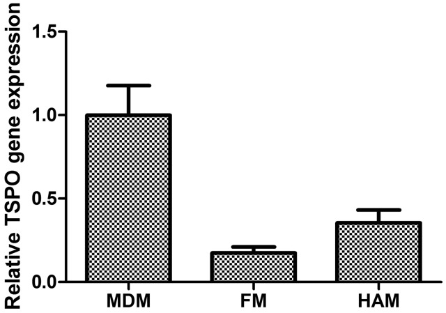
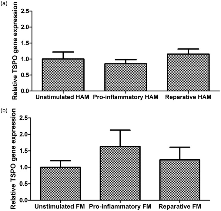
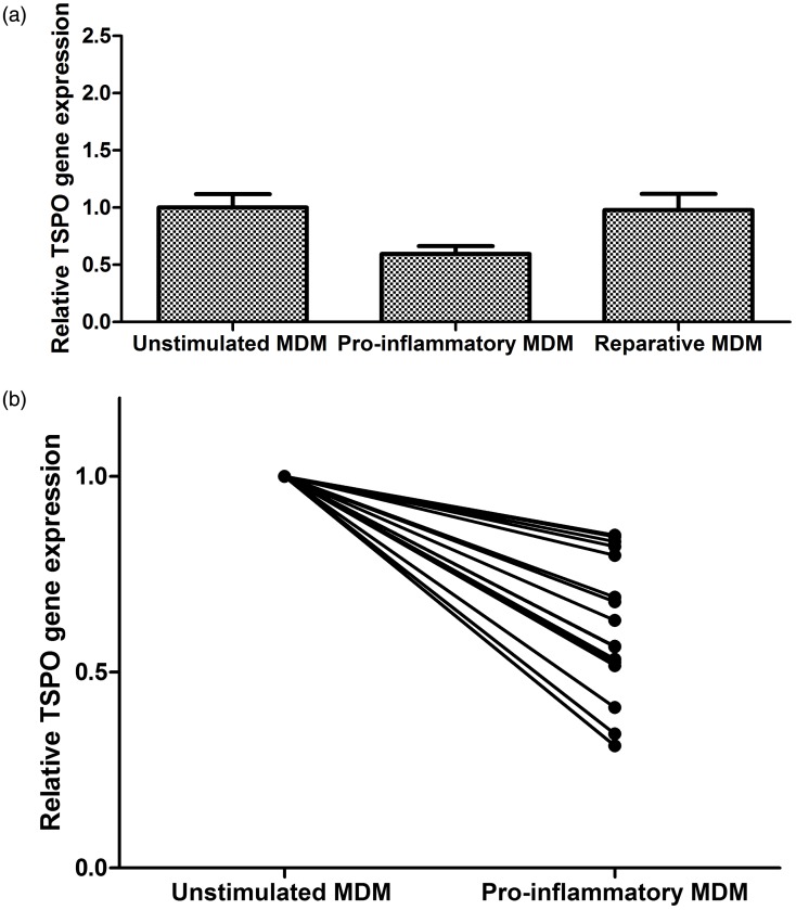
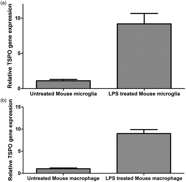
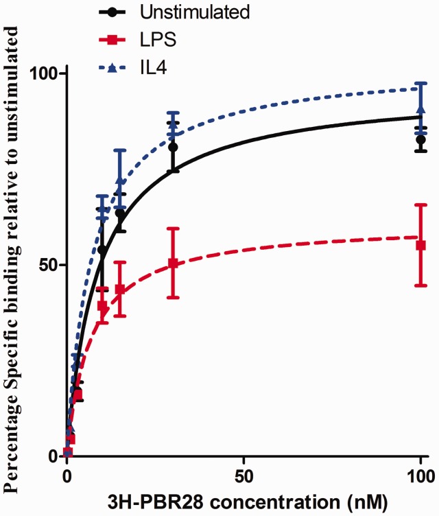
The 18kDa Translocator Protein (TSPO) is the most commonly used tissue-specific marker of inflammation in positron emission tomography (PET) studies. It is expressed in myeloid cells such as microglia and macrophages, and in rodent myeloid cells expression increases with cellular activation. We assessed the effect of myeloid cell activation on TSPO gene expression in both primary human and rodent microglia and macrophages in vitro, and also measured TSPO radioligand binding with 3 H-PBR28 in primary human macrophages. As observed previously, we found that TSPO expression increases (∼9-fold) in rodent-derived macrophages and microglia upon pro-inflammatory stimulation. However, TSPO expression does not increase with classical pro-inflammatory activation in primary human microglia (fold change 0.85 [95% CI 0.58–1.12], p = 0.47). In contrast, pro-inflammatory activation of human monocyte-derived macrophages is associated with a reduction of both TSPO gene expression (fold change 0.60 [95% CI 0.45–0.74], p = 0.02) and TSPO binding site abundance (fold change 0.61 [95% CI 0.49–0.73], p < 0.0001). These findings have important implications for understanding the biology of TSPO in activated macrophages and microglia in humans. They are also clinically relevant for the interpretation of PET studies using TSPO targeting radioligands, as they suggest changes in TSPO expression may reflect microglial and macrophage density rather than activation phenotype.
Overview
The claims and statistics of this paper haven’t been checked yet.
Run the check — free, no account needed. It reads the open-access text and checks the sources it cites.
Integrity signals
Registry
Cites
Statistics
Figures
Verification
Claims the paper makes
The claims and statistics of this paper haven’t been checked yet.
Run the check — free, no account needed. It reads the open-access text and checks the sources it cites.
Claims other papers make about this one
Each is checked against this paper’s own text: does it say what the citing paper says it does?
The source backs part of the claim, not all of it — or backs it for a different population, condition or comparison.
In the citing paper
“Recently, we have shown in vitro that, although TSPO gene and protein expression and TSPO radioligand binding increases in rodent microglia and macrophages with inflammatory stimulation, relative TSPO gene expression in human microglia and TSPO ligand binding in human monocyte-derived macrophages are reduced with pro-inflammatory activation (Owen et al., 2017).”
From A quantitative neuropathological assessment of translocator protein expression in multiple sclerosis (2019) · cites this paper as Owen et al., 2017
Experimental — accuracy not yet measuredThe source backs part of the claim, not all of it — or backs it for a different population, condition or comparison.
In the citing paper
“Recently, we have shown in vitro that, although TSPO gene and protein expression and TSPO radioligand binding increases in rodent microglia and macrophages with inflammatory stimulation, relative TSPO gene expression in human microglia and TSPO ligand binding in human monocyte-derived macrophages are reduced with pro-inflammatory activation (Owen et al., 2017).”
From A quantitative neuropathological assessment of translocator protein expression in multiple sclerosis (2019) · cites this paper as Owen et al., 2017
Experimental — accuracy not yet measuredThe cited source says what the claim attributes to it.
In the citing paper
“However, in humans, TSPO is not upregulated in either pro-inflammatory macrophages (Narayan et al., 2017) or primary microglia (Owen et al., 2017).”
From A quantitative neuropathological assessment of translocator protein expression in multiple sclerosis (2019) · cites this paper as Owen et al., 2017
The cited source says what the claim attributes to it.
In the citing paper
“This new observation thus confirms in vitro data suggesting that the TSPO expression signal does not distinguish between microglial phenotypes in human tissue (Owen et al., 2017).”
From A quantitative neuropathological assessment of translocator protein expression in multiple sclerosis (2019) · cites this paper as Owen et al., 2017
Self-check
Percentages, sums and reported differences re-computed from the paper’s own numbers.
The self-check needs the paper’s full text.
Citations
Cites
Amyloid oligomers exacerbate tau pathology in a mouse model of tauopathy
Selenica, M.-L. B., Brownlow, M., Jimenez, J. P., Lee, D. C., Pena, G., Dickey, C. A., Gordon, M. N., & Morgan, D. (2013) · Neurodegenerative Diseases · doi:10.1159/000337230 · Cited by 10,359
Identification of a unique TGF-β–dependent molecular and functional signature in microglia
Butovsky, O., Jedrychowski, M. P., Moore, C. S., Cialic, R., Lanser, A. J., Gabriely, G., Koeglsperger, T., Dake, B., Wu, P. M., Doykan, C. E., et al. (2014) · Nature Neuroscience · doi:10.1038/nn.3599 · Cited by 2,543 · cited 1×
“Furthermore, many of the markers with differential expression profiles in human microglia relative to human macrophages are not expressed by rodent myeloid cells.(37,38) Our findings add to these differences and suggest that extrapolating TSPO biology from rodent to human myeloid cells should be done with extreme caut…”
BackgroundSelf-citationMitochondrial reactive oxygen species trigger hypoxia-induced transcription
Chandel, N. S., Maltepe, E., Goldwasser, E., Mathieu, C. E., Simon, M. C., & Schumacker, P. T. (1998) · Proceedings of the National Academy of Sciences · doi:10.1073/pnas.95.20.11715 · Cited by 1,926
Microglial physiology: unique stimuli, specialized responses
Ransohoff, R. M., & Perry, V. H. (2009) · Annual Review of Immunology · doi:10.1146/annurev.immunol.021908.132528 · Cited by 1,858 · cited 3×
“Microglia also express receptors for neurotransmitters including g-aminobutyric acid (GABA) and glutamate, providing another pathway for microglial phenotypic regulation.(1,50,51) Caution should therefore be used when extrapolating these findings in vivo and further work will therefore examine directly how TSPO expres…”
BackgroundInnate immune activation in neurodegenerative disease
Heneka, M. T., Kummer, M. P., & Latz, E. (2014) · Nature Reviews Immunology · doi:10.1038/nri3705 · Cited by 1,437 · cited 3×
“Dysregulated or inappropriate microglial activation is thought to contribute to many neurodegenerative diseases.(4) Modulating microglial phenotype has therefore been hypothesised as a potential therapeutic strategy for diseases such as Alzheimer’s and Parkinson’s Disease.”
BackgroundMethodInfluentialMicroglia in physiology and disease
Wolf, S. A., Boddeke, H. W. G. M., & Kettenmann, H. (2017) · Annual Review of Physiology · doi:10.1146/annurev-physiol-022516-034406 · Cited by 1,426 · cited 1×
“Rodent microglia are used extensively given the scarcity of primary human microglia; however, there are several important differences between the two cell types.(34,35) For example, in rodent but not human microglia, transforming growth Figure 2.”
In vivo imaging of microglial activation with [11C](R)-PK11195 PET in idiopathic Parkinson's disease
Gerhard, A., Pavese, N., Hotton, G., Turkheimer, F., Es, M., Hammers, A., Eggert, K., Oertel, W., Banati, R. B., & Brooks, D. J. (2006) · Neurobiology of Disease · doi:10.1016/j.nbd.2005.08.002 · Cited by 1,200
Owen, D. R., Yeo, A. J., Gunn, R. N., Song, K., Wadsworth, G., Lewis, A., Rhodes, C., Pulford, D. J., Bennacef, I., Parker, C. A., et al. (2012) · Journal of Cerebral Blood Flow & Metabolism · doi:10.1038/jcbfm.2011.147 · Cited by 779
Self-citationThe peripheral benzodiazepine binding site in the brain in multiple sclerosis
Banati, R. B., Newcombe, J., Gunn, R. N., Cagnin, A., Turkheimer, F., Heppner, F., Price, G., Wegner, F., Giovannoni, G., Miller, D. H., et al. (2000) · Brain · doi:10.1093/brain/123.11.2321 · Cited by 661
High‐yield isolation of murine microglia by mild trypsinization
Saura, J., Tusell, J. M., & Serratosa, J. (2003) · Glia · doi:10.1002/glia.10274 · Cited by 592 · cited 1×
“At confluency, cultures were subjected to mild trypsinization as described.(32) Following removal of the astrocyte monolayer, microglia were re-plated at a density of 2 10(5) cells mL 1 in DMEMþ 10% FBS, 100 U mL 1 penicillin and 100 mg mL 1 streptomycin Owen et al.”
MethodEdison, P., Archer, H. A., Gerhard, A., Hinz, R., Pavese, N., Turkheimer, F. E., Hammers, A., Tai, Y. F., Fox, N., Kennedy, A., et al. (2008) · Neurobiology of Disease · doi:10.1016/j.nbd.2008.08.001 · Cited by 513
Comparison of polarization properties of human adult microglia and blood‐derived macrophages
Durafourt, B. A., Moore, C. S., Zammit, D., Johnson, T., Zaguia, F., Guiot, M., Bar‐Or, A., & Antel, J. P. (2012) · Glia · doi:10.1002/glia.22298 · Cited by 470 · cited 4×
“The proportion of microglia in the culture, determined using CD11c staining by flow cytometry, was 90%.(3,28) Human foetal CNS tissue (cerebral hemispheres, n1⁄4 8) was obtained from the human foetal tissue repository (Albert Einstein College of Medicine, Bronx, NY), following approved institutional and Canadian Insti…”
BackgroundMethodInfluentialSelf-citationRoles of microglia in brain development, tissue maintenance and repair
Michell‐Robinson, M. A., Touil, H., Healy, L. M., Owen, D. R., Durafourt, B. A., Bar‐Or, A., Antel, J. P., & Moore, C. S. (2015) · Brain · doi:10.1093/brain/awv066 · Cited by 435 · cited 1×
“Loss of this inhibition induces the characteristic microglial activation cascade.(49) Microglia also express receptors for neurotransmitters including g-aminobutyric acid (GABA) and glutamate, providing another pathway for microglial phenotypic regulation.”
BackgroundSelf-citationImaging robust microglial activation after lipopolysaccharide administration in humans with PET
Sandiego, C. M., Gallezot, J.-D., Pittman, B., Nabulsi, N., Lim, K., Lin, S.-F., Matuskey, D., Lee, J.-Y., O’Connor, K. C., Huang, Y., et al. (2015) · Proceedings of the National Academy of Sciences · doi:10.1073/pnas.1511003112 · Cited by 356 · cited 1×
“Likewise, data showing a robust increase in CNS (11)C-PBR28 PET signal following systemic LPS administration to both humans and nonhuman primates may need to be reassessed.(57,58) In these experiments, the increased TSPO PET signal was interpreted as evidence of microglial activation.”
BackgroundRole of mitochondria in steroidogenesis
Papadopoulos, V., & Miller, W. L. (2012) · Best Practice & Research Clinical Endocrinology & Metabolism · doi:10.1016/j.beem.2012.05.002 · Cited by 261 · cited 1×
“Its functions in steroidogenic cells likely are related to de novo steroid synthesis, which it appears able to modulate, although it may not be a necessary part of the steroidogenic machinery.(39,40) However, rodent microglia(26) and human microglia (data not shown) lack CYP11A1, the enzyme necessary to synthesize ste…”
BackgroundSmith, A. M., & Dragunow, M. (2014) · Trends in Neurosciences · doi:10.1016/j.tins.2013.12.001 · Cited by 256 · cited 2×
“Production of nitric oxide, an important molecule in the inflammatory response, requires greater stimulation in human compared with rodent microglia, and even so human cells produce much lower levels of it.(34) Furthermore, many of the markers with differential expression profiles in human microglia relative to human…”
BackgroundPositron emission tomography and functional characterization of a complete PBR/TSPO knockout
Banati, R. B., Middleton, R. J., Chan, R., Hatty, C. R., Wai-Ying Kam, W., Quin, C., Graeber, M. B., Parmar, A., Zahra, D., Callaghan, P., et al. (2014) · Nature Communications · doi:10.1038/ncomms6452 · Cited by 241 · cited 3×
“Various potentially relevant alternative functional roles have been described, including roles in the regulation of the efficiency of mitochondrial oxidative phosphorylation and the production of ROS.23,25 The latter might contribute to potential immunoregulatory functions, e.g. through modulation of ROS activation of…”
BackgroundTu, L. N., Morohaku, K., Manna, P. R., Pelton, S. H., Butler, W. R., Stocco, D. M., & Selvaraj, V. (2014) · Journal of Biological Chemistry · doi:10.1074/jbc.m114.578286 · Cited by 239 · cited 1×
“Its functions in steroidogenic cells likely are related to de novo steroid synthesis, which it appears able to modulate, although it may not be a necessary part of the steroidogenic machinery.(39,40) However, rodent microglia(26) and human microglia (data not shown) lack CYP11A1, the enzyme necessary to synthesize ste…”
BackgroundBenarroch, E. E. (2013) · Neurology · doi:10.1212/wnl.0b013e3182a4a577 · Cited by 232 · cited 1×
“This involves a myriad of changes to morphology, metabolism, surface marker expression and cytokine release, but the range of activation phenotypes is broad and microglia can generate both pro-inflammatory and reparative effector functions.(2,3) Dysregulated or inappropriate microglial activation is thought to contrib…”
BackgroundKarlstetter, M., Nothdurfter, C., Aslanidis, A., Moeller, K., Horn, F., Scholz, R., Neumann, H., Weber, B. H. F., Rupprecht, R., & Langmann, T. (2014) · Journal of Neuroinflammation · doi:10.1186/1742-2094-11-3 · Cited by 224 · cited 2×
“TSPO expression has been reported to increase in activated rodent myeloid cells, with very similar fold increases in both gene and protein expression.(13,15,21,26) Our rodent data are consistent with the literature and showed an approximately 10-fold increase in TSPO gene expression following activation, in both macro…”
BackgroundResultmiR‐155 as a multiple sclerosis–relevant regulator of myeloid cell polarization
Moore, C. S., Rao, V. T. S., Durafourt, B. A., Bedell, B. J., Ludwin, S. K., Bar‐Or, A., & Antel, J. P. (2013) · Annals of Neurology · doi:10.1002/ana.23967 · Cited by 219 · cited 1×
“Both stimulation protocols reliably generate the respective phenotypes.(3,28,30) To confirm pro-inflammatory activation, TNF-a production or TNF-a gene expression was measured by ELISA (BD Biosciences, as per manufacturer’s protocol) or qPCR (methods below) respectively in a subset of samples (12/26).”
MethodSelf-citationThe 18 kDa Translocator Protein, Microglia and Neuroinflammation
Liu, G., Middleton, R. J., Hatty, C. R., Kam, W. W.-Y., Chan, R. H. Y., Pham, T. Q., Harrison‐Brown, M., Dodson, E., Veale, K., & Banati, R. B. (2014) · Brain Pathology · doi:10.1111/bpa.12196 · Cited by 218 · cited 1×
“Increases in the PET signal suggesting increased TSPO density generally have been interpreted in terms of increases in TSPO expression in microglia with activation.(42,48) However, under the assumptions Figure 3.”
BackgroundMicroglia express GABA(B) receptors to modulate interleukin release
Kuhn, S. A., van Landeghem, F. K. H., Zacharias, R., Färber, K., Rappert, A., Pavlovic, S., Hoffmann, A., Nolte, C., & Kettenmann, H. (2004) · Molecular and Cellular Neuroscience · doi:10.1016/j.mcn.2003.10.023 · Cited by 218 · cited 1×
“Microglia also express receptors for neurotransmitters including g-aminobutyric acid (GABA) and glutamate, providing another pathway for microglial phenotypic regulation.(1,50,51) Caution should therefore be used when extrapolating these findings in vivo and further work will therefore examine directly how TSPO expres…”
BackgroundHannestad, J. O., Gallezot, J., Schafbauer, T., Lim, K., Kloczynski, T., Morris, E. D., Carson, R. E., Ding, Y., & Cosgrove, K. (2012) · NeuroImage · doi:10.1016/j.neuroimage.2012.06.055 · Cited by 218 · cited 1×
“Likewise, data showing a robust increase in CNS (11)C-PBR28 PET signal following systemic LPS administration to both humans and nonhuman primates may need to be reassessed.(57,58) In these experiments, the increased TSPO PET signal was interpreted as evidence of microglial activation.”
BackgroundP2Y12 expression and function in alternatively activated human microglia
Moore, C. S., Ase, A. R., Kinsara, A., Rao, V. T. S., Michell-Robinson, M., Leong, S. Y., Butovsky, O., Ludwin, S. K., Séguéla, P., Bar-Or, A., et al. (2015) · Neurology Neuroimmunology & Neuroinflammation · doi:10.1212/nxi.0000000000000080 · Cited by 212 · cited 2×
“The proportion of microglia in the culture, determined using CD11c staining by flow cytometry, was 90%.(3,28) Human foetal CNS tissue (cerebral hemispheres, n1⁄4 8) was obtained from the human foetal tissue repository (Albert Einstein College of Medicine, Bronx, NY), following approved institutional and Canadian Insti…”
MethodSelf-citationWang, M., Wang, X., Zhao, L., Ma, W., Rodríguez, I. R., Fariss, R. N., & Wong, W. T. (2014) · Journal of Neuroscience · doi:10.1523/jneurosci.3153-13.2014 · Cited by 211 · cited 3×
“While the precise function of TSPO is unknown, preliminary studies suggest TSPO may alter myeloid phenotype by modulating the increases in reactive oxygen species (ROS) production and bioenergetics shifts that accompany myeloid cellular activation.(15,25) In vitro, both mRNA and protein expression of TSPO increase sub…”
BackgroundResultOwen, D. R., Howell, O. W., Tang, S.-P., Wells, L. A., Bennacef, I., Bergström, M., Gunn, R. N., Rabiner, E. A., Wilkins, M. R., Reynolds, R., et al. (2010) · Journal of Cerebral Blood Flow & Metabolism · doi:10.1038/jcbfm.2010.63 · Cited by 211
Self-citationCazareth, J., Guyon, A., Heurteaux, C., Chabry, J., & Petit-Paitel, A. (2014) · Journal of Neuroinflammation · doi:10.1186/1742-2094-11-132 · Cited by 206 · cited 2×
“For example, adherence of circulating leucocytes to the vascular endothelium and recruitment of peripheral monocytes into the parenchyma are both rapid processes that would increase the TSPO PET signal in the CNS.(59,60) Endothelial cells also express both TSPO and Toll-like receptor 4 (TLR4, an LPS receptor).”
BackgroundTranslational evaluation of translocator protein as a marker of neuroinflammation in schizophrenia
Notter, T., Coughlin, J. M., Gschwind, T., Weber‐Stadlbauer, U., Wang, Y., Kassiou, M., Vernon, A. C., Benke, D., Pomper, M. G., Sawa, A., et al. (2017) · Molecular Psychiatry · doi:10.1038/mp.2016.248 · Cited by 200
Imaging Microglial Activation with TSPO PET: Lighting Up Neurologic Diseases?
Vivash, L. E., & O'Brien, T. J. (2015) · Journal of Nuclear Medicine · doi:10.2967/jnumed.114.141713 · Cited by 192 · cited 2×
“For this reason, TSPO is the most commonly used marker of inflammation in positron emission tomography (PET) studies in clinical populations.(10) Furthermore, preclinical experiments show that TSPO ligands provide protection in animal models of neuroinflammation and degeneration.”
MethodGershen, L. D., Zanotti‐Fregonara, P., Dustin, I. H., Liow, J., Hirvonen, J., Kreisl, W. C., Jenko, K. J., Inati, S. K., Fujita, M., Morse, C. L., et al. (2015) · JAMA Neurology · doi:10.1001/jamaneurol.2015.0941 · Cited by 178
Ligand for Translocator Protein Reverses Pathology in a Mouse Model of Alzheimer's Disease
Barron, A. M., García‐Segura, L. M., Caruso, D., Jayaraman, A., Lee, J.-W., Melcangi, R. C., & Pike, C. J. (2013) · Journal of Neuroscience · doi:10.1523/jneurosci.1350-13.2013 · Cited by 159
A TSPO ligand is protective in a mouse model of multiple sclerosis
Daugherty, D. J., Selvaraj, V., Chechneva, O. V., Liu, X., Pleasure, D., & Deng, W. (2013) · EMBO Molecular Medicine · doi:10.1002/emmm.201202124 · Cited by 158
Chen, M., Baidoo, K. E., Verina, T., & Guilarte, T. R. (2004) · Brain · doi:10.1093/brain/awh161 · Cited by 140
Torres, S. R., Fröde, T. S., Nardi, G. M., Vita, N., Reeb, R., Ferrara, P., Ribeiro‐do‐Valle, R. M., & Farges, R. C. (2000) · European Journal of Pharmacology · doi:10.1016/s0014-2999(00)00760-3 · Cited by 138
Zavala, F., Taupin, V., & Descamps‐Latscha, B. (1990) · Journal of Pharmacology and Experimental Therapeutics · doi:10.1016/s0022-3565(25)22922-6 · Cited by 133
Translocator Protein 18 kDa Negatively Regulates Inflammation in Microglia
Bae, K.-R., Shim, H.-J., Balu, D., Kim, S. R., & Yu, S. (2014) · Journal of Neuroimmune Pharmacology · doi:10.1007/s11481-014-9540-6 · Cited by 124 · cited 2×
“TSPO expression has been reported to increase in activated rodent myeloid cells, with very similar fold increases in both gene and protein expression.(13,15,21,26) Our rodent data are consistent with the literature and showed an approximately 10-fold increase in TSPO gene expression following activation, in both macro…”
BackgroundResultChoi, J., Ifuku, M., Noda, M., & Guilarte, T. R. (2010) · Glia · doi:10.1002/glia.21091 · Cited by 124
Richardson, R. P., Rhyne, C. D., Fong, Y., HESSE, D. G., Tracey, K. J., Marano, M. A., Lowry, S. F., Antonacci, A. C., & Calvano, S. E. (1989) · Annals of Surgery · doi:10.1097/00000658-198908000-00018 · Cited by 124 · cited 2×
“For example, adherence of circulating leucocytes to the vascular endothelium and recruitment of peripheral monocytes into the parenchyma are both rapid processes that would increase the TSPO PET signal in the CNS.(59,60) Endothelial cells also express both TSPO and Toll-like receptor 4 (TLR4, an LPS receptor).”
BackgroundHernández‐Rabaza, V., Cabrera‐Pastor, A., Taoro‐González, L., González‐Usano, A., Agustí, A., Balzano, T., Llansola, M., & Felipo, V. (2016) · Journal of Neuroinflammation · doi:10.1186/s12974-016-0549-z · Cited by 120 · cited 1×
“Microglia also express receptors for neurotransmitters including g-aminobutyric acid (GABA) and glutamate, providing another pathway for microglial phenotypic regulation.(1,50,51) Caution should therefore be used when extrapolating these findings in vivo and further work will therefore examine directly how TSPO expres…”
BackgroundTranslocator Protein 18 kDa (TSPO): An Old Protein with New Functions?
Li, F., Liu, J., Liu, N., Kuhn, L. A., Garavito, R. M., & Ferguson-Miller, S. M. (2016) · Biochemistry · doi:10.1021/acs.biochem.6b00142 · Cited by 119 · cited 3×
“TSPO is a highly conserved protein, richly expressed in steroidogenic and myeloid cells.(5) Its functions in steroidogenic cells likely are related to de novo steroid synthesis, which it appears able to modulate, although it may not be a necessary part of the steroidogenic machinery.”
BackgroundInfluentialIn Vivo Assessment of Brain White Matter Inflammation in Multiple Sclerosis with18F-PBR111 PET
Colasanti, A., Guo, Q., Muhlert, N., Giannetti, P., Onega, M., Newbould, R. D., Ciccarelli, O., Rison, S., Thomas, C., Nicholas, R., et al. (2014) · Journal of Nuclear Medicine · doi:10.2967/jnumed.113.135129 · Cited by 100
Self-citationBrain microglia express steroid-converting enzymes in the mouse
Gottfried‐Blackmore, A., Sierra, A., Jellinck, P. H., McEwen, B. S., & Bulloch, K. (2008) · The Journal of Steroid Biochemistry and Molecular Biology · doi:10.1016/j.jsbmb.2007.12.013 · Cited by 96 · cited 2×
“TSPO expression has been reported to increase in activated rodent myeloid cells, with very similar fold increases in both gene and protein expression.(13,15,21,26) Our rodent data are consistent with the literature and showed an approximately 10-fold increase in TSPO gene expression following activation, in both macro…”
BackgroundResultRyu, J. K., Choi, H. B., & McLarnon, J. G. (2005) · Neurobiology of Disease · doi:10.1016/j.nbd.2005.04.010 · Cited by 93
Cell cycle synchronization of animal cells and nuclei by centrifugal elutriation
Bánfalvi, G. (2008) · Nature Protocols · doi:10.1038/nprot.2008.34 · Cited by 91 · cited 1×
“For quantification of TSPO binding signal, very large cell numbers are required and therefore peripheral blood monocytes were isolated from blood cones of healthy donors purchased from the National Blood Service (Colindale, Edgware, London UK) using density gradient centrifugation, and centrifugal elutriation performe…”
MethodThalamic inflammation after brain trauma is associated with thalamo-cortical white matter damage
Scott, G., Hellyer, P. J., Ramlackhansingh, A. F., Brooks, D. J., Matthews, P. M., & Sharp, D. J. (2015) · Journal of Neuroinflammation · doi:10.1186/s12974-015-0445-y · Cited by 81
Self-citationImaging neuroinflammation in multiple sclerosis using TSPO-PET
Airas, L. M., Rissanen, E., & Rinne, J. O. (2015) · Clinical and Translational Imaging · doi:10.1007/s40336-015-0147-6 · Cited by 78 · cited 1×
“Increases in the PET signal suggesting increased TSPO density generally have been interpreted in terms of increases in TSPO expression in microglia with activation.(42,48) However, under the assumptions Figure 3.”
BackgroundKalk, N. J., Guo, Q., Owen, D. R., Cherian, R., Erritzøe, D., Gilmour, A.-M., Ribeiro, A., McGonigle, J., Waldman, A. D., Matthews, P. M., et al. (2017) · Translational Psychiatry · doi:10.1038/tp.2016.264 · Cited by 76 · cited 1×
“Furthermore, recent studies in conditions characterised by microglial activation but without peripheral monocyte recruitment (such as alcohol withdrawal and schizophrenia) have not demonstrated the expected increase in TSPO PET signal.(55,56) These unexpected findings merit reconsideration in light of the data we pres…”
BackgroundSelf-citationGulyás, B. Z., Makkai, B., Kása, P., Gulya, K., Bakota, L., Várszegi, S., Beliczai, Z., Andersson, J. D., Csiba, L., Thiele, A., et al. (2008) · Neurochemistry International · doi:10.1016/j.neuint.2008.10.001 · Cited by 74
Translocator protein (18 kDa) is involved in the regulation of reactive gliosis
Veiga, S., Carrero, P., Pernia, O., Azcoitia, I., & Garcia‐Segura, L. M. (2007) · Glia · doi:10.1002/glia.20558 · Cited by 62
Dendritic Cell Differentiation Signals Induce Anti-Inflammatory Properties in Human Adult Microglia
Lambert, C., Desbarats, J., Arbour, N., Hall, J. A., Olivier, A., Bar-Or, A., & Antel, J. P. (2008) · The Journal of Immunology · doi:10.4049/jimmunol.181.12.8288 · Cited by 50 · cited 1×
“Cells were isolated as previously described.(29) Briefly, brain tissue (gestational age 14–20 weeks) was minced and treated with DNase/trypsin.”
MethodSelf-citationDistinct migratory and cytokine responses of human microglia and macrophages to ATP
Lambert, C., Ase, A. R., Séguéla, P., & Antel, J. P. (2010) · Brain Behavior and Immunity · doi:10.1016/j.bbi.2010.02.010 · Cited by 43 · cited 1×
“The surgical brain tissue was processed as described previously.(3,27) Briefly, tissue was obtained in pieces <1 mm(3) and treated with DNase (Roche, Nutley, NJ) and trypsin (Invitrogen, Carlsbad, CA) for 30min at 37 C.”
MethodSelf-citationSmith, A. M., Graham, E. S., Feng, S. X., Oldfield, R. L., Bergin, P. M., Mee, E. W., Faull, R. L. M., Curtis, M. A., & Dragunow, M. (2013) · PLoS ONE · doi:10.1371/journal.pone.0080463 · Cited by 38 · cited 1×
“factor b1 (TGFb1) inhibits IFNg-induced microglial HLA expression.(36) Production of nitric oxide, an important molecule in the inflammatory response, requires greater stimulation in human compared with rodent microglia, and even so human cells produce much lower levels of it.”
BackgroundThe peripheral benzodiazepine receptor ligand PK 11195 inhibits arthritis in the MRL-lpr mouse model
Waterfield, J. D., McGeer, E. G., & McGeer, P. L. (1999) · Lara D. Veeken · doi:10.1093/rheumatology/38.11.1068 · Cited by 37
Translocator protein 18 kDa (TSPO) expression in multiple sclerosis patients
Harberts, E., Datta, D., Chen, S., Wohler, J. E., Oh, U., & Jacobson, S. (2013) · Journal of Neuroimmune Pharmacology · doi:10.1007/s11481-012-9397-5 · Cited by 36
Mitochondrial ligand inhibits store‐operated calcium influx and COX‐2 production in human microglia
Hong, S. H., Choi, H. B., Kim, S. U., & McLarnon, J. G. (2006) · Journal of Neuroscience Research · doi:10.1002/jnr.20829 · Cited by 33
Ligands of the peripheral benzodiazepine receptor have therapeutic effects in pneumopathies in vivo
Bribes, E., Bourrié, B., & Casellas, P. (2003) · Immunology Letters · doi:10.1016/s0165-2478(03)00083-x · Cited by 27
Diazepam inhibits HIV-1 Tat-induced migration of human microglia
Lokensgard, J. R. (2001) · Journal of NeuroVirology · doi:10.1080/135502801753170345 · Cited by 27
Differential transcriptional response profiles in human myeloid cell populations
Healy, L. M., Perron, G., Won, S., Rao, V. T. S., Guiot, M., Moore, C. S., Bar‐Or, A., & Antel, J. P. (2016) · Clinical Immunology · doi:10.1016/j.clim.2016.04.006 · Cited by 22 · cited 1×
“Furthermore, many of the markers with differential expression profiles in human microglia relative to human macrophages are not expressed by rodent myeloid cells.(37,38) Our findings add to these differences and suggest that extrapolating TSPO biology from rodent to human myeloid cells should be done with extreme caut…”
BackgroundSelf-citationBazzichi, L. M., Betti, L., Giannaccini, G., Rossi, A., & Lucacchini, A. (2003) · Clinical Biochemistry · doi:10.1016/s0009-9120(02)00408-3 · Cited by 22
Branley, H. M., du Bois, R. M., Wells, A. U., & Jones, H. A. (2007) · Nuclear Medicine and Biology · doi:10.1016/j.nucmedbio.2007.03.005 · Cited by 12
Cited by
Sorted by citations
The Bidirectional Relationship of Depression and Inflammation: Double Trouble
Beurel, E., Toups, M., & Nemeroff, C. B. (2020) · Neuron · doi:10.1016/j.neuron.2020.06.002 · Cited by 2,136
“It is, however, important to note that the expression of TSPO initially thought to represent microglia activation, has been recently proposed to also measure local myeloid cell proliferation, or monocytes infiltration (Owen et al., 2017).”
BackgroundEnache, D., Pariante, C. M., & Mondelli, V. (2019) · Brain, Behavior, and Immunity · doi:10.1016/j.bbi.2019.06.015 · Cited by 615
Brain glial activation in fibromyalgia – A multi-site positron emission tomography investigation
Albrecht, D. S., Forsberg, A., Sandström, A., Bergan, C., Kadetoff, D., Protsenko, E., Lampa, J., Lee, Y. C., Höglund, C. O., Catana, C., et al. (2019) · Brain, Behavior, and Immunity · doi:10.1016/j.bbi.2018.09.018 · Cited by 310
“immune challenges induce TSPO upregulation in anti-inflammatory M2-like macrophages, and TSPO reduction in inflammatory M1-like macrophages (Narayan et al., 2017; Owen et al., 2017).”
Neuroinflammation in frontotemporal dementia
Bright, F., Werry, E. L., Dobson-Stone, C., Piguet, O., Ittner, L. M., Halliday, G. M., Hodges, J. R., Kiernan, M. C., Loy, C. T., Kassiou, M., et al. (2019) · Nature Reviews Neurology · doi:10.1038/s41582-019-0231-z · Cited by 280
Werry, E. L., Bright, F., Piguet, O., Ittner, L. M., Halliday, G. M., Hodges, J. R., Kiernan, M. C., Loy, C. T., Kril, J. J., & Kassiou, M. (2019) · International Journal of Molecular Sciences · doi:10.3390/ijms20133161 · Cited by 298
Metadata
- Authors
- Owen, D. R., Narayan, N., Wells, L., Healy, L., Smyth, E., Rabiner, E. A., Galloway, D., Williams, J. B., Lehr, J., Mandhair, H., Peferoen, L. A., Taylor, P. C., Amor, S., Antel, J. P., Matthews, P. M., & Moore, C. S.
- Affiliations
- Hammersmith Hospital (GB)
- University of Oxford (GB)
- Montreal Neurological Institute and Hospital (CA)
- King's College London (GB)
- Memorial University of Newfoundland (CA)
- Vrije Universiteit Amsterdam (NL)
- Queen Mary University of London (GB)
- Corresponding author
- Owen, David R
- ORCID
- Venue
- Journal of Cerebral Blood Flow & Metabolism
- Publisher
- SAGE Publications
- Published
- 2017-05-22
- Volume
- 37
- Issue
- 8
- Pages
- 2679-2690
- ISSN
- 0271-678X (ISSN-L)
- 1559-7016
- PMC
- PMC5536262
- OpenAlex
- W2619298813
- Cited by
- 194
- References
- 61
- Citations by year
- 2026: 10 · 2025: 12 · 2024: 22 · 2023: 25 · 2022: 26 · 2021: 24 · 2020: 35 · 2019: 18 · 2018: 17 · 2017: 5
- FWCI
- 10.22
- Type
- journal article
- Language
- en
- Open access
- hybrid
- doi.org
- Locations
- Journal of Cerebral Blood Flow & Metabolism (publishedVersion, cc-by)
- PubMed Central (publishedVersion, cc-by)
- PubMed (publishedVersion)
- Research Portal (King's College London) (publishedVersion, other-oa)
- Amsterdam UMC (VU Amsterdam) - Institutional Repository (publishedVersion)
- Pure Amsterdam UMC (submittedVersion, cc-by)
- Spiral (Imperial College London) (submittedVersion, other-oa)
- Data Archiving and Networked Services (DANS) (submittedVersion)
- Funders
- National Institute for Health and Care Research (NF-SI-0514-10022)
- Multiple Sclerosis Society
- Canadian Institutes of Health Research
- Medical Research Council (MR/N008219/1, MR/N026934/1)
- Topics
- Life Sciences › Neuroscience › Neurology › Neuroinflammation and Neurodegeneration Mechanisms
- Life Sciences › Pharmacology, Toxicology and Pharmaceutics › Pharmacology › Pharmacological Receptor Mechanisms and Effects
- Health Sciences › Medicine › Radiology, Nuclear Medicine and Imaging › Medical Imaging Techniques and Applications
- Keywords
- TSPO, microglia, macrophages, pro-inflammatory activation, myeloid cell activation, monocyte-derived macrophages
- MeSH terms
- Acetamides, Adult, Animals, Brain, Cells, Cultured, Humans, Interferon-gamma, Lipopolysaccharides, Macrophages, Mice, Inbred C57BL, Middle Aged, Protein Binding, Pyridines, Species Specificity, Gene Expression, Granulocyte-Macrophage Colony-Stimulating Factor, Microglia, Receptors, GABA, Myeloid Cells, Young Adult
About these checks
Registry, retraction, Cites and statistics results are computed, not judged, with one exception: a printed reference that no index lists is read once by a model before it counts as a citation error. Claims are extracted by a model and kept only when their quote appears verbatim in the text; each cited source is then checked, with a model’s help, for whether it says what the claim relies on. Those verdicts can be wrong — read the claim’s quote and the cited source before relying on one.
Statistics and arithmetic are recomputed, not judged.
Confidence bands are shown only for fields calibrated on our annotated test set. None is calibrated yet, so no band is shown.
What counts as a claim ›How we measure confidence ›How we check this ›
Are you an author, or do you see a problem with this page? Request a review
