

The DOI matches its Crossref record; 86 of the 86 references it cites were found in a registry or an index.
Classically and alternatively activated bone marrow derived macrophages differ in cytoskeletal functions and migration towards specific CNS cell types
Open accessAbstract
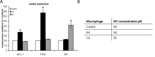
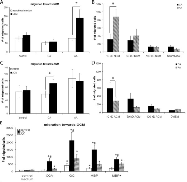
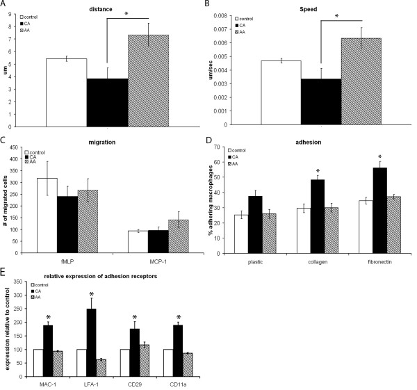
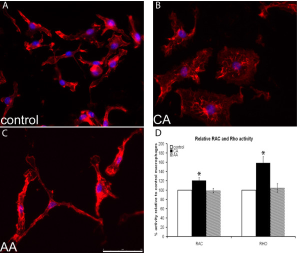
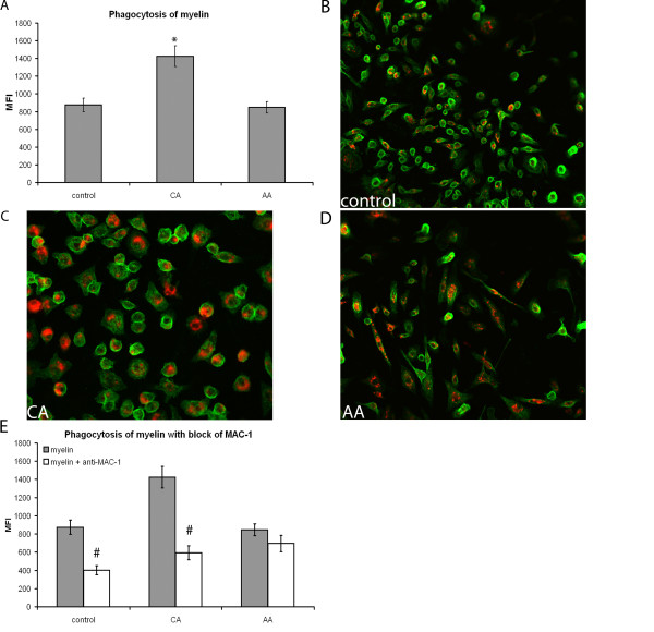
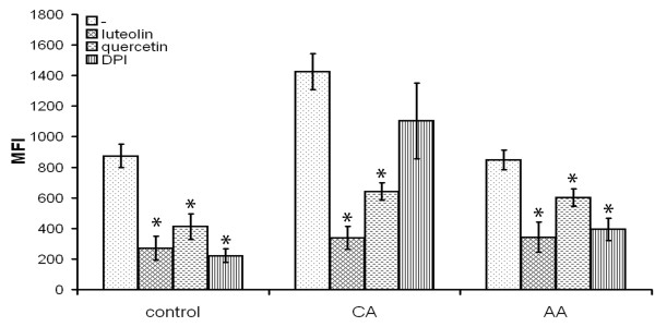
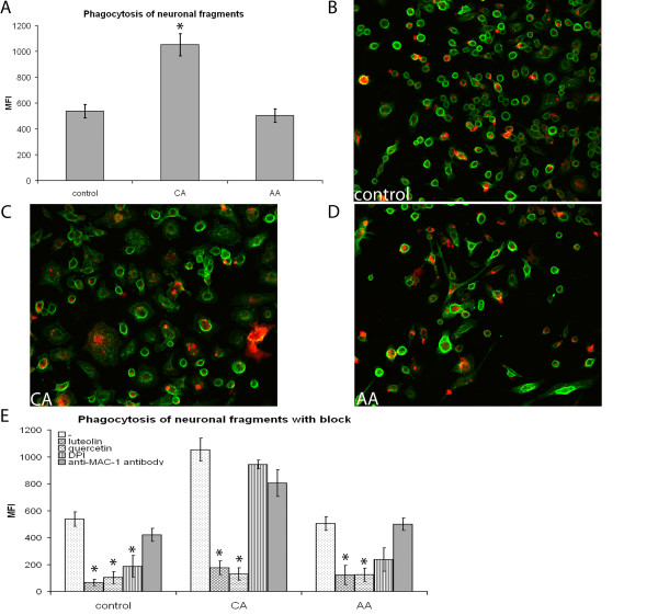
Background: Macrophages play an important role in neuroinflammatory diseases such as multiple sclerosis (MS) and spinal cord injury (SCI), being involved in both damage and repair. The divergent effects of macrophages might be explained by their different activation status: classically activated (CA/M1), pro-inflammatory, macrophages and alternatively activated (AA/M2), growth promoting, macrophages. Little is known about the effect of macrophages with these phenotypes in the central nervous system (CNS) and how they influence pathogenesis. The aim of this study was therefore to determine the characteristics of these phenotypically different macrophages in the context of the CNS in an in vitro setting. Results: Here we show that bone marrow derived CA and AA macrophages have a distinct migratory capacity towards medium conditioned by various cell types of the CNS. AA macrophages were preferentially attracted by the low weight (< 10 kD) fraction of neuronal conditioned medium, while CA macrophages were attracted in higher numbers by astrocyte- and oligodendrocyte conditioned medium. Intrinsic motility was twice as high in AA macrophages compared to CA macrophages. The adhesion to extracellular matrix molecules (ECM) was significantly enhanced in CA macrophages compared to control and AA macrophages. The actin cytoskeleton was differentially organized between CA and AA macrophages, possibly due to greater activity of the GTPases RhoA and Rac in CA macrophages. Phagocytosis of myelin and neuronal fragments was increased in CA macrophages compared to AA macrophages. The increase in myelin phagocytosis was associated with higher expression of CR3/MAC-1 in CA macrophages. Conclusion: In conclusion, since AA macrophages are more motile and are attracted by NCM, they are prone to migrate towards neurons in the CNS. CA macrophages have a lower motility and a stronger adhesion to ECM. In neuroinflammatory diseases the restricted migration and motility of CA macrophages might limit lesion size due to bystander damage.
Overview
The claims and statistics of this paper haven’t been checked yet.
Run the check — free, no account needed. It reads the open-access text and checks the sources it cites.
Integrity signals
Registry
Cites
Statistics
Figures
Verification
Claims the paper makes
The claims and statistics of this paper haven’t been checked yet.
Run the check — free, no account needed. It reads the open-access text and checks the sources it cites.
Claims other papers make about this one
Each is checked against this paper’s own text: does it say what the citing paper says it does?
The source backs part of the claim, not all of it — or backs it for a different population, condition or comparison.
In the citing paper
“Previous studies indicated that pro-inflammatory M1 macrophages appeared large, round-shape morphology, while anti-inflammatory M2 macrophages showed elongated shape [49, 50].”
From A tailored bioactive 3D porous poly(lactic-acid)-exosome scaffold with osteo-immunomodulatory and osteogenic differentiation properties (2022) · cites this paper as [50]
Experimental — accuracy not yet measured
Self-check
Percentages, sums and reported differences re-computed from the paper’s own numbers.
The self-check needs the paper’s full text.
Citations
Cites
Exploring the full spectrum of macrophage activation
Mosser, D. M., & Edwards, J. P. (2008) · Nature Reviews Immunology · doi:10.1038/nri2448 · Cited by 9,144 · cited 5×
“Together these features determine the wound healing and growth promoting phenotype of AA macrophages [1,2].”
BackgroundInfluentialAxonal transection in the lesions of multiple sclerosis
Trapp, B. D., Peterson, J., Ransohoff, R. M., Rudick, R., Mörk, S., & Bö, L. (1998) · New England Journal of Medicine · doi:10.1056/nejm199801293380502 · Cited by 4,029 · cited 2×
“A correlation was found between location and amount of axonal damage and loss and cellular infiltrates containing macrophages [25-27].”
BackgroundMonocyte Chemoattractant Protein-1 (MCP-1): An Overview
Deshmane, S. L., Kremlev, S., Amini, S., & Sawaya, B. E. (2009) · Journal of Interferon & Cytokine Research · doi:10.1089/jir.2008.0027 · Cited by 3,889 · cited 1×
“Both fMLP and MCP-1 are potent chemoattractants [57,58].”
BackgroundKigerl, K. A., Gensel, J. C., Ankeny, D. P., Alexander, J. K., Donnelly, D. J., & Popovich, P. G. (2009) · The Journal of Neuroscience · doi:10.1523/jneurosci.3257-09.2009 · Cited by 2,130 · cited 2×
“found that conditioned medium from CA macrophages was toxic to neurons, while conditioned medium from AA macrophages was not and could even induce axonal outgrowth across a gradient of inhibitory substrate [39].”
BackgroundMartinez, F. O., Gordon, S., Locati, M., & Mantovani, A. (2006) · The Journal of Immunology · doi:10.4049/jimmunol.177.10.7303 · Cited by 2,125 · cited 1×
“Interestingly, human AA macrophages have increased expression levels of CXCR4, the receptor for CXCL12 which is expressed in cultured neurons [52] and in normal brain [53], while CA macrophages have higher expression levels of CCR7 [54].”
BackgroundStein, M., Keshav, S., Harris, N., & Gordon, S. (1992) · The Journal of experimental medicine · doi:10.1084/jem.176.1.287 · Cited by 1,960 · cited 3×
“MR is a longstanding marker for the AA phenotype [3].”
BackgroundMethodNathan, C., & Shiloh, M. U. (2000) · Proceedings of the National Academy of Sciences · doi:10.1073/pnas.97.16.8841 · Cited by 1,469 · cited 1×
“The CA macrophages produce nitric oxide (NO) and reactive oxygen species (ROS) making them cytotoxic [1,4].”
BackgroundTumor necrosis factor mediates myelin and oligodendrocyte damage in vitro
Selmaj, K., & Raine, C. S. (1988) · Annals of Neurology · doi:10.1002/ana.410230405 · Cited by 1,417 · cited 1×
“Activated macrophages secrete many products that could contribute to axonal and oligodendrocyte damage seen in MS lesions, such as matrix metalloproteinases (MMPs) [15], NO and glutamate, [16-22].”
BackgroundKerschensteiner, M., Gallmeier, E., Behrens, L., Vargas-Leal, V., Misgeld, T., Klinkert, W. E. F., Kolbeck, R., Hoppe, E., Oropeza-Wekerle, R. L., Bartke, I., et al. (1999) · The Journal of Experimental Medicine · doi:10.1084/jem.189.5.865 · Cited by 1,019 · cited 2×
“Moreover, activated macrophages/microglia are found to be sources of neurotrophins and growth factors, such as brain derived neurotrophic factor (BDNF) in MS lesions [30,31] and elimination of infiltrating macrophages reduced remyelination in demyelinating models [32].”
BackgroundButovsky, O., Ziv, Y., Schwartz, A., Landa, G., Talpalar, A. E., Pluchino, S., Martino, G., & Schwartz, M. (2006) · Molecular and Cellular Neuroscience · doi:10.1016/j.mcn.2005.10.006 · Cited by 923
Acute axonal injury in multiple sclerosis: Correlation with demyelination and inflammation
Bitsch, A. (2000) · Brain · doi:10.1093/brain/123.6.1174 · Cited by 898 · cited 1×
“A correlation was found between location and amount of axonal damage and loss and cellular infiltrates containing macrophages [25-27].”
BackgroundBiochemical and functional characterization of three activated macrophage populations
Edwards, J. P., Zhang, X., Frauwirth, K. A., & Mosser, D. M. (2006) · Journal of Leukocyte Biology · doi:10.1189/jlb.0406249 · Cited by 863 · cited 6×
“Together these features determine the wound healing and growth promoting phenotype of AA macrophages [1,2].”
BackgroundInfluentialKuhlmann, T., Lingfeld, G., Bitsch, A., Schuchardt, J., & Brück, W. (2002) · Brain : a journal of neurology · doi:10.1093/brain/awf235 · Cited by 851 · cited 1×
“A correlation was found between location and amount of axonal damage and loss and cellular infiltrates containing macrophages [25-27].”
BackgroundImplantation of stimulated homologous macrophages results in partial recovery of paraplegic rats
Rapalino, O., Lazarov-Spiegler, O., Agranov, E., Velan, G. J., Yoles, E., Fraidakis, M., Soloman, A., Gepstein, R., Katz, A., Belkin, M., et al. (1998) · Nature Medicine · doi:10.1038/nm0798-814 · Cited by 788 · cited 7×
“However, macrophages can also create a growth-permissive environment in which axonal regeneration can take place during SCI [36-38].”
BackgroundInfluentialMyelin Impairs CNS Remyelination by Inhibiting Oligodendrocyte Precursor Cell Differentiation
Kotter, M. R., Li, W.-W., Zhao, C., & Franklin, R. J. M. (2006) · The Journal of Neuroscience · doi:10.1523/jneurosci.2615-05.2006 · Cited by 768 · cited 1×
“Phagocytosis of myelin debris is important for axonal repair/ regrowth and remyelination, since myelin components are known to be growth inhibiting [28,29].”
BackgroundExperimental autoimmune encephalomyelitis repressed by microglial paralysis
Heppner, F. L., Greter, M., Marino, D., Falsig, J., Raivich, G., Hövelmeyer, N., Waisman, A., Rülicke, T., Prinz, M., Priller, J., et al. (2005) · Nature Medicine · doi:10.1038/nm1177 · Cited by 732 · cited 9×
“51. van Zwam M, Huizinga R, Melief MJ, Wierenga-Wolf AF, van Meurs M, Voerman JS, Biber KP, Boddeke HW, Hopken UE, Meisel C, Meisel A, Bechmann I, Hintzen RQ, ’t Hart BA, Amor S, Laman JD, Boven LA: Brain antigens in functionally distinct antigen-presenting cell populations in cervical lymph nodes in MS and EAE.”
BackgroundInfluentialHesse, M., Modolell, M., Flamme, A. C. L., Schito, M. L., Fuentes, J. M., Cheever, A. W., Pearce, E. J., & Wynn, T. A. (2001) · The Journal of Immunology · doi:10.4049/jimmunol.167.11.6533 · Cited by 723 · cited 10×
“Due to activation of arginase, arginine is converted to ornithine, a precursor for collagen, which contributes to the production of extracellular matrix molecules (ECM) [8-11] and promotes cell growth [10,12].”
BackgroundInfluentialPopovich, P. G., Guan, Z., Wei, P. Y., Huitinga, I., van Rooijen, N., & Stokes, B. T. (1999) · Experimental Neurology · doi:10.1006/exnr.1999.7118 · Cited by 692 · cited 1×
“Elimination of infiltrating macrophages was observed to increase axonal repair and functional outcome [33-35].”
BackgroundModolell, M., Corraliza, I. M., Link, F., Soler, G., & Eichmann, K. (1995) · European Journal of Immunology · doi:10.1002/eji.1830250436 · Cited by 554 · cited 1×
“Due to activation of arginase, arginine is converted to ornithine, a precursor for collagen, which contributes to the production of extracellular matrix molecules (ECM) [8-11] and promotes cell growth [10,12].”
BackgroundKim, H. K., Cheon, B. S., Kim, Y. H., Kim, S. Y., & Kim, H. P. (1999) · Biochemical Pharmacology · doi:10.1016/s0006-2952(99)00160-4 · Cited by 547 · cited 1×
“Furthermore, they inhibit nuclear factor-kappa B, thereby inhibiting pro-inflammatory cytokine secretion and NO production [70,71].”
BackgroundThe role of nitric oxide in multiple sclerosis
Smith, K. J., & Lassmann, H. (2002) · The Lancet Neurology · doi:10.1016/s1474-4422(02)00102-3 · Cited by 535
Stirling, D. P., Khodarahmi, K., Liu, J., McPhail, L. T., McBride, C. B., Steeves, J. D., Ramer, M. S., & Tetzlaff, W. (2004) · Journal of Neuroscience · doi:10.1523/jneurosci.5275-03.2004 · Cited by 506 · cited 1×
“Elimination of infiltrating macrophages was observed to increase axonal repair and functional outcome [33-35].”
BackgroundHuitinga, I., van Rooijen, N., de Groot, C. J., Uitdehaag, B. M., & Dijkstra, C. D. (1990) · The Journal of experimental medicine · doi:10.1084/jem.172.4.1025 · Cited by 506 · cited 9×
“51. van Zwam M, Huizinga R, Melief MJ, Wierenga-Wolf AF, van Meurs M, Voerman JS, Biber KP, Boddeke HW, Hopken UE, Meisel C, Meisel A, Bechmann I, Hintzen RQ, ’t Hart BA, Amor S, Laman JD, Boven LA: Brain antigens in functionally distinct antigen-presenting cell populations in cervical lymph nodes in MS and EAE.”
BackgroundInfluentialSelf-citationType II monocytes modulate T cell–mediated central nervous system autoimmune disease
Weber, M. S., Prod'homme, T., Youssef, S., Dunn, S. E., Rundle, C. D., Lee, L., Patarroyo, J. C., Stüve, O., Sobel, R. A., Steinman, L., et al. (2007) · Nature Medicine · doi:10.1038/nm1620 · Cited by 447 · cited 9×
“51. van Zwam M, Huizinga R, Melief MJ, Wierenga-Wolf AF, van Meurs M, Voerman JS, Biber KP, Boddeke HW, Hopken UE, Meisel C, Meisel A, Bechmann I, Hintzen RQ, ’t Hart BA, Amor S, Laman JD, Boven LA: Brain antigens in functionally distinct antigen-presenting cell populations in cervical lymph nodes in MS and EAE.”
BackgroundInfluentialSong, E., Ouyang, N., Hörbelt, M., Antus, B., Wang, M., & Exton, M. S. (2000) · Cellular Immunology · doi:10.1006/cimm.2000.1687 · Cited by 447 · cited 2×
“They produce growth factors, such as platelet derived growth factor (PDGF), vascular endothelial growth factor (VEGF) and fibroblast growth factor (FGF) [5-7].”
BackgroundStadelmann, C., Kerschensteiner, M., Misgeld, T., Brück, W., Hohlfeld, R., & Lassmann, H. (2002) · Brain · doi:10.1093/brain/awf015 · Cited by 425
Simpson, J. E., Newcombe, J., Cuzner, M. L., & Woodroofe, M. N. (1998) · Journal of Neuroimmunology · doi:10.1016/s0165-5728(97)00208-7 · Cited by 403 · cited 1×
“MCP-1 expression is mainly found in reactive astrocytes and macrophages [60,61].”
BackgroundPonomarev, E. D., Maresz, K., Tan, Y., & Dittel, B. N. (2007) · The Journal of Neuroscience · doi:10.1523/jneurosci.1922-07.2007 · Cited by 396 · cited 9×
“51. van Zwam M, Huizinga R, Melief MJ, Wierenga-Wolf AF, van Meurs M, Voerman JS, Biber KP, Boddeke HW, Hopken UE, Meisel C, Meisel A, Bechmann I, Hintzen RQ, ’t Hart BA, Amor S, Laman JD, Boven LA: Brain antigens in functionally distinct antigen-presenting cell populations in cervical lymph nodes in MS and EAE.”
BackgroundInfluentialExpression profiles for macrophage alternative activation genes in AD and in mouse models of AD.
(2006) · Journal of Neuroinflammation · doi:10.1186/1742-2094-3-27 · Cited by 393 · cited 1×
“Similar results have been found in a mouse model of Alzheimer’s disease, since mRNA levels for markers of the CA and AA phenotype were observed to be upregulated in microglial cells, indicating that these microglia express functional characteristics of both CA and AA macrophages [86].”
ResultKotter, M. R., Setzu, A., Sim, F. J., Van Rooijen, N., & Franklin, R. J. M. (2001) · Glia · doi:10.1002/glia.1085 · Cited by 385 · cited 1×
“Furthermore, remyelination correlated with the number of oligodendrocytes and macrophages [55,56].”
BackgroundVerreck, F. A. W., de Boer, T., Langenberg, D. M. L., van der Zanden, L., & Ottenhoff, T. H. M. (2005) · Journal of Leukocyte Biology · doi:10.1189/jlb.0105015 · Cited by 383 · cited 1×
“Human M2 macrophages, analogous to AA macrophages, appeared stretched with a spindle-like morphology, while human M1, analogous to CA macrophages, macrophages appeared rounder with a “friedegg” morphology [66].”
BackgroundSpecific inflammatory cytokines regulate the expression of human monocyte 15-lipoxygenase.
Conrad, D. J., Kühn, H., Mulkins, M. A., Highland, E., & Sigal, E. (1992) · Proceedings of the National Academy of Sciences · doi:10.1073/pnas.89.1.217 · Cited by 379 · cited 1×
“Activated macrophages secrete many products that could contribute to axonal and oligodendrocyte damage seen in MS lesions, such as matrix metalloproteinases (MMPs) [15], NO and glutamate, [16-22].”
BackgroundAlternatively activated macrophages in helminth infections
Kreider, T., Anthony, R. M., Urban, J. F., & Gause, W. C. (2007) · Current Opinion in Immunology · doi:10.1016/j.coi.2007.07.002 · Cited by 362 · cited 10×
“Due to activation of arginase, arginine is converted to ornithine, a precursor for collagen, which contributes to the production of extracellular matrix molecules (ECM) [8-11] and promotes cell growth [10,12].”
BackgroundInfluentialMikita, J., Dubourdieu-Cassagno, N., Deloire, M. S., Vekris, A., Biran, M., Raffard, G., Brochet, B., Canron, M.-H., Franconi, J.-M., Boiziau, C., et al. (2011) · Multiple Sclerosis Journal · doi:10.1177/1352458510379243 · Cited by 358 · cited 1×
“Administration of AA macrophages reduced the development of relapses during the relapsing EAE model [82].”
BackgroundMantovani, A., Sica, A., Allavena, P., Garlanda, C., & Locati, M. (2009) · Human Immunology · doi:10.1016/j.humimm.2009.02.008 · Cited by 354 · cited 2×
“They produce growth factors, such as platelet derived growth factor (PDGF), vascular endothelial growth factor (VEGF) and fibroblast growth factor (FGF) [5-7].”
BackgroundTemporal expression of different pathways of 1-arginine metabolism in healing wounds.
Albina, J. E., Mills, C. D., Henry, W. L., & Caldwell, M. D. (1990) · The Journal of Immunology · doi:10.4049/jimmunol.144.10.3877 · Cited by 351 · cited 10×
“Due to activation of arginase, arginine is converted to ornithine, a precursor for collagen, which contributes to the production of extracellular matrix molecules (ECM) [8-11] and promotes cell growth [10,12].”
BackgroundInfluentialKotter, M. R., Zhao, C., van Rooijen, N., & Franklin, R. J. M. (2005) · Neurobiology of Disease · doi:10.1016/j.nbd.2004.09.019 · Cited by 331 · cited 1×
“Moreover, activated macrophages/microglia are found to be sources of neurotrophins and growth factors, such as brain derived neurotrophic factor (BDNF) in MS lesions [30,31] and elimination of infiltrating macrophages reduced remyelination in demyelinating models [32].”
BackgroundIn Vitro-Derived Alternatively Activated Macrophages Reduce Colonic Inflammation in Mice
Hunter, M. M., Wang, A., Parhar, K. K. S., Johnston, M. J. G., van Rooijen, N., Beck, P. L., & McKay, D. M. (2010) · Gastroenterology · doi:10.1053/j.gastro.2009.12.041 · Cited by 313 · cited 1×
“In a model for colitis the injection of AA macrophages ameliorated the disease [78].”
BackgroundStaging of multiple sclerosis (MS) lesions: pathology of the time frame of MS
van der Valk, P., & Groot, C. J. A. D. (2000) · Neuropathology and Applied Neurobiology · doi:10.1046/j.1365-2990.2000.00217.x · Cited by 304 · cited 1×
“Major neuropathological hallmarks of MS are inflammatory demyelinating lesions associated with perivascular infiltrates containing macrophages and lymphocytes [13,14].”
BackgroundWadsworth, T. L., & Koop, D. R. (1999) · Biochemical Pharmacology · doi:10.1016/s0006-2952(99)00002-7 · Cited by 302 · cited 1×
“Furthermore, they inhibit nuclear factor-kappa B, thereby inhibiting pro-inflammatory cytokine secretion and NO production [70,71].”
BackgroundGratchev, A., Guillot, P., Hakiy, N., Politz, O., Orfanos, C. E., Schledzewski, K., & Goerdt, S. (2001) · Scandinavian Journal of Immunology · doi:10.1046/j.1365-3083.2001.00885.x · Cited by 300 · cited 10×
“Due to activation of arginase, arginine is converted to ornithine, a precursor for collagen, which contributes to the production of extracellular matrix molecules (ECM) [8-11] and promotes cell growth [10,12].”
BackgroundInfluentialOligodendrocyte precursor cells in the demyelinated multiple sclerosis spinal cord
Wolswijk, G. (2002) · Brain · doi:10.1093/brain/awf031 · Cited by 268 · cited 1×
“Furthermore, remyelination correlated with the number of oligodendrocytes and macrophages [55,56].”
BackgroundPrewitt, C. M., Niesman, I. R., Kane, C. J. M., & Houlé, J. D. (1997) · Experimental Neurology · doi:10.1006/exnr.1997.6694 · Cited by 260 · cited 7×
“However, macrophages can also create a growth-permissive environment in which axonal regeneration can take place during SCI [36-38].”
BackgroundInfluentialRequirement of Myeloid Cells for Axon Regeneration
Barrette, B., Hébert, M.-A., Filali, M., Lafortune, K., Vallières, N., Gowing, G., Julien, J., & Lacroix, S. (2008) · Journal of Neuroscience · doi:10.1523/jneurosci.1447-08.2008 · Cited by 256 · cited 7×
“However, macrophages can also create a growth-permissive environment in which axonal regeneration can take place during SCI [36-38].”
BackgroundInfluentialCuzner, M. L., Gveric, D. O., Strand, C., Loughlin, J., Paemen, L. R., Opdenakker, G., & Newcombe, J. (1996) · Journal of Neuropathology & Experimental Neurology · doi:10.1097/00005072-199612000-00002 · Cited by 251 · cited 1×
“Activated macrophages secrete many products that could contribute to axonal and oligodendrocyte damage seen in MS lesions, such as matrix metalloproteinases (MMPs) [15], NO and glutamate, [16-22].”
BackgroundMacrophages and neurodegeneration
Hendriks, J. J. A., Teunissen, C. E., de Vries, H. E., & Dijkstra, C. D. (2005) · Brain Research Reviews · doi:10.1016/j.brainresrev.2004.12.008 · Cited by 247 · cited 1×
“Activated macrophages secrete many products that could contribute to axonal and oligodendrocyte damage seen in MS lesions, such as matrix metalloproteinases (MMPs) [15], NO and glutamate, [16-22].”
BackgroundSelf-citationBerman, J. W., Guida, M. P., Warren, J. S., Amat, J., & Brosnan, C. F. (1996) · The Journal of Immunology · doi:10.4049/jimmunol.156.8.3017 · Cited by 245 · cited 1×
“MCP-1 expression is mainly found in reactive astrocytes and macrophages [60,61].”
BackgroundLaminin-2/integrin interactions enhance myelin membrane formation by oligodendrocytes.
Buttery, P., & ffrench-Constant, C. (1999) · Molecular and cellular neurosciences · doi:10.1006/mcne.1999.0781 · Cited by 217 · cited 1×
“Briefly, oligodendrocyte precursors were cultured on poly-l-lysine coated cell culture plates for 2 days in a defined SATO medium [45] containing platelet derived growth factor-AA (PDGF-AA) and fibroblast growth factor-2 (FGF-2) in order to synchronize precursors to the bipolar oligodendrocyte-type II astrocyte (O2A)…”
MethodBaer, A. S., Syed, Y. A., Kang, S. U., Mitteregger, D., Vig, R., ffrench-Constant, C., Franklin, R. J. M., Altmann, F., Lubec, G., & Kotter, M. R. (2009) · Brain · doi:10.1093/brain/awn334 · Cited by 211
Varin, A., Mukhopadhyay, S., Herbein, G., & Gordon, S. (2009) · Blood · doi:10.1182/blood-2009-08-236711 · Cited by 194 · cited 1×
“AA macrophages were prepared by exposure to 10 ng/ml IL-4 (Invitrogen) in the culture medium [3,43].”
MethodHendriks, J. J. A., Alblas, J., van der Pol, S. M. A., van Tol, E. A. F., Dijkstra, C. D., & de Vries, H. E. (2004) · The Journal of Experimental Medicine · doi:10.1084/jem.20040819 · Cited by 191 · cited 7×
“RhoA and Rac activity in differently activated macrophages was determined using G-LISA activation assay kit (Cytoskeleton, Denver, USA) according to the manufacturer’s description.”
BackgroundInfluentialSelf-citationRemyelination in multiple sclerosis
Lassmann, H., Brück, W., Lucchinetti, C., & Rodriguez, M. (1997) · Multiple Sclerosis Journal · doi:10.1177/135245859700300213 · Cited by 191
Neuronal Chemokines: Versatile Messengers In Central Nervous System Cell Interaction
Haas, A. H. D., van Weering, H. R. J., de Jong, E. K., Boddeke, H. W. G. M., & Biber, K. P. H. (2007) · Molecular Neurobiology · doi:10.1007/s12035-007-0036-8 · Cited by 187 · cited 1×
“Interestingly, human AA macrophages have increased expression levels of CXCR4, the receptor for CXCL12 which is expressed in cultured neurons [52] and in normal brain [53], while CA macrophages have higher expression levels of CCR7 [54].”
BackgroundDifferences in angiogenic potential of classically vs alternatively activated macrophages
Kodelja, V., Müller, C., Tenorio, S., Schebesch, C., Orfanos, C. E., & Goerdt, S. (1997) · Immunobiology · doi:10.1016/s0171-2985(97)80080-0 · Cited by 186 · cited 3×
“They produce growth factors, such as platelet derived growth factor (PDGF), vascular endothelial growth factor (VEGF) and fibroblast growth factor (FGF) [5-7].”
BackgroundReactive oxygen species are required for the phagocytosis of myelin by macrophages
van der Goes, A., Brouwer, J. P., Hoekstra, K., Roos, D., van den Berg, T. K., & Dijkstra, C. D. (1998) · Journal of Neuroimmunology · doi:10.1016/s0165-5728(98)00175-1 · Cited by 180 · cited 1×
“In rat macrophages myelin phagocytosis was ROS dependent [68].”
BackgroundSelf-citationExcitatory Amino Acids and Multiple Sclerosis: Evidence From Cerebrospinal Fluid
Sarchielli, P., Greco, L., Floridi, A., Floridi, A., & Gallai, V. (2003) · Archives of Neurology · doi:10.1001/archneur.60.8.1082 · Cited by 168 · cited 1×
“Activated macrophages secrete many products that could contribute to axonal and oligodendrocyte damage seen in MS lesions, such as matrix metalloproteinases (MMPs) [15], NO and glutamate, [16-22].”
Backgroundvan der Laan, L. J. W., Ruuls, S. R., Weber, K. S., Lodder, I. J., Döpp, E. A., & Dijkstra, C. D. (1996) · Journal of Neuroimmunology · doi:10.1016/s0165-5728(96)00110-5 · Cited by 166 · cited 1×
“CR3 is implicated in myelin phagocytosis [67].”
Self-citationReactive oxygen species enhance the migration of monocytes across the blood‐brain barrier in vitro
van der Goes, A., Wouters, D., van der Pol, S. M. A., Huizinga, R., Ronken, E., Adamson, P., Greenwood, J., Dijkstra, C. D., & de Vries, H. E. (2001) · The FASEB Journal · doi:10.1096/fj.00-0881fje · Cited by 162 · cited 1×
“Macrophage adhesion to plastic and several extracellular matrix molecules (ECM) was determined as described previously [47].”
BackgroundSelf-citationVarga, G., Ehrchen, J. M., Tsianakas, A., Tenbrock, K., Rattenholl, A., Seeliger, S., Mack, M., Roth, J., & Sunderkötter, C. H. (2008) · Journal of Leukocyte Biology · doi:10.1189/jlb.1107768 · Cited by 160 · cited 1×
“Our findings were in line with a previous study, observing that glucocorticoid stimulated murine monocytes, which display some features of AA macrophages, adhered less and were more motile than control cells [64].”
Resultvan Zwam, M., Huizinga, R., Melief, M., Wierenga‐Wolf, A. F., van Meurs, M., Voerman, J. S. A., Biber, K. P. H., Boddeke, H. W. G. M., Höpken, U. E., Meisel, C., et al. (2008) · Journal of Molecular Medicine · doi:10.1007/s00109-008-0421-4 · Cited by 148 · cited 1×
“since macrophages in cervical lymph nodes of MS patients contained neuronal antigens [51].”
BackgroundBusch, S. A., Hamilton, J. A., Horn, K. P., Cuascut, F. X., Cutrone, R., Lehman, N., Deans, R., Ting, A. E., Mays, R. W., & Silver, J. (2011) · Journal of Neuroscience · doi:10.1523/jneurosci.3566-10.2011 · Cited by 145 · cited 2×
“In vitro these multipotent adult progenitor cells induced an AA phenotype in macrophages [79].”
BackgroundNewman, T. A. (2001) · Brain · doi:10.1093/brain/124.11.2203 · Cited by 143
Gleichmann, M., Gillen, C., Czardybon, M., Bosse, F., Greiner–Petter, R., Auer, J., & Müller, H. W. (2000) · European Journal of Neuroscience · doi:10.1046/j.1460-9568.2000.00048.x · Cited by 133 · cited 1×
“Interestingly, human AA macrophages have increased expression levels of CXCR4, the receptor for CXCL12 which is expressed in cultured neurons [52] and in normal brain [53], while CA macrophages have higher expression levels of CCR7 [54].”
BackgroundLee, W.-J., Wu, L.-F., Chen, W.-K., Wang, C., & Tseng, T. (2006) · Chemico-Biological Interactions · doi:10.1016/j.cbi.2006.01.002 · Cited by 131 · cited 1×
“Luteolin has effects on the actin cytoskeleton [73,74] through Rho and Rac [75] and could thereby affect phagocytosis.”
BackgroundFlavonoids inhibit myelin phagocytosis by macrophages; a structure–activity relationship study
Hendriks, J. J. A., de Vries, H. E., van der Pol, S. M. A., van den Berg, T. K., van Tol, E. A. F., & Dijkstra, C. D. (2003) · Biochemical Pharmacology · doi:10.1016/s0006-2952(02)01609-x · Cited by 115 · cited 2×
“Previously, myelin phagocytosis was blocked after exposure to quercetin and luteolin in a macrophage cell line [69].”
BackgroundSelf-citationSchiffmann, E. (1982) · Annual Review of Physiology · doi:10.1146/annurev.ph.44.030182.003005 · Cited by 107 · cited 1×
“Both fMLP and MCP-1 are potent chemoattractants [57,58].”
BackgroundActivating ROCK1 somatic mutations in human cancer
Lochhead, P. A., Wickman, G., Mezna, M., & Olson, M. F. (2010) · Oncogene · doi:10.1038/onc.2010.3 · Cited by 94 · cited 1×
“Adhesion can also be influenced by the cytoskeletal organization [62,63].”
BackgroundLourbopoulos, A. I., Grigoriadis, N., Lagoudaki, R., Touloumi, O., Polyzoidou, E., Mavromatis, I., Tascos, N., Breuer, A., Ovadia, H., Karussis, D., et al. (2011) · Brain Research · doi:10.1016/j.brainres.2011.03.020 · Cited by 91 · cited 1×
“2-arachidonoylglycerol [81], which were found to skew macrophages to an alternatively activated phenotype, increased the presence of AA macrophages in the lesions and ameliorated EAE disease course.”
BackgroundChristophi, G. P., Panos, M., Hudson, C. A., Christophi, R. L., Gruber, R. C., Mersich, A. T., Blystone, S. D., Jubelt, B., & Massa, P. T. (2009) · Laboratory Investigation · doi:10.1038/labinvest.2009.32 · Cited by 85 · cited 9×
“AA macrophages were prepared by exposure to 10 ng/ml IL-4 (Invitrogen) in the culture medium [3,43].”
BackgroundInfluentialTransfused Macrophages Ameliorate Pancreatic and Renal Injury in Murine Diabetes Mellitus
Zheng, D., Wang, Y., Cao, Q., Lee, V. W., Zheng, G., Sun, Y., Tan, T. K., Wang, Y., Alexander, S. I., & Harris, D. C. H. (2011) · Nephron Experimental Nephrology · doi:10.1159/000321034 · Cited by 82 · cited 1×
“In vitro generated AA macrophages reduced renal and pancreatic injury in a model for murine diabetes [77].”
BackgroundHeeroma, J. H., Roelandse, M., Wierda, K. D. B., van Aerde, K. I., Toonen, R. F. G., Hensbroek, R. A., Brussaard, A. B., Matus, A. I., & Verhage, M. (2004) · European Journal of Neuroscience · doi:10.1111/j.1460-9568.2004.03503.x · Cited by 71 · cited 1×
“Neuronal fragments were made by roughly pipetting 2 day old neuronal cultures, prepared as described previously [48], and labeling them with the lipophilic fluorescent dye 1,1’,di-octadecyl-3,3,3’3’-tetramethylindocarbocyanine perchlorate (DiI) (SigmaAldrich).”
MethodMaier, O., van der Heide, T., van Dam, A., Baron, W., de Vries, H., & Hoekstra, D. (2004) · Molecular and Cellular Neuroscience · doi:10.1016/j.mcn.2004.09.012 · Cited by 69 · cited 1×
“rat oligodendrocytes were cultured as described previously [44].”
MethodSelf-citationSHP-1 deficiency and increased inflammatory gene expression in PBMCs of multiple sclerosis patients
Christophi, G. P., Hudson, C. A., Gruber, R. C., Christophi, C. P., Mihai, C., Mejico, L. J., Jubelt, B., & Massa, P. T. (2008) · Laboratory Investigation · doi:10.1038/labinvest.3700720 · Cited by 67 · cited 9×
“AA macrophages were prepared by exposure to 10 ng/ml IL-4 (Invitrogen) in the culture medium [3,43].”
BackgroundInfluentialInhibitory effect of luteolin on osteoclast differentiation and function
Lee, J., Ahn, J.-Y., Hasegawa, S., Cha, B., Yonezawa, T., Nagai, K., Seo, H.-J., Jeon, W., & Woo, J. (2009) · Cytotechnology · doi:10.1007/s10616-010-9253-5 · Cited by 64 · cited 1×
“Luteolin has effects on the actin cytoskeleton [73,74] through Rho and Rac [75] and could thereby affect phagocytosis.”
BackgroundIannotti, C. A., Clark, M., Horn, K. P., van Rooijen, N., Silver, J., & Steinmetz, M. P. (2010) · Experimental Neurology · doi:10.1016/j.expneurol.2010.03.010 · Cited by 63
A method for the derivation and continuous propagation of cloned murine bone marrow macrophages
Johnson, C. R., Kitz, D. J., & Little, J. R. (1983) · Journal of Immunological Methods · doi:10.1016/0022-1759(83)90127-8 · Cited by 63 · cited 1×
“Bone marrow derived macrophages were generated as described previously [42].”
MethodDogan, R.-N. E., Long, N. C., Forde, E., Dennis, K. L., Kohm, A. P., Miller, S. D., & Karpus, W. J. (2010) · Journal of Leukocyte Biology · doi:10.1189/jlb.0810442 · Cited by 59 · cited 1×
“Treatment with anti-CCL22 [80] and Vereyken et al.”
MethodJee, Y., Yoon, W. K., Okura, Y., Tanuma, N., & Matsumoto, Y. (2002) · Journal of Neuroimmunology · doi:10.1016/s0165-5728(02)00147-9 · Cited by 55
BCL2 inhibits cell adhesion, spreading, and motility by enhancing actin polymerization
Ke, H., Parron, V. I., Reece, J. M., Zhang, J. Y., Akiyama, S. K., & French, J. E. (2010) · Cell Research · doi:10.1038/cr.2010.21 · Cited by 45 · cited 1×
“Adhesion can also be influenced by the cytoskeletal organization [62,63].”
BackgroundGoebeler, M., Roth, J., Kunz, M., & Sorg, C. (1993) · Immunobiology · doi:10.1016/s0171-2985(11)80495-x · Cited by 26 · cited 1×
“The increased expression of adhesion receptors in macrophages stimulated with IFN-g and LPS is in line with previous studies [49,65] and with our current finding that CA macrophages adhere better when compared to AA macrophages.”
ResultHespanhol, M. R., & Mantovani, B. (2002) · Brazilian Journal of Medical and Biological Research · doi:10.1590/s0100-879x2002000300015 · Cited by 11 · cited 1×
“Quercetin could also mediate its effects due to the inhibition of protein tyrosine kinases, which are involved in the signaling of the engulfment phase of CR3 mediated phagocytosis [72].”
BackgroundPourshafie, M. R., & Sonnenfeld, G. (1997) · Journal of Interferon & Cytokine Research · doi:10.1089/jir.1997.17.69 · Cited by 8 · cited 8×
“The expression of Mac-1, F4/80 and MR was determined using FACS analysis.”
BackgroundResultInfluentialBos, H. J., Boorsma, D. M., Tuk, C. W., de Veld, J. C., van den Muysenberg, A. J. C., Helmerhorst, T. J. M., Struijk, D. G., van Bronswijk, H., & Beelen, R. H. J. (1990) · European Journal of Clinical Investigation · doi:10.1111/j.1365-2362.1990.tb01901.x · Cited by 7
Bos, H. J., BOORSMA, D. M., Tuk, C. W., et al. (1990) · European Journal of Clinical Investigation · doi:10.1111/j.1365-2362.1990.tb01926.x · Cited by 6 · cited 1×
“The migratory capacity of CA and AA macrophages was studied using a 48-wells micro chemotaxis chamber (Neuro Probe, Gaithersburg, USA) as described previously [46] with some modifications.”
MethodZujovic (2000) · doi:10.1002/(sici)1098-1136(20000215)29:4
Research works printed but not linked: 1
- van der Laan LJ, Ruuls SR, Weber KS, Lodder IJ, Dopp EA, Dijkstra CD: Macrophage phagocytosis of myelin in vitro determined by flow cytometry: phagocytosis is mediated by CR3 and induces production of tumor necrosis factor-alpha and nitric oxide. J Neuroimmunol. 1996, 70: 145-152. 10.1016/S0165-5728(96)00110-5. · DOI
Cited by
Sorted by citations
Human macrophage polarization in vitro: maturation and activation methods compared.
Vogel, D., Glim, J. E., Stavenuiter, A. W., Breur, M., Heijnen, P., Amor, S., Dijkstra, C., & Beelen, R. (2014) · Immunobiology · doi:10.1016/j.imbio.2014.05.002 · Cited by 431
Self-citationPhysical and mechanical regulation of macrophage phenotype and function
McWhorter, F. Y., Davis, C. T., & Liu, W. F. (2015) · Cellular and Molecular Life Sciences · doi:10.1007/s00018-014-1796-8 · Cited by 437
“LPS or M1-like activation has been demonstrated to enhance cell adhesion in both human and murine macrophages [49, 64, 67].”
BackgroundResultInfluentialMacrophage biology in the peripheral nervous system after injury
Zigmond, R. E., & Echevarría, F. D. (2018) · Progress in Neurobiology · doi:10.1016/j.pneurobio.2018.12.001 · Cited by 383
“With respect to Wallerian degeneration, Vereyken et al. (2011) showed that M(LPS-IFNγ) macrophages consumed a greater amount of fluorescent myelin than M(IL-4) macrophages, suggesting that M(LPS-IFNγ) have a greater propensity for myelin phagocytosis.”
Function of microglia and macrophages in secondary damage after spinal cord injury
Ren, Y., Zhou, X., & He, X. (2014) · Neural Regeneration Research · doi:10.4103/1673-5374.143423 · Cited by 328
“Several macrophage subsets have been classified based on the expression of cell surface markers, intracellular enzymes, and secreted molecules, including M1 (classical activation), M2 (alternative activation), regulatory macrophages, tumor-associated macrophages (TAM), myeloid-derived suppressor cells (MDSC), and so f…”
Natrajan, M. S., de la Fuente, A. G., Crawford, A. H., Linehan, E., Núñez, V., Johnson, K. R., Wu, T., Fitzgerald, D. C., Ricote, M., Bielekova, B., et al. (2015) · Brain · doi:10.1093/brain/awv289 · Cited by 225
Metadata
- Authors
- Vereyken, E. J., Heijnen, P. D., Baron, W., de Vries, E. H., Dijkstra, C. D., & Teunissen, C. E.
- Affiliations
- Amsterdam UMC Location Vrije Universiteit Amsterdam (NL)
- University Medical Center Groningen (NL)
- Corresponding author
- Dijkstra, Christine D
- Venue
- Journal of Neuroinflammation
- Published
- 2011-05-26
- Volume
- 8
- Issue
- 1
- Pages
- 58-58
- ISSN
- 1742-2094 (ISSN-L)
- PMC
- PMC3123187
- OpenAlex
- W2165155335
- Cited by
- 108
- References
- 85
- Citations by year
- 2026: 6 · 2025: 5 · 2024: 5 · 2023: 11 · 2022: 6 · 2021: 11 · 2020: 8 · 2019: 5 · 2018: 8 · 2017: 7 · 2016: 5 · 2015: 7 · 2014: 9 · 2013: 6 · 2012: 6
- FWCI
- 4.06
- Type
- journal article
- Language
- en
- Open access
- gold
- doi.org
- Licence
- cc-by
- Locations
- Journal of Neuroinflammation (publishedVersion, cc-by)
- PubMed Central (publishedVersion, cc-by)
- PubMed (publishedVersion)
- Amsterdam UMC (VU Amsterdam) - Institutional Repository (publishedVersion, other-oa)
- University of Groningen research database (University of Groningen / Centre for Information Technology) (publishedVersion, cc-by)
- Digital Academic REpository of VU University Amsterdam (Vrije Universiteit Amsterdam) (submittedVersion)
- DOAJ (DOAJ: Directory of Open Access Journals) (submittedVersion, cc-by-sa)
- Pure Amsterdam UMC (submittedVersion, other-oa)
- University of Groningen research database (University of Groningen / Centre for Information Technology) (submittedVersion, cc-by)
- Data Archiving and Networked Services (DANS) (submittedVersion, other-oa)
- Funders
- Nederlandse Organisatie voor Wetenschappelijk Onderzoek (016.056.024)
- Topics
- Life Sciences › Neuroscience › Neurology › Neuroinflammation and Neurodegeneration Mechanisms
- Health Sciences › Medicine › Clinical Neurology › Multiple Sclerosis Research Studies
- Life Sciences › Immunology and Microbiology › Immunology › Immune cells in cancer
- Keywords
- macrophage polarization, M1 macrophages, M2 macrophages, bone marrow-derived macrophages, macrophage migration, conditioned medium, actin cytoskeleton, RhoA, Rac GTPase, extracellular matrix adhesion, phagocytosis, myelin phagocytosis, astrocytes, oligodendrocytes, neurons, neuroinflammation, multiple sclerosis, spinal cord injury
- MeSH terms
- Actins, Animals, Cell Movement, Cells, Cultured, Central Nervous System, Cytoskeleton, Flavonoids, Macrophages, Mice, Inbred C57BL, Myelin Sheath, Neuroglia, Neurons, Phagocytosis, Phenotype, Culture Media, Conditioned, Mice
About these checks
Registry, retraction, Cites and statistics results are computed, not judged, with one exception: a printed reference that no index lists is read once by a model before it counts as a citation error. Claims are extracted by a model and kept only when their quote appears verbatim in the text; each cited source is then checked, with a model’s help, for whether it says what the claim relies on. Those verdicts can be wrong — read the claim’s quote and the cited source before relying on one.
Statistics and arithmetic are recomputed, not judged.
Confidence bands are shown only for fields calibrated on our annotated test set. None is calibrated yet, so no band is shown.
What counts as a claim ›How we measure confidence ›How we check this ›
Are you an author, or do you see a problem with this page? Request a review
