

The DOI matches its Crossref record; 8 of the 8 references it cites were found in a registry or an index.
Flipping the switches: CD40 and CD45 modulation of microglial activation states in HIV associated dementia (HAD)
Abstract
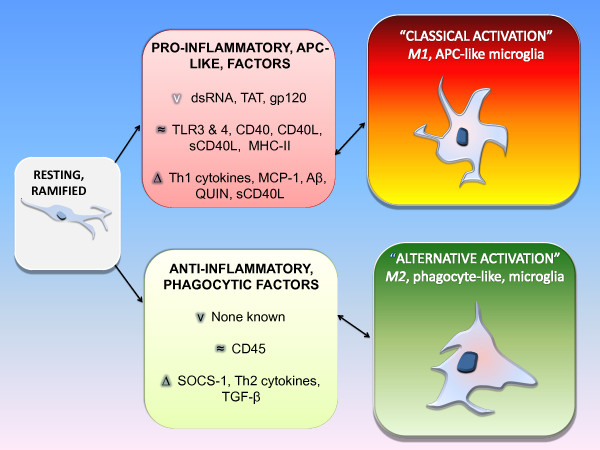
Microglial dysfunction is associated with the pathogenesis and progression of a number of neurodegenerative disorders including HIV associated dementia (HAD). HIV promotion of an M1 antigen presenting cell (APC) - like microglial phenotype, through the promotion of CD40 activity, may impair endogenous mechanisms important for amyloid- beta (Aβ) protein clearance. Further, a chronic pro-inflammatory cycle is established in this manner. CD45 is a protein tyrosine phosphatase receptor which negatively regulates CD40L-CD40-induced microglial M1 activation; an effect leading to the promotion of an M2 phenotype better suited to phagocytose and clear Aβ. Moreover, this CD45 mediated activation state appears to dampen harmful cytokine production. As such, this property of microglial CD45 as a regulatory "off switch" for a CD40-promoted M1, APC-type microglia activation phenotype may represent a critical therapeutic target for the prevention and treatment of neurodegeneration, as well as microglial dysfunction, found in patients with HAD.
Overview
The claims and statistics of this paper haven’t been checked yet.
Run the check — free, no account needed. It reads the open-access text and checks the sources it cites.
Integrity signals
Registry
Cites
Statistics
Figures
Verification
Claims the paper makes
The claims and statistics of this paper haven’t been checked yet.
Run the check — free, no account needed. It reads the open-access text and checks the sources it cites.
Claims other papers make about this one
Each is checked against this paper’s own text: does it say what the citing paper says it does?
The source backs part of the claim, not all of it — or backs it for a different population, condition or comparison.
In the citing paper
“The M1 phenotype is a pro-inflammatory state and induces neuropathology, whereas M2 is anti-inflammatory state that could have a neuroprotective role [36].”
From miR-155 induction in microglial cells suppresses Japanese encephalitis virus replication and negatively modulates innate immune responses (2014) · cites this paper as [36]
Experimental — accuracy not yet measuredThe source backs part of the claim, not all of it — or backs it for a different population, condition or comparison.
In the citing paper
“It is reported that CD45 can negatively regulate CD40L-CD40-induced microglial M1 activation; an effect leading to the promotion of the M2 phenotype.”
From miR-155 induction in microglial cells suppresses Japanese encephalitis virus replication and negatively modulates innate immune responses (2014) · cites this paper as [36]
Experimental — accuracy not yet measuredIn the citing paper
“Moreover, this CD45-mediated activation state appears to decrease harmful cytokine production [36].”
From miR-155 induction in microglial cells suppresses Japanese encephalitis virus replication and negatively modulates innate immune responses (2014) · cites this paper as [36]
In the citing paper
“In hematopoietic cell lineage CD45 (a Src-homology 2 domain (SH2)-containing protein tyrosine phosphatase) plays an important role in regulating cytokine receptor mediated signal [36].”
From miR-155 induction in microglial cells suppresses Japanese encephalitis virus replication and negatively modulates innate immune responses (2014) · cites this paper as [36]
Self-check
Percentages, sums and reported differences re-computed from the paper’s own numbers.
The self-check needs the paper’s full text.
Citations
Cites
The many faces of macrophage activation
Mosser, D. M. (2003) · Journal of Leukocyte Biology · doi:10.1189/jlb.0602325 · Cited by 1,902
Reduction of β‐amyloid peptide42 in the cerebrospinal fluid of patients with Alzheimer's disease
Motter, R., Vigo‐Pelfrey, C., Kholodenko, D., Barbour, R., Johnson‐Wood, K., Galasko, D., Chang, L., Miller, B., Clark, C., Green, R., et al. (1995) · Annals of Neurology · doi:10.1002/ana.410380413 · Cited by 701
Microglia and inflammatory mechanisms in the clearance of amyloid β peptide
Rogers, J., Strohmeyer, R., Kovelowski, C. J., & Li, R. (2002) · Glia · doi:10.1002/glia.10153 · Cited by 381
Brain deposition of beta-amyloid is a common pathologic feature in HIV positive patients
Green, D. A., Masliah, E., Vinters, H. V., Beizai, P., Moore, D. J., & Achim, C. L. (2005) · AIDS · doi:10.1097/01.aids.0000161770.06158.5c · Cited by 299
Takashima, A., Noguchi, K., Michel, G., Mercken, M., Hoshi, M., Ishiguro, K., & Imahori, K. (1996) · Neuroscience Letters · doi:10.1016/0304-3940(95)12257-5 · Cited by 260
Cosenza;, M. A., Zhao;, M., Si;, Q., & Lee, S. C. (2002) · Brain Pathology · doi:10.1111/j.1750-3639.2002.tb00461.x · Cited by 238
Rogers, J., & Lue, L.-F. (2001) · Neurochemistry International · doi:10.1016/s0197-0186(01)00040-7 · Cited by 236
Suo, Z., Tan, J., Placzek, A., Crawford, F., Fang, C., & Mullan, M. (1998) · Brain Research · doi:10.1016/s0006-8993(98)00780-x · Cited by 116
Self-citation
Research works printed but not linked: 131
- Benveniste EN: Role of macrophages/microglia in multiple sclerosis and experimental allergic encephalomyelitis. J Mol Med. 1997, 75 (3): 165-73. 10.1007/s001090050101. · DOI
- Banati RB, Gehrmann J, Schubert P, Kreutzberg GW: Cytotoxicity of microglia. Glia. 1993, 7 (1): 111-8. 10.1002/glia.440070117. · DOI
- Walker DG, Kim SU, McGeer PL: Complement and cytokine gene expression in cultured microglial derived from postmortem human brains. J Neurosci Res. 1995, 40 (4): 478-93. 10.1002/jnr.490400407. · DOI
- Panek RB, Benveniste EN: Class II MHC gene expression in microglia. Regulation by the cytokines IFN-gamma, TNF-alpha, and TGF-beta. J Immunol. 1995, 154 (6): 2846-54. · DOI
- Frei K, Siepl C, Groscurth P, Bodmer S, Schwerdel C, Fontana A: Antigen presentation and tumor cytotoxicity by interferon-gamma-treated microglial cells. Eur J Immunol. 1987, 17 (9): 1271-8. 10.1002/eji.1830170909. · DOI
- Suzumura A, Mezitis SG, Gonatas NK, Silberberg DH: MHC antigen expression on bulk isolated macrophage-microglia from newborn mouse brain: induction of Ia antigen expression by gamma-interferon. J Neuroimmunol. 1987, 15 (3): 263-78. 10.1016/0165-5728(87)90121-4. · DOI
- Williams K, Bar-Or A, Ulvestad E, Olivier A, Antel JP, Yong VW: Biology of adult human microglia in culture: comparisons with peripheral blood monocytes and astrocytes. J Neuropathol Exp Neurol. 1992, 51 (5): 538-49. 10.1097/00005072-199209000-00009. · DOI
- Gordon S, Taylor PR: Monocyte and macrophage heterogeneity. Nat Rev Immunol. 2005, 5 (12): 953-64. 10.1038/nri1733. · DOI · Paper page
- Bruce-Keller AJ, Barger SW, Moss NI, Pham JT, Keller JN, Nath A: Pro-inflammatory and pro-oxidant properties of the HIV protein Tat in a microglial cell line: attenuation by 17 beta-estradiol. J Neurochem. 2001, 78 (6): 1315-24. 10.1046/j.1471-4159.2001.00511.x. · DOI
- Suh HS, Zhao ML, Choi N, Belbin TJ, Brosnan CF, Lee SC: TLR3 and TLR4 are innate antiviral immune receptors in human microglia: role of IRF3 in modulating antiviral and inflammatory response in the CNS. Virology. 2009, 392 (2): 246-59. 10.1016/j.virol.2009.07.001. · DOI
- Akhtar LN, Tahir MY, Ahmed F, Ul-Haq I, Salim KP: Suppressor of cytokine signaling 3 inhibits antiviral IFN-beta signaling to enhance HIV-1 replication in macrophages. J Immunol. 185 (4): 2393-404. 10.4049/jimmunol.0903563. · DOI
- Qin H, Wilson CA, Lee SJ, Benveniste EN: IFN-beta-induced SOCS-1 negatively regulates CD40 gene expression in macrophages and microglia. Faseb J. 2006, 20 (7): 985-7. 10.1096/fj.05-5493fje. · DOI
- Minagar A, Shapshak P, Fujimura R, Ownby R, Heyes M, Eisdorfer C: The role of macrophage/microglia and astrocytes in the pathogenesis of three neurologic disorders: HIV-associated dementia, Alzheimer disease, and multiple sclerosis. J Neurol Sci. 2002, 202 (1-2): 13-23. 10.1016/S0022-510X(02)00207-1. · DOI
- Ma N, Streilein JW: T cell immunity induced by allogeneic microglia in relation to neuronal retina transplantation. J Immunol. 1999, 162 (8): 4482-9. · DOI
- Ford AL, Foulcher E, Lemckert FA, Sedgwick JD: Microglia induce CD4 T lymphocyte final effector function and death. J Exp Med. 1996, 184 (5): 1737-45. 10.1084/jem.184.5.1737. · DOI
- Genis P, Jett M, Bernton EW, Boyle T, Gelbard HA, Dzenko K, Keane RW, Resnick L, Mizrachi Y, Volsky DJ, et al: Cytokines and arachidonic metabolites produced during human immunodeficiency virus (HIV)-infected macrophage-astroglia interactions: implications for the neuropathogenesis of HIV disease. J Exp Med. 1992, 176 (6): 1703-18. 10.1084/jem.176.6.1703. · DOI
- Persidsky Y, Gendelman HE: Mononuclear phagocyte immunity and the neuropathogenesis of HIV-1 infection. J Leukoc Biol. 2003, 74 (5): 691-701. 10.1189/jlb.0503205. · DOI
- Adle-Biassette HF, Chretien L, Wingertsmann C, Hery T, Ereau F, Scaravilli M, Tardieu Gray F: Neuronal apoptosis does not correlate with dementia in HIV infection but is related to microglial activation and axonal damage. Neuropathol Appl Neurobiol. 1999, 25: 123-133. 10.1046/j.1365-2990.1999.00167.x. · DOI
- Liu BHJ: Role of microglia in inflammation-mediated neurodegenerative diseases: mechanisms and strategies for therapeutic intervention. J Pharm Exp Ther. 2003, 304: 1-7. 10.1124/jpet.102.035048. · DOI · Paper page
- BlockML Z.L, Hong JS: Microglia-mediated neurotoxicity: uncovering the molecular mechanisms. Nat Rev Neurosci. 2007, 8: 57-69. 10.1038/nrn2038. · DOI · Paper page
- Alirezaei MKW, Flynn CT, Brady NR, Fox HS: Disruption of neuronal autophagy by infected microglia results in neurodegeneration. PLoS ONE. 2008, 3: e2906-10.1371/journal.pone.0002906. · DOI
- Alirezaei MKW, Fox HS: Decreased neuronal autophagy in HAD: a mechanism of indirect neurotoxicity. Autophagy. 2008, 1 (4(7)): 963-969. · DOI
- Avison MJ, Nath A, Greene-Avison R, Schmitt FA, Greenberg RN, Berger JR: Neuroimaging correlates of HIV-associated BBB compromise. J Neuroimmunol. 2004, 157 (1-2): 140-6. 10.1016/j.jneuroim.2004.08.025. · DOI
- Thind K, Sabbagh MN: Pathological correlates of cognitive decline in Alzheimer's disease. Panminerva Med. 2007, 49 (4): 191-5.
- Sulkava R, Erkinjuntti T, Palo J: Head injuries in Alzheimer's disease and vascular dementia. Neurology. 1985, 35 (12): 1804- · DOI
- D'Aversa TG, Eugenin E.A, Berman JW: NeuroAIDS: contributions of the human immunodeficiency virus-1 proteins Tat and gp120 as well as CD40 to microglial activation. J Neurosci Res. 2005, 81 (3): 436-46. · DOI
- Avison MJ, Nath A, Greene-Avison R, Schmitt FA, Bales RA, Ethisham A, Greenberg RN, Berger JR: Inflammatory changes and breakdown of microvascular integrity in early human immunodeficiency virus dementia. J Neurovirol. 2004, 10 (4): 223-32. 10.1080/13550280490463532. · DOI
- Schneider JA, Boyle PA, Arvanitakis Z, Bienias JL, Bennett DA: Subcortical infarcts, Alzheimer's disease pathology, and memory function in older persons. Ann Neurol. 2007, 62 (1): 59-66. 10.1002/ana.21142. · DOI · Paper page
- Hachiya NS, Kozuka Y, Kaneko K: Mechanical stress and formation of protein aggregates in neurodegenerative disorders. Med Hypotheses. 2008, 70 (5): 1034-7. 10.1016/j.mehy.2007.06.043. · DOI
- Alisky JM: The coming problem of HIV-associated Alzheimer's disease. Med Hypotheses. 2007, 69 (5): 1140-3. 10.1016/j.mehy.2007.02.030. · DOI
- Simone MJ, Appelbaum J: HIV in older adults. Geriatrics. 2008, 63 (12): 6-12.
- Repetto MJ, Petitto JM: Psychopharmacology in HIV-infected patients. Psychosom Med. 2008, 70 (5): 585-92. 10.1097/PSY.0b013e3181777190. · DOI
- Keblesh JP, Reiner BC, Liu J, Xiong H: Pathogenesis of Human Immunodeficiency Virus Type-1 (HIV-1)-Associated Dementia: Role of Voltage-Gated Potassium Channels. Retrovirology. 2008, 2: 1-10.
- Hult B, Chana G, Masliah E, Everall I: Neurobiology of HIV. Int Rev Psychiatry. 2008, 20 (1): 3-13. 10.1080/09540260701862086. · DOI
- Giunta B, Zhou Y, Hou H, Rrapo E, Fernandez F, Tan J: HIV-1 TAT inhibits microglial phagocytosis of Abeta peptide. Int J Clin Exp Pathol. 2008, 1 (3): 260-75.
- Valcour V, Shikuma C, Shiramizu B, Watters M, Poff P, Selnes OA, Grove J, Liu Y, Abdul-Majid KB, Gartner S, Sacktor N: Age, apolipoprotein E4, and the risk of HIV dementia: the Hawaii Aging with HIV Cohort. J Neuroimmunol. 2004, 157 (1-2): 197-202. 10.1016/j.jneuroim.2004.08.029. · DOI
- Esiri MM, Biddolph S.C, Morris CS: Prevalence of Alzheimer plaques in AIDS. J Neurol Neurosurg Psychiatry. 1998, 65 (1): 29-33. 10.1136/jnnp.65.1.29. · DOI
- Giunta B, Hou H, Zhu Y, Rrapo E, Tian J, Takashi M, Commins D, Singer E, He J, Fernandez F, Tan J: HIV-1 Tat contributes to Alzheimer's disease-like pathology in PSAPP mice. Int J Clin Exp Pathol. 2009, 2 (5): 433-43.
- Everall IP, Luthert PJ, Lantos PL: Neuronal number and volume alterations in the neocortex of HIV infected individuals. J Neurol Neurosurg Psychiatry. 1993, 56 (5): 481-6. 10.1136/jnnp.56.5.481. · DOI
- Bell JE: The neuropathology of adult HIV infection. Rev Neurol (Paris). 1998, 154 (12): 816-29.
- Adle-Biassette H, Levy Y, Colombel M, Poron F, Natchev S, Keohane C, Gray F: Neuronal apoptosis in HIV infection in adults. Neuropathol Appl Neurobiol. 1995, 21 (3): 218-27. 10.1111/j.1365-2990.1995.tb01053.x. · DOI
- Galasko D: CSF tau and Abeta42: logical biomarkers for Alzheimer's disease?. Neurobiol Aging. 1998, 19 (2): 117-9. 10.1016/S0197-4580(98)00021-9. · DOI
- Brew BJ, Pemberton L, Blennow K, Wallin A, Hagberg L: CSF amyloid beta42 and tau levels correlate with AIDS dementia complex. Neurology. 2005, 65 (9): 1490-2. 10.1212/01.wnl.0000183293.95787.b7. · DOI
- Andreasen N, Minthon L, Vanmechelen E, Vanderstichele H, Davidsson P, Winblad B, Blennow K: Cerebrospinal fluid tau and Abeta42 as predictors of development of Alzheimer's disease in patients with mild cognitive impairment. Neurosci Lett. 1999, 273 (1): 5-8. 10.1016/S0304-3940(99)00617-5. · DOI
- Navia BA, Jordan BD, Price RW: The AIDS dementia complex: I. Clinical features. Ann Neurol. 1986, 19 (6): 517-24. 10.1002/ana.410190602. · DOI
- Johnson RT, McArthur JC, Narayan O: The neurobiology of human immunodeficiency virus infections. Faseb J. 1988, 2 (14): 2970-81. · DOI
- Guillemin GJ, Brew BJ: Implications of the kynurenine pathway and quinolinic acid in Alzheimer's disease. Redox Rep. 2002, 7 (4): 199-206. 10.1179/135100002125000550. · DOI
- Snyder SH: Nitric oxide: first in a new class of neurotransmitters. Science. 1992, 257 (5069): 494-6. 10.1126/science.1353273. · DOI
- Heyes MP, Brew BJ, Martin A, Price RW, Salazar AM, Sidtis JJ, Yergey JA, Mouradian MM, Sadler AE, Keilp J, et al: Quinolinic acid in cerebrospinal fluid and serum in HIV-1 infection: relationship to clinical and neurological status. Ann Neurol. 1991, 29 (2): 202-9. 10.1002/ana.410290215. · DOI
- Heyes MP, Mefford IN, Quearry BJ, Dedhia M, Lackner A: Increased ratio of quinolinic acid to kynurenic acid in cerebrospinal fluid of D retrovirus-infected rhesus macaques: relationship to clinical and viral status. Ann Neurol. 1990, 27 (6): 666-75. 10.1002/ana.410270614. · DOI
- Heyes MP, Rubinow D, Lane C, Markey SP: Cerebrospinal fluid quinolinic acid concentrations are increased in acquired immune deficiency syndrome. Ann Neurol. 1989, 26 (2): 275-7. 10.1002/ana.410260215. · DOI
- Heyes MP, Saito K, Crowley JS, Davis LE, Demitrack MA, Der M, Dilling LA, Elia J, Kruesi MJ, Lackner A, et al: Quinolinic acid and kynurenine pathway metabolism in inflammatory and non-inflammatory neurological disease. Brain. 1992, 115 (Pt 5): 1249-73. 10.1093/brain/115.5.1249. · DOI
- Nath A, Geiger J: Neurobiological aspects of human immunodeficiency virus infection: neurotoxic mechanisms. Prog Neurobiol. 1998, 54 (1): 19-33. 10.1016/S0301-0082(97)00053-1. · DOI
- Wilt SG, Milward E, Zhou JM, Nagasato K, Patton H, Rusten R, Griffin DE, O'Connor M, Dubois-Dalcq M: In vitro evidence for a dual role of tumor necrosis factor-alpha in human immunodeficiency virus type 1 encephalopathy. Ann Neurol. 1995, 37 (3): 381-94. 10.1002/ana.410370315. · DOI
- Wesselingh SL, Takahashi K, Glass JD, McArthur JC, Griffin JW, Griffin DE: Cellular localization of tumor necrosis factor mRNA in neurological tissue from HIV-infected patients by combined reverse transcriptase/polymerase chain reaction in situ hybridization and immunohistochemistry. J Neuroimmunol. 1997, 74 (1-2): 1-8. 10.1016/S0165-5728(96)00160-9. · DOI
- Byrne GI, Lehmann LK, Kirschbaum JG, Borden EC, Lee CM, Brown RR: Induction of tryptophan degradation in vitro and in vivo: a gamma-interferon-stimulated activity. J Interferon Res. 1986, 6 (4): 389-96. · DOI
- Fuchs D, Shearer GM, Boswell RN, Lucey DR, Clerici M, Reibnegger G, Werner ER, Zajac RA, Wachter H: Negative correlation between blood cell counts and serum neopterin concentration in patients with HIV-1 infection. Aids. 1991, 5 (2): 209-12. 10.1097/00002030-199102000-00012. · DOI
- Werner ER, Bitterlich G, Fuchs D, Hausen A, Reibnegger G, Szabo G, Dierich MP, Wachter H: Human macrophages degrade tryptophan upon induction by interferon-gamma. Life Sci. 1987, 41 (3): 273-80. 10.1016/0024-3205(87)90149-4. · DOI
- Schmidtmayerova H, Nottet HS, Nuovo G, Raabe T, Flanagan CR, Dubrovsky L, Gendelman HE, Cerami A, Bukrinsky M, Sherry B: Human immunodeficiency virus type 1 infection alters chemokine beta peptide expression in human monocytes: implications for recruitment of leukocytes into brain and lymph nodes. Proc Natl Acad Sci USA. 1996, 93 (2): 700-4. 10.1073/pnas.93.2.700. · DOI
- Kure K, Weidenheim KM, Lyman WD, Dickson DW: Morphology and distribution of HIV-1 gp41-positive microglia in subacute AIDS encephalitis. Pattern of involvement resembling a multisystem degeneration. Acta Neuropathol. 1990, 80 (4): 393-400. 10.1007/BF00307693. · DOI
- Gullotta F, Kuchelmeister K, Masini T, Ghidoni P, Cappricci E: [The morphology of HIV encephalopathy]. Zentralbl Allg Pathol. 1989, 135 (1): 5-13.
- Fischer-Smith T, Bell C, Croul S, Lewis M, Rappaport J: Monocyte/macrophage trafficking in acquired immunodeficiency syndrome encephalitis: lessons from human and nonhuman primate studies. J Neurovirol. 2008, 14 (4): 318-26. 10.1080/13550280802132857. · DOI
- Gras G, Kaul M: Molecular mechanisms of neuroinvasion by monocytes-macrophages in HIV-1 infection. Retrovirology. 7: 30-10.1186/1742-4690-7-30. · DOI
- Edelman M, Birkenhauer MC, Steinberg JJ, Dickson DW, Casadevall A, Lee SC: Microglial nodule encephalitis: limited CNS infection despite disseminated systemic cryptococcosis. Clin Neuropathol. 1996, 15 (1): 30-3.
- Persidsky Y, Ghorpade A, Rasmussen J, Limoges J, Liu XJ, Stins M, Fiala M, Way D, Kim KS, Witte MH, Weinand M, Carhart L, Gendelman HE: Microglial and astrocyte chemokines regulate monocyte migration through the blood-brain barrier in human immunodeficiency virus-1 encephalitis. Am J Pathol. 1999, 155 (5): 1599-611. · DOI
- Nelson PT, Soma LA, Lavi E: Microglia in diseases of the central nervous system. Ann Med. 2002, 34 (7-8): 491-500. 10.1080/078538902321117698. · DOI
- Michaels J, Price RW, Rosenblum MK: Microglia in the giant cell encephalitis of acquired immune deficiency syndrome: proliferation, infection and fusion. Acta Neuropathol. 1988, 76 (4): 373-9. 10.1007/BF00686974. · DOI
- Liner KJ, Hall CD, Robertson KR: Effects of antiretroviral therapy on cognitive impairment. Curr HIV/AIDS Rep. 2008, 5 (2): 64-71. 10.1007/s11904-008-0011-7. · DOI
- Ferrando SJ: Diagnosis and treatment of HIV-associated neurocognitive disorders. New Dir Ment Health Serv. 2000, 25-35. 10.1002/yd.23320008705. 87 · DOI
- Boisse L, Gill MJ, Power C: HIV infection of the central nervous system: clinical features and neuropathogenesis. Neurol Clin. 2008, 26 (3): 799-819. 10.1016/j.ncl.2008.04.002. x · DOI
- Lorenzo A, Yankner BA: Amyloid fibril toxicity in Alzheimer's disease and diabetes. Ann N Y Acad Sci. 1996, 777: 89-95. 10.1111/j.1749-6632.1996.tb34406.x. · DOI
- Lorenzo A, Yankner BA: Beta-amyloid neurotoxicity requires fibril formation and is inhibited by congo red. Proc Natl Acad Sci USA. 1994, 91 (25): 12243-7. 10.1073/pnas.91.25.12243. · DOI · Paper page
- Giunta B, Obregon D, Hou H, Zeng J, Sun N, Nikolic V, Ehrhart J, Shytle D, Fernandez F, Tan J: EGCG mitigates neurotoxicity mediated by HIV-1 proteins gp120 and Tat in the presence of IFN-gamma: role of JAK/STAT1 signaling and implications for HIV-associated dementia. Brain Res. 2006, 1123 (1): 216-25. 10.1016/j.brainres.2006.09.057. · DOI
- Giulian D, Haverkamp LJ, Li J, Karshin WL, Yu J, Tom D, Li X, Kirkpatrick JB: Senile plaques stimulate microglia to release a neurotoxin found in Alzheimer brain. Neurochem Int. 1995, 27 (1): 119-37. 10.1016/0197-0186(95)00067-I. · DOI · Paper page
- Tan J, Town T, Mullan M: CD45 inhibits CD40L-induced microglial activation via negative regulation of the Src/p44/42 MAPK pathway. J Biol Chem. 2000, 275 (47): 37224-31. 10.1074/jbc.M002006200. · DOI · Paper page
- McGeer PL, Itagaki S, Boyes BE, McGeer EG: Reactive microglia are positive for HLA-DR in the substantia nigra of Parkinson's and Alzheimer's disease brains. Neurology. 1988, 38 (8): 1285-91. · DOI · Paper page
- McGeer EG, McGeer PL: The role of the immune system in neurodegenerative disorders. Mov Disord. 1997, 12 (6): 855-8. 10.1002/mds.870120604. · DOI
- McGeer PL, McGeer EG, Kawamata T, Yamada T, Akiyama H: Reactions of the immune system in chronic degenerative neurological diseases. Can J Neurol Sci. 1991, 18 (3 Suppl): 376-9. · DOI
- Rozemuller JM, Eikelenboom P, Stam FC: Role of microglia in plaque formation in senile dementia of the Alzheimer type. An immunohistochemical study. Virchows Arch B Cell Pathol Incl Mol Pathol. 1986, 51 (3): 247-54. 10.1007/BF02899034. · DOI
- Rozemuller JM, Eikelenboom P, Pals ST, Stam FC: Microglial cells around amyloid plaques in Alzheimer's disease express leucocyte adhesion molecules of the LFA-1 family. Neurosci Lett. 1989, 101 (3): 288-92. 10.1016/0304-3940(89)90547-8. · DOI
- Kiebala M, Polesskaya O, Yao Z, Perry SW, Maggirwar SB: Nuclear factor-kappa B family member RelB inhibits human immunodeficiency virus-1 Tat-induced tumor necrosis factor-alpha production. PLoS One. 5 (7): e11875-10.1371/journal.pone.0011875. · DOI
- Rappaport J, Joseph J, Croul S, Alexander G, Del Valle L, Amini S, Khalili K: Molecular pathway involved in HIV-1-induced CNS pathology: role of viral regulatory protein, Tat. J Leukoc Biol. 1999, 65 (4): 458-65. · DOI
- Wisniewski HM, Wegiel J, Wang KC, Kujawa M, Lach B: Ultrastructural studies of the cells forming amyloid fibers in classical plaques. Can J Neurol Sci. 1989, 16 (4 Suppl): 535-42. · DOI
- Klegeris A, Walker DG, McGeer PL: Interaction of Alzheimer beta-amyloid peptide with the human monocytic cell line THP-1 results in a protein kinase C-dependent secretion of tumor necrosis factor-alpha. Brain Res. 1997, 747 (1): 114-21. 10.1016/S0006-8993(96)01229-2. · DOI
- Klegeris A, McGeer PL: beta-amyloid protein enhances macrophage production of oxygen free radicals and glutamate. J Neurosci Res. 1997, 49 (2): 229-35. 10.1002/(SICI)1097-4547(19970715)49:2<229::AID-JNR11>3.0.CO;2-W. · DOI
- Klegeris A, Walker DG, McGeer PL: Activation of macrophages by Alzheimer beta amyloid peptide. Biochem Biophys Res Commun. 1994, 199 (2): 984-91. 10.1006/bbrc.1994.1326. · DOI
- McDonald DR, Brunden KR, Landreth GE: Amyloid fibrils activate tyrosine kinase-dependent signaling and superoxide production in microglia. J Neurosci. 1997, 17 (7): 2284-94. · DOI
- McDonald DR, Bamberger ME, Combs CK, Landreth GE: beta-Amyloid fibrils activate parallel mitogen-activated protein kinase pathways in microglia and THP1 monocytes. J Neurosci. 1998, 18 (12): 4451-60. · DOI
- Combs CK, Johnson DE, Cannady SB, Lehman TM, Landreth GE: Identification of microglial signal transduction pathways mediating a neurotoxic response to amyloidogenic fragments of beta-amyloid and prion proteins. J Neurosci. 1999, 19 (3): 928-39. · DOI
- Townsend KP, Town T, Mori T, Lue LF, Shytle D, Sanberg PR, Morgan D, Fernandez F, Flavell RA, Tan J: CD40 signaling regulates innate and adaptive activation of microglia in response to amyloid beta-peptide. Eur J Immunol. 2005, 35 (3): 901-10. 10.1002/eji.200425585. · DOI · Paper page
- Lorton D, Kocsis JM, King L, Madden K, Brunden KR: beta-Amyloid induces increased release of interleukin-1 beta from lipopolysaccharide-activated human monocytes. J Neuroimmunol. 1996, 67 (1): 21-9. · DOI
- Yankner BA, Duffy LK, Kirschner DA: Neurotrophic and neurotoxic effects of amyloid beta protein: reversal by tachykinin neuropeptides. Science. 1990, 250 (4978): 279-82. 10.1126/science.2218531. · DOI · Paper page
- Turchan-Cholewo J, Dimayuga VM, Gupta S, Gorospe RM, Keller JN, Bruce-Keller AJ: NADPH oxidase drives cytokine and neurotoxin release from microglia and macrophages in response to HIV-Tat. Antioxid Redox Signal. 2009, 11 (2): 193-204. 10.1089/ars.2008.2097. · DOI
- Shatrov VA, Ratter F, Gruber A, Droge W, Lehmann V: HIV type 1 glycoprotein 120 amplifies tumor necrosis factor-induced NF-kappa B activation in Jurkat cells. AIDS Res Hum Retroviruses. 1996, 12 (13): 1209-16. 10.1089/aid.1996.12.1209. · DOI
- Turchan-Cholewo J, Dimayuga FO, Gupta S, Keller JN, Knapp PE, Hauser KF, Bruce-Keller AJ: Morphine and HIV-Tat increase microglial-free radical production and oxidative stress: possible role in cytokine regulation. J Neurochem. 2009, 108 (1): 202-15. 10.1111/j.1471-4159.2008.05756.x. · DOI
- Aksenov MY, Hasselrot U, Wu G, Nath A, Anderson C, Mactutus CF, Booze RM: Temporal relationships between HIV-1 Tat-induced neuronal degeneration, OX-42 immunoreactivity, reactive astrocytosis, and protein oxidation in the rat striatum. Brain Res. 2003, 987 (1): 1-9. 10.1016/S0006-8993(03)03194-9. · DOI
- Kornbluth RS: The emerging role of CD40 ligand in HIV infection. J Leukoc Biol. 2000, 68 (3): 373-82. · DOI
- Giunta B, Rezai-Zadeh K, Tan J: Impact of the CD40-CD40L dyad in Alzheimer's disease. CNS Neurol Disord Drug Targets. 9 (2): 149-55. · DOI
- Giunta B, Figueroa KP, Town T, Tan J: Soluble Cd40 Ligand in Dementia. Drugs Future. 2009, 34 (4): 333-340. 10.1358/dof.2009.034.04.1358595. · DOI
- Heeschen C, Dimmeler S, Hamm CW, van den Brand MJ, Boersma E, Zeiher AM, Simoons ML, CAPTURE Study Investigators: Soluble CD40 ligand in acute coronary syndromes. N Engl J Med. 2003, 348 (12): 1104-11. 10.1056/NEJMoa022600. · DOI
- Tsakiris DA, Tschöpl M, Wolf F, Labs KH, Jäger KA, Marbet GA: Platelets and cytokines in concert with endothelial activation in patients with peripheral arterial occlusive disease. Blood Coagul Fibrinolysis. 2000, 11 (2): 165-73. · DOI
- Devaraj S, Glaser N, Griffen S, Wang-Polagruto J, Miguelino E, Jialal I: Increased monocytic activity and biomarkers of inflammation in patients with type 1 diabetes. Diabetes. 2006, 55 (3): 774-9. 10.2337/diabetes.55.03.06.db05-1417. · DOI
- Chai H, Yan S, Wang H, Zhang R, Lin PH, Yao Q, Chen C: CD40 ligand increases expression of its receptor CD40 in human coronary artery endothelial cells. Surgery. 2006, 140 (2): 236-42. 10.1016/j.surg.2006.03.016. · DOI
- Gerritse K, Laman JD, Noelle RJ, Aruffo A, Ledbetter JA, Boersma WJ, Claassen E: CD40-CD40 ligand interactions in experimental allergic encephalomyelitis and multiple sclerosis. Proc Natl Acad Sci USA. 1996, 93 (6): 2499-504. 10.1073/pnas.93.6.2499. · DOI
- Shapshak P, Duncan R, Minagar A, Rodriguez de la Vega P, Stewart RV, Goodkin K: Elevated expression of IFN-gamma in the HIV-1 infected brain. Front Biosci. 2004, 9: 1073-81. 10.2741/1271. · DOI
- Aloisi F, Penna G, Polazzi E, Minghetti L, Adorini L: CD40-CD154 interaction and IFN-gamma are required for IL-12 but not prostaglandin E2 secretion by microglia during antigen presentation to Th1 cells. J Immunol. 1999, 162 (3): 1384-91. · DOI
- Nguyen VT, Walker WS, Benveniste EN: Post-transcriptional inhibition of CD40 gene expression in microglia by transforming growth factor-beta. Eur J Immunol. 1998, 28 (8): 2537-48. 10.1002/(SICI)1521-4141(199808)28:08<2537::AID-IMMU2537>3.0.CO;2-1. · DOI
- Togo T, Akiyama H, Kondo H, Ikeda K, Kato M, Iseki E, Kosaka K: Expression of CD40 in the brain of Alzheimer's disease and other neurological diseases. Brain Res. 2000, 885 (1): 117-21. 10.1016/S0006-8993(00)02984-X. · DOI
- Tan J, Town T, Paris D, Mori T, Suo Z, Crawford F, Mattson MP, Flavell RA, Mullan M: Microglial activation resulting from CD40-CD40L interaction after beta-amyloid stimulation. Science. 1999, 286 (5448): 2352-5. 10.1126/science.286.5448.2352. · DOI
- Tan J, Placzek A, Crawford F, Fang C, Mullan M: Induction of CD40 on human endothelial cells by Alzheimer's beta-amyloid peptides. Brain Res Bull. 1999, 50 (2): 143-8. 10.1016/S0361-9230(99)00122-7. · DOI
- Hollenbaugh D, Mischel-Petty N, Edwards CP, Simon JC, Denfeld RW, Kiener PA, Aruffo A: Expression of functional CD40 by vascular endothelial cells. J Exp Med. 1995, 182 (1): 33-40. 10.1084/jem.182.1.33. · DOI
- Karmann K, Hughes CC, Schechner J, Fanslow WC, Pober JS: CD40 on human endothelial cells: inducibility by cytokines and functional regulation of adhesion molecule expression. Proc Natl Acad Sci USA. 1995, 92 (10): 4342-6. 10.1073/pnas.92.10.4342. · DOI
- Moses AV, Williams SE, Strussenberg JG, Heneveld ML, Ruhl RA, Bakke AC, Bagby GC, Nelson JA: HIV-1 induction of CD40 on endothelial cells promotes the outgrowth of AIDS-associated B-cell lymphomas. Nat Med. 1997, 3 (11): 1242-9. 10.1038/nm1197-1242. · DOI
- Sui Z, Sniderhan LF, Schifitto G, Phipps RP, Gelbard HA, Dewhurst S, Maggirwar SB: Functional synergy between CD40 ligand and HIV-1 Tat contributes to inflammation: implications in HIV type 1 dementia. J Immunol. 2007, 178 (5): 3226-36. · DOI
- Eilers M, Roy U, Mondal D: MRP (ABCC) transporters-mediated efflux of anti-HIV drugs, saquinavir and zidovudine, from human endothelial cells. Exp Biol Med (Maywood). 2008, 233 (9): 1149-60. 10.3181/0802-RM-59. · DOI
- Sipsas NV, Sfikakis pp, Kontos A, Kordossis T: Levels of soluble CD40 ligand (CD154) in serum are increased in human immunodeficiency virus type 1-infected patients and correlate with CD4(+) T-cell counts. Clin Diagn Lab Immunol. 2002, 9 (3): 558-61.
- Piguet PF, Kan CD, Vesin C, Rochat A, Donati Y, Barazzone C: Role of CD40-CVD40L in mouse severe malaria. Am J Pathol. 2001, 159 (2): 733-42. · DOI
- Ishikawa M, Vowinkel T, Stokes KY, Arumugam TV, Yilmaz G, Nanda A, Granger DN: CD40/CD40 ligand signaling in mouse cerebral microvasculature after focal ischemia/reperfusion. Circulation. 2005, 111 (13): 1690-6. 10.1161/01.CIR.0000160349.42665.0C. · DOI
- Sitati E, McCandless EE, Klein RS, Diamond MS: CD40-CD40 ligand interactions promote trafficking of CD8+ T cells into the brain and protection against West Nile virus encephalitis. J Virol. 2007, 81 (18): 9801-11. 10.1128/JVI.00941-07. · DOI
- Ramirez SH, Fan S, Dykstra H, Reichenbach N, Del Valle L, Potula R, Phipps RP, Maggirwar SB, Persidsky Y: Dyad of CD40/CD40 ligand fosters neuroinflammation at the blood-brain barrier and is regulated via JNK signaling: implications for HIV-1 encephalitis. J Neurosci. 30 (28): 9454-64. · DOI
- Tan J, Town T, Saxe M, Paris D, Wu Y, Mullan M: Ligation of microglial CD40 results in p44/42 mitogen-activated protein kinase-dependent TNF-alpha production that is opposed by TGF-beta 1 and IL-10. J Immunol. 1999, 163 (12): 6614-21. · DOI
- Todd Roach J, Volmar CH, Dwivedi S, Town T, Crescentini R, Crawford F, Tan J, Mullan M: Behavioral effects of CD40-CD40L pathway disruption in aged PSAPP mice. Brain Res. 2004, 1015 (1-2): 161-8. 10.1016/j.brainres.2004.05.004. · DOI
- Tan J, Town T, Mori T, Wu Y, Saxe M, Crawford F, Mullan M: CD45 opposes beta-amyloid peptide-induced microglial activation via inhibition of p44/42 mitogen-activated protein kinase. J Neurosci. 2000, 20 (20): 7587-94. · DOI
- Irie-Sasaki J, Sasaki T, Penninger JM: CD45 regulated signaling pathways. Curr Top Med Chem. 2003, 3 (7): 783-96. 10.2174/1568026033452339. · DOI
- Irie-Sasaki J, Sasaki T, Matsumoto W, Opavsky A, Cheng M, Welstead G, Griffiths E, Krawczyk C, Richardson CD, Aitken K, Iscove N, Koretzky G, Johnson P, Liu P, Rothstein DM, Penninger JM: CD45 is a JAK phosphatase and negatively regulates cytokine receptor signalling. Nature. 2001, 409 (6818): 349-54. 10.1038/35053086. · DOI
- Baur A, Garber S, Peterlin BM: Effects of CD45 on NF-kappa B. Implications for replication of HIV-1. J Immunol. 1994, 152 (3): 976-83. · DOI · Paper page
- Koronyo-Hamaoui M, Ko MK, Koronyo Y, Azoulay D, Seksenyan A, Kunis G, Pham M, Bakhsheshian J, Rogeri P, Black KL, Farkas DL, Schwartz M: Attenuation of AD-like neuropathology by harnessing peripheral immune cells: local elevation of IL-10 and MMP-9. J Neurochem. 2009, 111 (6): 1409-24. 10.1111/j.1471-4159.2009.06402.x. · DOI
- Townsend KP, Vendrame M, Ehrhart J, Faza B, Zeng J, Town T, Tan J: CD45 isoform RB as a molecular target to oppose lipopolysaccharide-induced microglial activation in mice. Neurosci Lett. 2004, 362 (1): 26-30. 10.1016/j.neulet.2004.01.082. · DOI
- Mahalingam M, Pozniak A, McManus TJ, Senaldi G, Vergani D, Peakman M: Abnormalities of CD45 isoform expression in HIV infection. Clin Immunol Immunopathol. 1996, 81 (2): 210-4. 10.1006/clin.1996.0178. · DOI
- Guntermann C, Amft N, Murphy BJ, Nye KE: Impaired CD45-associated tyrosine phosphatase activity during HIV-1 infection: implications for CD3 and CD4 receptor signalling. Biochem Biophys Res Commun. 1998, 252 (1): 69-77. 10.1006/bbrc.1998.9595. · DOI
- Kim MO, Suh HS, Si Q, Terman BI, Lee SC: Anti-CD45RO suppresses human immunodeficiency virus type 1 replication in microglia: role of Hck tyrosine kinase and implications for AIDS dementia. J Virol. 2006, 80 (1): 62-72. 10.1128/JVI.80.1.62-72.2006. · DOI
Cited by
Sorted by citations
Pareek, S., Roy, S., Kumari, B., Jain, P., Banerjee, A., & Vrati, S. (2014) · Journal of Neuroinflammation · doi:10.1186/1742-2094-11-97 · Cited by 121
Vigil, F. A., Bozdemir, E., Bugay, V., Chun, S. H., Hobbs, M., Sanchez, I., Hastings, S. D., Veraza, R. J., Holstein, D. M., Sprague, S. M., et al. (2020) · Journal of Cerebral Blood Flow & Metabolism · doi:10.1177/0271678x19857818 · Cited by 51
Metadata
- Authors
- Salemi, J., Obregon, D. F., Cobb, A., Reed, S., Sadic, E., Jin, J., Fernandez, F., Tan, J., & Giunta, B.
- Venue
- Molecular Neurodegeneration
- Published
- 2011-01-11
- Volume
- 6
- Issue
- 1
- ISSN
- 1750-1326
- PMC
- PMC3030526
- Cited by
- 28
- References
- 8
- Language
- en
About these checks
Registry, retraction, Cites and statistics results are computed, not judged, with one exception: a printed reference that no index lists is read once by a model before it counts as a citation error. Claims are extracted by a model and kept only when their quote appears verbatim in the text; each cited source is then checked, with a model’s help, for whether it says what the claim relies on. Those verdicts can be wrong — read the claim’s quote and the cited source before relying on one.
Statistics and arithmetic are recomputed, not judged.
Confidence bands are shown only for fields calibrated on our annotated test set. None is calibrated yet, so no band is shown.
What counts as a claim ›How we measure confidence ›How we check this ›
Are you an author, or do you see a problem with this page? Request a review
