

The DOI matches its Crossref record; 77 of the 77 references it cites were found in a registry or an index.
Sex- and brain region-specific acceleration of β-amyloidogenesis following behavioral stress in a mouse model of Alzheimer's disease
Open accessAbstract
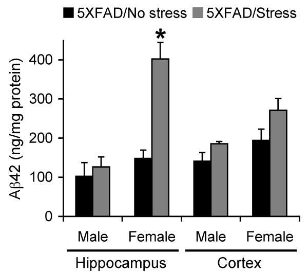
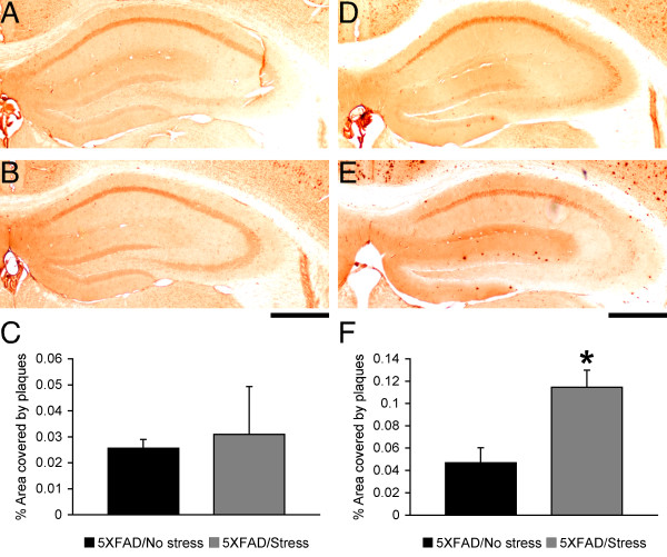
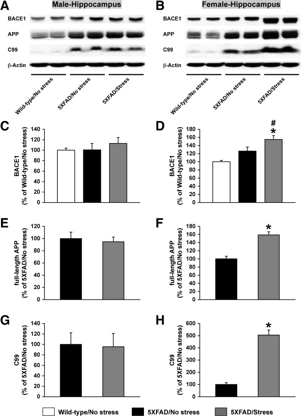
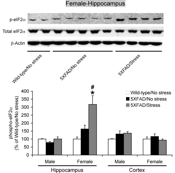
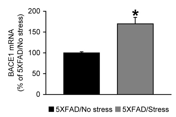
BACKGROUND: It is hypothesized that complex interactions between multiple environmental factors and genetic factors are implicated in sporadic Alzheimer's disease (AD); however, the underlying mechanisms are poorly understood. Importantly, recent evidence reveals that expression and activity levels of the β-site APP cleaving enzyme 1 (BACE1), which initiates amyloid-β (Aβ) production, are elevated in AD brains. In this study, we investigated a molecular mechanism by which sex and stress interactions may accelerate β-amyloidogenesis and contribute to sporadic AD. RESULTS: We applied 5-day restraint stress (6 h/day) to the male and female 5XFAD transgenic mouse model of AD at the pre-pathological stage of disease, which showed little amyloid deposition under non-stressed control conditions. Exposure to the relatively brief behavioral stress increased levels of neurotoxic Aβ42 peptides, the β-secretase-cleaved C-terminal fragment (C99) and plaque burden in the hippocampus of female 5XFAD mice but not in that of male 5XFAD mice. In contrast, significant changes in the parameters of β-amyloidosis were not observed in the cerebral cortex of stressed male or female 5XFAD mice. We found that this sex- and brain region-specific acceleration of β-amyloidosis was accounted for by elevations in BACE1 and APP levels in response to adverse stress. Furthermore, not only BACE1 mRNA but also phosphorylation of the translation initiation factor eIF2α (a proposed mediator of the post-transcriptional upregulation of BACE1) was elevated in the hippocampus of stressed female 5XFAD mice. CONCLUSIONS: Our results suggest that the higher prevalence of sporadic AD in women may be attributable to the vulnerability of female brains (especially, the hippocampus) to stressful events, which alter APP processing to favor the β-amyloidogenesis through the transcriptional and translational upregulation of BACE1 combined with elevations in its substrate APP.
Overview
The claims and statistics of this paper haven’t been checked yet.
Run the check — free, no account needed. It reads the open-access text and checks the sources it cites.
Integrity signals
Registry
Cites
Statistics
Figures
Verification
Claims the paper makes
The claims and statistics of this paper haven’t been checked yet.
Run the check — free, no account needed. It reads the open-access text and checks the sources it cites.
Claims other papers make about this one
Each is checked against this paper’s own text: does it say what the citing paper says it does?
In the citing paper
“Females also appear to have increased BACE1 activity, a β-secretase processing enzyme which cleaves pAPP and produces pathogenic Aβ fragments [40,41], ultimately leading to increased amyloid plaque burden compared to male 5XFAD mice [41,42].”
From Centella Asiatica Improves Memory and Promotes Antioxidative Signaling in 5XFAD Mice (2019) · cites this paper as [41]
Self-check
Percentages, sums and reported differences re-computed from the paper’s own numbers.
The self-check needs the paper’s full text.
Citations
Cites
The mouse brain in stereotaxic coordinates
Konsman, J. (2003) · Psychoneuroendocrinology · doi:10.1016/s0306-4530(03)00088-x · Cited by 16,796
The Mouse Brain in Stereotaxic Coordinates
Paxinos, G. (2001) · Cited by 12,859
Alzheimer's Disease: Genes, Proteins, and Therapy
Selkoe, D. J. (2001) · Physiological Reviews · doi:10.1152/physrev.2001.81.2.741 · Cited by 6,063
Soluble protein oligomers in neurodegeneration: lessons from the Alzheimer's amyloid β-peptide
Haass, C., & Selkoe, D. J. (2007) · Nature Reviews Molecular Cell Biology · doi:10.1038/nrm2101 · Cited by 4,744
Shankar, G. M., Li, S., Mehta, T. H., Garcia-Munoz, A., Shepardson, N. E., Smith, I., Brett, F. M., Farrell, M. A., Rowan, M. J., Lemere, C. A., et al. (2008) · Nature Medicine · doi:10.1038/nm1782 · Cited by 3,685
Oakley, H., Cole, S. L., Logan, S., Maus, E., Shao, P., Craft, J., Guillozet-Bongaarts, A., Ohno, M., Disterhoft, J., Van Eldik, L., et al. (2006) · The Journal of Neuroscience · doi:10.1523/jneurosci.1202-06.2006 · Cited by 3,530 · cited 3×
“Specifically, 5XFAD transgenic model mice at 3 months of age, which exhibit little or only faint amyloid pathology under normal conditions [ 23 ], were exposed to 5-day restraint stress, and we compared levels of BACE1, APP, its b-cleavage products and plaque burden between stressed and non-stressed subjects.”
MethodInfluentialSelf-citationBlennow, K., de Leon, M. J., & Zetterberg, H. (2006) · The Lancet · doi:10.1016/s0140-6736(06)69113-7 · Cited by 3,134
The stressed hippocampus, synaptic plasticity and lost memories
Kim, J. J., & Diamond, D. M. (2002) · Nature Reviews Neuroscience · doi:10.1038/nrn849 · Cited by 1,543
Amyloid, the presenilins and Alzheimer’s disease
Hardy, J. (1997) · Trends in Neurosciences · doi:10.1016/s0166-2236(96)01030-2 · Cited by 1,370
Epidemiology of Alzheimer's disease: occurrence, determinants, and strategies toward intervention
Qiu, C., Kivipelto, M., & von Strauss, E. (2009) · Dialogues in Clinical Neuroscience · doi:10.31887/dcns.2009.11.2/cqiu · Cited by 1,261
Mandelkow, E. (1998) · Trends in Cell Biology · doi:10.1016/s0962-8924(98)01368-3 · Cited by 980
Elevated β-secretase expression and enzymatic activity detected in sporadic Alzheimer disease
Yang, L.-B., Lindholm, K., Yan, R., Citron, M., Xia, W., Yang, X.-L., Beach, T., Sue, L., Wong, P., Price, D., et al. (2003) · Nature Medicine · doi:10.1038/nm0103-3 · Cited by 729
Sex influences on the neurobiology of learning and memoryPreprint
Andreano, J. M., & Cahill, L. F. (2009) · Learning & Memory · doi:10.1101/lm.918309 · Cited by 714
Animal models of Alzheimer's disease and frontotemporal dementia
Götz, J., & Ittner, L. M. (2008) · Nature Reviews Neuroscience · doi:10.1038/nrn2420 · Cited by 676
Gender differences in the incidence of AD and vascular dementia: The EURODEM Studies
Andersen, K., Launer, L. J., Dewey, M. E., Letenneur, L., Ott, A., Copeland, J. R. M., Dartigues, J.-F., Kragh–Sorensen, P., Baldereschi, M., Brayne, C., et al. (1999) · Neurology · doi:10.1212/wnl.53.9.1992 · Cited by 650
Glucocorticoids increase amyloid-β and tau pathology in a mouse model of alzheimer’s disease
Green, K. N., Billings, L. M., Roozendaal, B., McGaugh, J. L., & LaFerla, F. M. (2006) · The Journal of Neuroscience · doi:10.1523/jneurosci.2797-06.2006 · Cited by 623
β-Secretase Protein and Activity Are Increased in the Neocortex in Alzheimer Disease
Fukumoto, H. (2002) · Archives of Neurology · doi:10.1001/archneur.59.9.1381 · Cited by 575
Aβ42 Is Essential for Parenchymal and Vascular Amyloid Deposition in Mice
McGowan, E., Pickford, F., Kim, J., Onstead, L., Eriksen, J., Yu, C., Skipper, L., Murphy, M. P., Beard, J., Das, P., et al. (2005) · Neuron · doi:10.1016/j.neuron.2005.06.030 · Cited by 565
Increased expression of the amyloid precursor β‐secretase in Alzheimer's disease
Holsinger, R. M. D., McLean, C. A., Beyreuther, K., Masters, C. L., & Evin, G. (2002) · Annals of Neurology · doi:10.1002/ana.10208 · Cited by 562
Aging renders the brain vulnerable to amyloid β-protein neurotoxicity
Guela, C., Wu, C.-K., Saroff, D., Lorenzo, A., Yuan, M., & Yankner, B. A. (1998) · Nature Medicine · doi:10.1038/nm0798-827 · Cited by 520
Li, R., Lindholm, K., Yang, L., Yue, X., Citron, M., Yan, R., Beach, T. G., Sue, L. I., Sabbagh, M. N., Cai, H., et al. (2004) · Proceedings of the National Academy of Sciences · doi:10.1073/pnas.0205689101 · Cited by 505
Shors, T. J., Chua, C., & Falduto, J. (2001) · The Journal of Neuroscience · doi:10.1523/jneurosci.21-16-06292.2001 · Cited by 496
Plasma Cortisol and Progression of Dementia in Subjects With Alzheimer-Type Dementia
Csernansky, J. G., Dong, H., Fagan, A. M., Wang, L., Xiong, C., Holtzman, D. M., & Morris, J. C. (2006) · American Journal of Psychiatry · doi:10.1176/ajp.2006.163.12.2164 · Cited by 433
O’Connor, T., Sadleir, K. R., Maus, E., Velliquette, R. A., Zhao, J., Cole, S. L., Eimer, W. A., Hitt, B. D., Bembinster, L. A., Lammich, S., et al. (2008) · Neuron · doi:10.1016/j.neuron.2008.10.047 · Cited by 429
Plasma Cortisol and Progression of Dementia in Subjects With Alzheimer-Type Dementia
Csernansky, J. G., Dong, H., Fagan, A. M., Wang, L., Xiong, C., Holtzman, D. M., & Morris, J. C. (2006) · American Journal of Psychiatry · doi:10.1176/appi.ajp.163.12.2164 · Cited by 426
Zhao, J., Fu, Y., Yasvoina, M., Shao, P., Hitt, B., O'Connor, T., Logan, S., Maus, E., Citron, M., Berry, R., et al. (2007) · The Journal of Neuroscience · doi:10.1523/jneurosci.4396-06.2007 · Cited by 365
Nalbantoglu, J. N., Santiago, G. T., Lahsaïni, A., Poirier, J., Goncalves, O., Verge, G. M., Momoli, F., Welner, S. A., Massicotte, G., Julien, J., et al. (1997) · Nature · doi:10.1038/387500a0 · Cited by 342
Dong, H., Goico, B., Martin, M., Csernansky, C. A., Bertchume, A., & Csernansky, J. G. (2004) · Neuroscience · doi:10.1016/j.neuroscience.2004.05.040 · Cited by 342 · cited 2×
“Consistent with the clinical observations, recent studies demonstrated that exposure to adverse behavioral stress accelerates the development of amyloid pathology and worsens memory decline in transgenic mouse models of AD [8-11], although the underlying molecular mechanisms have not been investigated in detail.”
BackgroundResultJin, C., & Reed, J. C. (2002) · Nature Reviews Molecular Cell Biology · doi:10.1038/nrm832 · Cited by 340
The role of amyloid β peptide 42 in Alzheimer's disease
Findeis, M. A. (2007) · Pharmacology & Therapeutics · doi:10.1016/j.pharmthera.2007.06.006 · Cited by 338
Temporal memory deficits in Alzheimer’s mouse models: Rescue by genetic deletion of BACE1
Ohno, M., Chang, L., Tseng, W., Oakley, H., Citron, M., Klein, W. L., Vassar, R., & Disterhoft, J. F. (2006) · European Journal of Neuroscience · doi:10.1111/j.1460-9568.2005.04551.x · Cited by 298
Self-citationBACE1 gene deletion prevents neuron loss and memory deficits in 5XFAD APP/PS1 transgenic mice
Ohno, M., Cole, S. L., Yasvoina, M., Zhao, J., Citron, M., Berry, R., Disterhoft, J. F., & Vassar, R. (2007) · Neurobiology of Disease · doi:10.1016/j.nbd.2006.12.008 · Cited by 290
Self-citationChang, R. C. C., Wong, A. K. Y., Ng, H.-K., & Hugon, J. (2002) · NeuroReport · doi:10.1097/00001756-200212200-00011 · Cited by 289
A decade of modeling Alzheimer's disease in transgenic mice
MCGOWAN, E., ERIKSEN, J., & HUTTON, M. (2006) · Trends in Genetics · doi:10.1016/j.tig.2006.03.007 · Cited by 287
Götz, J., Streffer, J. R., David, D. C., Schild, A., Hoerndli, F. J., Pennanen, L., Kurosinski, P., & Chen, F. (2004) · Molecular Psychiatry · doi:10.1038/sj.mp.4001508 · Cited by 284
Chronic Psychological Distress and Risk of Alzheimer’s Disease in Old Age
Wilson, R. S., Arnold, S. E., Schneider, J. A., Kelly, J. F., Tang, Y., & Bennett, D. A. (2006) · Neuroepidemiology · doi:10.1159/000095761 · Cited by 266
Weiser, M. J., & Handa, R. J. (2009) · Neuroscience · doi:10.1016/j.neuroscience.2008.12.058 · Cited by 250
Jeong, Y. H., Park, C. H., Yoo, J., Shin, K. Y., Ahn, S., Kim, H., Lee, S. H., Emson, P. C., & Suh, Y. (2006) · The FASEB Journal · doi:10.1096/fj.05-4265fje · Cited by 246
MRI and CSF studies in the early diagnosis of Alzheimer's disease
de Leon, M. J., DeSanti, S., Zinkowski, R. P., Mehta, P. D., Praticò, D., Segal, S., Clark, C. M., Kerkman, D. J., DeBernardis, J. F., Li, J., et al. (2004) · Journal of Internal Medicine · doi:10.1111/j.1365-2796.2004.01381.x · Cited by 226
Adverse Stress, Hippocampal Networks, and Alzheimer’s Disease
Rothman, S. M., & Mattson, M. P. (2009) · NeuroMolecular Medicine · doi:10.1007/s12017-009-8107-9 · Cited by 206
Roßner, S., Sastre, M., Bourne, K. Z., & Lichtenthaler, S. F. (2006) · Progress in Neurobiology · doi:10.1016/j.pneurobio.2006.06.001 · Cited by 202
Proneness to psychological distress and risk of Alzheimer disease in a biracial community
Wilson, R. S., Barnes, L. L., Bennett, D. A., Li, Y., Bienias, J. L., de Leon, C. F. M., & Evans, D. A. (2005) · Neurology · doi:10.1212/01.wnl.0000149525.53525.e7 · Cited by 201
Transcriptional Regulation of β-Secretase by p25/cdk5 Leads to Enhanced Amyloidogenic Processing
Wen, Y., Yu, W. H., Maloney, B., Bailey, J. A., Ma, J., Marié, I., Maurin, T., Wang, L., Figueroa, H. Y., Herman, M., et al. (2008) · Neuron · doi:10.1016/j.neuron.2008.02.024 · Cited by 201
Gene structure and organization of the human β‐secretase ( BACE ) promoter
Sambamurti, K., Kinsey, R., Maloney, B., Ge, Y., & Lahiri, D. K. (2004) · The FASEB Journal · doi:10.1096/fj.03-1378fje · Cited by 182
The amyloidogenic potential and behavioral correlates of stress
Catania, C., Sotiropoulos, I., Silva, R., Onofri, C., Breen, K. C., Sousa, N., & Almeida, O. F. X. (2007) · Molecular Psychiatry · doi:10.1038/sj.mp.4002101 · Cited by 177
Expression of APP pathway mRNAs and proteins in Alzheimer’s disease
Matsui, T., Ingelsson, M., Fukumoto, H., Ramasamy, K., Kowa, H., Frosch, M. P., Irizarry, M. C., & Hyman, B. T. (2007) · Brain Research · doi:10.1016/j.brainres.2007.05.050 · Cited by 162
Hy, L. X., & Keller, D. M. (2000) · Neurology · doi:10.1212/wnl.55.2.198 · Cited by 161
Relevance of Transgenic Mouse Models to Human Alzheimer Disease
Morrissette, D. A., Parachikova, A. I., Green, K. N., & LaFerla, F. M. (2008) · Journal of Biological Chemistry · doi:10.1074/jbc.r800030200 · Cited by 150
Lee, K., Kim, J., Seo, J., Kim, T., Im, J., Baek, I., Kim, K., Lee, J., & Han, P. (2008) · Journal of Neurochemistry · doi:10.1111/j.1471-4159.2008.05769.x · Cited by 149
Kimura, R., Devi, L., & Ohno, M. (2010) · Journal of Neurochemistry · doi:10.1111/j.1471-4159.2010.06608.x · Cited by 147
Self-citationDevi, L., & Ohno, M. (2010) · PLoS ONE · doi:10.1371/journal.pone.0012974 · Cited by 140 · cited 5×
“Increases in phospho-eIF2a levels occur in brains of sporadic AD and advanced pathological stages of APP transgenic mice including the 5XFAD model [24-26,45,46] and are shown to correlate with BACE1 elevation [25,26].”
BackgroundResultInfluentialSelf-citationAlzheimer’s disease: Insights from epidemiology
McDowell, I. (2001) · Aging Clinical and Experimental Research · doi:10.1007/bf03351474 · Cited by 140
BACE1 and BACE2 enzymatic activities in Alzheimer’s disease
Ahmed, R. R., Holler, C. J., Webb, R. L., Li, F., Beckett, T. L., & Murphy, M. P. (2009) · Journal of Neurochemistry · doi:10.1111/j.1471-4159.2009.06528.x · Cited by 133
Swanwick, G., Kirby, M., Bruce, I., Buggy, F., Coen, R. F., Coakley, D., & LAWLOR, B. A. (1998) · American Journal of Psychiatry · doi:10.1176/ajp.155.2.286 · Cited by 132
Sex, Hormones, and Alzheimer's Disease
Baum, L. (2005) · The Journals of Gerontology Series A · doi:10.1093/gerona/60.6.736 · Cited by 127
Brioni, J. D., Esbenshade, T. A., Garrison, T. R., Bitner, S. R., & Cowart, M. (2010) · Cited by 125
Zhang, X., Cai, Y., Xiong, K., Cai, H., Luo, X., Feng, J., Clough, R. W., Struble, R. G., Patrylo, P. R., & Yan, X. (2009) · European Journal of Neuroscience · doi:10.1111/j.1460-9568.2009.07017.x · Cited by 120
Page, G., Rioux Bilan, A., Ingrand, S., Lafay-Chebassier, C., Pain, S., Perault Pochat, M. C., Bouras, C., Bayer, T., & Hugon, J. (2006) · Neuroscience · doi:10.1016/j.neuroscience.2006.01.047 · Cited by 116
Dong, H., Yuede, C. M., Yoo, H.-S., Martin, M. V., Deal, C., Mace, A. G., & Csernansky, J. G. (2008) · Neuroscience · doi:10.1016/j.neuroscience.2008.05.017 · Cited by 112
Stress and glucocorticoid footprints in the brain—The path from depression to Alzheimer's disease
Sotiropoulos, I., Cerqueira, J., Catania, C., Takashima, A., Sousa, N., & Almeida, O. F. X. (2008) · Neuroscience & Biobehavioral Reviews · doi:10.1016/j.neubiorev.2008.05.007 · Cited by 105
The hippocampus is necessary for enhancements and impairments of learning following stress
Bangasser, D. A., & Shors, T. J. (2007) · Nature Neuroscience · doi:10.1038/nn1973 · Cited by 97
The Environment, Epigenetics and Amyloidogenesis
Wu, J., Basha, R. M., & Zawia, N. H. (2007) · Journal of Molecular Neuroscience · doi:10.1007/s12031-007-0009-4 · Cited by 96
CHOI, D., Whittier, P. S., Oshima, J., & Funk, W. D. (2001) · The FASEB Journal · doi:10.1096/fj.00-0104com · Cited by 85
The increased activity of BACE1 correlates with oxidative stress in Alzheimer's disease
Borghi, R., Patriarca, S., Traverso, N., Piccini, A., Storace, D., Garuti, A., Cirmena, G., Odetti, P. R., & Tabaton, M. (2006) · Neurobiology of Aging · doi:10.1016/j.neurobiolaging.2006.05.004 · Cited by 82
Towards a molecular understanding of sex differences in memory formation
Mizuno, K., & Giese, K.-P. (2010) · Trends in Neurosciences · doi:10.1016/j.tins.2010.03.001 · Cited by 79
Waddell, J., Bangasser, D. A., & Shors, T. J. (2008) · Journal of Neuroscience · doi:10.1523/jneurosci.1129-08.2008 · Cited by 78
BACE1 mRNA Expression in Alzheimer's Disease Postmortem Brain Tissue
Coulson, D. T. R., Beyer, N., Quinn, J. G., Brockbank, S., Hellemans, J., Irvine, G. B., Ravid, R., & Johnston, J. A. (2011) · Journal of Alzheimer s Disease · doi:10.3233/jad-2010-101254 · Cited by 69
Devi, L., & Ohno, M. (2009) · European Journal of Neuroscience · doi:10.1111/j.1460-9568.2009.07031.x · Cited by 67
Self-citationβ-Secretase (BACE) and GSK-3 mRNA levels in Alzheimer’s disease
Preece, P., Virley, D. J., Costandi, M., Coombes, R., Moss, S. J., Mudge, A. W., JAZIN, E. E., & Cairns, N. J. (2003) · Molecular Brain Research · doi:10.1016/s0169-328x(03)00233-x · Cited by 64
Li, W., Li, W.-P., Yao, Y., Zhang, W., Yin, Y., Wu, G., & Gong, H. (2009) · European Journal of Pharmacology · doi:10.1016/j.ejphar.2009.11.045 · Cited by 63
Swedish amyloid precursor protein mutation increases phosphorylation of eIF2α in vitro and in vivo
Kim, H., Choi, Y., Shin, K. Y., Joo, Y., Lee, Y., Jung, S. Y., Suh, Y., & Kim, J. (2007) · Journal of Neuroscience Research · doi:10.1002/jnr.21267 · Cited by 49
Choi, S. H., Park, C. H., Koo, J. W., Seo, J., Kim, H., Jeong, S., Lee, J., Kim, S. S., & Suh, Y. (2001) · The FASEB Journal · doi:10.1096/fj.00-0859fje · Cited by 49
Lee, K.-W., Im, J., Song, J.-S., Lee, S. H., Lee, H.-J., Ha, H.-Y., Koh, J., Gwag, B. J., Yang, S.-D., Paik, S., et al. (2005) · Neurobiology of Disease · doi:10.1016/j.nbd.2005.09.011 · Cited by 48
High plasma levels of cortisol in patients with senile dementia of the Alzheimer's type.
Masugi, F., Ogihara, T., Sakaguchi, K., Otsuka, A., Tsuchiya, Y., Morimoto, S., Kumahara, Y., Saeki, S., & Nishide, M. (1989) · PubMed · Cited by 48
Transcriptional Profiling of Small Samples in the Central Nervous System
Ginsberg, S. D. (2008) · Methods in molecular biology · doi:10.1007/978-1-59745-188-8_10 · Cited by 43
Self-citationAlldred, M. J., Che, S., & Ginsberg, S. D. (2008) · International Journal of Molecular Sciences · doi:10.3390/ijms9112091 · Cited by 34
Self-citationScreening for Depression in Elderly Patients
SWANWICK, G. R. J., & Wrigley, M. (1998) · American Journal of Psychiatry · doi:10.1176/ajp.155.7.994 · Cited by 28
Cited by
Sorted by citations
Practical considerations for choosing a mouse model of Alzheimer’s disease
Jankowsky, J. L., & Zheng, H. (2017) · Molecular Neurodegeneration · doi:10.1186/s13024-017-0231-7 · Cited by 449
Bisht, K., Sharma, K. P., & Tremblay, M. (2018) · Neurobiology of Stress · doi:10.1016/j.ynstr.2018.05.003 · Cited by 366
Sex differences in Alzheimer’s disease and common neuropathologies of aging
Oveisgharan, S., Arvanitakis, Z., Yu, L., Farfel, J., Schneider, J. A., & Bennett, D. A. (2018) · Acta Neuropathologica · doi:10.1007/s00401-018-1920-1 · Cited by 330
Devi, L., & Ohno, M. (2012) · Neuropsychopharmacology · doi:10.1038/npp.2011.191 · Cited by 282
Self-citationSexual dimorphism in predisposition to Alzheimer's disease
Fisher, D. W., Bennett, D. A., & Dong, H. (2018) · Neurobiology of Aging · doi:10.1016/j.neurobiolaging.2018.04.004 · Cited by 270
Metadata
- Authors
- Devi, L., Alldred, M. J., Ginsberg, S. D., & Ohno, M.
- Affiliations
- Nathan Kline Institute for Psychiatric Research (US)
- Corresponding author
- Ohno, Masuo
- Venue
- Molecular Brain
- Published
- 2010-11-08
- Volume
- 3
- Issue
- 1
- Pages
- 34-34
- ISSN
- 1756-6606 (ISSN-L)
- PMC
- PMC2988063
- OpenAlex
- W2171661607
- Cited by
- 129
- References
- 77
- Citations by year
- 2026: 1 · 2025: 9 · 2024: 11 · 2023: 11 · 2022: 7 · 2021: 10 · 2020: 8 · 2019: 13 · 2018: 11 · 2017: 9 · 2016: 5 · 2015: 8 · 2014: 6 · 2013: 7 · 2012: 7
- FWCI
- 4.33
- Type
- journal article
- Language
- en
- Open access
- gold
- doi.org
- Licence
- cc-by
- Locations
- Molecular Brain (publishedVersion, cc-by)
- PubMed Central (publishedVersion, cc-by)
- PubMed (publishedVersion)
- DOAJ (DOAJ: Directory of Open Access Journals) (submittedVersion, cc-by-sa)
- Funders
- Alzheimer's Association (IIRG-08-91231)
- National Institutes of Health
- National Institute on Aging (P01AG014449, R01AG043375, P01AG017617)
- National Institute of Mental Health (R01MH067251)
- Topics
- Life Sciences › Neuroscience › Cellular and Molecular Neuroscience › Alzheimer's disease research and treatments
- Health Sciences › Medicine › Psychiatry and Mental health › Dementia and Cognitive Impairment Research
- Life Sciences › Neuroscience › Neurology › Neuroinflammation and Neurodegeneration Mechanisms
- Keywords
- Alzheimer's disease, behavioral stress, restraint stress, sex differences, 5xFAD mice, BACE1, amyloid-beta 42, APP processing, eIF2α phosphorylation, hippocampus, cerebral cortex, amyloid plaques
- MeSH terms
- Alzheimer Disease, Animals, Behavior, Animal, Brain, Disease Models, Animal, Female, Humans, Male, Mice, Transgenic, Sex Factors, Stress, Psychological, Aspartic Acid Endopeptidases, Amyloid beta-Protein Precursor, Mice, Amyloid Precursor Protein Secretases
About these checks
Registry, retraction, Cites and statistics results are computed, not judged, with one exception: a printed reference that no index lists is read once by a model before it counts as a citation error. Claims are extracted by a model and kept only when their quote appears verbatim in the text; each cited source is then checked, with a model’s help, for whether it says what the claim relies on. Those verdicts can be wrong — read the claim’s quote and the cited source before relying on one.
Statistics and arithmetic are recomputed, not judged.
Confidence bands are shown only for fields calibrated on our annotated test set. None is calibrated yet, so no band is shown.
What counts as a claim ›How we measure confidence ›How we check this ›
Are you an author, or do you see a problem with this page? Request a review
