

The DOI matches its Crossref record; 148 of the 150 references it cites were found in a registry or an index.
Regulation of skeletal muscle growth by the IGF1-Akt/PKB pathway: insights from genetic models
Open accessAbstract
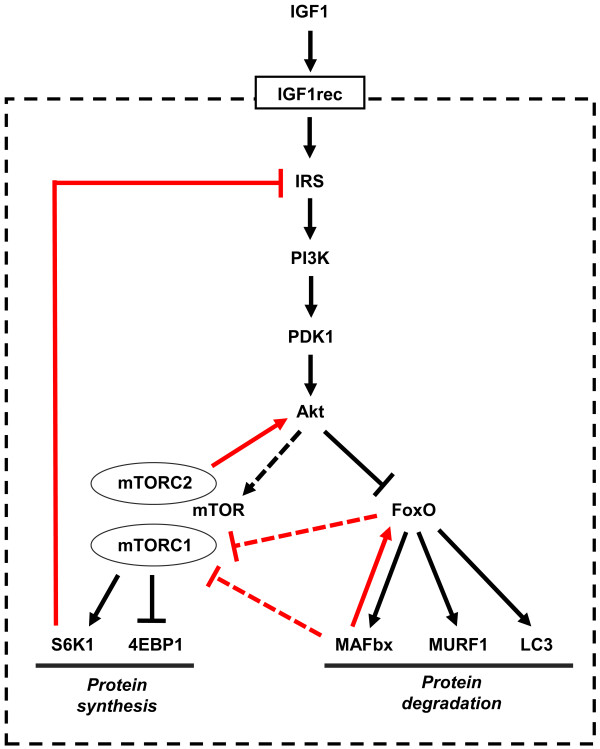
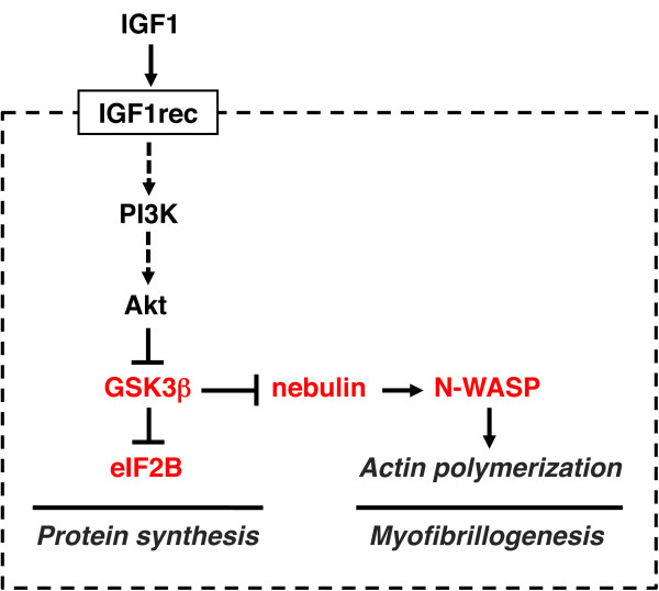
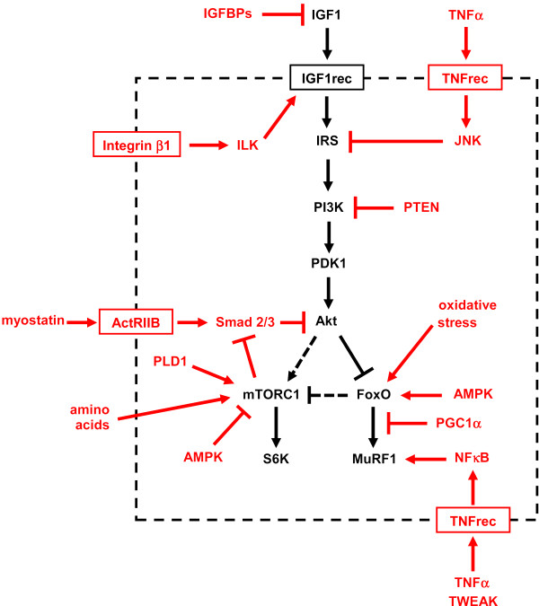
A highly conserved signaling pathway involving insulin-like growth factor 1 (IGF1), and a cascade of intracellular components that mediate its effects, plays a major role in the regulation of skeletal muscle growth. A central component in this cascade is the kinase Akt, also called protein kinase B (PKB), which controls both protein synthesis, via the kinases mammalian target of rapamycin (mTOR) and glycogen synthase kinase 3β (GSK3β), and protein degradation, via the transcription factors of the FoxO family. In this paper, we review the composition and function of this pathway in skeletal muscle fibers, focusing on evidence obtained in vivo by transgenic and knockout models and by muscle transient transfection experiments. Although this pathway is essential for muscle growth during development and regeneration, its role in adult muscle response to mechanical load is less clear. A full understanding of the operation of this pathway could help to design molecularly targeted therapeutics aimed at preventing muscle wasting, which occurs in a variety of pathologic contexts and in the course of aging.
Overview
The claims and statistics of this paper haven’t been checked yet.
Run the check — free, no account needed. It reads the open-access text and checks the sources it cites.
Integrity signals
Registry
Cites
Statistics
Figures
Verification
Claims the paper makes
The claims and statistics of this paper haven’t been checked yet.
Run the check — free, no account needed. It reads the open-access text and checks the sources it cites.
Claims other papers make about this one
Each is checked against this paper’s own text: does it say what the citing paper says it does?
The cited source says what the claim attributes to it.
In the citing paper
“The Akt‐mTOR pathway is central to the regulation of muscle mass because it promotes protein synthesis and inhibits protein degradation (Schiaffino & Mammucari, 2011).”
From Parkin overexpression protects from ageing‐related loss of muscle mass and strength (2019) · cites this paper as Schiaffino & Mammucari, 2011
In the citing paper
“Additionally, it has been discovered that PEs present in cosmetic preparation protect skin from pigmentation and aging163.”
From The Phytochemical, Biological, and Medicinal Attributes of Phytoecdysteroids: An Updated Review (2021) · cites this paper as [163]
Self-check
Percentages, sums and reported differences re-computed from the paper’s own numbers.
The self-check needs the paper’s full text.
Citations
Cites
Phosphorylation and regulation of Akt/PKB by the rictor-mTOR complex
Sarbassov, D. D., Guertin, D. A., Ali, S. M., & Sabatini, D. M. (2005) · Science · doi:10.1126/science.1106148 · Cited by 6,216
AKT/PKB signaling: navigating downstream
Manning, B. D., & Cantley, L. C. (2007) · Cell · doi:10.1016/j.cell.2007.06.009 · Cited by 6,115 · cited 1×
“transcription factors of the FoxO family, and stimulates protein synthesis via the mammalian target of rapamycin (mTOR) and glycogen synthase kinase 3b (GSK3b) [4].”
BackgroundRegulation of skeletal muscle mass in mice by a new TGF-p superfamily member
McPherron, A. C., Lawler, A. M., & Lee, S.-J. (1997) · Nature · doi:10.1038/387083a0 · Cited by 4,013
mTOR: from growth signal integration to cancer, diabetes and ageing
Zoncu, R., Efeyan, A., & Sabatini, D. M. (2011) · Nature Reviews Molecular Cell Biology · doi:10.1038/nrm3025 · Cited by 3,941 · cited 15×
“Deletion of S6K1 is sufficient to reproduce this atrophic phenotype.”
BackgroundInfluentialAMPK Phosphorylation of Raptor Mediates a Metabolic Checkpoint
Gwinn, D. M., Shackelford, D. B., Egan, D. F., Mihaylova, M. M., Mery, A., Vasquez, D. S., Turk, B. E., & Shaw, R. J. (2008) · Molecular Cell · doi:10.1016/j.molcel.2008.03.003 · Cited by 3,849 · cited 13×
“(AMPK), an energy status sensor activated by ATP depletion, inhibits mTORC1 by phosphorylating and inactivating Raptor [23], and by phosphorylating and activating TSC2 [24].”
BackgroundInfluentialIdentification of ubiquitin ligases required for skeletal muscle atrophy.
Bodine, S. C., Latres, E., Baumhueter, S., Lai, V. K.-M., Nunez, L., Clarke, B. A., Poueymirou, W. T., Panaro, F. J., Na, E., Dharmarajan, K., et al. (2001) · Science · doi:10.1126/science.1065874 · Cited by 3,521 · cited 5×
“Their role in muscle-mass regulation is supported by the finding that muscle atrophy induced by denervation is partially prevented in both MuRF1 null and atrogin1/MAFbx null mice [ 92 ].”
BackgroundInfluentialTSC2 is phosphorylated and inhibited by Akt and suppresses mTOR signalling
Inoki, K., Li, Y., Zhu, T., Wu, J., & Guan, K.-L. (2002) · Nature Cell Biology · doi:10.1038/ncb839 · Cited by 3,083 · cited 3×
“Rictor-dependent phosphorylation of Akt at serine 473 is required for Akt-mediated phosphorylation of FoxO but not of TSC2, thus it does not affect activation of S6K [5].”
BackgroundInfluentialSandri, M., Sandri, C., Gilbert, A., Skurk, C., Calabria, E., Picard, A., Walsh, K., Schiaffino, S., Lecker, S. H., & Goldberg, A. L. (2004) · Cell · doi:10.1016/s0092-8674(04)00400-3 · Cited by 3,012 · cited 5×
“Small interfering RNA to FoxO1 and FoxO3 Knockdown of FoxO1 and FoxO3 by RNA interference Prevention of atrogin-1 reporter upregulation induced by starvation [ 91 ]”
BackgroundInfluentialSelf-citationLiu, J.-P., Baker, J. C., Perkins, A. S., Robertson, E. J., & Efstratiadis, A. (1993) · Cell · doi:10.1016/s0092-8674(05)80084-4 · Cited by 2,854
The Rag GTPases Bind Raptor and Mediate Amino Acid Signaling to mTORC1
Sancak, Y., Peterson, T. R., Shaul, Y. D., Lindquist, R. A., Thoreen, C. C., Bar-Peled, L., & Sabatini, D. M. (2008) · Science · doi:10.1126/science.1157535 · Cited by 2,773 · cited 13×
“(AMPK), an energy status sensor activated by ATP depletion, inhibits mTORC1 by phosphorylating and inactivating Raptor [23], and by phosphorylating and activating TSC2 [24].”
BackgroundInfluential
Cited by
Sorted by citations
Items 201–300 of 817
Parafati, M., Giza, S., Shenoy, T., Mojica-Santiago, J., Hopf, M. E., Malany, L. K., Platt, D., Moore, I., Jacobs, Z. A., Kuehl, P., et al. (2023) · npj Microgravity · doi:10.1038/s41526-023-00322-y · Cited by 42
Margolis, L. M., McClung, H. L., Murphy, N. E., Carrigan, C. T., & Pasiakos, S. M. (2017) · Frontiers in Physiology · doi:10.3389/fphys.2017.00182 · Cited by 42
“Binding of IGF-1 to its receptor phosphorylates IRS which stimulates Akt-dependent activation of mTORC1 (Schiaffino and Mammucari, 2011).”
BackgroundPharaoh, G. A., Owen, D. B., Yeganeh, A., Premkumar, P., Farley, J. A., Bhaskaran, S., Ashpole, N. M., Kinter, M. T., Remmen, H. V., & Logan, S. (2019) · Molecular Neurobiology · doi:10.1007/s12035-019-01821-4 · Cited by 44
The Emerging Role of MicroRNAs and Other Non-Coding RNAs in Cancer Cachexia
Santos, J., da Silva, S. P., da Costa, R. M. G., & Medeiros, R. (2020) · Cancers · doi:10.3390/cancers12041004 · Cited by 42
“The IGF1-PI3K/Akt pathway controls protein synthesis and degradation, and knockdown of the insulin receptor substrate 1 (IRS) decreases IGF1, leading to weigh loss in mice [52,53].”
BackgroundMarceca, G. P., Nigita, G., Calore, F., & Croce, C. M. (2020) · Frontiers in Oncology · doi:10.3389/fonc.2020.607196 · Cited by 45
Runx2-Twist1 interaction coordinates cranial neural crest guidance of soft palate myogenesis
Han, X., Feng, J., Guo, T., Loh, Y. E., Yuan, Y., Ho, T., Cho, C. K., Li, J., Jing, J., Janečková, E., et al. (2021) · eLife · doi:10.7554/elife.62387 · Cited by 41
Skeletal muscle heme oxygenase-1 activity regulates aerobic capacity
de Souza, R. W. A., Gallo, D. J., Lee, G. R., Katsuyama, E., Schaufler, A., Weber, J., Csizmadia, E., Tsokos, G. C., Koch, L. G., Britton, S. L., et al. (2021) · Cell Reports · doi:10.1016/j.celrep.2021.109018 · Cited by 44
Genetic basis of muscle‐related traits in sheep: A review
Talebi, R., Ghaffari, M. R., Zeinalabedini, M., Abdoli, R., & Mardi, M. (2022) · Animal Genetics · doi:10.1111/age.13266 · Cited by 41
Kim, J., Lee, J.-Y., & Kim, C. Y. (2023) · Nutrients · doi:10.3390/nu15112625 · Cited by 45
“In a Chinese cohort study, high levels of TNF-α (>11.15 pg/mL) were associated with a 7.6-fold increased risk of sarcopenia [49].”
BackgroundInfluentialMuscle wasting: an overview of recent developments in basic research
Palus, S., von Haehling, S., & Springer, J. (2014) · Journal of Cachexia Sarcopenia and Muscle · doi:10.1007/s13539-014-0157-7 · Cited by 42
“Cytokines like interleukine-1 (IL-1), IL-6, TNF-α, and interferon-γ lead to a receptor-mediated activation of NFκB and FOXO in muscle [32]; the latter can also be activated by glucocorticoids [19].”
BackgroundSharlo, K. A., Paramonova, I. I., Turtikova, O. V., Tyganov, S. A., & Shenkman, B. S. (2019) · Journal of Applied Physiology · doi:10.1152/japplphysiol.00029.2019 · Cited by 41
“5) may be associated with GSK-3 inactivation, which is known to inhibit protein synthesis (40) along with upregulation of fast MyHC mRNA expression and partial prevention of unloading-induced MuRF-1 expression”
BackgroundAldiss, P., Lewis, J. E., Lupini, I., Bloor, I. D., Chavoshinejad, R., Boocock, D. J., Miles, A. K., Ebling, F. J. P., Budge, H., & Symonds, M. E. (2020) · Frontiers in Endocrinology · doi:10.3389/fendo.2020.00097 · Cited by 48
Reconstruction of Muscle Fascicle‐Like Tissues by Anisotropic 3D Patterning
Jin, Y., Jeon, E. J., Jeong, S., Min, S., Choi, Y. S., Ys, K., Lee, J. S., Shin, J., Yu, J. H., Ahn, D., et al. (2021) · Advanced Functional Materials · doi:10.1002/adfm.202006227 · Cited by 41
Myogenic, genomic and non‐genomic influences of the vitamin D axis in skeletal muscle
Bollen, S. E., & Atherton, P. J. (2020) · Cell Biochemistry and Function · doi:10.1002/cbf.3595 · Cited by 43
“THEM4 inhibits the phosphorylation and subsequent activation of Akt, resulting in muscle protein breakdown (MPB) due to cessation of Akt's inhibition and activation of members of the FOXO family and mTOR, respectively.(61) Treatment with 1,25(OH)2D3 caused upregulation of THEM4 which, paired with the fact that it has…”
BackgroundXiao, L.-Q., Jiang, W., Wu, P., Yang, L., Ren, H., Tang, L., Li, S., Zhong, C.-B., Zhang, R.-N., Feng, L., et al. (2023) · Aquaculture · doi:10.1016/j.aquaculture.2023.740005 · Cited by 40
Cancer-associated muscle weakness: What's bone got to do with it?
Waning, D. L., & Guise, T. A. (2015) · BoneKEy Reports · doi:10.1038/bonekey.2015.59 · Cited by 40
“In contrast to the negative effects possible from activin, myostatin and TGFb signaling in muscle, IGF-1 and BMP-2 signaling results in muscle hypertrophy.(42,52,53) IGF-1 is a major regulator of muscle mass because of its effect on myogenic cell proliferation and differentiation.”
BackgroundVassilakos, G., Lei, H., Yang, Y., Puglise, J. M., Matheny, M. E., Durzyńska, J., Ozery, M., Bennett, K., Spradlin, R. A., Bonanno, H., et al. (2018) · The FASEB Journal · doi:10.1096/fj.201800459r · Cited by 40
“Although IGF-IR phosphorylation was undetectable in resting muscles without exogenous stimulation by its ligand, we examined the resting phosphorylation levels of Akt and Erk (p-Akt and p-Erk1/2) as representative downstream elements of the pathway (29).”
BackgroundLecce, E., Amoruso, P., Felici, F., & Bazzucchi, I. (2025) · The Journal of Physiology · doi:10.1113/jp289716 · Cited by 56
“These peptides bind the IGF-1 receptor on adjacent fibres, inducing phosphorylation of insulin receptor substrate 1 (IRS-1) and recruitment of PI3K to the membrane, where it converts PIP 2 to PIP 3 (Ji et al., 2025; Rommel et al., 2001; Schiaffino & Mammucari, 2011).”
ChainRank, a chain prioritisation method for contextualisation of biological networks
Tényi, Á., de Atauri, P., Gómez-Cabrero, D., Cano, I., Clarke, K., Falciani, F., Cascante, M., Roca, J., & Maier, D. (2016) · BMC Bioinformatics · doi:10.1186/s12859-015-0864-x · Cited by 39
“2 Changes in the representation of IGF-Akt pathway. a shows the protein synthesis regulation related part of IGF-Akt pathway [21].”
BackgroundMethodKetogenic Diet and Skeletal Muscle Hypertrophy: a Frenemy Relationship?
Paoli, A., Cancellara, P., Pompei, P., & Moro, T. (2019) · Journal of Human Kinetics · doi:10.2478/hukin-2019-0071 · Cited by 41
“As described above, skeletal muscle protein synthesis following exercise is mainly regulated by mTORC1 that is part of the IGF-AktmTOR signalling pathway (Schiaffino and Mammucari, 2011; Wackerhage et al., 2019).”
BackgroundInfluentialMuscle specific miRNAs are induced by testosterone and independently upregulated by age
Nielsen, S. J., Hvid, T., Kelly, M., Lindegaard, B., Dethlefsen, C., Winding, K. M., Mathur, N., Schéele, C., Pedersen, B. K., & Laye, M. J. (2014) · Frontiers in Physiology · doi:10.3389/fphys.2013.00394 · Cited by 39
Villars, F. O., Pietra, C., Giuliano, C., Lutz, T. A., & Riediger, T. (2017) · International Journal of Molecular Sciences · doi:10.3390/ijms18050986 · Cited by 37
Song, C., Yang, Z., Dong, D., Xu, J., Wang, J., Li, H., Huang, Y., Lan, X., Lei, C., Ma, Y., et al. (2018) · Journal of Cellular Physiology · doi:10.1002/jcp.27672 · Cited by 36
“The findings of previous studies show that IGF1 regulates skeletal muscle development through the PI3K/AKT signaling pathway (Schiaffino & Mammucari, 2011).”
BackgroundPhysiological Systems in Promoting Frailty
Perazza, L. R., Brown‐Borg, H. M., & Thompson, L. V. (2022) · Comprehensive physiology · doi:10.1002/cphy.c210034 · Cited by 69
Kim, S., Welsh, D. A., Ravussin, É., Welsch, M. A., Cherry, K. E., Myers, L., & Jazwinski, S. M. (2013) · The Journals of Gerontology Series A · doi:10.1093/gerona/glt150 · Cited by 37
“IgF1 induces skeletal muscle growth and regeneration by activating the Akt-mammalian target of rapamycin pathway (25,26).”
BackgroundConserved and species-specific molecular denominators in mammalian skeletal muscle aging
Mercken, E. M., Capri, M., Carboneau, L., Conte, M., Heidler, J., Santoro, A., Martín‐Montalvo, A., González‐Freire, M., Khraiwesh, H. M., González-Reyes, J. A., et al. (2017) · npj Aging and Mechanisms of Disease · doi:10.1038/s41514-017-0009-8 · Cited by 38
Indoprofen prevents muscle wasting in aged mice through activation of PDK1/AKT pathway
Kim, H., Cho, S. C., Jeong, H., Lee, H., Jeong, M., Pyun, J., Ryu, D., Kim, M., Lee, Y., Kim, M. S., et al. (2020) · Journal of Cachexia Sarcopenia and Muscle · doi:10.1002/jcsm.12558 · Cited by 37
Circadian regulation of muscle growth independent of locomotor activity
Kelu, J. J., Pipalia, T. G., & Hughes, S. M. (2020) · Proceedings of the National Academy of Sciences · doi:10.1073/pnas.2012450117 · Cited by 35
Lee, E. M., Kim, A.-Y., Lee, E. H., Park, J., Lee, M., Hwang, M., Kim, C.-Y., Kim, S., & Jeong, K. (2014) · Cell Transplantation · doi:10.3727/096368914x678599 · Cited by 36
Chen, Z., Hagen, D. E., Ji, T., Elsik, C. G., & Rivera, R. M. (2017) · Scientific Reports · doi:10.1038/s41598-017-13012-z · Cited by 35
Alsolmei, F. A., Li, H., Pereira, S. L., Krishnan, P., Johns, P. W., & Siddiqui, R. A. (2019) · Nutrients · doi:10.3390/nu11051077 · Cited by 36
“The signaling pathway IGF-1/PI3K/Akt (Insulin like growth factor -1/phosphatidyl inositol 3-kinase/protein kinase) is considered the main mediator of normal muscle development and one of the most studied signaling molecular systems involved in muscle metabolism [61].”
BackgroundLu, Y., Bradley, J. S., McCoski, S. R., Gonzalez, J. M., Ealy, A. D., & Johnson, S. E. (2017) · American Journal of Physiology-Regulatory, Integrative and Comparative Physiology · doi:10.1152/ajpregu.00400.2016 · Cited by 35
Regulatory role of exercise-induced autophagy for sarcopenia
Liang, J., Zeng, Z., Zhang, Y., & Chen, N. (2020) · Experimental Gerontology · doi:10.1016/j.exger.2019.110789 · Cited by 33
Pathogenesis of Musculoskeletal Deficits in Children and Adults with Inflammatory Bowel Disease
Steell, L., Gray, S. R., Russell, R. K., MacDonald, J. C., Seenan, J. P., Wong, S. C., & Gaya, D. R. (2021) · Nutrients · doi:10.3390/nu13082899 · Cited by 35
“In skeletal muscle, the anabolic actions of GH are mediated by systemic and local IGF-1, which stimulate muscle protein synthesis and reduce protein degradation [92].”
BackgroundKang, M. S., Yu, Y., Park, R., Heo, H. J., Lee, S. H., Hong, S. W., Kim, Y. H., & Han, D. (2024) · Nano-Micro Letters · doi:10.1007/s40820-023-01293-1 · Cited by 35
“Tissue regeneration in the wound was evaluated at the designated time points.”
BackgroundHindi, S. M., Paul, P. K., Dahiya, S., Mishra, V., Bhatnagar, S., Kuang, S., Choi, Y., & Kumar, A. (2012) · Molecular and Cellular Biology · doi:10.1128/mcb.00717-12 · Cited by 41
Systems for Muscle Cell Differentiation: From Bioengineering to Future Food
Lee, K.-Y., Loh, H. X., & Wan, A. C. A. (2021) · Micromachines · doi:10.3390/mi13010071 · Cited by 35
Differential induction of muscle atrophy pathways in two mouse models of spinal muscular atrophy
Deguise, M., Boyer, J. G., McFall, E. R., Yazdani, A., Repentigny, Y. D., & Kothary, R. (2016) · Scientific Reports · doi:10.1038/srep28846 · Cited by 33
BIO101 stimulates myoblast differentiation and improves muscle function in adult and old mice
Serova, M., Didry‐Barca, B., Deloux, R., Foucault, A., Veillet, S., Lafont, R., Dilda, P. J., & Latil, M. (2024) · Journal of Cachexia, Sarcopenia and Muscle · doi:10.1002/jcsm.13326 · Cited by 35
“9 AMPK emerges as a master regulator of mitochondrial homeostasis, coupling mitochondrial dynamics to mitophagy which is involved in removal of dysfunctional mitochondria to preserve skeletal muscle function.”
BackgroundAn Overview of Sarcopenia: Focusing on Nutritional Treatment Approaches
Barone, M., Baccaro, P., & Molfino, A. (2025) · Nutrients · doi:10.3390/nu17071237 · Cited by 43
“Muscle synthesis is intrinsically linked to muscle atrophy via the PI3k–Akt pathway, which is central to both processes [25].”
Webster, J. M., Fenton, C. G., LANGEN, R. C. J., & Hardy, R. (2019) · International Journal of Molecular Sciences · doi:10.3390/ijms20225768 · Cited by 36
“mTOR activation suppresses proteolytic, forkhead box class O family member proteins (FOXOs) and glycogen synthase kinase-3 beta (GSK-3β) pathways [91,92].”
BackgroundRodent Model of Muscular Atrophy for Sarcopenia Study
Baek, K., Jung, Y., Kim, J., Park, J. S., Hah, Y., Kim, S.-J., & Yoo, J. (2020) · Journal of Bone Metabolism · doi:10.11005/jbm.2020.27.2.97 · Cited by 40
Vav2 catalysis-dependent pathways contribute to skeletal muscle growth and metabolic homeostasis
Rodríguez‐Fdez, S., Lorenzo‐Martín, L. F., Fernández‐Pisonero, I., Porteiro, B., Veyrat‐Durebex, C., Beiroa, D., Al–Massadi, O., Abad, A., Diéguez, C., Coppari, R., et al. (2020) · Nature Communications · doi:10.1038/s41467-020-19489-z · Cited by 33
Bawa, S., Piccirillo, R., & Geisbrecht, E. R. (2021) · Biomolecules · doi:10.3390/biom11030408 · Cited by 35
Parvalbumin Affects Skeletal Muscle Trophism Through Modulation of Mitochondrial Calcium Uptake
Butera, G., Reane, D. V., Canato, M., Pietrangelo, L., Boncompagni, S., Protasi, F., Rizzuto, R., Reggiani, C., & Raffaello, A. (2020) · Cell Reports · doi:10.1016/j.celrep.2021.109087 · Cited by 38
MEAT SCIENCE AND MUSCLE BIOLOGY SYMPOSIUM: Stem cell niche and postnatal muscle growth1,2
Bi, P., & Kuang, S. (2011) · Journal of Animal Science · doi:10.2527/jas.2011-4594 · Cited by 34
“…on adipogenesis was shown to act through several mechanisms, including microRNA, IGF-1, and Notch signaling, which are potent regulators of MuSC and myogenesis as well (Nueda et al., 2007b, 2008; Andersen et al., 2010; Mayeuf and Relaix, 2011; Porrello et al., 2011; Schiaffino and Mammucari, 2011).”
BackgroundRejuvenating stem cells to restore muscle regeneration in aging
Bengal, E., Perdiguero, E., Serrano, A. L., & Muñoz‐Cánoves, P. (2017) · F1000Research · doi:10.12688/f1000research.9846.1 · Cited by 30
Effects of training intensity in electromyostimulation on human skeletal muscle
Natsume, T., Ozaki, H., Kakigi, R., Kobayashi, H., & Naıto, H. (2018) · European Journal of Applied Physiology · doi:10.1007/s00421-018-3866-3 · Cited by 36
Taurine Attenuates Catabolic Processes Related to the Onset of Sarcopenia
Barbiera, A., Sorrentino, S., Lepore, E., Carfì, A., Sica, G., Dobrowolny, G., & Scicchitano, B. M. (2020) · International Journal of Molecular Sciences · doi:10.3390/ijms21228865 · Cited by 34
“Besides, it has been demonstrated that NF-kB can induce the expression of FoxO transcription factors by modulating the Akt/mTOR signaling cascade [21,22], ultimately leading to skeletal muscle loss and functional impairment.”
BackgroundRana, K., Chiu, M. W. S., Russell, P. K., Skinner, J. P. J., Lee, N. K., Fam, B. C., Zajac, J. D., & MacLean, H. E. (2016) · Journal of Molecular Endocrinology · doi:10.1530/jme-15-0320 · Cited by 33
“1985), regulation of myoblast differentiation (Igf2) (Wilson & Rotwein 2006, Schiaffino & Mammucari 2011) and myogenic differentiation or activation of satellite cells (Fzd4) (Rudnicki & Williams 2015).”
BackgroundBorner, T., Loi, L., Pietra, C., Giuliano, C., Lutz, T. A., & Riediger, T. (2016) · American Journal of Physiology-Regulatory, Integrative and Comparative Physiology · doi:10.1152/ajpregu.00044.2016 · Cited by 32
Potential Roles of Vascular Endothelial Growth Factor During Skeletal Muscle Hypertrophy
Huey, K. A. (2018) · Exercise and Sport Sciences Reviews · doi:10.1249/jes.0000000000000152 · Cited by 32
Jin, C., Zhuo, Y., Wang, J., Zhao, Y., Xuan, Y., Mou, D., Liu, H., Zhou, P., Fang, Z., Che, L., et al. (2018) · Journal of Animal Physiology and Animal Nutrition · doi:10.1111/jpn.12933 · Cited by 32
Protective effects of Liuwei dihuang water extracts on diabetic muscle atrophy
Tseng, Y., Chang, W., Lin, C., Chang, F., Wu, P., & Lo, Y. (2018) · Phytomedicine · doi:10.1016/j.phymed.2018.09.032 · Cited by 32
Europa, T. A., Nel, M., & Heckmann, J. M. (2019) · Neuromuscular Disorders · doi:10.1016/j.nmd.2019.03.009 · Cited by 32
Nur77 deletion impairs muscle growth during developmental myogenesis and muscle regeneration in mice
Cortez-Toledo, O., Schnair, C., Sangngern, P., METZGER, D., & Chao, L. C. (2017) · PLoS ONE · doi:10.1371/journal.pone.0171268 · Cited by 34
Renna, L. V., Bosè, F., Brigonzi, E., Fossati, B., Meola, G., & Cardani, R. (2019) · PLoS ONE · doi:10.1371/journal.pone.0214254 · Cited by 34
IGF-1 prevents simvastatin-induced myotoxicity in C2C12 myotubes
Bonifacio, A., Sanvee, G. M., Brecht, K., Kratschmar, D. V., Odermatt, A., Bouitbir, J., & Krähenbühl, S. (2016) · Archives of Toxicology · doi:10.1007/s00204-016-1871-z · Cited by 31
“1, AKT controls both skeletal muscle protein synthesis by activating mTORC1 and protein degradation by suppressing the activation of forkhead members of the O class (FoxO) transcription factors (Schiaffino and Mammucari 2011).”
BackgroundPreclinical insights into the gut‐skeletal muscle axis in chronic gastrointestinal diseases
Ehlers, L., Bannert, K., Rohde, S., Berlin, P., Reiner, J., Wiese, M. L., Doller, J., Lerch, M. M., Aghdassi, A. A., Meyer, F., et al. (2020) · Journal of Cellular and Molecular Medicine · doi:10.1111/jcmm.15554 · Cited by 33
Mediators and clinical treatment for cancer cachexia: a systematic review
Cao, Z., Scott, A. M., Hoogenraad, N. J., & Osellame, L. D. (2021) · JCSM Rapid Communications · doi:10.1002/rco2.30 · Cited by 31
Sales, F., Pacheco, D., Blair, H. T., Kenyon, P. R., & McCoard, S. A. (2013) · SpringerPlus · doi:10.1186/2193-1801-2-483 · Cited by 31
“Protein synthesis in muscle is controlled by specific signalling pathways, including phosphatidylinositol-3 kinase (Schiaffino and Mammucari 2011), 5′-AMP-activated protein kinase (Bolster et al. 2002) and mitogen-activated protein kinase (Williamson et al. 2003).”
BackgroundDevelopmental changes of Insulin-like growth factors in the liver and muscle of chick embryos
Liu, Y., Guo, W., Pu, Z., Li, X., Lei, X., Yao, J., & Yang, X. (2016) · Poultry Science · doi:10.3382/ps/pew043 · Cited by 31
“, 1991), and many signaling pathways involving IGF1 were related to muscle growth (Christoffolete et al., 2015; Dupont and Holzenberger, 2003; Schiaffino and Mammucari, 2011).”
BackgroundLi, X., Zhu, Y., Zhang, H., Ma, G., Wu, G., Xiang, A., Shi, X., Yang, G., & Sun, S. (2018) · Genes · doi:10.3390/genes9070333 · Cited by 31
“PI3K (p85α), encoded by PIK3R1 gene, is a key protein involved in the PI3K/AKT signaling pathway [24], which is essential for myogenic differentiation [25,26].”
BackgroundAlterations of signaling pathways in muscle tissues of patients with amyotrophic lateral sclerosis
Yin, F., Ye, F., Tan, L., Liu, K. Y., Xuan, Z., Zhang, J., Wang, W., Zhang, Y., Jiang, X., & Zhang, D. Y. (2012) · Muscle & Nerve · doi:10.1002/mus.23411 · Cited by 30
“Akt has been reported to play a key role in muscle atrophy and hypertrophy.(28) Muscle hypertrophy is correlated with upregulation of Akt, which is activated by insulin-like growth factor-1 (IGF-1).”
BackgroundLunar gravity prevents skeletal muscle atrophy but not myofiber type shift in mice
Hayashi, T., Fujita, R., Okada, R., Hamada, M., Suzuki, R., Fuseya, S., Leckey, J., Kanai, M., Inoue, Y., Sadaki, S., et al. (2023) · Communications Biology · doi:10.1038/s42003-023-04769-3 · Cited by 29
Overload-induced skeletal muscle hypertrophy is not impaired in STZ-diabetic rats
Fortes, M. A. S., Pinheiro, C. H. D. J., Guimarães‐Ferreira, L., Vitzel, K. F., de Vasconcelos, D. A. A., & Curi, R. (2015) · Physiological Reports · doi:10.14814/phy2.12457 · Cited by 29
Wetzel, M. D., & Wenke, J. C. (2019) · Journal of Translational Medicine · doi:10.1186/s12967-018-1753-7 · Cited by 29
Tyganov, S. A., Mochalova, E. P., Belova, S. P., Sharlo, K. A., Rozhkov, S. V., Vilchinskaya, N. A., Paramonova, I. I., Mirzoev, T. M., & Shenkman, B. S. (2019) · Frontiers in Physiology · doi:10.3389/fphys.2019.01252 · Cited by 31
Sahm, A., Hoffmann, S., Koch, P., Henning, Y., Bens, M., Groth, M., Burda, H., Begall, S., Ting, S. C., Goetz, M., et al. (2021) · eLife · doi:10.7554/elife.57843 · Cited by 29
Yang, S., Yang, G., Wu, H., Kang, L., Xiang, J., Zheng, P., Qiu, S., Liang, Z., Lü, Y., & Jia, L. (2021) · Diabetologia · doi:10.1007/s00125-021-05616-y · Cited by 29
“Consistent with this, the genetic activation of IGF1 or Akt in skeletal muscle is sufficient to increase muscle mass [11].”
BackgroundMolecular mechanisms of post‐burn muscle wasting and the therapeutic potential of physical exercise
Dombrecht, D., Daele, U. V., Asbroeck, B. V., Schieffelers, D. R., Guns, P., Gebruers, N., Meirte, J., & van Breda, E. (2023) · Journal of Cachexia Sarcopenia and Muscle · doi:10.1002/jcsm.13188 · Cited by 28
“Two mTORC complexes exist, that is, mTORC1 and mTORC2.(28) The mechanisms of mTORC2 are not fully unravelled, but in vivo research in mice did not reveal a structural impact on skeletal muscle by mTORC2 inhibition.”
BackgroundPaco, S., Kalko, S. G., Jou, C., Rodríguez, M. A., Corbera, J., Francesco Muntoni, A., Feng, L., Rivas, E., Torner, F., Gualandi, F., et al. (2013) · PLoS ONE · doi:10.1371/journal.pone.0077430 · Cited by 29
“Insulin –like growth factor I (IGF-I), whose expression is known to be up-regulated in regenerating muscle in vivo, positively regulates the proliferation and differentiation of satellite cells/ myoblasts in vitro via the Akt/PKB pathway [45] and calcineurin [46].”
BackgroundDang, K., Li, Y.-Z., Gong, L.-C., Xue, W., Wang, H., Goswami, N., & Gao, Y. (2016) · Biology Open · doi:10.1242/bio.015776 · Cited by 29
“It is worth noting that after 14-days, HU atrogin-1 and MuRF1 mRNA expression levels were upregulated, while the phosphorylation level of FoxO1 was significantly increased.”
BackgroundMethodInfluentialShibu, M. A., Hung, T.-H., Chiang, W., & Lin, W. (2018) · Journal of Medicinal Food · doi:10.1089/jmf.2017.3941 · Cited by 29
“4 Natural products such as acidic polysaccharide and hydroxypropyl methyl cellulose can suppress HFD-induced disease risk.”
Muscle wasting: An overview of recent developments in basic research
Palus, S., von Haehling, S., & Springer, J. (2014) · International Journal of Cardiology · doi:10.1016/j.ijcard.2014.08.086 · Cited by 29
Yoshimoto, Y., & Oishi, Y. (2023) · Pharmacology & Therapeutics · doi:10.1016/j.pharmthera.2023.108357 · Cited by 28
Ranasinghe, T., Seo, Y., Park, H., Choe, S., & Cha, S. (2024) · Journal of Hazardous Materials · doi:10.1016/j.jhazmat.2024.136215 · Cited by 27
Margolis, L. M., Berryman, C. E., Murphy, N. E., Carrigan, C. T., Young, A., Carbone, J. W., & Pasiakos, S. M. (2018) · Physiological Reports · doi:10.14814/phy2.13931 · Cited by 26
“When miR-19b-3p, miR-222-3p, miR-324-3p, and miR-486-5p expression is higher in the muscle, there is lower PTEN expression, increased activity of p-AKTThr473, and lower activity of p-FOXO1Thr24 and MURF1 expression.”
BackgroundInfluentialDiet and redox state in maintaining skeletal muscle health and performance at high altitude
Rathor, R., Suryakumar, G., & Singh, S. N. (2021) · Free Radical Biology and Medicine · doi:10.1016/j.freeradbiomed.2021.07.024 · Cited by 28
Role of MicroRNAs and Long Non-Coding RNAs in Sarcopenia
Lee, J., & Kang, H. (2022) · Cells · doi:10.3390/cells11020187 · Cited by 28
“The IGF-1 signaling pathway plays an important role in skeletal muscle growth, differentiation, and regeneration [46,47].”
BackgroundGiannos, P., Prokopidis, K., Raleigh, S. M., Kelaiditi, E., & Hill, M. W. (2022) · Scientific Reports · doi:10.1038/s41598-022-15578-9 · Cited by 28
Fernández, T., Briceño, F. A. C., Espinosa‐Ramírez, M., García-Valdés, P., López‐Fuenzalida, A., Riquelme, A. J., Arab, J. P., Cabrera, D., Arrese, M. A., & Francisco Barrera, M. (2020) · Expert Review of Gastroenterology & Hepatology · doi:10.1080/17474124.2020.1810563 · Cited by 28
“and important pathways of protein catabolism such as MuRF1 and atrogin-1 [64].”
BackgroundLi, Z., Abdalla, B. A., Zheng, M., He, X., Cai, B., Han, P., Ouyang, H., Chen, B., Nie, Q., & Zhang, X. (2017) · Molecular Genetics and Genomics · doi:10.1007/s00438-017-1364-7 · Cited by 27
“The kinase Akt/protein kinase B (PKB) targets mTOR and glycogen synthase kinase 3β, thus regulating protein degradation, and also targets a FOXO family transcription factor, thus regulating protein synthesis (Schiaffino and Mammucari 2011).”
BackgroundLambert, B. S., Shimkus, K. L., Fluckey, J. D., Riechman, S. E., Greene, N. P., Cardin, J. M., & Crouse, S. F. (2014) · American Journal of Physiology-Endocrinology and Metabolism · doi:10.1152/ajpendo.00689.2013 · Cited by 28
de la serrana, D. G., Fuentes, E. N., Martín, S., Johnston, I. A., & Macqueen, D. J. (2017) · General and Comparative Endocrinology · doi:10.1016/j.ygcen.2017.01.017 · Cited by 27
“The hormones Igf-I and Igf-II act as endocrine factors (Laron, 58 1996; Wood et al. 2005), but are also released locally by tissues, including skeletal muscle 59 (Schiaffino and Mammucari, 2011).”
Extracellular matrix in skeletal muscle injury and atrophy: mechanisms and therapeutic implications
Ge, X., Jin, Y., He, J., Jia, Z., Liu, Y., & Xu, Y. X. (2025) · Journal of Orthopaedic Translation · doi:10.1016/j.jot.2025.03.004 · Cited by 33
“Numerous signaling cascades are instrumental in the modulation of muscle wasting, such as the insulin/IGF-1-Protein Kinase B (AKT)-mammalian Target of Rapamycin (mTOR) axis, which facilitates protein anabolism and suppresses proteolysis [25].”
Time course of diaphragm function recovery after controlled mechanical ventilation in rats
Thomas, D., Maes, K., Agten, A., Heunks, L. M. A., Dekhuijzen, R. P. N., Decramer, M., van Hees, H. W. H., & Gayan‐Ramirez, G. (2013) · Journal of Applied Physiology · doi:10.1152/japplphysiol.00302.2012 · Cited by 26
Interspecies Organogenesis for Human Transplantation
Crane, A. T., Aravalli, R. N., Asakura, A., Grande, A. W., Krishna, V. D., Carlson, D. F., Cheeran, M. C. ‐., Danczyk, G., Dutton, J. R., Hackett, P. B., et al. (2019) · Cell Transplantation · doi:10.1177/0963689719845351 · Cited by 26
Skeletal muscle IGF-1 is lower at rest and after resistance exercise in humans with obesity
Sullivan, B. P., Weiss, J. A., Nie, Y., Garner, R. T., Drohan, C. J., Kuang, S., Stout, J., & Gavin, T. P. (2020) · European Journal of Applied Physiology · doi:10.1007/s00421-020-04509-z · Cited by 26
Tyganov, S. A., Mochalova, E. P., Belova, S. P., Sharlo, K. A., Rozhkov, S. V., Kalashnikov, V. E., Turtikova, O. V., Mirzoev, T. M., & Shenkman, B. S. (2021) · Scientific Reports · doi:10.1038/s41598-021-89362-6 · Cited by 26
Graça, F. A., Sheffield, N., Puppa, M., Finkelstein, D., Hunt, L. C., & Demontis, F. (2021) · PLoS Genetics · doi:10.1371/journal.pgen.1009926 · Cited by 27
“Altogether, these findings indicate that Gsk3, similar to Deaf1, regulates muscle mass in Drosophila (Fig 3), as observed in mammals [14,41,56].”
BackgroundMethodXu, J., Strasburg, G. M., Reed, K. M., & Velleman, S. G. (2022) · PLoS ONE · doi:10.1371/journal.pone.0262576 · Cited by 28
“As an upstream signal, IGF-1 can interact with IGF-1 receptor, activating mTOR/S6K signal transduction via PI3K/Akt pathway [38, 49, 91].”
BackgroundChycki, J., Czuba, M., Gołaś, A., Zając, A., Fidos-Czuba, O., Młynarz, A., & Smółka, W. (2016) · Journal of Human Kinetics · doi:10.1515/hukin-2016-0013 · Cited by 26
Rinninella, E., Cintoni, M., Raoul, P., Pozzo, C., Strippoli, A., Ponziani, F., Pompili, M., Bria, E., Tortora, G., Gasbarrini, A., et al. (2020) · Nutrients · doi:10.3390/nu12103101 · Cited by 26
“The insulin/IGF-1- Akt pathway increases skeletal muscle protein synthesis via inhibiting glycogen synthase kinase 3β and activating the mechanistic target of rapamycin complex 1 (mTORC1) signaling [9].”
BackgroundAncel, S., Michaud, J., Migliavacca, E., Jomard, C., Fessard, A., Garcia, P., Karaz, S., Raja, S., Jacot, G. E., Desgeorges, T., et al. (2024) · Journal of Clinical Investigation · doi:10.1172/jci163648 · Cited by 25
“The AKT serine/threonine kinase controls myogenic differentiation and is a central regulator of anabolic processes and muscle protein synthesis (44), which were regulated in hMPs in response to PN (Figure 1G).”
Li, Y., Zhang, F., Modrak, S., Little, A., & Zhang, H. (2019) · Alcoholism Clinical and Experimental Research · doi:10.1111/acer.14221 · Cited by 25
“Activation of IGFR-1 by IGF-1 recruits and activates insulin receptor substrate (IRS), which in turn recruits and activates phosphatidylinositol-3 kinase (PI3K) to generate phosphoinositide-3,4,5-triphosphate (PIP3).”
BackgroundInfluentialEffect of GLP-1 receptor agonist, liraglutide, on muscle in spontaneously diabetic torii fatty rats
Yamada, S., Ogura, Y., Inoue, K., Tanabe, J., Sugaya, T., Ohata, K., Nagai, Y., Natsuki, Y., Hoshino, S., Watanabe, S., et al. (2022) · Molecular and Cellular Endocrinology · doi:10.1016/j.mce.2021.111472 · Cited by 25
Wang, S., Tan, B., Xiao, L., Zhao, X., Zeng, J., Hong, L., Yang, J., Cai, G., Zheng, E., Wu, Z., et al. (2022) · International Journal of Molecular Sciences · doi:10.3390/ijms23094600 · Cited by 25
McIntosh, M. C., Sexton, C. L., Godwin, J. S., Ruple, B. A., Michel, J. M., Plotkin, D. L., Ziegenfuss, T. N., Lopez, H. L., Smith, R. C., Dwaraka, V. B., et al. (2023) · Cells · doi:10.3390/cells12060898 · Cited by 28
Hayakawa, N., Fukumura, J., Yasuno, H., Fujimoto-Ouchi, K., & Kitamura, H. (2015) · Biomedical Research · doi:10.2220/biomedres.36.71 · Cited by 25
“IGF-1 promotes phosphorylation of AKT and the activated AKT downregulates MAFbx and MuRF1 via downstream FOXO inhibition (39).”
Background
Metadata
- Authors
- Schiaffino, S., & Mammucari, C.
- Affiliations
- Veneto Institute of Molecular Medicine (IT)
- University of Padua (IT)
- Corresponding author
- Schiaffino, Stefano
- Venue
- Skeletal Muscle
- Published
- 2011
- Volume
- 1
- Issue
- 1
- Pages
- 4
- ISSN
- 2044-5040 (ISSN-L)
- PMC
- PMC3143906
- OpenAlex
- W2150059080
- Cited by
- 816
- References
- 150
- Citations by year
- 2026: 42 · 2025: 70 · 2024: 68 · 2023: 63 · 2022: 55 · 2021: 59 · 2020: 56 · 2019: 69 · 2018: 68 · 2017: 57 · 2016: 42 · 2015: 46 · 2014: 48 · 2013: 44 · 2012: 23
- FWCI
- 15.73
- Type
- journal article
- Language
- en
- Open access
- gold
- doi.org
- Licence
- cc-by
- Locations
- Skeletal Muscle (publishedVersion, cc-by)
- Europe PMC (PubMed Central) (publishedVersion, cc-by)
- PubMed Central (publishedVersion, cc-by)
- PubMed (publishedVersion)
- Padua Research Archive (University of Padova) (submittedVersion, cc-by)
- DOAJ (DOAJ: Directory of Open Access Journals) (submittedVersion)
- Funders
- European Commission
- Università degli Studi di Padova
- Topics
- Life Sciences › Biochemistry, Genetics and Molecular Biology › Molecular Biology › Muscle Physiology and Disorders
- Health Sciences › Nursing › Nutrition and Dietetics › Muscle metabolism and nutrition
- Life Sciences › Biochemistry, Genetics and Molecular Biology › Molecular Biology › Metabolism, Diabetes, and Cancer
- Keywords
- skeletal muscle growth, IGF-1 signaling, mTOR, GSK3β, FOXO transcription factors, muscle regeneration, myogenesis, mechanical loading, muscle atrophy, muscle protein synthesis, protein degradation
About these checks
Registry, retraction, Cites and statistics results are computed, not judged, with one exception: a printed reference that no index lists is read once by a model before it counts as a citation error. Claims are extracted by a model and kept only when their quote appears verbatim in the text; each cited source is then checked, with a model’s help, for whether it says what the claim relies on. Those verdicts can be wrong — read the claim’s quote and the cited source before relying on one.
Statistics and arithmetic are recomputed, not judged.
Confidence bands are shown only for fields calibrated on our annotated test set. None is calibrated yet, so no band is shown.
What counts as a claim ›How we measure confidence ›How we check this ›
Are you an author, or do you see a problem with this page? Request a review
