

The DOI matches its Crossref record; 148 of the 150 references it cites were found in a registry or an index.
Regulation of skeletal muscle growth by the IGF1-Akt/PKB pathway: insights from genetic models
Open accessAbstract
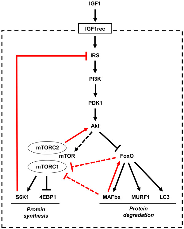
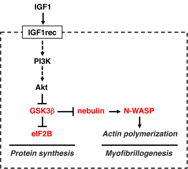
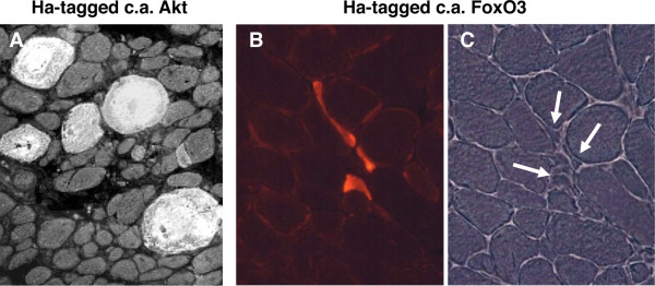
A highly conserved signaling pathway involving insulin-like growth factor 1 (IGF1), and a cascade of intracellular components that mediate its effects, plays a major role in the regulation of skeletal muscle growth. A central component in this cascade is the kinase Akt, also called protein kinase B (PKB), which controls both protein synthesis, via the kinases mammalian target of rapamycin (mTOR) and glycogen synthase kinase 3β (GSK3β), and protein degradation, via the transcription factors of the FoxO family. In this paper, we review the composition and function of this pathway in skeletal muscle fibers, focusing on evidence obtained in vivo by transgenic and knockout models and by muscle transient transfection experiments. Although this pathway is essential for muscle growth during development and regeneration, its role in adult muscle response to mechanical load is less clear. A full understanding of the operation of this pathway could help to design molecularly targeted therapeutics aimed at preventing muscle wasting, which occurs in a variety of pathologic contexts and in the course of aging.
Overview
The claims and statistics of this paper haven’t been checked yet.
Run the check — free, no account needed. It reads the open-access text and checks the sources it cites.
Integrity signals
Registry
Cites
Statistics
Figures
Verification
Claims the paper makes
The claims and statistics of this paper haven’t been checked yet.
Run the check — free, no account needed. It reads the open-access text and checks the sources it cites.
Claims other papers make about this one
Each is checked against this paper’s own text: does it say what the citing paper says it does?
The cited source says what the claim attributes to it.
In the citing paper
“The Akt‐mTOR pathway is central to the regulation of muscle mass because it promotes protein synthesis and inhibits protein degradation (Schiaffino & Mammucari, 2011).”
From Parkin overexpression protects from ageing‐related loss of muscle mass and strength (2019) · cites this paper as Schiaffino & Mammucari, 2011
In the citing paper
“Additionally, it has been discovered that PEs present in cosmetic preparation protect skin from pigmentation and aging163.”
From The Phytochemical, Biological, and Medicinal Attributes of Phytoecdysteroids: An Updated Review (2021) · cites this paper as [163]
Self-check
Percentages, sums and reported differences re-computed from the paper’s own numbers.
The self-check needs the paper’s full text.
Citations
Cites
Phosphorylation and regulation of Akt/PKB by the rictor-mTOR complex
Sarbassov, D. D., Guertin, D. A., Ali, S. M., & Sabatini, D. M. (2005) · Science · doi:10.1126/science.1106148 · Cited by 6,216
AKT/PKB signaling: navigating downstream
Manning, B. D., & Cantley, L. C. (2007) · Cell · doi:10.1016/j.cell.2007.06.009 · Cited by 6,115 · cited 1×
“transcription factors of the FoxO family, and stimulates protein synthesis via the mammalian target of rapamycin (mTOR) and glycogen synthase kinase 3b (GSK3b) [4].”
BackgroundRegulation of skeletal muscle mass in mice by a new TGF-p superfamily member
McPherron, A. C., Lawler, A. M., & Lee, S.-J. (1997) · Nature · doi:10.1038/387083a0 · Cited by 4,013
mTOR: from growth signal integration to cancer, diabetes and ageing
Zoncu, R., Efeyan, A., & Sabatini, D. M. (2011) · Nature Reviews Molecular Cell Biology · doi:10.1038/nrm3025 · Cited by 3,941 · cited 15×
“Deletion of S6K1 is sufficient to reproduce this atrophic phenotype.”
BackgroundInfluentialAMPK Phosphorylation of Raptor Mediates a Metabolic Checkpoint
Gwinn, D. M., Shackelford, D. B., Egan, D. F., Mihaylova, M. M., Mery, A., Vasquez, D. S., Turk, B. E., & Shaw, R. J. (2008) · Molecular Cell · doi:10.1016/j.molcel.2008.03.003 · Cited by 3,849 · cited 13×
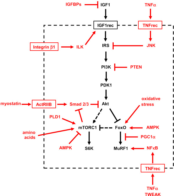
“(AMPK), an energy status sensor activated by ATP depletion, inhibits mTORC1 by phosphorylating and inactivating Raptor [23], and by phosphorylating and activating TSC2 [24].”
BackgroundInfluentialIdentification of ubiquitin ligases required for skeletal muscle atrophy.
Bodine, S. C., Latres, E., Baumhueter, S., Lai, V. K.-M., Nunez, L., Clarke, B. A., Poueymirou, W. T., Panaro, F. J., Na, E., Dharmarajan, K., et al. (2001) · Science · doi:10.1126/science.1065874 · Cited by 3,521 · cited 5×
“Their role in muscle-mass regulation is supported by the finding that muscle atrophy induced by denervation is partially prevented in both MuRF1 null and atrogin1/MAFbx null mice [ 92 ].”
BackgroundInfluentialTSC2 is phosphorylated and inhibited by Akt and suppresses mTOR signalling
Inoki, K., Li, Y., Zhu, T., Wu, J., & Guan, K.-L. (2002) · Nature Cell Biology · doi:10.1038/ncb839 · Cited by 3,083 · cited 3×
“Rictor-dependent phosphorylation of Akt at serine 473 is required for Akt-mediated phosphorylation of FoxO but not of TSC2, thus it does not affect activation of S6K [5].”
BackgroundInfluentialSandri, M., Sandri, C., Gilbert, A., Skurk, C., Calabria, E., Picard, A., Walsh, K., Schiaffino, S., Lecker, S. H., & Goldberg, A. L. (2004) · Cell · doi:10.1016/s0092-8674(04)00400-3 · Cited by 3,012 · cited 5×
“Small interfering RNA to FoxO1 and FoxO3 Knockdown of FoxO1 and FoxO3 by RNA interference Prevention of atrogin-1 reporter upregulation induced by starvation [ 91 ]”
BackgroundInfluentialSelf-citationLiu, J.-P., Baker, J. C., Perkins, A. S., Robertson, E. J., & Efstratiadis, A. (1993) · Cell · doi:10.1016/s0092-8674(05)80084-4 · Cited by 2,854
The Rag GTPases Bind Raptor and Mediate Amino Acid Signaling to mTORC1
Sancak, Y., Peterson, T. R., Shaul, Y. D., Lindquist, R. A., Thoreen, C. C., Bar-Peled, L., & Sabatini, D. M. (2008) · Science · doi:10.1126/science.1157535 · Cited by 2,773 · cited 13×
“(AMPK), an energy status sensor activated by ATP depletion, inhibits mTORC1 by phosphorylating and inactivating Raptor [23], and by phosphorylating and activating TSC2 [24].”
BackgroundInfluential
Cited by
Sorted by citations
Items 601–700 of 817
Wang, L., Zhang, L., Yang, Y., Luo, Y., Wang, L., & Patil, S. (2024) · Frontiers in Nutrition · doi:10.3389/fnut.2024.1493502 · Cited by 3
“Patients with TDT necessitate lifelong, periodic blood transfusions and iron chelation therapy, subjecting them to distinctive health considerations, with particular implications for nutritional status and muscle mass composition (6, 7).”
BackgroundMoon, H. Y. (2014) · In Vitro Cellular & Developmental Biology - Animal · doi:10.1007/s11626-014-9811-9 · Cited by 3
“…factor/PI3K/AKT pathway is well known for increasing muscle mass through the survival and proliferation of muscle cells, while myostatin negatively regulates muscle mass by inhibiting the proliferation and differentiation of muscle cells (Ohsawa et al. 2006; Schiaffino and Mammucari 2011).”
BackgroundVaughn, M. A., Phelps, K. J., & Gonzalez, J. M. (2017) · animal · doi:10.1017/s1751731117003329 · Cited by 3
“The growth factor IGF-1 promotes myogenic differentiation and protein synthesis through binding to the IGF-1R to trigger a PI3-K pathway signaling phosphorylation cascade (Engert et al., 1996; Schiaffino and Mammucari, 2011).”
BackgroundBigard, X. A. (2013) · Acta Physiologica · doi:10.1111/apha.12113 · Cited by 3
“Recent studies markedly improved our knowledge on the cellular and molecular events involved in the control of muscle mass (Schiaffino & Mammucari 2011).”
BackgroundFrailty, sarcopenia and immunesenescence: shared mechanisms and clinical insights
Wilson, D. V. (2018) · University of Birmingham Institutional Research Archive (University of Birmingham) · Cited by 3
Therapeutic potential of 4-hexylresorcinol in reducing sarcopenia in diabetic masseter muscle
Gaida, D., Park, Y., Kang, Y.-J., & Kim, S. (2025) · Maxillofacial Plastic and Reconstructive Surgery · doi:10.1186/s40902-025-00457-w · Cited by 2
“In diabetes, the nuclear factor kappa B (NF-κB) signaling pathway is activated [7], and the expression level of tumor necrosis factor-α (TNF-α) is increased [8], both of which contribute to muscle inflammation and degradation.”
Ghanemi, A., Yoshioka, M., & St‐Amand, J. (2025) · Biology · doi:10.3390/biology14040398 · Cited by 2
Craner, A. J., Dahlen, C. R., Hurlbert, J. L., Menezes, A. C. B., Banerjee, P., Baumgaertner, F., Bochantin-Winders, K. A., Amat, S., Sedivec, K. K. K., Swanson, K. C., et al. (2025) · Animals · doi:10.3390/ani15182664 · Cited by 2
“The PI3K-Akt, AMPK, and FoxO pathways are interconnected to maintain energy homeostasis [61,62].”
Karekezi, J., Kim, H., Dusabimana, T., Nugroho, T. A., Ndahigwa, E. N., So, Y. J., Kim, J., Kim, T., Sohn, M., Miao, J., et al. (2025) · Nutrients · doi:10.3390/nu17193156 · Cited by 2
Chang, Y.-C., Chan, Y., Chen, Y. J., Huang, C.-Y., & Chang, S.-J. (2025) · Experimental Gerontology · doi:10.1016/j.exger.2025.112954 · Cited by 2
Xu, Z., Zhuo, M., Xu, G., Chen, Y., Xiao, H., Wei, J., Sun, L., Qiang, J., Wang, D., & Tao, W. (2026) · Water Biology and Security · doi:10.1016/j.watbs.2026.100582 · Cited by 2
Therapeutic potential of exerkines in neurodegenerative and mental disorders: a narrative review
Yang, S., Sang, H.-W., Kim, S., Cho, E., Choi, Y., Kang, D., Jang, Y. C., Park, D.-H., Kwak, H., & Yook, J. S. (2026) · Frontiers in Physiology · doi:10.3389/fphys.2026.1793043 · Cited by 2
Kobayashi, H., Nishio, M., Umetani, M., Shigetomi, H., Imanaka, S., & Hashimoto, H. (2026) · Reproduction · doi:10.1093/reprod/xaag044 · Cited by 2
Giacomazzi, G., Sampaolesi, M., & Quattrocelli, M. (2016) · Current Stem Cell Research & Therapy · doi:10.2174/1574888x10666150113111320 · Cited by 2
Molecular Mechanisms Regulating Skeletal Muscle Mass
Akın, Ş., Özerkliğ, B., Türkel, İ., & Demirel, H. A. (2019) · Turkish Journal of Sports Medicine · doi:10.5152/tjsm.2019.125 · Cited by 2
TAZ and myostatin involved in muscle atrophy of congenital neurogenic clubfoot
Sun, J., Yang, Z., Xie, L., Wang, B., Bai, N., & Ai-lu, C. (2019) · World Journal of Clinical Cases · doi:10.12998/wjcc.v7.i16.2238 · Cited by 2
VIAS DE SINALIZAÇÃO CELULAR ENVOLVIDAS NO CRESCIMENTO E MANUTENÇÃO DO MÚSCULO ESTRIADO ESQUELÉTICO
dos Santos, V. C., Vechetti, I. J., Sant’Ana, D. D. M. G., & de Almeida, F. L. A. (2019) · Arquivos do Mudi · doi:10.4025/arqmudi.v23i3.51539 · Cited by 2
Mangrulkar, S. V., Chaple, D., Korewar, S., Mazumdar, P., & Twinkle, C. (2020) · IP International Journal of Comprehensive and Advanced Pharmacology · doi:10.18231/j.ijcaap.2020.017 · Cited by 2
How Muscle Activity Controls Slow Myosin Expression
Shenkman, E. A., & Sharlo, K. A. (2021) · Journal of Evolutionary Biochemistry and Physiology · doi:10.1134/s002209302103011x · Cited by 3
Saghiv, M. S., & Sagiv, M. S. (2020) · Springer eBooks · doi:10.1007/978-3-030-48806-2_8 · Cited by 2
Effects of the purified dry extract of fermented ginseng BST204 on muscle fiber regeneration
Jo, S. I., Park, Y. S., Chang, Y., Moon, J.-H., Lee, S. B., Lee, H., Kim, M., Kim, D. Y., Bae, S., Park, S. Y., et al. (2023) · Biochemistry and Biophysics Reports · doi:10.1016/j.bbrep.2023.101525 · Cited by 2
“In brief, FRET images were obtained using a Nikon Ti-E inverted microscope (Nikon, Tokyo, Japan) equipped with a PFS and CoolSNAP HQ camera (Roper Scientific, Trenton, NJ, USA) with excitation and emission filter wheels.”
MethodTastekin, B., Pelit, A., Sapmaz, T., Çelenk, A., Majeed, M. A., Mundkur, L., & Nagabhushanam, K. (2023) · Journal of Diabetes Research · doi:10.1155/2023/6657869 · Cited by 4
Sasako, T. (2023) · Diabetology International · doi:10.1007/s13340-023-00677-3 · Cited by 2
Advancing cancer cachexia diagnosis with -omics technology and exercise as molecular medicine
Hesketh, S. J. (2024) · Sports Medicine and Health Science · doi:10.1016/j.smhs.2024.01.006 · Cited by 3
Quinine inhibits myogenic differentiation by disrupting AKT signaling pathway
Byun, M. R., Kim, S. H., Woo, R., Noh, S. J., Joo, S. H., Jung, Y., & Choi, J. (2024) · Toxicological Research · doi:10.1007/s43188-024-00273-w · Cited by 2
“Skeletal muscle formation is intricately regulated by signaling pathways involving IGF and AKT, which play pivotal roles in promoting or inhibiting myogenic differentiation and regeneration [14].”
Hirase, S., Nagano, A. J., Nohara, K., Kikuchi, K., & Kokita, T. (2024) · Molecular Ecology · doi:10.1111/mec.17599 · Cited by 2
The Roles of myomiRs in the Pathogenesis of Sarcopenia: From Literature to In Silico Analysis
Jia, H., Nurgulsim, K., Khan, R., & Ayari‐Akkari, A. (2025) · Molecular Biotechnology · doi:10.1007/s12033-025-01373-0 · Cited by 2
β-Alanine decreases plasma taurine but improves nitrogen utilization efficiency in beef steers
Zhang, S., Liu, Y., Hu, J., Liu, C., Li, M., & Zhao, G. (2025) · Animal nutrition · doi:10.1016/j.aninu.2025.05.001 · Cited by 4
Skeletal Muscle Homeostasis and Aging in Drosophila
Puppa, M., & Demontis, F. (2015) · Healthy ageing and longevity · doi:10.1007/978-3-319-18326-8_5 · Cited by 2
Insulin signaling pathway in the masseter muscle of dexamethasone-treated rats
de França, I. R., Santos, D. M., Moreira, G., Lima, F. B., Carvalho, C. R. D. O., & Marçal, A. C. (2018) · Interventional Medicine and Applied Science · doi:10.1556/1646.10.2018.44 · Cited by 2
“Akt is an important central component of the signaling pathway and is involved in protein synthesis (via the activation of mammalian target of rapamycin kinase and of the glycogen synthase kinase 3β pathway) [32, 38] and in intracellular protein degradation (mediated by the transcription of proteins belonging to the F…”
BackgroundControl of muscle protein synthesis in response to exercise and amino acids
Nakai, N., Kawano, F., & Ohira, Y. (2012) · The Journal of Physical Fitness and Sports Medicine · doi:10.7600/jpfsm.1.297 · Cited by 2
Morelli, L., Perry, G. L., & Tagliavini, F. (2012) · International Journal of Alzheimer s Disease · doi:10.1155/2012/709613 · Cited by 2
“It plays a certain role in stroke pathology [49], pathophysiology of itch [50], attenuates central functions of baroreceptors [51], food intake, hormonal release, cardiovascular regulation, thermoregulation, stress [52], and anxiolytic response [53].”
BackgroundInfluentialDifferential effects of pregnancy on contractile behavior of rat fast and slow skeletal muscles
Virgen‐Ortiz, A., Muñiz, J., & Apolinar-Iribe, A. (2014) · Bratislavské lekárske listy/Bratislava medical journal · doi:10.4149/bll_2014_077 · Cited by 2
“392 proteins degradation is regulated by the transcription factors of the FoxO family (24).”
BackgroundGholipour, M., Seifabadi, M., & Asad, M. R. (2022) · Sport Sciences for Health · doi:10.1007/s11332-021-00888-8 · Cited by 2
Li, Y., Nong, X., Deng, J., & Yang, G. (2022) · Physiology International · doi:10.1556/2060.2022.00191 · Cited by 2
Zeng, M., Yan, S., Yang, P., Li, Q., Li, J., Fan, X., Liu, X., Yao, Y., Wang, W., Chen, R., et al. (2023) · International Journal of Biological Macromolecules · doi:10.1016/j.ijbiomac.2023.126322 · Cited by 3
The Signaling Pathways Involved in the Regulation of Skeletal Muscle Plasticity
Moreira‐Pais, A., Amado, F. M. L., Vitorino, R. M. P., Coriolano, H.-J. A., Duarte, J. A., & Ferreira, R. M. (2020) · Springer eBooks · doi:10.1007/978-3-030-44436-5_14 · Cited by 2
Wang, T., Crenshaw, M. A., Hasan, M. S., Wu, G., & Liao, S. F. (2017) · InTech eBooks · doi:10.5772/intechopen.68545 · Cited by 2
“Therefore, they exert cell signaling effects through binding to their corresponding receptors on the cell membranes, where they further activate cell signaling cascades to regulate gene expression and protein turnover [7, 12].”
BackgroundCarson, J. A., & Puppa, M. (2012) · Springer eBooks · doi:10.1007/978-1-4614-4493-0_5 · Cited by 2
Cellular regulators of myoblast migration and myogenesis
Dimchev, G. A. (2026) · e-space (Manchester Metropolitan University) · doi:10.83056/mmu.32434632 · Cited by 2
Parkulo, T. D. (2014) · The Research Repository @ WVU (West Virginia University) · doi:10.33915/etd.265 · Cited by 2
Mogilicherla, K., Ashwini, R., Chatterjee, R., & Bhattacharya, T. K. (2021) · Research Square · doi:10.21203/rs.3.rs-346155/v1 · Cited by 2
“Insulin-like growth factor 1 (IGF1) and a cascade of intracellular components (protein kinase B, mTOR, GSK3β, and FoxO) play a major role in the regulation of skeletal muscle growth during development and regeneration [12].”
BackgroundLogesh, R., Hari, B., Chidambaram, K., & Das, N. (2024) · Gene · doi:10.1016/j.gene.2024.148216 · Cited by 1
Orthobiologics in Muscle Injury
Droz, L. G., Blaber, O. K., & Hurley, E. T. (2024) · Clinics in Sports Medicine · doi:10.1016/j.csm.2024.10.005 · Cited by 1
Sporda Dayanıklılık ve Aerobik Dayanıklılık Testleri
Atasever, G., & Kıyıcı, F. (2024) · Özgür Yayınları eBooks · doi:10.58830/ozgur.pub640.c2773 · Cited by 1
Egzersizde Protein Sentezi ve Etkileyen Faktörler
Akkuş, E. (2024) · Özgür Yayınları eBooks · doi:10.58830/ozgur.pub640.c2771 · Cited by 1
Lu, Y., Xu, K., Kong, J., & Liu, C. (2025) · Molecular & cellular biomechanics · doi:10.62617/mcb1298 · Cited by 1
“For instance, mTORC2 triggers Akt activity through phosphorylation, while myostatin serves as a negative regulator by restricting Akt, hence governing muscle growth [26].”
InfluentialHeparanase, a host gene that potently restricts retrovirus transcription
Waxman, B., Salka, K., Timilsina, U., Umthong, S., Shukla, D., & Stavrou, S. (2025) · mBio · doi:10.1128/mbio.03252-24 · Cited by 1
“IGF1-FLAG (Addgene, 175170), a known activator of the AKT pathway (60), served as a positive control.”
Taguchi, A., Kai, S., Matsukawa, S., Seo, H., & Egi, M. (2025) · Anesthesia & Analgesia · doi:10.1213/ane.0000000000007466 · Cited by 1
The titin N2A-MARP signalosome constrains muscle longitudinal hypertrophy in response to stretch
van der Pijl, R. J., Gohlke, J., Strom, J. G., Peters, E. L., Shen, S., Conijn, S., Hourani, Z., Lange, S., Chen, J., Langlais, P. R., et al. (2026) · eLife · doi:10.7554/elife.107597.3 · Cited by 1
Viola, M., Cirrone, M., Mekala, K., & Guccione, A. (2025) · Springer eBooks · doi:10.1007/978-3-031-92079-0_8 · Cited by 1
Two undescribed phytoecdysteroids from Dianthus superbus L.
Liu, Y.-H., Zhang, Q., Zhang, D.-Z., Fu, X.-Y., Liu, W.-Y., & Tian, J. (2025) · Phytochemistry Letters · doi:10.1016/j.phytol.2025.103015 · Cited by 1
Zagarin, A. Y., Шитикова, А. В., Selionova, M. I., Akchurin, S. V., & Gladkikh, M. Y. (2025) · Animals · doi:10.3390/ani15172574 · Cited by 1
“The physiological function of this gene and its product, which ensures the growth of skeletal muscles, allows IGF1 to be considered one of the most promising candidate genes for assessing meat productivity traits in chickens [41,42].”
MTY001 probiotic improves muscle mass and function in a dexamethasone-induced atrophy mouse model
Park, M., Ryu, S.-Y., Hwang, I., Kim, Y.-R., Yoo, J.-Y., & Park, J. (2025) · Journal of Functional Foods · doi:10.1016/j.jff.2025.107017 · Cited by 1
Jeong, H. Y., Jeun, J., Yoon, Y., & Jang, Y. J. (2025) · Food Science and Biotechnology · doi:10.1007/s10068-025-02014-7 · Cited by 1
“This process plays a role in promoting muscle cell differentiation by increasing the expression of myogenin (Schiaffino and Mammucari, 2011).”
Mahrous, E. S., Husseiny, W. A., & Elaswad, A. H. (2025) · Aquaculture · doi:10.1016/j.aquaculture.2025.743531 · Cited by 1
Aghaei, F., Arabzadeh, E., Feizolahi, F., Bezdi, A. N., Abasi, M., & Zargani, M. (2025) · Tissue and Cell · doi:10.1016/j.tice.2025.103290 · Cited by 1
Choi, D.-I., Lee, H., Heo, S., Hong, J.-A., Bae, D., & Choi, C.-Y. (2025) · Applied Sciences · doi:10.3390/app152412978 · Cited by 1
Curiel-Cervantes, V. (2026) · Physiologia · doi:10.3390/physiologia6010005 · Cited by 1
“Hypertrophy and regeneration are primarily regulated by a signaling pathway induced by IGF-1 [20].”
Lin, C.-H., Chang, P., Yang, J.-J., Chang, G., & Chen, C. (2026) · Biomedical Journal · doi:10.1016/j.bj.2026.100951 · Cited by 1
“Akt is a central signaling molecule that regulates skeletal muscle growth by activating mTOR and its downstream effectors to promote protein synthesis while suppressing proteolytic pathways through inhibition of FoxO family transcription factors [25].”
Lee, H., Seo, H. Y., Ryu, H. W., Kim, J. K., Kwon, E.-Y., Lee, J. H., Ra, J.-E., & Seo, W. D. (2026) · Journal of Agriculture and Food Research · doi:10.1016/j.jafr.2026.102713 · Cited by 1
Dong, C., Liu, S., Wu, L., Huoshen, W., Zhu, Y., & Luo, P. (2026) · iScience · doi:10.1016/j.isci.2026.115477 · Cited by 1
da Silva, M. T., Joshi, A. S., Roy, A., Hornberger, T. A., & Kumar, A. (2026) · The FASEB Journal · doi:10.1096/fj.202601212r · Cited by 1
“In addition, mTORC1 phosphorylates p70 ribosomal protein S6 kinase beta-1 (p70S6K1), which enhances protein synthesis by phosphorylating ribosomal protein S6 (rpS6), a component of the 40S ribosomal subunit [4, 7–11].”
Zhu, T., Chen, H., Zheng, Z., Guo, H., Liu, B., Zhu, K., Zhang, N., Xian, L., Yu, Y., Liu, Y., et al. (2026) · Animals · doi:10.3390/ani16142205 · Cited by 1
“…accumulation of AKT2, MAPK1, EIF4E, and CSNK2A1) [67], driving robust ribosome biogenesis (via RPLs and RPSs up-regulation) and ribosome-driven translational intensity [62,68] to yield massive myofibrillar protein accretion ( myh4 , tnnt2 ) in the hyper-anabolic muscle microenvironment [55,61].”
Choi, D., Lee, H., Heo, S., Cho, S., Kim, P., & Choi, C. (2026) · Frontiers in Physiology · doi:10.3389/fphys.2026.1860064 · Cited by 1
Protein Kinases in the Pathogenesis of Muscle Wasting
Penna, F., Costamagna, D., Camperi, A., Muscaritoli, M., Baccino, F., & Costelli, P. (2012) · InTech eBooks · doi:10.5772/38476 · Cited by 1
“Once phosphorylated, mTOR may participate to two different protein complexes, the Raptor-containing TORC1, sensitive to inhibition by rapamycin, and the Rictor-containing TORC2, which cannot be blocked by rapamycin (reviewed in Schiaffino & Mammucari, 2011).”
BackgroundCell senescence is a cause of frailty
Wan, T., Miwa, S., & von Zglinicki, T. (2021) · Elsevier eBooks · doi:10.1016/b978-0-12-822514-1.00013-4 · Cited by 1
Kim, M. J., Roh, S., Seo, S.-W., Kim, K., & Shin, M.-R. (2024) · Journal of Nutrition and Health · doi:10.4163/jnh.2024.57.1.27 · Cited by 1
Tangbjerg, M., Damgaard, A., Karlsen, A., Svensson, R. B., Schjerling, P., Gelabert‐Rebato, M., Pankratova, S., Sangild, P. T., Kjær, M., & Mackey, A. L. (2024) · Experimental Physiology · doi:10.1113/ep092010
“To sustain the growing muscle, satellite cells (SCs) must proliferate and differentiate to be incorporated as myonuclei in the existing muscle fibres (Rudar et al., 2019; Schiaffino & Mammucari, 2011).”
BackgroundCaminiti, G., Volterrani, M., Iellamo, F., Marazzi, G., Silvestrini, M., Giamundo, D. M., Morsella, V., Biasio, D. D., Franchini, A., & Perrone, M. A. (2024) · Monaldi Archives for Chest Disease · doi:10.4081/monaldi.2024.3030 · Cited by 1
From Liver to Muscle: Crosstalk Mechanisms and Interventions in Sarcopenia
Wang, Q., Meng, Y., Wang, X., Lan, X., Fan, G., & Xiang, Y. (2025) · Health and Metabolism · doi:10.53941/hm.2025.100022 · Cited by 1
Basic Principles of Muscle Healing
Laumonier, T., Ménétrey, J., & Huard, J. (2014) · Springer eBooks · doi:10.1007/978-3-319-03722-6_2 · Cited by 1
“IGF-1 is implicated in the regulation of muscle growth [ 38 ].”
BackgroundJacques, H. (2015) · International Journal of Food and Nutritional Science · doi:10.15436/2377-0619.15.025 · Cited by 1
“2 atrophying process following injury[5].”
InfluentialGene therapy strategies for X-linked myotubular myopathy
Pierson, C. R. (2018) · Expert Opinion on Orphan Drugs · doi:10.1080/21678707.2018.1443807 · Cited by 1
“that mediate protein degradation and autophagy [37].”
The Bioengineering of Exogenic Organs and/or Cells for Use in Regenerative Medicine
Aravalli, R. N., Shiao, M. L., Lu, W.-C., Xie, H. M., Pearce, C., Toman, N. G., Danczyk, G., Sipe, C. J., Miller, Z. D., Crane, A. T., et al. (2018) · Elsevier eBooks · doi:10.1016/b978-0-12-813068-1.00015-4 · Cited by 1
Bone-Muscle Crosstalk in Advanced Cancer and Chemotherapy
Waning, D. L. (2022) · Springer eBooks · doi:10.1007/978-3-031-09518-4_9 · Cited by 1
Fortes, M. A. S. (2014) · Digital Library of Theses and Dissertations (Universidade de São Paulo) · doi:10.11606/d.42.2014.tde-10072014-160702 · Cited by 1
The Effects of Poor Maternal Nutrition on Muscle Development in Lambs
Raja, J. (2013) · OpenCommons - UConn (University of Connecticut) · Cited by 1
Rudnicki, M. A., & Pasut, A. (2013) · Faculty Opinions – Post-Publication Peer Review of the Biomedical Literature · doi:10.3410/f.717968882.793468564 · Cited by 1
Wang, Q., Hao, X., Anastasiadi, D., Liu, K., Li, S., Feng, B., Wang, R., Ritchie, M. G., Liu, Y., Wang, H., et al. (2025) · bioRxiv (Cold Spring Harbor Laboratory) · doi:10.1101/2025.04.14.648707 · Cited by 1
Llano‐Diez, M. (2012) · KTH Publication Database DiVA (KTH Royal Institute of Technology) · Cited by 1
de Moraes, W. M. A. M. (2011) · Digital Library of Theses and Dissertations (Universidade de São Paulo) · doi:10.11606/d.39.2011.tde-14022012-145955
Effects of Hormonal Therapy in Patients with Heart Failure
Caminiti, G., FOSSATI, C., Battaglia, D., & Volterrani, M. (2014) · Journal of Cardiology and Therapy
Critical illness myopathy : understanding different effects on muscle fibre function
Ogilvie, H. (2015) · Figshare
Puppa, M. (2014) · Scholar Commons (University of South Carolina)
NF-κB and p38 MAPK: Major regulators in skeletal muscle development
Ma, Y., Zhang, Y., & Zhu, Y. (2012) · International Conference on Information Technology in Medicine and Education · doi:10.1109/itime.2012.6291353
“Moreover, circulating cytokine TNF-related weak inducer of apoptosis (TWEAK), could also activate MAFbx and MuRFl through the same mechanism [22].”
BackgroundEffects of Heregulin on Muscle: A Biochemical and Histological Analysis
Moss, N. (2014) · Dominican Scholar (Dominican University of California) · doi:10.33015/dominican.edu/2014.bio.04
“AKT is linked to muscle cell survival, differentiation through the stimulation of growth factors, fiber type switching, and glucose metabolism (Blaauw et al., 2009; Blaauw et al., 2008; Schiaffino & Mammucari, 2011).”
Backgroundde Cardiologia, P., & Paulo, S. (2013)
Pöllänen, E. (2011) · Jyväskylä University Digital Archive (University of Jyväskylä)
Rodrigues, C. (2015) · Digital Library of Theses and Dissertations (Universidade de São Paulo) · doi:10.11606/t.42.2015.tde-07122015-184843
Marin, T. (2013) · eScholarship (California Digital Library)
Effec ts of Heregulin on Muscle: A Biochemical and Histological Analysis
Scholar, D. (2014)
Molecular Mechanism of Stress Regulation in Cardiac Dysfunction and Hematopathologies
Ali, T. (2015) · HEC National Digital Library
Boonyarom, O. (2014) · Journal of Sports Science and Technology
Metadata
- Authors
- Schiaffino, S., & Mammucari, C.
- Affiliations
- Veneto Institute of Molecular Medicine (IT)
- University of Padua (IT)
- Corresponding author
- Schiaffino, Stefano
- Venue
- Skeletal Muscle
- Published
- 2011
- Volume
- 1
- Issue
- 1
- Pages
- 4
- ISSN
- 2044-5040 (ISSN-L)
- PMC
- PMC3143906
- OpenAlex
- W2150059080
- Cited by
- 816
- References
- 150
- Citations by year
- 2026: 42 · 2025: 70 · 2024: 68 · 2023: 63 · 2022: 55 · 2021: 59 · 2020: 56 · 2019: 69 · 2018: 68 · 2017: 57 · 2016: 42 · 2015: 46 · 2014: 48 · 2013: 44 · 2012: 23
- FWCI
- 15.73
- Type
- journal article
- Language
- en
- Open access
- gold
- doi.org
- Licence
- cc-by
- Locations
- Skeletal Muscle (publishedVersion, cc-by)
- Europe PMC (PubMed Central) (publishedVersion, cc-by)
- PubMed Central (publishedVersion, cc-by)
- PubMed (publishedVersion)
- Padua Research Archive (University of Padova) (submittedVersion, cc-by)
- DOAJ (DOAJ: Directory of Open Access Journals) (submittedVersion)
- Funders
- European Commission
- Università degli Studi di Padova
- Topics
- Life Sciences › Biochemistry, Genetics and Molecular Biology › Molecular Biology › Muscle Physiology and Disorders
- Health Sciences › Nursing › Nutrition and Dietetics › Muscle metabolism and nutrition
- Life Sciences › Biochemistry, Genetics and Molecular Biology › Molecular Biology › Metabolism, Diabetes, and Cancer
- Keywords
- skeletal muscle growth, IGF-1 signaling, mTOR, GSK3β, FOXO transcription factors, muscle regeneration, myogenesis, mechanical loading, muscle atrophy, muscle protein synthesis, protein degradation
About these checks
Registry, retraction, Cites and statistics results are computed, not judged, with one exception: a printed reference that no index lists is read once by a model before it counts as a citation error. Claims are extracted by a model and kept only when their quote appears verbatim in the text; each cited source is then checked, with a model’s help, for whether it says what the claim relies on. Those verdicts can be wrong — read the claim’s quote and the cited source before relying on one.
Statistics and arithmetic are recomputed, not judged.
Confidence bands are shown only for fields calibrated on our annotated test set. None is calibrated yet, so no band is shown.
What counts as a claim ›How we measure confidence ›How we check this ›
Are you an author, or do you see a problem with this page? Request a review
