

The DOI matches its Crossref record; 148 of the 150 references it cites were found in a registry or an index.
Regulation of skeletal muscle growth by the IGF1-Akt/PKB pathway: insights from genetic models
Open accessAbstract
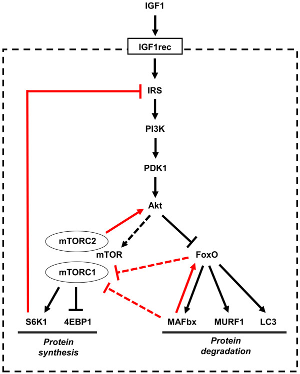
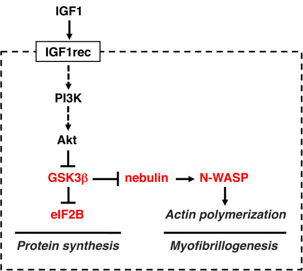
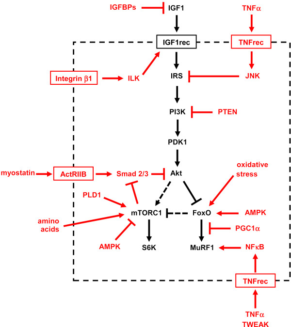
A highly conserved signaling pathway involving insulin-like growth factor 1 (IGF1), and a cascade of intracellular components that mediate its effects, plays a major role in the regulation of skeletal muscle growth. A central component in this cascade is the kinase Akt, also called protein kinase B (PKB), which controls both protein synthesis, via the kinases mammalian target of rapamycin (mTOR) and glycogen synthase kinase 3β (GSK3β), and protein degradation, via the transcription factors of the FoxO family. In this paper, we review the composition and function of this pathway in skeletal muscle fibers, focusing on evidence obtained in vivo by transgenic and knockout models and by muscle transient transfection experiments. Although this pathway is essential for muscle growth during development and regeneration, its role in adult muscle response to mechanical load is less clear. A full understanding of the operation of this pathway could help to design molecularly targeted therapeutics aimed at preventing muscle wasting, which occurs in a variety of pathologic contexts and in the course of aging.
Overview
The claims and statistics of this paper haven’t been checked yet.
Run the check — free, no account needed. It reads the open-access text and checks the sources it cites.
Integrity signals
Registry
Cites
Statistics
Figures
Verification
Claims the paper makes
The claims and statistics of this paper haven’t been checked yet.
Run the check — free, no account needed. It reads the open-access text and checks the sources it cites.
Claims other papers make about this one
Each is checked against this paper’s own text: does it say what the citing paper says it does?
The cited source says what the claim attributes to it.
In the citing paper
“The Akt‐mTOR pathway is central to the regulation of muscle mass because it promotes protein synthesis and inhibits protein degradation (Schiaffino & Mammucari, 2011).”
From Parkin overexpression protects from ageing‐related loss of muscle mass and strength (2019) · cites this paper as Schiaffino & Mammucari, 2011
In the citing paper
“Additionally, it has been discovered that PEs present in cosmetic preparation protect skin from pigmentation and aging163.”
From The Phytochemical, Biological, and Medicinal Attributes of Phytoecdysteroids: An Updated Review (2021) · cites this paper as [163]
Self-check
Percentages, sums and reported differences re-computed from the paper’s own numbers.
The self-check needs the paper’s full text.
Citations
Cites
Phosphorylation and regulation of Akt/PKB by the rictor-mTOR complex
Sarbassov, D. D., Guertin, D. A., Ali, S. M., & Sabatini, D. M. (2005) · Science · doi:10.1126/science.1106148 · Cited by 6,216
AKT/PKB signaling: navigating downstream
Manning, B. D., & Cantley, L. C. (2007) · Cell · doi:10.1016/j.cell.2007.06.009 · Cited by 6,115 · cited 1×
“transcription factors of the FoxO family, and stimulates protein synthesis via the mammalian target of rapamycin (mTOR) and glycogen synthase kinase 3b (GSK3b) [4].”
BackgroundRegulation of skeletal muscle mass in mice by a new TGF-p superfamily member
McPherron, A. C., Lawler, A. M., & Lee, S.-J. (1997) · Nature · doi:10.1038/387083a0 · Cited by 4,013
mTOR: from growth signal integration to cancer, diabetes and ageing
Zoncu, R., Efeyan, A., & Sabatini, D. M. (2011) · Nature Reviews Molecular Cell Biology · doi:10.1038/nrm3025 · Cited by 3,941 · cited 15×
“Deletion of S6K1 is sufficient to reproduce this atrophic phenotype.”
BackgroundInfluentialAMPK Phosphorylation of Raptor Mediates a Metabolic Checkpoint
Gwinn, D. M., Shackelford, D. B., Egan, D. F., Mihaylova, M. M., Mery, A., Vasquez, D. S., Turk, B. E., & Shaw, R. J. (2008) · Molecular Cell · doi:10.1016/j.molcel.2008.03.003 · Cited by 3,849 · cited 13×
“(AMPK), an energy status sensor activated by ATP depletion, inhibits mTORC1 by phosphorylating and inactivating Raptor [23], and by phosphorylating and activating TSC2 [24].”
BackgroundInfluentialIdentification of ubiquitin ligases required for skeletal muscle atrophy.
Bodine, S. C., Latres, E., Baumhueter, S., Lai, V. K.-M., Nunez, L., Clarke, B. A., Poueymirou, W. T., Panaro, F. J., Na, E., Dharmarajan, K., et al. (2001) · Science · doi:10.1126/science.1065874 · Cited by 3,521 · cited 5×
“Their role in muscle-mass regulation is supported by the finding that muscle atrophy induced by denervation is partially prevented in both MuRF1 null and atrogin1/MAFbx null mice [ 92 ].”
BackgroundInfluentialTSC2 is phosphorylated and inhibited by Akt and suppresses mTOR signalling
Inoki, K., Li, Y., Zhu, T., Wu, J., & Guan, K.-L. (2002) · Nature Cell Biology · doi:10.1038/ncb839 · Cited by 3,083 · cited 3×
“Rictor-dependent phosphorylation of Akt at serine 473 is required for Akt-mediated phosphorylation of FoxO but not of TSC2, thus it does not affect activation of S6K [5].”
BackgroundInfluentialSandri, M., Sandri, C., Gilbert, A., Skurk, C., Calabria, E., Picard, A., Walsh, K., Schiaffino, S., Lecker, S. H., & Goldberg, A. L. (2004) · Cell · doi:10.1016/s0092-8674(04)00400-3 · Cited by 3,012 · cited 5×
“Small interfering RNA to FoxO1 and FoxO3 Knockdown of FoxO1 and FoxO3 by RNA interference Prevention of atrogin-1 reporter upregulation induced by starvation [ 91 ]”
BackgroundInfluentialSelf-citationLiu, J.-P., Baker, J. C., Perkins, A. S., Robertson, E. J., & Efstratiadis, A. (1993) · Cell · doi:10.1016/s0092-8674(05)80084-4 · Cited by 2,854
The Rag GTPases Bind Raptor and Mediate Amino Acid Signaling to mTORC1
Sancak, Y., Peterson, T. R., Shaul, Y. D., Lindquist, R. A., Thoreen, C. C., Bar-Peled, L., & Sabatini, D. M. (2008) · Science · doi:10.1126/science.1157535 · Cited by 2,773 · cited 13×
“(AMPK), an energy status sensor activated by ATP depletion, inhibits mTORC1 by phosphorylating and inactivating Raptor [23], and by phosphorylating and activating TSC2 [24].”
BackgroundInfluential
Cited by
Sorted by citations
Items 401–500 of 817
Chardulo, L. A. L., Baldassini, W. A., Curi, R. A., Pereira, G. L., Neto, O. R. M., Dal‐Pai‐Silva, M., Vechetti, I. J., Malheiros, J. M., & Enríquez-Valencia, C. E. (2019) · Animal Biotechnology · doi:10.1080/10495398.2019.1688168 · Cited by 15
“Each gene of the myosin family (MYH) encodes a specific isoform.(9) The MyHC-I, IIa and IIx/d isoforms(10,11) encoded by the MYH7, MYH2 and MYH1 genes, respectively,(12) are the main isoforms found in skeletal muscle of adult cattle.”
BackgroundWilson, R. J., Lyons, S. P., Koves, T. R., Bryson, V. G., Zhang, H., Li, T., Crown, S. B., Ding, J.-D., Grimsrud, P. A., Rosenberg, P. B., et al. (2021) · Molecular Metabolism · doi:10.1016/j.molmet.2021.101429 · Cited by 15
Mogilicherla, K., Ashwini, R., CHATTERJEE, R. N., & Bhattacharya, T. K. (2022) · Frontiers in Genetics · doi:10.3389/fgene.2022.990849 · Cited by 19
“In addition, MYOZ1 and MYOZ2 genes expressed in mice significantly reduced calcineurin gene expression (Frey et al., 2004, 2008; Schulz and Yutzey, 2004).”
BackgroundThe inflammatory response, a mixed blessing for muscle homeostasis and plasticity
Bouredji, Z., Argaw, A., & Frenette, J. (2022) · Frontiers in Physiology · doi:10.3389/fphys.2022.1032450 · Cited by 19
Oh, H., Jin, H., Lee, J., & Lee, B.-Y. (2023) · Cells · doi:10.3390/cells12182257 · Cited by 15
Yan, X., Fu, P., Zhang, Y., Ling, D., Reynolds, L., Hua, W., Wang, Z., Ma, F., Li, B., Yu, J., et al. (2024) · Molecules · doi:10.3390/molecules29030712 · Cited by 15
“…in diabetic muscle atrophy is still unclear, evidence has demonstrated that insulin and insulin-like growth factor-1 (IGF-1) signaling pathways play an important role in the phosphorylation of phosphoinositide 3-kinase (PI3K)/protein kinase B (Akt)-mediated apoptosis and protein synthesis [53,54].”
BackgroundWood, N., Critchlow, A. J., Cheng, C. W., Straw, S., Hendrickse, P. W., Pereira, M. G., Wheatcroft, S. B., Egginton, S., Witte, K. K., Roberts, L. D., et al. (2024) · Circulation Heart Failure · doi:10.1161/circheartfailure.123.011471 · Cited by 17
Extraocular muscle regeneration in zebrafish requires late signals from Insulin-like growth factors
Saera-Vila, A., Louie, K. W., Sha, C., Kelly, R. M., Kish, P. E., & Kahana, A. (2018) · PLoS ONE · doi:10.1371/journal.pone.0192214 · Cited by 17
Pharmacology of doping agents—mechanisms promoting muscle hypertrophy
Parr, M. K., & Laughlin, A. M. M. (2018) · AIMS molecular science · doi:10.3934/molsci.2018.2.131 · Cited by 14
Endocrine Regulation of Epimorphic Regeneration
Easterling, M. R., Engbrecht, K. M., & Crespi, E. J. (2019) · Endocrinology · doi:10.1210/en.2019-00321 · Cited by 15
Growth and Differentiation of Circulating Stem Cells After Extensive Ex Vivo Expansion
Barbon, S., Rajendran, S., Bertalot, T., Piccione, M., Gasparella, M., Parnigotto, P. P., Liddo, R. D., & Conconi, M. T. (2021) · Tissue Engineering and Regenerative Medicine · doi:10.1007/s13770-021-00330-7 · Cited by 15
“Transgenic mice over-expressing IGF-I in skeletal muscles are characterized by hypertrophy and an enhanced muscle regeneration via activation of muscle satellite cells [44].”
BackgroundXiang, R., Ghanipoor-Samami, M., Johns, W. H., Eindorf, T., Rutley, D. L., Kruk, Z. A., Fitzsimmons, C., Thomsen, D. A., Roberts, C. T., Burns, B. M., et al. (2013) · PLoS ONE · doi:10.1371/journal.pone.0053402 · Cited by 15
“Further examples of genespecific genetic and epigenetic regulatory mechanisms that could explain effects of maternal and paternal genomes on fetal muscle phenotype observed in the present study are found in the IGF1AKT/PKB pathway [58].”
BackgroundMuta, Y., Tanaka, T., Hamaguchi, Y., Hamanoue, N., Motonaga, R., Tanabe, M., Nomiyama, T., Nawata, H., & Yanase, T. (2019) · Biochemistry and Biophysics Reports · doi:10.1016/j.bbrep.2019.01.006 · Cited by 15
“Phosphorylation of Akt and Erk, which are upstream regulators of mTORC1 [17,22], was not changed by administration of S42.”
BackgroundGeneration of Novel Genetic Models to Dissect Resistance to Thyroid Hormone Receptor α in Zebrafish
Han, C. R., Holmsen, E., Carrington, B., Bishop, K. S., Zhu, Y. J., Starost, M. F., Meltzer, P. S., Sood, R. B., Liu, P., & Cheng, S.-Y. (2020) · Thyroid · doi:10.1089/thy.2019.0598 · Cited by 15
“We next analyzed the IGF-1 downstream AKT-p70S6 pathway (30,33).”
BackgroundWei, Y., Gong, L.-C., Fu, W., Xu, S., Wang, Z., Zhang, J., Ning, E., Chang, H., Wang, H., & Gao, Y. (2018) · Journal of Cellular Physiology · doi:10.1002/jcp.26751 · Cited by 15
“The IGF1/Akt/mTOR pathway acts as a master regulator of protein synthesis in muscle cell growth and cell cycle progression (Erbay, Park, Nuzzi, Schoenherr, & Chen, 2003; Han, Tong, Zhu, Ma, & Du, 2008; Schiaffino & Mammucari, 2011).”
BackgroundMolecular and metabolic insights of creatine supplementation on resistance training
Bonilla, D. A., & Moreno, Y. (2016) · Revista Colombiana de Química · doi:10.15446/rev.colomb.quim.v44n1.53978 · Cited by 14
Li, J., Zhang, Y., Yu, F., Pan, Y., Zhang, Z., He, Y., Yang, H., & Zhou, P. (2023) · ACS Omega · doi:10.1021/acsomega.3c03513 · Cited by 14
Mechanisms of endurance and resistance exercise in type 2 diabetes mellitus: A Narrative review
Zhao, X., Huang, F., Sun, Y., & Li, L. (2025) · Biochemical and Biophysical Research Communications · doi:10.1016/j.bbrc.2025.151731 · Cited by 14
Molecular Biology of ACL Graft Healing: Early Mechanical Loading Perspective.
Stańczak, M., & Kacprzak, B. (2025) · Orthopedic Reviews · doi:10.52965/001c.140716 · Cited by 16
Exercise-induced inflammatory and metabolic adaptations in ageing: A meta-analytic compendium
Sheldon, R., Gan, A., Tasong, J., Yap, A., Ahmad, M., Júnior, P. R. H., Viswanathan, K., Setti, N. S. M., Jarman, O., Hoque-Uddin, M. S., et al. (2025) · Ageing Research Reviews · doi:10.1016/j.arr.2025.102974 · Cited by 16
Mei, S., & Zhu, H. (2015) · Scientific Reports · doi:10.1038/srep17983 · Cited by 15
Laskin, G. R., Cabrera, A. R., Greene, N. P., Tomko, R. J., Vied, C., & Gordon, B. S. (2024) · American Journal of Physiology-Cell Physiology · doi:10.1152/ajpcell.00444.2023 · Cited by 14
Čurović, I., Rhodes, D., Alexander, J., & Harper, D. J. (2024) · Sports Medicine · doi:10.1007/s40279-024-02039-8 · Cited by 14
“In reverse order of signalling pathways, anabolic mediators such as mTOR1, insulin-like growth factor-1 and ser-ine-threonine protein kinase (Akt) after resistance exercise do seem to suppress catabolic processes in human organisms [66–69].”
BackgroundFish Protein Promotes Skeletal Muscle Hypertrophy via the Akt/mTOR Signaling Pathways
Morisasa, M., Yoshida, E., Fujitani, M., Kimura, K., Uchida, K., Kishida, T., Mori, T., & Goto‐Inoue, N. (2022) · Journal of Nutritional Science and Vitaminology · doi:10.3177/jnsv.68.23 · Cited by 14
Sirt2 positively regulates muscle regeneration after Notexin-induced muscle injury
Lee, E. H., Lee, M., Park, S., & Jeong, K. (2022) · Experimental and Molecular Pathology · doi:10.1016/j.yexmp.2022.104798 · Cited by 15
Dexamethasone and BCAA Failed to Modulate Muscle Mass and mTOR Signaling in GH-Deficient Rats
Nishida, H., Ikegami, A., Kaneko, C., Kakuma, H., Nishi, H., Tanaka, N., Aoyama, M., Usami, M., & Okimura, Y. (2015) · PLoS ONE · doi:10.1371/journal.pone.0128805 · Cited by 14
“Furthermore, we found that, in the SDR muscles, BCAA did not stimulate the phosphorylation of p70S6K or 4E-BP1, both of which are important for enhancing protein synthesis [23].”
BackgroundEffects of alkali supplementation and vitamin D insufficiency on rat skeletal muscle
Ceglia, L., Rivas, D. A., Pojednic, R. M., Price, L. L., Harris, S. S., Smith, D. E., Fielding, R. A., & Dawson‐Hughes, B. (2013) · Endocrine · doi:10.1007/s12020-013-9976-0 · Cited by 14
S1P3receptor influences key physiological properties of fast-twitch extensor digitorum longus muscle
Germinario, E., Bondı̀, M., Cencetti, F., Donati, C., Nocella, M., Colombini, B., Betto, R., Bruni, P., Bagni, M. A., & Danieli‐Betto, D. (2015) · Journal of Applied Physiology · doi:10.1152/japplphysiol.00345.2015 · Cited by 14
Sheng, X., Wang, L., Ni, H., Wang, L., Qi, X., Xing, S., & Guo, Y. (2016) · PLoS ONE · doi:10.1371/journal.pone.0156780 · Cited by 14
“Two major signaling pathways control protein synthesis: the insulin-like growth factor 1–phosphoinositide3-kinase/Akt (protein kinase B)–mammalian target of rapamycin (IGF1—Akt—mTOR) pathway, which acts as a positive regulator [5]; and the myostatin—Smad2/3 pathway, which acts as a negative regulator [6, 7].”
BackgroundOgilvie, H., Cacciani, N., Akkad, H., & Larsson, L. (2016) · Frontiers in Physiology · doi:10.3389/fphys.2016.00417 · Cited by 14
“Band densities were quantified with Image Studio Lite analysis software (LI-COR Biosciences) and normalized to actin.”
BackgroundMethodLong-Term PGC1β Overexpression Leads to Apoptosis, Autophagy and Muscle Wasting
Sopariwala, D. H., Yadav, V., Badin, P.-M., Likhite, N., Sheth, M., Lorca, S., Vila, I. K., Kim, E. R., Tong, Q., Song, M. S., et al. (2017) · Scientific Reports · doi:10.1038/s41598-017-10238-9 · Cited by 14
C-reactive protein is associated with the development of tongue squamous cell carcinoma
Du, J., Hu, W., Yang, C., Wang, Y., Wang, X., & Yang, P.-S. (2018) · Acta Biochimica et Biophysica Sinica · doi:10.1093/abbs/gmy004 · Cited by 14
“Akt serves as an essential regulator of both protein synthesis and degradation through modulating mTOR pathway activity [32].”
BackgroundEscalante-Rojas, M., Peña, E., Hernández, C., Llera‐Herrera, R., & Garcı́a-Gasca, A. (2018) · Aquaculture Research · doi:10.1111/are.13628 · Cited by 16
Crosstalk between Mitochondrial Ca2+ Uptake and Autophagy in Skeletal Muscle
Gherardi, G., Marco, G. D., Rizzuto, R., & Mammucari, C. (2019) · Oxidative Medicine and Cellular Longevity · doi:10.1155/2019/1845321 · Cited by 15
“This is associated with increased activity of well-known hypertrophic signaling routes, like the Akt pathway, previously reported to play an inhibitory role on autophagy [3] and to the reduced activity of the protein synthesis inhibitor and autophagy activator, GSK3 β [20, 21].”
Self-citationThe Role of Natural Products in the Improvement of Cancer-Associated Cachexia
Han, Y., Kim, H. I., & Park, J. (2023) · International Journal of Molecular Sciences · doi:10.3390/ijms24108772 · Cited by 13
Rodrigues, C., Murata, G. M., Gerlinger‐Romero, F., Nachbar, R. T., Marzuca-Nassr, G. N., Gorjão, R., Vitzel, K. F., Hirabara, S. M., Pithon‐Curi, T. C., & Curi, R. (2023) · Nutrients · doi:10.3390/nu15224711 · Cited by 13
Insights into Muscle Degeneration from Heritable Inclusion Body Myopathies
Krause, S. (2015) · Frontiers in Aging Neuroscience · doi:10.3389/fnagi.2015.00013 · Cited by 13
“Moreover, in age-related muscle deterioration a critical role for Akt has also been demonstrated (reviewed by Glass, 2003; Schiaffino and Mammucari, 2011).”
Novel Metabolic Disorders in Skeletal Muscle of Lipodystrophic Bscl2/Seipin Deficient Mice
Xu, W.-Q., Zhou, H., Xuan, H.-Z., Saha, P., Wang, G.-X., & Chen, W.-Q. (2018) · Molecular and Cellular Endocrinology · doi:10.1016/j.mce.2018.12.001 · Cited by 13
“Inactivation of GSK-3β by phosphorylation at Ser9 is known to result in eIF2B activation and skeletal muscle hypertrophy (Schiaffino and Mammucari, 2011), suggesting a potential mechanism underlying muscle overgrowth in BSCL2 lipodystrophy.”
BackgroundDo neurogenic and cancer-induced muscle atrophy follow common or divergent paths?
Bouché, M., Lozanoska‐Ochser, B., Proietti, D., & Madaro, L. (2018) · European Journal of Translational Myology · doi:10.4081/ejtm.2018.7931 · Cited by 14
“- 396 The autophagy machinery in muscle atrophy, like the proteasome system, is controlled by the FoxO protein family, predominantly by the FoxO3 member.(6,68,70,71) FoxO3 activation increases the expression of many autophagy-related genes in myotubes, and its inactivation is incompletely compensated by the other fact…”
BackgroundEffect of YAP on an Immortalized Periodontal Ligament Stem Cell Line
Chen, X., Wang, Q., Gu, K., Li, A., Fu, X., Wang, Y., Gu, W., & Wen, Y. (2019) · Stem Cells International · doi:10.1155/2019/6804036 · Cited by 13
“But the total ERK and AKT remained unchanged, which means YAP-TEAD complex could affect the phosphorylation of ERK and AKT.”
BackgroundInfluentialModerators of skeletal muscle maintenance are compromised in sarcopenic obese mice
Brown, L. A., Perry, R. A., Haynie, W. S., Lee, D. E., Rosa‐Caldwell, M. E., Brown, J. L., Greene, N. P., Wolchok, J. C., & Washington, T. A. (2020) · Mechanisms of Ageing and Development · doi:10.1016/j.mad.2020.111404 · Cited by 13
Gene expression analysis of the liver and skeletal muscle of psyllium-treated mice
Togawa, N., Takahashi, R., Hirai, S., Fukushima, T., & Egashira, Y. (2012) · British Journal Of Nutrition · doi:10.1017/s0007114512001250 · Cited by 13
Kamanga‐Sollo, E., Thornton, K. J., White, M. E., & Dayton, W. R. (2016) · Domestic Animal Endocrinology · doi:10.1016/j.domaniend.2016.09.002 · Cited by 13
Aldiss, P., Lewis, J. E., Lupini, I., Boocock, D. J., Miles, A. K., Ebling, F. J. P., Budge, H., & Symonds, M. E. (2019) · bioRxiv (Cold Spring Harbor Laboratory) · doi:10.1101/649061 · Cited by 13
“Ribosome biogenesis is regulated by growth factors, and IGF1 specifically regulates muscle hypertrophy through both the PI3K-AKT-mTOR and MAPK signalling pathways, with the latter playing a key role in regulating ribosomal biogenesis [49, 50].”
BackgroundYakabe, M., Hosoi, T., Sasakawa, H., Akishita, M., & Ogawa, S. (2022) · BMC Complementary Medicine and Therapies · doi:10.1186/s12906-022-03812-w · Cited by 12
“Under overnutrient conditions, p70 inhibits insulin receptor substrate-1 (IRS-1) by phosphorylation at multiple sites, negatively regulating insulin signaling [44, 45].”
BackgroundNutritional approaches targeting mitochondria for the prevention of sarcopenia
Hahm, J., Nirmala, F. S., Ha, T. Y., & Ahn, J. (2023) · Nutrition Reviews · doi:10.1093/nutrit/nuad084 · Cited by 13
Kumar, S., Kushawaha, B., Mohanty, A., Kumari, A., Kumar, A., Beniwal, R., Kumar, P. K., Athar, M., Rao, D. K., & Rao, H. B. D. P. (2024) · Biochimica et Biophysica Acta (BBA) - Molecular Basis of Disease · doi:10.1016/j.bbadis.2024.167235 · Cited by 12
Sosnicki, A. A., Gonzalez, J. M., Fields, B., & Knap, P. W. (2024) · Meat Science · doi:10.1016/j.meatsci.2024.109676 · Cited by 12
A mechanistic basis of fast myofiber vulnerability to neuromuscular diseases
Santos, M. D., Bezprozvannaya, S., McAnally, J., Cai, C., Liu, N., & Olson, E. N. (2025) · Cell Reports · doi:10.1016/j.celrep.2025.115959 · Cited by 12
“7 For example, during muscle atrophy, the insulin-like growth factor 1 (IGF-1)/AKT signaling pathway is suppressed, 8 while the FoxO family of transcription factors (FoxO1, FoxO3, and FoxO4) is activated.”
Ringseis, R., Keller, J., Lukas, I., Spielmann, J., Most, E., Couturier, A., König, B., Hirche, F., Stangl, G. I., Wen, G., et al. (2012) · Biochimica et Biophysica Acta (BBA) - General Subjects · doi:10.1016/j.bbagen.2012.09.024 · Cited by 12
FMAj: a tool for high content analysis of muscle dynamics in Drosophila metamorphosis
Kuleesha, Y., Puah, W. C., Lin, F., & Wasser, M. (2014) · BMC Bioinformatics · doi:10.1186/1471-2105-15-s16-s6 · Cited by 12
“Most of our knowledge about muscle size control and atrophy is derived from C2C12 myoblast cell culture and mouse transgenic models [6].”
BackgroundFerrandi, P. J., Khan, M. M., Paez, H. G., Pitzer, C. R., Alway, S. E., & Mohamed, J. S. (2020) · Genes · doi:10.3390/genes11070726 · Cited by 12
Muscle Regeneration and RNA: New Perspectives for Ancient Molecules
Buonaiuto, G., Desideri, F., Taliani, V., & Ballarino, M. (2021) · Cells · doi:10.3390/cells10102512 · Cited by 11
Effect of irradiation on Akt signaling in atrophying skeletal muscle
Fix, D. K., Hardee, J. P., Bateman, T. A., & Carson, J. A. (2016) · Journal of Applied Physiology · doi:10.1152/japplphysiol.00218.2016 · Cited by 12
“In addition to its effects on protein synthesis, Akt can also inhibit catabolic processes through the phosphorylation and inhibition of the forkhead box O (FOXO) transcription factor, which regulates the expression of muscle-specific E3 ligases atrogin-1 and MuRF-1 (36, 43, 44).”
BackgroundInfluentialEffects of Vitamin C on Cytotherapy-Mediated Muscle Regeneration
Kim, A.-Y., Lee, E. M., Lee, E. H., Min, C., Kang, K., Park, J., Hong, I., Ishigami, A., Tremblay, J. P., & Jeong, K. (2013) · Cell Transplantation · doi:10.3727/096368912x657846 · Cited by 12
Bou, T., Ding, W., Ren, X., Liu, H., Gong, W., Jia, Z., Zhang, X., Dugarjaviin, M., & Bai, D. (2023) · Equine Veterinary Journal · doi:10.1111/evj.13968 · Cited by 11
Kamal, K. Y., Othman, M., Kim, J., & Lawler, J. M. (2024) · npj Microgravity · doi:10.1038/s41526-023-00320-0 · Cited by 14
Freeman, P., Bellomo, G., Ireland, L., Abudula, M., Luckett, T., Oberst, M. D., Stafferton, R., Ghaneh, P., Halloran, C. M., Schmid, M. C., et al. (2024) · Frontiers in Immunology · doi:10.3389/fimmu.2024.1382538 · Cited by 11
Bareja, A., Patel, S. R., Hodgkinson, C. P., Payne, A. J., & Dzau, V. J. (2018) · Cellular Signalling · doi:10.1016/j.cellsig.2018.02.013 · Cited by 11
“The Insulin-like Growth Factor 1 Receptor (IGF1R) plays an important role in a range of biological processes, including proliferation (1), lifespan (2), and muscle growth (3).”
BackgroundKousari, A., Moser, C., Olefsky, M., Brown, T. T., Currier, J. S., McComsey, G. A., Scherzinger, A., Stein, J. H., Lake, J. E., & Erlandson, K. M. (2021) · JAIDS Journal of Acquired Immune Deficiency Syndromes · doi:10.1097/qai.0000000000002776 · Cited by 14
Aleksova, A., Janjusevic, M., Gagno, G., Pierri, A., Padoan, L., Fluca, A. L., Carriere, C., Beltrami, A. P., & Sinagra, G. (2022) · Frontiers in Physiology · doi:10.3389/fphys.2021.794641 · Cited by 11
“…effect of exercise on signaling pathways, it is plausible that training could regulate cell growth by supporting protein synthesis and downregulating their catabolism through inhibition of the E3 ubiquitin ligases MuRF1 and MAFbx (Schiaffino and Mammucari, 2011; Bodine and Baehr, 2014).”
BackgroundInfluentialMendes, S., Leal, D. V., Baker, L. A., Ferreira, A., Smith, A. C., & Viana, J. L. (2023) · International Journal of Molecular Sciences · doi:10.3390/ijms24076017 · Cited by 12
Yu, B., Cai, Z., Liu, J., Zhang, T., Feng, X., Wang, C., Li, J., Gu, Y., & Zhang, J. (2024) · International Journal of Biological Macromolecules · doi:10.1016/j.ijbiomac.2024.130737 · Cited by 11
Sesamol Alleviates Sarcopenia via Activating AKT/mTOR/FoxO1 Signal Pathway in Aged Obese Mice
Yang, J., Wang, Z., Xie, Y., Tang, Y., Fu, Y., Xu, Z., Chen, J., & Qin, H. (2024) · Plant Foods for Human Nutrition · doi:10.1007/s11130-024-01199-2 · Cited by 11
“The synthesis and degradation of myoproteins are regulated by various signal pathways, with the AKT signal pathway plays a key role [24, 25].”
Relation between serum myokines and phase II cardiac rehabilitation
Han, D., Hsiao, M., Wang, T., Chen, S., & Yang, W. (2017) · Medicine · doi:10.1097/md.0000000000006579 · Cited by 12
“It affects the development of several systems, the musculoskeletal being one of the most important.([5]) IGF-1 and IL-6 share common pathways in physiologic regulation.”
BackgroundLi, X., Fan, L., Zhu, M., Jiang, H., Bai, W., & Kang, J. (2019) · Climacteric · doi:10.1080/13697137.2019.1660639 · Cited by 11
Tanabe, J., Ogura, Y., Kosaki, K., Nagai, Y., Sugaya, T., Ohata, K., Watanabe, S., Ichikawa, D., Inoue, K., Hoshino, S., et al. (2020) · Journal of Diabetes Research · doi:10.1155/2020/7614035 · Cited by 11
“As a critical downstream effector of the insulin/IGF-1 pathway, phosphorylation of Akt leads to activation of protein synthesis via phosphorylation of both mTOR and S6K or inhibition of protein degradation via FoxO1 phosphorylation, which results in downregulated expression levels of the E3 ligases Atrogin-1 and MuRF-…”
BackgroundInfluentialOh, H., Jin, H., Kim, B., Lee, O., & Lee, B.-Y. (2022) · Cells · doi:10.3390/cells11203245 · Cited by 12
Ding, Y., Li, J., Liu, Z., Liu, H., Li, H., & Li, Z. (2017) · Scientific Reports · doi:10.1038/srep43949 · Cited by 11
Sorrentino, S., Barbiera, A., Proietti, G., Sica, G., Adamo, S., & Scicchitano, B. M. (2019) · International Journal of Molecular Sciences · doi:10.3390/ijms20174188 · Cited by 11
O’Bryan, S. M., Lavin, K. M., Graham, Z., Drummer, D. J., Tuggle, S. C., Keuren‐Jensen, K. R. V., Reiman, R. A., Alsop, E. B., Kadakia, M., Craig, M. P., et al. (2024) · Journal of Applied Physiology · doi:10.1152/japplphysiol.00680.2023 · Cited by 11
Zeng, X., Zhou, X., Jiang, W., Wu, P., Yang, L., Ma, Y.-B., Tang, L., Li, S.-W., Kuang, S., & Feng, L. (2024) · Food Bioscience · doi:10.1016/j.fbio.2024.105537 · Cited by 10
Tian, Y., Lucena-Cacace, A., Tani, K., Elvandari, A. P., Osorio, R. S. A., Narita, M., Matsumura, Y., Paixao, I. C., Miyoshi, Y., Inagaki, A., et al. (2025) · Nature Communications · doi:10.1038/s41467-025-60934-8 · Cited by 10
Wang, N., Zheng, A., Yan, Y., He, T., Wu, X., Xian, M., Luo, J., Li, C., Wei, J., Wang, Y., et al. (2025) · Journal of Advanced Research · doi:10.1016/j.jare.2025.07.004 · Cited by 11
Crossland, H., Timmons, J. A., & Atherton, P. J. (2017) · The FASEB Journal · doi:10.1096/fj.201700329r · Cited by 11
“…biogenesis in human primary myocyte size regulation in vitro , 2 ) to determine whether ribosomal biogenesis is required for skeletal muscle hypertrophy in response to an established in vivo trophic signal (IGF-1) (19 – 21), and 3 ) to identify the signaling mediators driving these changes.”
BackgroundLee, M., Choi, J., Choi, Y., & Nam, T. (2018) · Marine Drugs · doi:10.3390/md16090328 · Cited by 10
“IGF-I is a circulating growth factor produced locally in many tissues, including skeletal muscle [13].”
BackgroundCoelho, P. D. O., Guarnier, F. A., Figueiredo, L. B., Zaramela, L. S., Pacini, E. S. A., Godinho, R. O., & Gomes, M. D. (2019) · Archives of Biochemistry and Biophysics · doi:10.1016/j.abb.2019.01.009 · Cited by 10
Rivas, D. A., Rice, N. P., Ezzyat, Y., McDonald, D., Cooper, B. E., & Fielding, R. A. (2019) · American Journal of Physiology-Cell Physiology · doi:10.1152/ajpcell.00455.2018 · Cited by 10
“The cellular mechanisms associated with contractioninduced protein synthesis include the activation of Akt/mammalian target of rapamycin (mTOR) signaling and changes in expression of noncoding RNA like microRNA (miRNA) and can occur independent of IGF-1 receptor signaling (39).”
BackgroundMethodFaraldi, M., Sansoni, V., Perego, S., Gomarasca, M., Gerosa, L., Ponzetti, M., Rucci, N., Banfi, G., & Lombardi, G. (2022) · Frontiers in Molecular Biosciences · doi:10.3389/fmolb.2022.915080 · Cited by 10
“The PI3K pathway (Schiaffino and Mammucari, 2011), angiogenesis (Bloor, 2005), p53 pathway (Feng and Levine, 2010), and inflammatory pathways (inflammation mediated by chemokines and cytokine signaling pathway; toll receptor signaling pathway) (Gomarasca et al., 2020) were enriched in t-miRNA and…”
BackgroundAlterations in insulin‐like growth factor system in spinal muscular atrophy
Kaymaz, A. Y., Bal, S. K., Bora, G., Talim, B., Özön, A., Alikaşifoğlu, A., Topaloğlu, H., & Erdem‐Yurter, H. (2022) · Muscle & Nerve · doi:10.1002/mus.27715 · Cited by 10
Effects of Turmeric Extract on Age-Related Skeletal Muscle Atrophy in Senescence-Accelerated Mice
Lyu, W., Kousaka, M., Jia, H., & Kato, H. (2023) · Life · doi:10.3390/life13040941 · Cited by 11
Kong, W. G. B. B.-W. (2014) · Biochemistry & Physiology Open Access · doi:10.4172/2168-9652.1000125 · Cited by 12
“The mammalian target of rapamycin (mTOR) in conjunction with Akt (also known as protein kinase B) are required via the phosphatidyl inositol 3- kinase (PI3K)/Akt/mTOR pathway for skeletal muscle cell development [4-7].”
BackgroundForesto, C. S., Paula-Gomes, S., Silveira, W. A., Graça, F. A., Kettelhut, Í. D. C., Gonçalves, D. A. P., & Mattiello‐Sverzut, A. C. (2016) · Journal of Applied Physiology · doi:10.1152/japplphysiol.00687.2015 · Cited by 10
Oh, S.-L., Lee, S.-R., & Kim, J. (2017) · Physical Activity and Nutrition · doi:10.20463/jenb.2017.0028 · Cited by 10
“When IGF1 binds to its receptor, insulin receptor substrate-1 (IRS-1) is activated, followed by PI3K activation through conversion of intramembranous phosphoinositide-(4,5)-biphosphate to phosphoinositide-(3,4,5)-triphosphate (PIP3)4.”
BackgroundGenes and response to aerobic training
Cagnin, S., Chemello, F., & Ahmetov, I. I. (2019) · Elsevier eBooks · doi:10.1016/b978-0-12-816193-7.00008-7 · Cited by 10
A novel treatment strategy targeting cellular pathways with natural products to alleviate sarcopenia
Lee, D. H., Lee, H. J., Yang, G., Kim, D. Y., Kim, J. U., Yook, T. H., Lee, J. H., & Kim, H. J. (2024) · Phytotherapy Research · doi:10.1002/ptr.8301 · Cited by 13
“This activation leads to the stimulation of ribosomal protein S6 and eukaryotic transcription initiation factor eIF-4E, promoting protein synthesis (Dennis et al., 2012; Schiaffino & Mammucari, 2011).”
MicroRNAs and their Modulatory Effect on the Hallmarks of Osteosarcopenia
Silva, W. J., Cruz, A., & Duque, G. (2024) · Current Osteoporosis Reports · doi:10.1007/s11914-024-00880-4 · Cited by 9
“Its potential as an inducer of atrophy is significant; even ectopic expression in healthy tissues is sufficient to induce muscle atrophy [92, 93].”
Temiz, K., Gül, A., & Göv, E. (2025) · NeuroMolecular Medicine · doi:10.1007/s12017-025-08847-z · Cited by 9
“Pathways, such as tyrosine kinase signaling, which regulates muscle repair via IGF-1 (Schiaffino & Mammucari, 2011), and RNA degradation which is linked to TDP-43-me-diated RNA processing defects (Buratti et al., 2010), further highlight key mechanisms driving muscle atrophy and dysregulation in ALS.”
Cytokine networks in immune-mediated muscle regeneration
Dziki, J. L., Velayutham, M., Hussey, G. S., & Turnquist, H. R. (2018) · Journal of Immunology and Regenerative Medicine · doi:10.1016/j.regen.2018.03.001 · Cited by 9
González-Franquesa, A., Peijs, L., Cervone, D. T., Koçana, C., Zierath, J. R., & Deshmukh, A. S. (2021) · Proteomes · doi:10.3390/proteomes9030037 · Cited by 9
“Insulin likely promotes the release of Igfbp proteins as part of a negative feedback loop to regulate Igfsignaling, which partly overlaps with cellular targets of insulin receptor activation [55,56].”
BackgroundDietary Alaska Pollack Protein Induces Acute and Sustainable Skeletal Muscle Hypertrophy in Rats
Uchida, K., Fujitani, M., Mizushige, T., Kawabata, F., Hayamizu, K., Uozumi, K., Hara, Y., Sawai, M., Uehigashi, R., Okada, S., et al. (2022) · Nutrients · doi:10.3390/nu14030547 · Cited by 9
Essex, A. L., Deosthale, P. J., Huot, J. R., Davis, H. M., Momeni, N., Bonetto, A., & Plotkin, L. I. (2022) · Biology of Sex Differences · doi:10.1186/s13293-022-00465-9 · Cited by 9
“On the other hand, the ratio of phosphorylated ERK1/2 (pT185/pY187) and Akt (S473) to total protein levels for these kinases known to be involved in skeletal muscle mass and homeostasis [29, 30], was, respectively, 54% higher and 33% lower in female OtmiR21Δ mice compared to miR21fl/fl controls (Fig.”
BackgroundTakeda, T., Takeuchi, M., Saitoh, M., & Takeda, S. (2018) · The Tohoku Journal of Experimental Medicine · doi:10.1620/tjem.245.107 · Cited by 9
“Since some kinases, such as PI3K and Akt, are considered to be closely associated with skeletal muscle growth (Luo et al. 2006; Glass 2010; Schiaffino and Mammucari 2011; Briata et al. 2012), the effects of nintedanib may inhibit further pulmonary fibrosis and improve muscular functions that regulate lung function tha…”
BackgroundAlpha-lipoic acid preserves skeletal muscle mass in type 2 diabetic OLETF rats
Hong, O., Son, J. W., Kwon, H., Lee, S., Kim, S., & Yoo, S. J. (2018) · Nutrition & Metabolism · doi:10.1186/s12986-018-0302-y · Cited by 9
“may be required to maintain muscle homeostasis and could play a role in muscle plasticity in response to both hypertrophic and atrophic stimuli [4, 5, 22, 23].”
BackgroundWang, W., Wu, B., Chen, G., Zhou, Y., Li, Z. H., Zhang, J., Ding, Y., Zhang, P., & Wang, J. Q. (2018) · Neoplasma · doi:10.4149/neo_2018_170720n491 · Cited by 9
Targeting myomiRs by tocotrienol-rich fraction to promote myoblast differentiation
Razak, A. M., Khor, S. C., Jaafar, F., Karim, N. A., & Makpol, S. (2018) · Genes & Nutrition · doi:10.1186/s12263-018-0618-2 · Cited by 9
Reitzner, S. M., Hengevoß, J., Isenmann, E., & Diel, P. R. (2019) · The Journal of Steroid Biochemistry and Molecular Biology · doi:10.1016/j.jsbmb.2019.03.023 · Cited by 10
Lorza‐Gil, E., García‐Arévalo, M., Favero‐Santos, B. C., Gomes‐Marcondes, M. C. C., & Oliveira, H. C. F. (2019) · Journal of Translational Medicine · doi:10.1186/s12967-019-2045-6 · Cited by 10
Casanova-Vallve, N., Constantin‐Teodosiu, D., Filer, A. D., Hardy, R., Greenhaff, P. L., & Chapman, V. (2020) · PLoS ONE · doi:10.1371/journal.pone.0235702 · Cited by 9
“Elevated levels of inflammatory cytokines have been implicated in aberrant phosphatidylinositol-3-kinase/Akt1 (PI3K/Akt1) signalling [8, 9], leading to net negative muscle protein balance and altered muscle metabolic flux [6, 7].”
BackgroundSandoval, C., Askelson, K., Lambo, C. A., Dunlap, K. A., & Satterfield, M. C. (2020) · Domestic Animal Endocrinology · doi:10.1016/j.domaniend.2020.106556 · Cited by 9
Metadata
- Authors
- Schiaffino, S., & Mammucari, C.
- Affiliations
- Veneto Institute of Molecular Medicine (IT)
- University of Padua (IT)
- Corresponding author
- Schiaffino, Stefano
- Venue
- Skeletal Muscle
- Published
- 2011
- Volume
- 1
- Issue
- 1
- Pages
- 4
- ISSN
- 2044-5040 (ISSN-L)
- PMC
- PMC3143906
- OpenAlex
- W2150059080
- Cited by
- 816
- References
- 150
- Citations by year
- 2026: 42 · 2025: 70 · 2024: 68 · 2023: 63 · 2022: 55 · 2021: 59 · 2020: 56 · 2019: 69 · 2018: 68 · 2017: 57 · 2016: 42 · 2015: 46 · 2014: 48 · 2013: 44 · 2012: 23
- FWCI
- 15.73
- Type
- journal article
- Language
- en
- Open access
- gold
- doi.org
- Licence
- cc-by
- Locations
- Skeletal Muscle (publishedVersion, cc-by)
- Europe PMC (PubMed Central) (publishedVersion, cc-by)
- PubMed Central (publishedVersion, cc-by)
- PubMed (publishedVersion)
- Padua Research Archive (University of Padova) (submittedVersion, cc-by)
- DOAJ (DOAJ: Directory of Open Access Journals) (submittedVersion)
- Funders
- European Commission
- Università degli Studi di Padova
- Topics
- Life Sciences › Biochemistry, Genetics and Molecular Biology › Molecular Biology › Muscle Physiology and Disorders
- Health Sciences › Nursing › Nutrition and Dietetics › Muscle metabolism and nutrition
- Life Sciences › Biochemistry, Genetics and Molecular Biology › Molecular Biology › Metabolism, Diabetes, and Cancer
- Keywords
- skeletal muscle growth, IGF-1 signaling, mTOR, GSK3β, FOXO transcription factors, muscle regeneration, myogenesis, mechanical loading, muscle atrophy, muscle protein synthesis, protein degradation
About these checks
Registry, retraction, Cites and statistics results are computed, not judged, with one exception: a printed reference that no index lists is read once by a model before it counts as a citation error. Claims are extracted by a model and kept only when their quote appears verbatim in the text; each cited source is then checked, with a model’s help, for whether it says what the claim relies on. Those verdicts can be wrong — read the claim’s quote and the cited source before relying on one.
Statistics and arithmetic are recomputed, not judged.
Confidence bands are shown only for fields calibrated on our annotated test set. None is calibrated yet, so no band is shown.
What counts as a claim ›How we measure confidence ›How we check this ›
Are you an author, or do you see a problem with this page? Request a review
