

The DOI matches its Crossref record; 148 of the 150 references it cites were found in a registry or an index.
Regulation of skeletal muscle growth by the IGF1-Akt/PKB pathway: insights from genetic models
Open accessAbstract
A highly conserved signaling pathway involving insulin-like growth factor 1 (IGF1), and a cascade of intracellular components that mediate its effects, plays a major role in the regulation of skeletal muscle growth. A central component in this cascade is the kinase Akt, also called protein kinase B (PKB), which controls both protein synthesis, via the kinases mammalian target of rapamycin (mTOR) and glycogen synthase kinase 3β (GSK3β), and protein degradation, via the transcription factors of the FoxO family. In this paper, we review the composition and function of this pathway in skeletal muscle fibers, focusing on evidence obtained in vivo by transgenic and knockout models and by muscle transient transfection experiments. Although this pathway is essential for muscle growth during development and regeneration, its role in adult muscle response to mechanical load is less clear. A full understanding of the operation of this pathway could help to design molecularly targeted therapeutics aimed at preventing muscle wasting, which occurs in a variety of pathologic contexts and in the course of aging.
Overview
The claims and statistics of this paper haven’t been checked yet.
Run the check — free, no account needed. It reads the open-access text and checks the sources it cites.
Integrity signals
Registry
Cites
Statistics
Figures
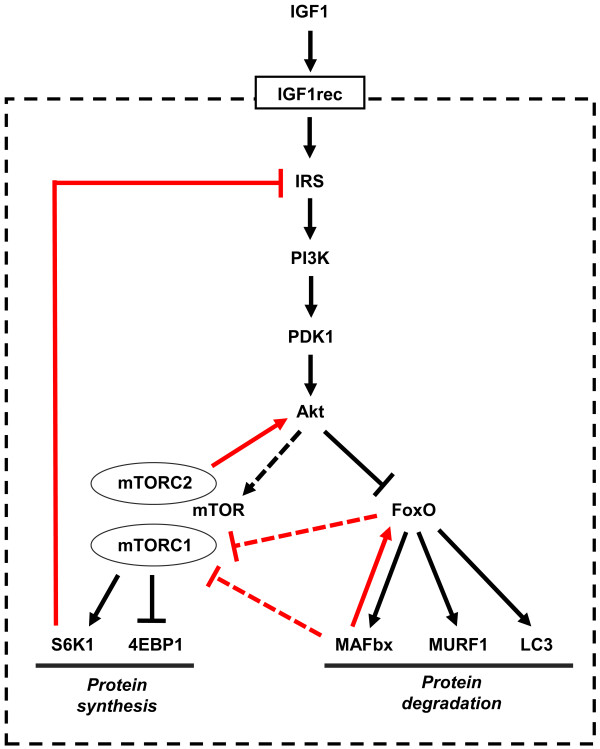
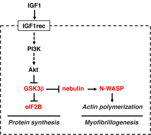
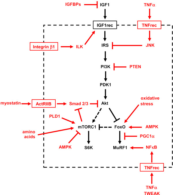
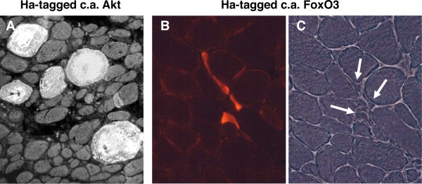
Verification
Claims the paper makes
The claims and statistics of this paper haven’t been checked yet.
Run the check — free, no account needed. It reads the open-access text and checks the sources it cites.
Claims other papers make about this one
Each is checked against this paper’s own text: does it say what the citing paper says it does?
The cited source says what the claim attributes to it.
In the citing paper
“The Akt‐mTOR pathway is central to the regulation of muscle mass because it promotes protein synthesis and inhibits protein degradation (Schiaffino & Mammucari, 2011).”
From Parkin overexpression protects from ageing‐related loss of muscle mass and strength (2019) · cites this paper as Schiaffino & Mammucari, 2011
In the citing paper
“Additionally, it has been discovered that PEs present in cosmetic preparation protect skin from pigmentation and aging163.”
From The Phytochemical, Biological, and Medicinal Attributes of Phytoecdysteroids: An Updated Review (2021) · cites this paper as [163]
Self-check
Percentages, sums and reported differences re-computed from the paper’s own numbers.
The self-check needs the paper’s full text.
Citations
Cites
Phosphorylation and regulation of Akt/PKB by the rictor-mTOR complex
Sarbassov, D. D., Guertin, D. A., Ali, S. M., & Sabatini, D. M. (2005) · Science · doi:10.1126/science.1106148 · Cited by 6,216
AKT/PKB signaling: navigating downstream
Manning, B. D., & Cantley, L. C. (2007) · Cell · doi:10.1016/j.cell.2007.06.009 · Cited by 6,115 · cited 1×
“transcription factors of the FoxO family, and stimulates protein synthesis via the mammalian target of rapamycin (mTOR) and glycogen synthase kinase 3b (GSK3b) [4].”
BackgroundRegulation of skeletal muscle mass in mice by a new TGF-p superfamily member
McPherron, A. C., Lawler, A. M., & Lee, S.-J. (1997) · Nature · doi:10.1038/387083a0 · Cited by 4,013
mTOR: from growth signal integration to cancer, diabetes and ageing
Zoncu, R., Efeyan, A., & Sabatini, D. M. (2011) · Nature Reviews Molecular Cell Biology · doi:10.1038/nrm3025 · Cited by 3,941 · cited 15×
“Deletion of S6K1 is sufficient to reproduce this atrophic phenotype.”
BackgroundInfluentialAMPK Phosphorylation of Raptor Mediates a Metabolic Checkpoint
Gwinn, D. M., Shackelford, D. B., Egan, D. F., Mihaylova, M. M., Mery, A., Vasquez, D. S., Turk, B. E., & Shaw, R. J. (2008) · Molecular Cell · doi:10.1016/j.molcel.2008.03.003 · Cited by 3,849 · cited 13×
“(AMPK), an energy status sensor activated by ATP depletion, inhibits mTORC1 by phosphorylating and inactivating Raptor [23], and by phosphorylating and activating TSC2 [24].”
BackgroundInfluentialIdentification of ubiquitin ligases required for skeletal muscle atrophy.
Bodine, S. C., Latres, E., Baumhueter, S., Lai, V. K.-M., Nunez, L., Clarke, B. A., Poueymirou, W. T., Panaro, F. J., Na, E., Dharmarajan, K., et al. (2001) · Science · doi:10.1126/science.1065874 · Cited by 3,521 · cited 5×
“Their role in muscle-mass regulation is supported by the finding that muscle atrophy induced by denervation is partially prevented in both MuRF1 null and atrogin1/MAFbx null mice [ 92 ].”
BackgroundInfluentialTSC2 is phosphorylated and inhibited by Akt and suppresses mTOR signalling
Inoki, K., Li, Y., Zhu, T., Wu, J., & Guan, K.-L. (2002) · Nature Cell Biology · doi:10.1038/ncb839 · Cited by 3,083 · cited 3×
“Rictor-dependent phosphorylation of Akt at serine 473 is required for Akt-mediated phosphorylation of FoxO but not of TSC2, thus it does not affect activation of S6K [5].”
BackgroundInfluentialSandri, M., Sandri, C., Gilbert, A., Skurk, C., Calabria, E., Picard, A., Walsh, K., Schiaffino, S., Lecker, S. H., & Goldberg, A. L. (2004) · Cell · doi:10.1016/s0092-8674(04)00400-3 · Cited by 3,012 · cited 5×
“Small interfering RNA to FoxO1 and FoxO3 Knockdown of FoxO1 and FoxO3 by RNA interference Prevention of atrogin-1 reporter upregulation induced by starvation [ 91 ]”
BackgroundInfluentialSelf-citationLiu, J.-P., Baker, J. C., Perkins, A. S., Robertson, E. J., & Efstratiadis, A. (1993) · Cell · doi:10.1016/s0092-8674(05)80084-4 · Cited by 2,854
The Rag GTPases Bind Raptor and Mediate Amino Acid Signaling to mTORC1
Sancak, Y., Peterson, T. R., Shaul, Y. D., Lindquist, R. A., Thoreen, C. C., Bar-Peled, L., & Sabatini, D. M. (2008) · Science · doi:10.1126/science.1157535 · Cited by 2,773 · cited 13×
“(AMPK), an energy status sensor activated by ATP depletion, inhibits mTORC1 by phosphorylating and inactivating Raptor [23], and by phosphorylating and activating TSC2 [24].”
BackgroundInfluential
Cited by
Sorted by citations
Items 301–400 of 817
Akkad, H., Corpeño, R., & Larsson, L. (2014) · PLoS ONE · doi:10.1371/journal.pone.0092622 · Cited by 25
“The highly conserved IGF-1/PI3K/Akt pathway plays an essential role in muscle protein synthesis [31].”
BackgroundWadosky, K. M., Rodriguez, J., Hite, R. L., Min, J.-N., Walton, B. L., & Willis, M. S. (2014) · American Journal of Physiology-Endocrinology and Metabolism · doi:10.1152/ajpendo.00326.2013 · Cited by 25
The role of sirtuins in the regulation of metabolic homeostasis in skeletal muscle
Ryall, J. G. (2012) · Current Opinion in Clinical Nutrition & Metabolic Care · doi:10.1097/mco.0b013e3283590914 · Cited by 24
Paoli, A., Cerullo, G., Bianco, A., Neri, M., Gennaro, F., Charrier, D., & Moro, T. (2024) · Journal of Human Kinetics · doi:10.5114/jhk/186660 · Cited by 29
Bucarey, J. L., Paules, E. M., Trujillo‐Gonzalez, I., & Espinosa, A. (2024) · Antioxidants · doi:10.3390/antiox13111363 · Cited by 27
What do mouse models of muscular dystrophy tell us about the DAPC and its components?
Whitmore, C. L., & Morgan, J. E. (2014) · International Journal of Experimental Pathology · doi:10.1111/iep.12095 · Cited by 24
“The IGF1-Akt signalling pathway is involved in regulation of skeletal muscle growth (reviewed in Schiaffino and Mammucari (2011)), and it is initiated by laminin binding to a-dystroglycan in the extracellular space, so a loss of this binding, seen in a number of these models, reduces Akt signalling…”
BackgroundMartínez‐García, M. Á., González, J. C. M., y Mateos, A. G.-D.-L., & Teijeira, S. (2019) · Clinical Nutrition · doi:10.1016/j.clnu.2019.05.027 · Cited by 26
Jiang, Q. L., Zhao, Y., Zhou, X., Wu, X., Xu, S.-X., Feng, L., Liu, Y., Jiang, W., Wu, P., Zhao, J., et al. (2021) · British Journal Of Nutrition · doi:10.1017/s0007114521002828 · Cited by 24
“The PIK3Ca/ AKT1/TOR signalling pathway plays a key role in regulating muscle protein synthesis(18,19).”
BackgroundCheng, L., Cheng, S.-H., Wu, C.-C., Huang, C.-L., Wen, P.-J., Chang, M.-Y., & Tsai, Y.-C. (2021) · Journal of Functional Foods · doi:10.1016/j.jff.2021.104651 · Cited by 24
Statins Aggravate the Risk of Insulin Resistance in Human Muscle
Grunwald, S. A., Haafke, S., Grieben, U. I., Kassner, U., Steinhagen‐Thiessen, E., & Spuler, S. (2022) · International Journal of Molecular Sciences · doi:10.3390/ijms23042398 · Cited by 24
Júnior, H. D. C. F., da Silva, D. L., de Carvalho, B. R., de Oliveira, H. C., Muniz, J. C. L., Alves, W. J., Pettigrew, J. E., Guimarães, S. E. F., Viana, G. D. S., & Hannas, M. I. (2022) · BMC Veterinary Research · doi:10.1186/s12917-022-03286-5 · Cited by 24
“The mTOR gene is an important marker of protein metabolism because it is involved in the stimulation of protein synthesis by activating p70S6K, which activates other factors involved in translation initiation and elongation [63].”
BackgroundMitochondrial transplantation as a possible therapeutic option for sarcopenia
Türkel, İ., Özerkliğ, B., Yılmaz, M., Ülger, Ö., Kubat, G. B., & Tuncer, M. (2023) · Journal of Molecular Medicine · doi:10.1007/s00109-023-02326-3 · Cited by 25
Han, D., Hsiao, M., Wang, T., Chen, S., & Yang, W. (2016) · BMC Neurology · doi:10.1186/s12883-016-0661-9 · Cited by 24
“It affects the development of multiple systems, with the musculo-skeletal as one of the most important [18, 19].”
BackgroundBiglari, S., Afousi, A. G., Mafi, F., & Shabkhiz, F. (2020) · Physiology International · doi:10.1556/2060.2020.00020 · Cited by 28
Aoyama, S., Jia, H., Nakazawa, K., Yamamura, J., Saito, K., & Kato, H. (2016) · The Journal of Nutrition · doi:10.3945/jn.115.226316 · Cited by 23
Russell, S. J., & Schneider, M. F. (2020) · Journal of Biological Chemistry · doi:10.1074/jbc.ra120.013634 · Cited by 23
“However, this may not be the full signaling pathway operative in adult skeletal muscle (43), which is subject to disuse, aging, and disease-dependent atrophy.”
BackgroundWhat Is Cardiometabolic HFpEF and How Can We Study it Preclinically?
Daou, D., Tong, D., Schiattarella, G. G., Gillette, T. G., & Hill, J. A. (2025) · JACC Basic to Translational Science · doi:10.1016/j.jacbts.2025.04.009 · Cited by 32
Milewska, M., Domoradzki, T., Majewska, A., Błaszczyk, M., Gajewska, M., Hulanicka, M., Ciecierska, A., & Grzelkowska‐Kowalczyk, K. (2019) · Journal of Cellular Physiology · doi:10.1002/jcp.28568 · Cited by 24
“Akt, also known as protein kinase B (PKB) is downstream effector of phosphatidyl‐inositol‐3 kinase (PI‐3K), implicated in skeletal muscle growth and hypertrophy (Schiaffino & Mammucari, 2011).”
BackgroundShuler, K. T., Wilson, B. E., Muñoz, E. R., Mitchell, A. D., Selsby, J. T., & Hudson, M. B. (2020) · Cells · doi:10.3390/cells9122544 · Cited by 22
Chan, S. M. H., Bernardo, I., Mastronardo, C., Mou, K., Luca, S. N. D., Seow, H. J., Dobric, A., Brassington, K., Selemidis, S., Bozinovski, S., et al. (2021) · British Journal of Pharmacology · doi:10.1111/bph.15482 · Cited by 21
“H2O2 exposure concentration-dependently decreased the phosphorylation level of S6 ribosomal protein and eukaryotic translation initiation factor 4E-binding protein 1 (4E-BP1; Figure 6a,c,d), which are the key downstream effectors of mTOR (Schiaffino & Mammucari, 2011).”
BackgroundLin, Y., Li, Y., Chang, Y.-C., Hsu, M., & Chen, S. (2021) · Scientific Reports · doi:10.1038/s41598-021-89039-0 · Cited by 23
Shin, J., Jeon, S.-H., Lee, S. J., & Choung, S. (2022) · Nutrients · doi:10.3390/nu14091747 · Cited by 23
Lashgari, N., Roudsari, N. M., Zadeh, S. S. T., Momtaz, S., Abbasifard, M., Reiner, Ž., Abdolghaffari, A. H., & Sahebkar, A. (2022) · Inflammopharmacology · doi:10.1007/s10787-022-01077-w · Cited by 23
“The AKT pathway induces protein synthesis and degradation by activation of mTOR (Schiaffino and Mammucari 2011), and forkhead box O inhabitation (Crossland et al.”
BackgroundCancer in growth hormone excess and growth hormone deficit
Guevara‐Aguirre, J., Peña, G., Acosta, W., Pazmiño, G., Saavedra, J., Soto, L., Lescano, D., Guevara, A., & Gavilanes, A. W. D. (2023) · Endocrine Related Cancer · doi:10.1530/erc-22-0402 · Cited by 24
Sharlo, K. A., Lvova, I. D., Tyganov, S. A., Zaripova, K. A., Belova, S. P., Kostrominova, T. Y., Shenkman, B. S., & Nemirovskaya, T. L. (2023) · Biomolecules · doi:10.3390/biom13091354 · Cited by 23
“Protein synthesis is regulated by p70S6k and eEF2 phosphorylation [46].”
PKCβ Positively Regulates RANKL-Induced Osteoclastogenesis by Inactivating GSK-3β
Shin, J., Jang, H., Lin, J., & Lee, S. Y. (2014) · Molecules and Cells · doi:10.14348/molcells.2014.0220 · Cited by 24
“It is well known that insulin and peptide growth factors such as insulin-like growth factor 1 and platelet derived growth factor regulate GSK-3 activity via the PI3K-Akt pathway (Doble and Woodgett, 2003; Fang et al., 2002; Schiaffino and Mammucari, 2011).”
Ropka‐Molik, K., Bereta, A., Żukowski, K., Tyra, M., Piórkowska, K., Żak, G., & Oczkowicz, M. (2018) · Asian-Australasian Journal of Animal Sciences · doi:10.5713/ajas.17.0714 · Cited by 23
“Schiaffino and Mammucari [31] showed that FOXO proteins can regulate skeletal muscle growth via interaction with the IGF1-Akt/PKB pathway.”
Kim, E., & Choi, Y. (2019) · Fisheries and aquatic sciences · doi:10.1186/s41240-019-0134-3 · Cited by 22
Kim, M.-J., & O’Connor, M. B. (2020) · Development · doi:10.1242/dev.190868 · Cited by 21
Ono, Y., Saito, M., Sakamoto, K., Maejima, Y., Misaka, S., Shimomura, K., Nakanishi, N., Inoue, S., & Kotani, J. (2022) · Frontiers in Pharmacology · doi:10.3389/fphar.2022.1031906 · Cited by 22
Intercellular adhesion molecule-1 expression by skeletal muscle cells augments myogenesis
Goh, Q., Dearth, C. L., Corbett, J. T., Pierre, P., Chadee, D. N., & Pizza, F. X. (2014) · Experimental Cell Research · doi:10.1016/j.yexcr.2014.09.032 · Cited by 22
“As protein synthesis in skeletal muscle cells is regulated by Akt and mTOR [7, 35], we analyzed levels of Akt and p70s6k (a downstream target of mTOR) in control, empty vector, and ICAM-1+ cells through 6 d of differentiation, as well as in ICAM-1+ cells treated with ICAM-1 peptide at 5 d of differentiation.”
BackgroundJang, J., Park, J., Chang, H., & Lim, K. (2016) · Applied Physiology Nutrition and Metabolism · doi:10.1139/apnm-2016-0094 · Cited by 22
“Lcarnitine administration can increase the level of insulin-like growth factor-1 (IGF-1) (Keller et al., 2013), which in turn activates the downstream PI3K/Akt/mTOR signaling pathway (Schiaffino and Mammucari 2011).”
BackgroundFrançois, J., Aïd, S., Chaker, Z., Lacube, P., Xu, J., Fayad, R., Côté, F., Even, P. C., & Holzenberger, M. (2017) · Endocrinology · doi:10.1210/en.2017-00261 · Cited by 22
Emerging Treatment Options for Sarcopenia in Chronic Liver Disease
Kim, Y. (2021) · Life · doi:10.3390/life11030250 · Cited by 26
Che, L., Peng, X., Hu, L., Wu, C.-M., Xu, Q., Fang, Z., Lin, Y., Xu, S., Li, J., Feng, B., et al. (2017) · Journal of Animal Science · doi:10.2527/jas.2016.0990 · Cited by 21
Cross talk between mineral metabolism and meat quality: a systems biology overview
Diniz, W. J. S., Banerjee, P., & Regitano, L. C. D. A. (2019) · Physiological Genomics · doi:10.1152/physiolgenomics.00072.2019 · Cited by 23
Non-coding RNAs in cancer-associated cachexia: clinical implications and future perspectives
Kottorou, A. E., Dimitrakopoulos, F.-I. D., & Tsezou, A. (2021) · Translational Oncology · doi:10.1016/j.tranon.2021.101101 · Cited by 21
Efficacy and Safety of Teprotumumab in Thyroid Eye Disease
Teo, H. M., Smith, T. J., & Joseph, S. S. (2021) · Therapeutics and Clinical Risk Management · doi:10.2147/tcrm.s303057 · Cited by 21
Muscle Hypertrophy in a Newly Developed Resistance Exercise Model for Rats
Al‐Sarraf, H., & Mouihate, A. (2022) · Frontiers in Physiology · doi:10.3389/fphys.2022.851789 · Cited by 21
Ballinger, T. J., Thompson, W. R., & Guise, T. A. (2022) · Breast Cancer Research · doi:10.1186/s13058-022-01576-2 · Cited by 22
“IGF1 signaling activates Akt in skeletal muscle, resulting in muscle hypertrophy and increased muscle force generation [39].”
BackgroundVitamin A injection at birth improves muscle growth in lambs
Song, P., Chen, X., Zhao, J., Li, Q., Li, X., Wang, Y., Wang, B., & Zhao, J. (2023) · Animal nutrition · doi:10.1016/j.aninu.2023.05.011 · Cited by 21
Ueno, S., Seino, Y., Hidaka, S., Nakatani, M., Hitachi, K., Murao, N., Maeda, Y., Fujisawa, H., Shibata, M., Takayanagi, T., et al. (2023) · Journal of Diabetes Investigation · doi:10.1111/jdi.14032 · Cited by 20
“The IGF-1 – protein kinase B pathway plays an important role in skeletal muscle growth 41 .”
BackgroundThe importance of serial sarcomere addition for muscle function and the impact of aging
Hinks, A., Hawke, T. J., Franchi, M. V., & Power, G. A. (2023) · Journal of Applied Physiology · doi:10.1152/japplphysiol.00205.2023 · Cited by 21
Side-chain cleaved phytoecdysteroid metabolites as activators of protein kinase B
Issaadi, H. M., Csábi, J., Hsieh, T., Gáti, T., Tóth, G., & Hunyadi, A. (2018) · Bioorganic Chemistry · doi:10.1016/j.bioorg.2018.10.049 · Cited by 22
Honda, M., Tsuchimochi, H., Hitachi, K., & Ohno, S. (2019) · Journal of Cellular Physiology · doi:10.1002/jcp.28239 · Cited by 24
“IGF‐1/PI3K signaling is a major modulator of protein synthesis and cell growth (Glass & Roubenoff, 2010; Schiaffino & Mammucari, 2011; Stitt et al., 2004; von Haehling et al., 2012).”
BackgroundHo, L., Yemul, S., Knable, L., Katsel, P., Zhao, R., Haroutunian, V., & Pasinetti, G. M. (2012) · International Journal of Alzheimer's Disease · doi:10.1155/2012/321280 · Cited by 21
“For example, activation of IR or insulin-like growth factor 1 receptor (IGF-1R) both lead to receptor-mediated tyrosine phosphorylation of adaptor proteins such as insulin receptor substrate proteins that, in turn, modulate the activation of Akt [45], GSK3 [50, 51], extracellular signal-regulated kinase (ERK) [52], an…”
Nakai, N., Kawano, F., & Nakata, K. (2015) · Molecular and Cellular Biochemistry · doi:10.1007/s11010-015-2446-7 · Cited by 22
“We further examined the phosphorylation of mTOR and Akt, which is involved in the regulation of muscle protein synthesis [1, 36, 45].”
Satellite cell function, intramuscular inflammation and exercise in chronic kidney disease
O’Sullivan, T. F., Smith, A. C., & Watson, E. L. (2018) · Clinical Kidney Journal · doi:10.1093/ckj/sfy052 · Cited by 22
“Following IGF-1 receptor binding, a cascade of intracellular signalling is initiated that represents a crossroads in protein metabolism, stimulating protein synthesis via downstream targets, such as Protein Kinase B and mammalian target of rapamycin, while suppressing degradation through phosphorylation of Forkhead bo…”
BackgroundKim, I., Lee, S. S., Ghosh, A., Ferguson, S. J., & Bar‐Nur, O. (2023) · Advanced Functional Materials · doi:10.1002/adfm.202300571 · Cited by 20
Barajaa, M. A., Ghosh, D., & Laurencin, C. T. (2023) · Regenerative Engineering and Translational Medicine · doi:10.1007/s40883-023-00328-8 · Cited by 24
Omega-3 polyunsatured fatty acids and physical performance across the lifespan: a narrative review
Azzolino, D., Bertoni, C., Cosmi, V. D., Spolidoro, G. C. I., Agostoni, C. V., Lucchi, T. A., & Mazzocchi, A. (2024) · Frontiers in Nutrition · doi:10.3389/fnut.2024.1414132 · Cited by 25
“In turn, IGF-1 stimulates muscle protein synthesis via mTORC1-dependent and independent pathways (47, 54).”
BackgroundLee, M., Wada, S., Oikawa, S., Suzuki, K., Ushida, T., & Akimoto, T. (2019) · Scientific Reports · doi:10.1038/s41598-018-37765-3 · Cited by 20
Aminoguanidine reduces diabetes‑associated cardiac fibrosis
Magdaleno, F., Blajszczak, C. C., Niño, C. L. C., Guadrón‐Llanos, A. M., V�zquez‐�lvarez, A., Miranda‐D�az, A., Nieto, N., Islas‐Carbajal, M., & Rincón‐Sánchez, A. R. (2019) · Experimental and Therapeutic Medicine · doi:10.3892/etm.2019.7921 · Cited by 21
Kelly, A. K., Waters, S. M., McGee, M., Browne, J. A., Magee, D. A., & Kenny, D. A. (2012) · Journal of Animal Science · doi:10.2527/jas.2012-5557 · Cited by 20
Use of mRNA expression signatures to discover small molecule inhibitors of skeletal muscle atrophy
Adams, C. M., Ebert, S. M., & Dyle, M. C. (2015) · Current Opinion in Clinical Nutrition & Metabolic Care · doi:10.1097/mco.0000000000000159 · Cited by 21
Chen, Y.-F., Lee, C., Li, Y.-S. J., Lin, W.-T., Chen, H.-Y., Chen, Y., Lin, C., Ho, J. H., Lu, L., Chien, S., et al. (2025) · Experimental & Molecular Medicine · doi:10.1038/s12276-025-01467-4 · Cited by 20
Yun, C., So, H.-K., Vuong, T. A., Na, M. W., Anh, S., Lee, H.-K., Kim, K. H., Kang, J., Bae, G., & Lee, S.-J. (2021) · Frontiers in Nutrition · doi:10.3389/fnut.2021.753643 · Cited by 20
“Other pathways more directly linked to the control of muscle mass are IGF1/Akt/mTOR and myostatin-Smad2/3 pathways controlling protein synthesis and degradation, respectively (13, 14).”
BackgroundGähler, A., Trufa, D. I., Chiriac, M. T., Tausche, P., Hohenberger, K., Brunst, A.-K., Rauh, M., Geppert, C. I., Rieker, R. J., Krammer, S., et al. (2022) · Frontiers in Oncology · doi:10.3389/fonc.2022.873293 · Cited by 22
Sáez-Arteaga, A., Wu, Y., Silva-Marrero, J. I., Rashidpour, A., Almajano, M. P., Fernández, F., Baanante, I. V., & Metón, I. (2022) · Aquaculture · doi:10.1016/j.aquaculture.2022.738221 · Cited by 19
Emerging Mechanisms of Skeletal Muscle Homeostasis and Cachexia: The SUMO Perspective
Khan, B., Gand, L. V., Amrute‐Nayak, M., & Nayak, A. (2023) · Cells · doi:10.3390/cells12040644 · Cited by 19
“In CIC, upregulation of dsRNA-dependent protein kinase (PKR) leads to phosphorylation of eIF2, which prevents its conversion to the active GTP-bound form, thereby repressing muscle protein synthesis [42,43].”
BackgroundEffects of risperidone on energy balance in female C57BL/6J mice
Li, X., Johnson, M. S., Smith, D. L., Li, Y., Kesterson, R. A., Allison, D. B., & Nagy, T. R. (2013) · Obesity · doi:10.1002/oby.20350 · Cited by 19
Paoli, A., Pacelli, Q. F., Neri, M., Toniolo, L., Cancellara, P., Canato, M., Moro, T., Quadrelli, M., Morra, A., Faggian, D., et al. (2014) · Journal of Medicinal Food · doi:10.1089/jmf.2014.0004 · Cited by 19
Shu, Y., Xia, J., Yu, Q., Wang, G., Zhang, J., He, J., Wang, H., Zhang, L., & Wu, H. (2018) · Gene · doi:10.1016/j.gene.2018.08.007 · Cited by 19
Wang, C., Liang, J., Ren, Y., Huang, J., Jin, B., Wang, G., & Chen, N. (2022) · Frontiers in Physiology · doi:10.3389/fphys.2022.930185 · Cited by 18
Global phosphoproteomic profiling of skeletal muscle in ovarian hormone-deficient mice
Peyton, M. P., Yang, T.-Y., Higgins, L., Markowski, T. W., Vue, C., Parker, L. L., & Lowe, D. A. (2022) · Physiological Genomics · doi:10.1152/physiolgenomics.00104.2022 · Cited by 18
Role of phosphoenolpyruvate carboxykinase 1 (pck1) in mediating nutrient metabolism in zebrafish
Lu, K., Wu, J., Zhang, Y., Zhuang, W., & Liang, X. (2023) · Functional & Integrative Genomics · doi:10.1007/s10142-023-00993-6 · Cited by 18
“In addition, previous research demonstrated that GH/IGF-1 signaling is crucial for regulating the skeletal muscle growth (Schiaffino and Mammucari, 2011).”
BackgroundIGF1 promotes human myotube differentiation toward a mature metabolic and contractile phenotype
Dreher, S. I., Grubba, P., von Toerne, C., Moruzzi, A., Maurer, J. A., Goj, T., Birkenfeld, A. L., Peter, A., Loskill, P., Hauck, S. M., et al. (2024) · American Journal of Physiology-Cell Physiology · doi:10.1152/ajpcell.00654.2023 · Cited by 20
Depletion of insulin receptors leads to β-cell hyperplasia in zebrafish
Yang, B., Zhai, G., Gong, Y., Su, J., Han, D. H., Yin, Z., & Xie, S. (2017) · 中国科学通报:英文版 · doi:10.1016/j.scib.2017.03.001 · Cited by 18
He, L., Roie, E. V., Bogaerts, A., Morse, C. I., Delecluse, C., Verschueren, S., & Thomis, M. A. (2018) · Experimental Gerontology · doi:10.1016/j.exger.2018.06.030 · Cited by 20
“AKT, also known as protein kinase B (PKB), is a critical regulator of muscle growth through the IGF1-AKT/ PKB pathway (Schiaffino and Mammucari, 2011).”
BackgroundHu, L., Han, F., Chen, L., Peng, X., Chen, D.-W., Wu, D., Che, L., & Zhang, K.-Y. (2018) · Genes & Nutrition · doi:10.1186/s12263-018-0612-8 · Cited by 18
Natural constituents from food sources: potential therapeutic agents against muscle wasting
Shen, S., Yu, H., Gan, L., Yang, Y., & Lin, L. (2019) · Food & Function · doi:10.1039/c9fo00912d · Cited by 20
Hernandez, S. J., Fote, G., Reyes-Ortiz, A. M., Steffan, J. S., & Thompson, L. M. (2021) · Matrix Biology Plus · doi:10.1016/j.mbplus.2021.100089 · Cited by 17
Angelica Sinensis promotes myotube hypertrophy through the PI3K/Akt/mTOR pathway
Yeh, T., Hsu, C.-C., Yang, S., Hsu, M., & Liu, J. (2014) · BMC Complementary and Alternative Medicine · doi:10.1186/1472-6882-14-144 · Cited by 19
“The phosphatidylinositol 3-kinase (PI3K)/ Akt (also termed PKB)/mammalian target of rapamycin (mTOR) pathway is recognized as a possible mechanism that regulates muscle mass [4-6].”
BackgroundSignaling networks governing skeletal muscle growth, atrophy, and cachexia
Joshi, A. S., da Silva, M. T., Kumar, S., & Kumar, A. (2025) · Skeletal Muscle · doi:10.1186/s13395-025-00397-z · Cited by 25
“Since TSC2 suppresses the activity of the Rheb, a selective activator of mTORC1, inhibition of TSC2 by Akt results in activation of mTORC1 [36].”
InfluentialGonzález-Luis, A., Llinares-Arvelo, V., Martínez‐Alberto, C. E., Hernández-Carballo, C., Fernández, C. M., Navarro‐González, J. F., & Donate‐Correa, J. (2025) · European Journal of Endocrinology · doi:10.1093/ejendo/lvaf223 · Cited by 20
Effects of insulin like growth factors on early embryonic chick limb myogenesis
Mohammed, R. H., Anderton, H., Brameld, J. M., & Sweetman, D. (2017) · PLoS ONE · doi:10.1371/journal.pone.0185775 · Cited by 17
Yan, X.-M., Zhang, Z., Liu, J., Li, N., Yang, G.-W., Luo, D., Zhang, Y., Yuan, B., Jiang, H., & Zhang, J. (2020) · Animal Bioscience · doi:10.5713/ajas.20.0317 · Cited by 16
“The core element of this cascade is the kinase Akt, which is also known as protein kinase B (PKB) and controls protein syn thesis through the kinases mTOR and GSK3b and regulates the degradation of FoxO family transcription factors [27].”
dos Santos, M. R., & Storer, T. W. (2022) · Endocrinology and Metabolism Clinics of North America · doi:10.1016/j.ecl.2021.11.012 · Cited by 17
Scaling of nuclear numbers and their spatial arrangement in skeletal muscle cell size regulation
Hansson, K., & Eftestøl, E. (2023) · Molecular Biology of the Cell · doi:10.1091/mbc.e22-09-0424 · Cited by 17
“, 2009), and thus influences Dp, while Sp and Dp are regulated by the extracellular cues mediated by the IGF-1 that leads to intracellular activation of the Akt/mTOR pathway that causes a net protein accumulation by promoting protein synthesis and inhibiting proteolysis (Schiaffino and Mammucari, 2011).”
BackgroundAshwagandha Ethanol Extract Attenuates Sarcopenia-Related Muscle Atrophy in Aged Mice
Ko, J.-S., Chang, B., Choi, Y., Choi, J., Kwon, H., Lee, J., Kim, S.-Y., & Choung, S. (2024) · Nutrients · doi:10.3390/nu16010157 · Cited by 19
“Numerous studies have demonstrated that heightened PI3K and AKT activity directly or indirectly promotes the induction of downstream myogenic proteins, such as Myogenin and MyoD, thereby expediting the differentiation and fusion of muscle cells [31,41,42].”
Ahmad, S. S., Ahmad, K., Lim, J. H., Shaikh, S., Lee, E. J., & Choi, I. (2024) · International Journal of Biological Macromolecules · doi:10.1016/j.ijbiomac.2024.131411 · Cited by 17
Virus integration and genome influence in approaches to stem cell based therapy for andro-urology
Li, L., Zhang, D., Li, P., Damaser, M. S., & Zhang, Y. (2014) · Advanced Drug Delivery Reviews · doi:10.1016/j.addr.2014.10.012 · Cited by 17
Skeletal muscle Ca2+ mishandling: Another effect of bone-to-muscle signaling
Regan, J. N., Waning, D. L., & Guise, T. A. (2015) · Seminars in Cell and Developmental Biology · doi:10.1016/j.semcdb.2015.11.007 · Cited by 16
Bareja, A., Hodgkinson, C. P., Soderblom, E. J., Waitt, G. M., & Dzau, V. J. (2018) · Journal of Biological Chemistry · doi:10.1074/jbc.ra118.002406 · Cited by 17
Developmental Programming and Postnatal Modulations of Muscle Development in Ruminants
Gundersen, K., & Anas, M. (2025) · Biology · doi:10.3390/biology14080929 · Cited by 19
“The IGF-1/mTOR signaling pathway is a key driver of muscle protein synthesis, responding to anabolic stimuli, such as growth hormones (GHs) and nutrient availability [21,22].”
Inferring the in vivo cellular program of developing bovine skeletal muscle from expression data
Hudson, N. J., Lyons, R. E., Reverter, A., Greenwood, P. L., & Dalrymple, B. P. (2013) · Gene Expression Patterns · doi:10.1016/j.gep.2013.02.001 · Cited by 18
“Insulin-like growth factors and receptors We observed very little change in the expression of IGF1 (a key driver of muscle growth) (Schiaffino and Mammucari, 2011), but detected developmental regulation of IGF2 (16-fold decline).”
BackgroundIdentification of novel pathways and immune profiles related to sarcopenia
Abdelrahman, Z., Wang, X., Wang, D., Zhang, T., Zhang, Y., Wang, X., & Chen, Z. (2023) · Frontiers in Medicine · doi:10.3389/fmed.2023.928285 · Cited by 16
“Animal studies suggested that the PI3k-Akt pathway is strongly linked to muscle synthesis (3).”
BackgroundMechanisms of Ovarian Cancer-Associated Cachexia
Callaway, C. S., Mouchantat, L. M., Bitler, B. G., & Bonetto, A. (2023) · Endocrinology · doi:10.1210/endocr/bqad176 · Cited by 16
Hour, T., Nhi, N. T. L., Lai, I., Chuu, C., Lin, P., Chang, H., Su, Y., Chen, C., & Chen, Y. (2024) · Molecular Nutrition & Food Research · doi:10.1002/mnfr.202300685 · Cited by 16
Sex-dependent regulation of vertebrate somatic growth and aging by germ cells
Abe, K., Ino, H., Niwa, T., Semmy, D., Takaochi, A., Nishimura, T., Mogi, C., Uenaka, M., Ishii, M., Tanaka, K. Y., et al. (2024) · Science Advances · doi:10.1126/sciadv.adi1621 · Cited by 19
“3G) ( 27 ), were consistently down-regulated in germ cell–removed females (Fig.”
BackgroundJang, J., Yun, H.-Y., Park, J.-H., & Lim, K. (2015) · Physical Activity and Nutrition · doi:10.5717/jenb.2015.15062704 · Cited by 16
“Inversely, a crucial regulatory factor for protein synthesis, mTOR, is inhibited by the activation of FoxOs activation, leading to protein degradation [37-38].”
BackgroundOliveira, T. R. D. P., Manhães‐de‐Castro, R., Silva, J. M., Cadena-Burbano, E. V., Cavalcanti, C. C. L., Benjamim, R. D. A. C., Lago, A. B., Tourneur, Y., Antônio-Santos, J., & Aragão, R. D. S. (2018) · Life Sciences · doi:10.1016/j.lfs.2018.11.011 · Cited by 16
“During postnatal development, the growth of muscle fibres involves the increase of myonuclei and hypertrophy, which are activated by some signalling pathways including obestatin, as cited above, and IGF-1-AKT-mTORpathway [37,41].”
BackgroundEndocrine regulation of regeneration: Linking global signals to local processes
Easterling, M. R., Engbrecht, K. M., & Crespi, E. J. (2019) · General and Comparative Endocrinology · doi:10.1016/j.ygcen.2019.113220 · Cited by 16
Weight-bearing exercise prevents skeletal muscle atrophy in ovariectomized rats
Tang, L., Cao, W., Zhao, T., Yu, K., Sun, L., Guo, J., Fan, X., & Ta, D. (2021) · Journal of Physiology and Biochemistry · doi:10.1007/s13105-021-00794-0 · Cited by 18
Genome-wide DNA methylation analysis in Chinese Chenghua and Yorkshire pigs
Wang, K., Wu, P., Wang, S., Ji, X., Chen, D., Jiang, A., Xiao, W., Gu, Y., Jiang, Y., Zeng, Y., et al. (2021) · BMC Genomic Data · doi:10.1186/s12863-021-00977-0 · Cited by 15
Ciemiński, K., Flis, D. J., Dzik, K. P., Kaczor, J. J., Bialobrodzka, E. G., Hałoń, M., Wieckowski, M. R., Antosiewicz, J., & Ziółkowski, W. (2021) · Scientific Reports · doi:10.1038/s41598-021-00319-1 · Cited by 16
Akt-mediated phosphorylation controls the activity of the Y-box protein MSY3 in skeletal muscle
Angelis, L. D., Balasubramanian, S., & Berghella, L. (2015) · Skeletal Muscle · doi:10.1186/s13395-015-0043-9 · Cited by 14
From QTL to QTN: Candidate Gene Set Approach and a Case Study in Porcine IGF1-FoxO Pathway
Wang, M., Wang, Q., & Pan, Y. (2013) · PLoS ONE · doi:10.1371/journal.pone.0053452 · Cited by 16
“functional elements correlated with the model animals’ growth [22,23,24].”
BackgroundFortes, M. A. S., Scervino, M. V. M., Marzuca-Nassr, G. N., Vitzel, K. F., Pinheiro, C. H. D. J., & Curi, R. (2017) · Frontiers in Physiology · doi:10.3389/fphys.2017.00830 · Cited by 16
Muscle injury, impaired muscle function and insulin resistance in Chromogranin A-knockout mice
Tang, K., Pasqua, T., Biswas, A., Mahata, S., Tang, J., Tang, A., Bandyopadhyay, G. K., Sinha‐Hikim, A. P., Chi, N., Webster, N. J. G., et al. (2016) · Journal of Endocrinology · doi:10.1530/joe-16-0370 · Cited by 16
“The activation of satellite cells under the basal lamina of muscle fibers is accelerated by insulin-like growth factor-I and mechano-growth factor induced in exhausted muscles after training (Machida & Booth 2004, Schiaffino & Mammucari 2011).”
Background
Metadata
- Authors
- Schiaffino, S., & Mammucari, C.
- Affiliations
- Veneto Institute of Molecular Medicine (IT)
- University of Padua (IT)
- Corresponding author
- Schiaffino, Stefano
- Venue
- Skeletal Muscle
- Published
- 2011
- Volume
- 1
- Issue
- 1
- Pages
- 4
- ISSN
- 2044-5040 (ISSN-L)
- PMC
- PMC3143906
- OpenAlex
- W2150059080
- Cited by
- 816
- References
- 150
- Citations by year
- 2026: 42 · 2025: 70 · 2024: 68 · 2023: 63 · 2022: 55 · 2021: 59 · 2020: 56 · 2019: 69 · 2018: 68 · 2017: 57 · 2016: 42 · 2015: 46 · 2014: 48 · 2013: 44 · 2012: 23
- FWCI
- 15.73
- Type
- journal article
- Language
- en
- Open access
- gold
- doi.org
- Licence
- cc-by
- Locations
- Skeletal Muscle (publishedVersion, cc-by)
- Europe PMC (PubMed Central) (publishedVersion, cc-by)
- PubMed Central (publishedVersion, cc-by)
- PubMed (publishedVersion)
- Padua Research Archive (University of Padova) (submittedVersion, cc-by)
- DOAJ (DOAJ: Directory of Open Access Journals) (submittedVersion)
- Funders
- European Commission
- Università degli Studi di Padova
- Topics
- Life Sciences › Biochemistry, Genetics and Molecular Biology › Molecular Biology › Muscle Physiology and Disorders
- Health Sciences › Nursing › Nutrition and Dietetics › Muscle metabolism and nutrition
- Life Sciences › Biochemistry, Genetics and Molecular Biology › Molecular Biology › Metabolism, Diabetes, and Cancer
- Keywords
- skeletal muscle growth, IGF-1 signaling, mTOR, GSK3β, FOXO transcription factors, muscle regeneration, myogenesis, mechanical loading, muscle atrophy, muscle protein synthesis, protein degradation
About these checks
Registry, retraction, Cites and statistics results are computed, not judged, with one exception: a printed reference that no index lists is read once by a model before it counts as a citation error. Claims are extracted by a model and kept only when their quote appears verbatim in the text; each cited source is then checked, with a model’s help, for whether it says what the claim relies on. Those verdicts can be wrong — read the claim’s quote and the cited source before relying on one.
Statistics and arithmetic are recomputed, not judged.
Confidence bands are shown only for fields calibrated on our annotated test set. None is calibrated yet, so no band is shown.
What counts as a claim ›How we measure confidence ›How we check this ›
Are you an author, or do you see a problem with this page? Request a review
