

The DOI matches its Crossref record; 34 of the 34 references it cites were found in a registry or an index; none of the 28 sources its claims cite is checked for support yet.
Arthropod steroid hormone (20-Hydroxyecdysone) suppresses IL-1β- induced catabolic gene expression in cartilage
Open accessAbstract
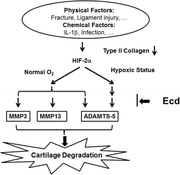
BACKGROUND: In osteoarthritis (OA), the imbalance of chondrocytes' anabolic and catabolic factors can induce cartilage destruction. Interleukin-1 beta (IL-1β) is a potent pro-inflammatory cytokine that is capable of inducing chondrocytes and synovial cells to synthesize MMPs. The hypoxia-inducible factor-2alpha (HIF-2alpha, encoded by Epas1) is the catabolic transcription factor in the osteoarthritic process. The purpose of this study is to validate the effects of ecdysteroids (Ecd) on IL-1β-induced cartilage catabolism and the possible role of Ecd in treatment or prevention of early OA. METHODS: Chondrocytes and articular cartilage was harvested from newborn ICR mice. Ecd effect on chondrocytes viability was tested and the optimal concentration was determined by MTT assay. The effect of HIF-2α (EPAS1) in cartilage catabolism simulated by IL-1β (5 ng/ml) was evaluated by articular cartilage explants culture. The effects of Ecd on IL-1β-induced inflammatory conditions and their related catabolic genes expression were analyzed. RESULTS: Interleukin-1β (IL-1β) treatment on primary mouse articular cartilage explants enhanced their Epas1, matrix metalloproteinases (MMP-3, MMP-13) and ADAMTS-5 genes expression and down-regulated collagen type II (Col2a1) gene expression. With the pre-treatment of 10(-8) M Ecd, the catabolic effects of IL-1β on articular cartilage were scavenged. CONCLUSION: In conclusions, Ecd can reduce the IL-1β-induced inflammatory effect of the cartilage. Ecd may suppress IL-1β-induced cartilage catabolism via HIF-2α pathway.
Overview
AI snapshot
Written from the paper’s full text.
TL;DR
In newborn ICR mouse chondrocytes and articular cartilage explants, IL-1β (5 ng/ml) increased Epas1, MMP-3, MMP-13, and ADAMTS-5 and decreased Col2a1; pretreatment with 10−8 M 20-hydroxyecdysone suppressed these catabolic changes. The authors conclude that Ecd has chondroprotective effects via the HIF-2α pathway, but call for further in vivo and translational work.
Key findings
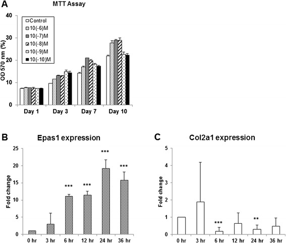
- Ecd enhanced chondrocyte viability, with maximal benefit at 10−8 M after 10 days of culture (p < 0.01); significant differences from control were observed at tested Ecd concentrations (p < 0.05), and 10−8 M was chosen for further experiments.
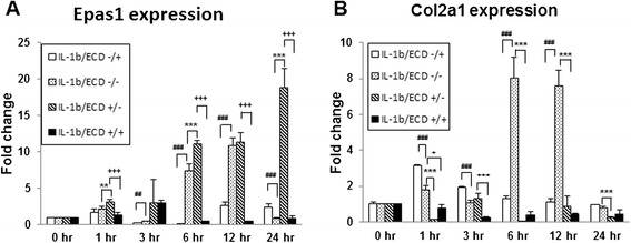
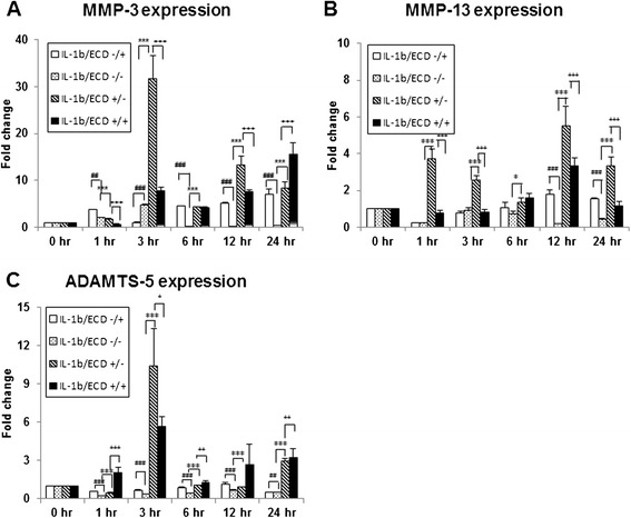
- IL-1β treatment enhanced Epas1 mRNA and suppressed Col2a1 in chondrocytes; in articular cartilage explants, IL-1β up-regulated MMP-3, MMP-13, and ADAMTS-5 and down-regulated Col2a1. Pretreatment with 10−8 M Ecd eliminated the IL-1β-enhanced Epas1 expression and down-regulated the catabolic genes, but did not affect Col2a1 in the conditions shown in Figure 2.
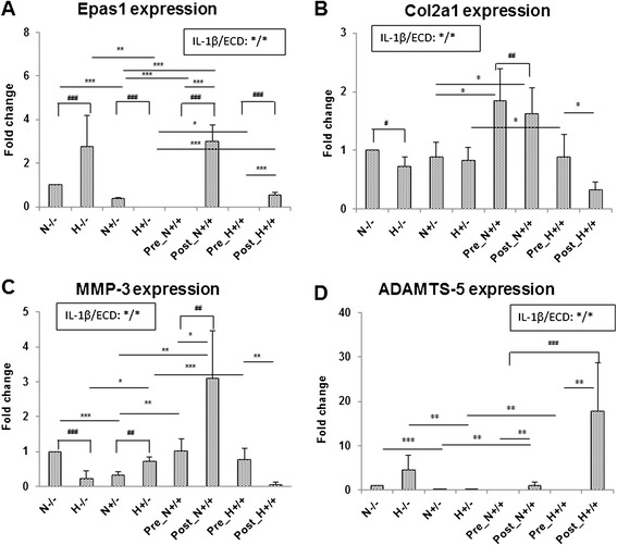
- Under hypoxic conditions (3.5% O2), Epas1 expression was higher; Ecd suppressed IL-1β-induced Epas1 and ADAMTS-5 under both hypoxic and normoxic conditions, while MMP-3 was down-regulated only under normoxic conditions. Under hypoxia, IL-1β-mediated ADAMTS-5 up-regulation was the main catabolic mediator, and no significant MMP13 expression was found (data not shown).
Methods
- Primary chondrocytes and articular cartilage explants were harvested from newborn ICR mice (3 days old); chondrocytes were isolated by collagenase digestion and cultured. Viability was tested by MTT assay with Ecd at 10−6 to 10−10 M on days 1, 3, 7, and 10 (n = 8).
- Cartilage explants were pre-incubated with or without Ecd (10−8 M), then treated with IL-1β (5 ng/ml); Ecd was added 24 hours before or after IL-1β, and explants were incubated under hypoxic (3.5% O2) or normoxic conditions.
- Gene expression was analyzed by RT-PCR for Col2a1, MMP-3, MMP-13, ADAMTS5, EPAS1, and β-actin at 0, 3, 6, 12, 24, and 36 hours after Ecd addition. Experiments were performed at least in triplicate; results were analyzed by two-way ANOVA with Dunnett’s test at p < 0.05.
Limitations
- The authors state that further in vivo HIF-1alpha gene knock-down and translational medicine studies are mandatory for possible clinical application.
- Evident from the text: the study is limited to in vitro mouse chondrocyte cultures and articular cartilage explants, with no in vivo osteoarthritis model, protein/activity assays, or histological outcomes reported.
- Evident from the text: Ecd did not affect Col2a1 in Figure 2, and MMP13 expression under hypoxic conditions was not shown, limiting conclusions about full phenotypic recovery and hypoxic MMP13 involvement.
Model-written summary. Check it against the paper before relying on it.
Integrity signals
Registry
Cites
Statistics
Figures
Verification
Claims the paper makes
22 Own result · 15 Not checked yet · 9 Own interpretation · 2 Own method · 1 No basis given
A result the paper reports from its own data — it cites no other source for it, so there is none to check. The quote shows it is in the paper.
In the paper
“Under hypoxic condition, IL-1β mediates HIF-2α–induced up-regulation of ADAMTS-5 gene (but not MMP3 or MMP13 genes) in articular cartilage (Figure 4C and D)”
§ Arthropod steroid hormone (20-Hydroxyecdysone) suppresses IL-1β- induced catabolic gene expression in cartilage > Results > Ecd protect cartilage explants both in hypoxic and normoxic condition
plaindirectn = 8- Asserted: passes — The authors put it forward as true.
- Checkable: passes — It can be checked against evidence.
- Load-bearing: passes — The paper’s conclusions depend on it.
- Based: passes — The paper gives a reason why it holds.
A result the paper reports from its own data — it cites no other source for it, so there is none to check. The quote shows it is in the paper.
In the paper
“there was no significant expression on MMP13 gene under hypoxic condition (data not shown).”
§ Arthropod steroid hormone (20-Hydroxyecdysone) suppresses IL-1β- induced catabolic gene expression in cartilage > Results > Ecd protect cartilage explants both in hypoxic and normoxic condition
plain- Asserted: passes — The authors put it forward as true.
- Checkable: passes — It can be checked against evidence.
- Load-bearing: passes — The paper’s conclusions depend on it.
- Based: passes — The paper gives a reason why it holds.
A result the paper reports from its own data — it cites no other source for it, so there is none to check. The quote shows it is in the paper.
In the paper
“with the pretreatment of 10−8M Ecd, the catabolic factor genes upregulated by IL-1β treatment are eliminated.”
§ Arthropod steroid hormone (20-Hydroxyecdysone) suppresses IL-1β- induced catabolic gene expression in cartilage > Results > Ecd pre-treatment reduce the catabolic effect of IL-1β on the cartilage explants
plaindirectn = 8 ·- Asserted: passes — The authors put it forward as true.
- Checkable: passes — It can be checked against evidence.
- Load-bearing: passes — The paper’s conclusions depend on it.
- Based: passes — The paper gives a reason why it holds.
A result the paper reports from its own data — it cites no other source for it, so there is none to check. The quote shows it is in the paper.
In the paper
“Under hypoxic condition, IL-1β mediates HIF-2α–induced up-regulation of ADAMTS-5 gene (but not MMP3 or MMP13 genes) in articular cartilage (Figure 4C and D)”
§ Arthropod steroid hormone (20-Hydroxyecdysone) suppresses IL-1β- induced catabolic gene expression in cartilage > Results > Ecd protect cartilage explants both in hypoxic and normoxic condition
plainn = 8- Asserted: passes — The authors put it forward as true.
- Checkable: passes — It can be checked against evidence.
- Load-bearing: passes — The paper’s conclusions depend on it.
- Based: passes — The paper gives a reason why it holds.
A result the paper reports from its own data — it cites no other source for it, so there is none to check. The quote shows it is in the paper.
In the paper
“Same as noted above, IL-1β enhances Epas1 mRNA expression and suppresses collagen type II (Col2a1) gene expression.”
§ Arthropod steroid hormone (20-Hydroxyecdysone) suppresses IL-1β- induced catabolic gene expression in cartilage > Results > Ecd pre-treatment reduce the catabolic effect of IL-1β on the cartilage explants
plaindirectn = 8- Asserted: passes — The authors put it forward as true.
- Checkable: passes — It can be checked against evidence.
- Load-bearing: passes — The paper’s conclusions depend on it.
- Based: passes — The paper gives a reason why it holds.
A result the paper reports from its own data — it cites no other source for it, so there is none to check. The quote shows it is in the paper.
In the paper
“with the pretreatment of 10−8M Ecd, the catabolic factor genes upregulated by IL-1β treatment are eliminated.”
§ Arthropod steroid hormone (20-Hydroxyecdysone) suppresses IL-1β- induced catabolic gene expression in cartilage > Results > Ecd pre-treatment reduce the catabolic effect of IL-1β on the cartilage explants
plaindirectn = 8 ·- Asserted: passes — The authors put it forward as true.
- Checkable: passes — It can be checked against evidence.
- Load-bearing: passes — The paper’s conclusions depend on it.
- Based: passes — The paper gives a reason why it holds.
A result the paper reports from its own data — it cites no other source for it, so there is none to check. The quote shows it is in the paper.
In the paper
“In this study, there is significant difference observed between treated cells with that of the control (p < 0.05).”
- Also stated in: § Ecd treatment enhance chondrocytes viability · plain
“Chondrocytes at every concentration proliferate actively, with significant statistical differences observed between chondrocytes treated with Ecd and that of the control.”
§ Arthropod steroid hormone (20-Hydroxyecdysone) suppresses IL-1β- induced catabolic gene expression in cartilage > Results > Ecd treatment enhance chondrocytes viability
plaindirectn = 8 ·- Asserted: passes — The authors put it forward as true.
- Checkable: passes — It can be checked against evidence.
- Load-bearing: unclear — It is unclear whether the paper’s conclusions depend on it.
- Based: passes — The paper gives a reason why it holds.
- Also stated in: § Ecd treatment enhance chondrocytes viability · plain
A result the paper reports from its own data — it cites no other source for it, so there is none to check. The quote shows it is in the paper.
In the paper
“We chose 10−8 M Ecd for the further evaluation because there is maximal viability of chondrocytes at this concentration (p < 0.01) at the 10th day’s culture.”
§ Arthropod steroid hormone (20-Hydroxyecdysone) suppresses IL-1β- induced catabolic gene expression in cartilage > Results > Ecd treatment enhance chondrocytes viability
plaincorrelationaln = 8 ·- Asserted: passes — The authors put it forward as true.
- Checkable: passes — It can be checked against evidence.
- Load-bearing: passes — The paper’s conclusions depend on it.
- Based: passes — The paper gives a reason why it holds.
A result the paper reports from its own data — it cites no other source for it, so there is none to check. The quote shows it is in the paper.
In the paper
“interleukin-1β (IL-1β) treatment of primary mouse articular chondrocyte cultures enhance Epas1 mRNA expression, while suppress collagen type II (Col2a1) gene expression (Figure 1B, C).”
§ Arthropod steroid hormone (20-Hydroxyecdysone) suppresses IL-1β- induced catabolic gene expression in cartilage > Results > IL-1β induce HIF-2α (encoded by Epas1) gene expression in chondrocytes
plaindirect- Asserted: passes — The authors put it forward as true.
- Checkable: passes — It can be checked against evidence.
- Load-bearing: passes — The paper’s conclusions depend on it.
- Based: passes — The paper gives a reason why it holds.
A result the paper reports from its own data — it cites no other source for it, so there is none to check. The quote shows it is in the paper.
In the paper
“while in hypoxic condition, Ecd pre-treatment eliminates the IL-1β-induced Col2a1 gene down-regulation (Figure 4B).”
§ Arthropod steroid hormone (20-Hydroxyecdysone) suppresses IL-1β- induced catabolic gene expression in cartilage > Results > Ecd protect cartilage explants both in hypoxic and normoxic condition
plaindirect- Asserted: passes — The authors put it forward as true.
- Checkable: passes — It can be checked against evidence.
- Load-bearing: passes — The paper’s conclusions depend on it.
- Based: passes — The paper gives a reason why it holds.
- The quote only partly states this claim. 1
- It may state two separate claims as one. 5
- Probably not a substantial claim. 13
A result the paper reports from its own data — it cites no other source for it, so there is none to check. The quote shows it is in the paper.
In the paper
“but there is also no effect on Col2a1 gene expression (Figure 2).”
- Also stated in: § Discussion · plain
“but there is no ability to recover the cartilage phenotype as Col2a1 gene was not affected (Figure 4).”
§ Arthropod steroid hormone (20-Hydroxyecdysone) suppresses IL-1β- induced catabolic gene expression in cartilage > Results > Ecd pre-treatment reduce the catabolic effect of IL-1β on the cartilage explants
plainn = 8- Asserted: passes — The authors put it forward as true.
- Checkable: passes — It can be checked against evidence.
- Load-bearing: passes — The paper’s conclusions depend on it.
- Based: passes — The paper gives a reason why it holds.
The quote only partly states this claim.
- Also stated in: § Discussion · plain
None of the sources cited for this claim have been checked yet.
In the paper
“In ovariectomized rats, pure Ecd supplementation significantly increased in the thickness of joint cartilage and presented beneficial effects in bones.”
§ Arthropod steroid hormone (20-Hydroxyecdysone) suppresses IL-1β- induced catabolic gene expression in cartilage > Background
What the sources say
- Not checked yetcites [13] Kapur P, Wuttke W, Jarry H, Seidlova-Wuttke D. Beneficial effects of beta-Ecdysone on the joint, epiphyseal cartilage tissue and trabecular bone in ovariectomized rats. Phytomedicine. 2010;17(5):350–5. source
- Not checked yetcites [14] Zhang X, Xu X, Xu T, Qin S. β-Ecdysterone suppresses interleukin-1β-induced apoptosis and inflammation in rat chondrocytes via inhibition of NF-κB signaling pathway. Drug Dev Res. 2014;75(3):195–201. source
- Not checked yetcites [15] He CC, Hui RR, Tezuka Y, Kadota S, Li JX. Osteoprotective effect of extract from Achyranthes bidentata in ovariectomized rats. J Ethnopharmacol. 2010;127(2):229–34. source
plaindirect- Asserted: passes — The authors put it forward as true.
- Checkable: passes — It can be checked against evidence.
- Load-bearing: passes — The paper’s conclusions depend on it.
- Based: passes — The paper gives a reason why it holds.
It may state two separate claims as one.
None of the sources cited for this claim have been checked yet.
In the paper
“Instead, IL-1β and certain other inflammatory mediators promote activation of latent ADAMTS-5, as does hypertrophic differentiation in chondrocytes [3,4].”
§ Arthropod steroid hormone (20-Hydroxyecdysone) suppresses IL-1β- induced catabolic gene expression in cartilage > Discussion
What the sources say
- Not checked yetcites [3] Saito T, Fukai A, Mabuchi A, Ikeda T, Yano F, Ohba S, et al. Transcriptional regulation of endochondral ossification by HIF-2alpha during skeletal growth and osteoarthritis development. Nat Med. 2010;16(6):678–86. source
- Not checked yetcites [4] Yang S, Kim J, Ryu JH, Oh H, Chun CH, Kim BJ, et al. Hypoxia-inducible factor-2alpha is a catabolic regulator of osteoarthritic cartilage destruction. Nat Med. 2010;16(6):687–93. source
plaindirect- Asserted: passes — The authors put it forward as true.
- Checkable: passes — It can be checked against evidence.
- Load-bearing: fails — The paper’s conclusions may not depend on it.
- Based: passes — The paper gives a reason why it holds.
Probably not a substantial claim. The paper’s conclusions may not depend on it.
In the paper
“In spite of a large variability of expression levels among different patients, MMP-3 appeared to be strongly expressed in normal and early degenerative cartilage and down-regulated in the late disease stages.”
§ Arthropod steroid hormone (20-Hydroxyecdysone) suppresses IL-1β- induced catabolic gene expression in cartilage > Discussion
What the sources say
- Not checked yetcites [31] Aigner T, Zien A, Hanisch D, Zimmer R. Gene expression in chondrocytes assessed with use of microarrays. J Bone Joint Surg Am. 2003;85-A Suppl 2:117–23. source
hedgedcorrelational- Asserted: passes — The authors put it forward as true.
- Checkable: passes — It can be checked against evidence.
- Load-bearing: fails — The paper’s conclusions may not depend on it.
- Based: passes — The paper gives a reason why it holds.
Probably not a substantial claim. The paper’s conclusions may not depend on it. It may state two separate claims as one.
In the paper
“The Ecd is frequently used by body builders and from these users it is known that Ecd has no known adverse effects even when taken up in gram (~1 g) quantities [16].”
§ Arthropod steroid hormone (20-Hydroxyecdysone) suppresses IL-1β- induced catabolic gene expression in cartilage > Background
What the sources say
- Not checked yetcites [16] Dinan L, Lafont R. Effects and applications of arthropod steroid hormones (ecdysteroids) in mammals. J Endocrinol. 2006;191(1):1–8. source
boosted- Asserted: passes — The authors put it forward as true.
- Checkable: passes — It can be checked against evidence.
- Load-bearing: fails — The paper’s conclusions may not depend on it.
- Based: passes — The paper gives a reason why it holds.
Probably not a substantial claim. The paper’s conclusions may not depend on it.
None of the sources cited for this claim have been checked yet.
In the paper
“It is reported that HIF-2α (like IL-1β) does not induce transcription of ADAMTS5 [3,4].”
§ Arthropod steroid hormone (20-Hydroxyecdysone) suppresses IL-1β- induced catabolic gene expression in cartilage > Discussion
What the sources say
- Not checked yetcites [3] Saito T, Fukai A, Mabuchi A, Ikeda T, Yano F, Ohba S, et al. Transcriptional regulation of endochondral ossification by HIF-2alpha during skeletal growth and osteoarthritis development. Nat Med. 2010;16(6):678–86. source
- Not checked yetcites [4] Yang S, Kim J, Ryu JH, Oh H, Chun CH, Kim BJ, et al. Hypoxia-inducible factor-2alpha is a catabolic regulator of osteoarthritic cartilage destruction. Nat Med. 2010;16(6):687–93. source
plain- Asserted: passes — The authors put it forward as true.
- Checkable: passes — It can be checked against evidence.
- Load-bearing: fails — The paper’s conclusions may not depend on it.
- Based: passes — The paper gives a reason why it holds.
Probably not a substantial claim. The paper’s conclusions may not depend on it.
None of the sources cited for this claim have been checked yet.
In the paper
“According to this paradigm, a stress-induced increase in the activity of HIF-2α will overshadow the beneficial effects of the closely related HIF-1α, then push the chondrocytes in the joint toward a more differentiated state known as hypertrophy, which then drives osteoarthritis changes [6,7].”
§ Arthropod steroid hormone (20-Hydroxyecdysone) suppresses IL-1β- induced catabolic gene expression in cartilage > Background
What the sources say
- Not checked yetcites [6] Husa M, Liu-Bryan R, Terkeltaub R. Shifting HIFs in osteoarthritis. Nat Med. 2010;16(6):641–4. source
- Not checked yetcites [7] Ryu JH, Shin Y, Huh YH, Yang S, Chun CH, Chun JS. Hypoxia-inducible factor-2α regulates Fas-mediated chondrocyte apoptosis during osteoarthritic cartilage destruction. Cell Death Differ. 2012;19(3):440–50. source
plaindirect- Asserted: passes — The authors put it forward as true.
- Checkable: passes — It can be checked against evidence.
- Load-bearing: passes — The paper’s conclusions depend on it.
- Based: passes — The paper gives a reason why it holds.
Probably not a substantial claim.
In the paper
“Together, these findings implicate hypoxic signaling as a central regulator of skeletal biology and disease [32].”
§ Arthropod steroid hormone (20-Hydroxyecdysone) suppresses IL-1β- induced catabolic gene expression in cartilage > Discussion
What the sources say
- Not checked yetcites [32] Rankin EB, Giaccia AJ, Schipani E. A central role for hypoxic signaling in cartilage, bone, and hematopoiesis. Curr Osteoporos Rep. 2011;9(2):46–52. source
hedgeddirect- Asserted: passes — The authors put it forward as true.
- Checkable: passes — It can be checked against evidence.
- Load-bearing: fails — The paper’s conclusions may not depend on it.
- Based: passes — The paper gives a reason why it holds.
Probably not a substantial claim. The paper’s conclusions may not depend on it.
A conclusion the paper draws from its own results — it cites no other source for it, so there is none to check. The quote shows it is in the paper.
In the paper
“The findings of our study are consistent with the observations that inflammation of articular cartilage stimulates ADAMTS5 activation.”
§ Arthropod steroid hormone (20-Hydroxyecdysone) suppresses IL-1β- induced catabolic gene expression in cartilage > Discussion
plaindirect- Asserted: passes — The authors put it forward as true.
- Checkable: passes — It can be checked against evidence.
- Load-bearing: fails — The paper’s conclusions may not depend on it.
- Based: passes — The paper gives a reason why it holds.
Probably not a substantial claim. The paper’s conclusions may not depend on it.
None of the sources cited for this claim have been checked yet.
In the paper
“During OA, ADAMTS-4 (aggrecanase 1) [28] and ADAMTS-5 (aggrecanase 2) [29,30] have been implicated prominently in cartilage breakdown.”
§ Arthropod steroid hormone (20-Hydroxyecdysone) suppresses IL-1β- induced catabolic gene expression in cartilage > Discussion
What the sources say
- Not checked yetcites [28] Poole AR, Nelson F, Dahlberg L, Tchetina E, Kobayashi M, Yasuda T, et al. Proteolysis of the collagen fibril in osteoarthritis. Biochem Soc Symp. 2003;70:115–23. source
- Not checked yetcites [29] Tortorella MD, Burn TC, Pratta MA, Abbaszade I, Hollis JM, Liu R, et al. Purification and cloning of aggrecanase-1: a member of the ADAMTS family of proteins. Science. 1999;284(5420):1664–6. source
- Not checked yetcites [30] Pattoli MA, MacMaster JF, Gregor KR, Burke JR. Collagen and aggrecan degradation is blocked in interleukin-1-treated cartilage explants by an inhibitor of IkappaB kinase through suppression of metalloproteinase expression. J Pharmacol Exp Ther. 2005;315(1):382–8. source
plaincorrelational- Asserted: passes — The authors put it forward as true.
- Checkable: passes — It can be checked against evidence.
- Load-bearing: fails — The paper’s conclusions may not depend on it.
- Based: passes — The paper gives a reason why it holds.
Probably not a substantial claim. The paper’s conclusions may not depend on it.
In the paper
“The degradation of aggrecan by aggrecanases has gained special interest, because it constitutes an early event in osteoarthritic cartilage damage and is followed by the irreversible loss of collagens [27].”
§ Arthropod steroid hormone (20-Hydroxyecdysone) suppresses IL-1β- induced catabolic gene expression in cartilage > Discussion
What the sources say
- Not checked yetcites [27] Brooks PM. The burden of musculoskeletal disease–a global perspective. Clin Rheumatol. 2006;25(6):778–81. source
plaindirect- Asserted: passes — The authors put it forward as true.
- Checkable: passes — It can be checked against evidence.
- Load-bearing: fails — The paper’s conclusions may not depend on it.
- Based: passes — The paper gives a reason why it holds.
Probably not a substantial claim. The paper’s conclusions may not depend on it. It may state two separate claims as one.
What the paper did — it cites no source for this, so there is none to check. The quote shows it is in the paper.
In the paper
“For each genes analysis, the experiments were repeated at least three times.”
§ Arthropod steroid hormone (20-Hydroxyecdysone) suppresses IL-1β- induced catabolic gene expression in cartilage > Methods > Gene expression analysis
plain- Asserted: passes — The authors put it forward as true.
- Checkable: passes — It can be checked against evidence.
- Load-bearing: fails — The paper’s conclusions may not depend on it.
- Based: passes — The paper gives a reason why it holds.
Probably not a substantial claim. The paper’s conclusions may not depend on it.
None of the sources cited for this claim have been checked yet.
In the paper
“In mammals, Ecd has protein anabolic effects and increases muscle mass of unexercised and exercised muscle [11,12].”
§ Arthropod steroid hormone (20-Hydroxyecdysone) suppresses IL-1β- induced catabolic gene expression in cartilage > Background
What the sources say
- Not checked yetcites [11] Báthori M, Tóth N, Hunyadi A, Márki A, Zádor E. Phytoecdysteroids and anabolic-androgenic steroids–structure and effects on humans. Curr Med Chem. 2008;15(1):75–91. source
- Not checked yetcites [12] Gorelick-Feldman J, Maclean D, Ilic N, Poulev A, Lila MA, Cheng D, et al. Phytoecdysteroids increase protein synthesis in skeletal muscle cells. J Agric Food Chem. 2008;56(10):3532–7. source
plaindirect- Asserted: passes — The authors put it forward as true.
- Checkable: passes — It can be checked against evidence.
- Load-bearing: fails — The paper’s conclusions may not depend on it.
- Based: passes — The paper gives a reason why it holds.
Probably not a substantial claim. The paper’s conclusions may not depend on it. It may state two separate claims as one.
What the paper did — it cites no source for this, so there is none to check. The quote shows it is in the paper.
In the paper
“All experiments were performed at least in triplicate.”
§ Arthropod steroid hormone (20-Hydroxyecdysone) suppresses IL-1β- induced catabolic gene expression in cartilage > Methods > Statistical analysis
plain- Asserted: passes — The authors put it forward as true.
- Checkable: passes — It can be checked against evidence.
- Load-bearing: fails — The paper’s conclusions may not depend on it.
- Based: passes — The paper gives a reason why it holds.
Probably not a substantial claim. The paper’s conclusions may not depend on it.
None of the sources cited for this claim have been checked yet.
In the paper
“In the early phases of OA, MMP-13 is predominantly expressed in the lower intermediate and deep layers of human and mouse cartilage, whereas aggrecanases are expressed in the superficial cartilage [25,26].”
§ Arthropod steroid hormone (20-Hydroxyecdysone) suppresses IL-1β- induced catabolic gene expression in cartilage > Discussion
What the sources say
- Not checked yetcites [25] Brew CJ, Clegg PD, Boot-Handford RP, Andrew JG, Hardingham T. Gene expression in human chondrocytes in late osteoarthritis is changed in both fibrillated and intact cartilage without evidence of generalised chondrocyte hypertrophy. Ann Rheum Dis. 2010;69(1):234–40. source
- Not checked yetcites [26] Little CB, Barai A, Burkhardt D, Smith SM, Fosang AJ, Werb Z, et al. Matrix metalloproteinase 13-deficient mice are resistant to osteoarthritic cartilage erosion but not chondrocyte hypertrophy or osteophyte development. Arthritis Rheum. 2009;60(12):3723–33. source
plaincorrelational- Asserted: passes — The authors put it forward as true.
- Checkable: passes — It can be checked against evidence.
- Load-bearing: fails — The paper’s conclusions may not depend on it.
- Based: passes — The paper gives a reason why it holds.
Probably not a substantial claim. The paper’s conclusions may not depend on it. It may state two separate claims as one.
The paper relies on this without support.
Epas1 gene expression is a valid proxy for HIF-2α activity.
“The hypoxia-inducible factor-2alpha (HIF-2α, encoded by Epas1) is a catabolic transcription factor in the osteoarthritic process.”
The paper relies on this without support.
Findings in mouse articular cartilage explants are relevant to human OA.
“The purpose of this study is to validate the effects of Ecd on IL-1β- induced cartilage destruction and also the possible role of Ecd in treatment or prevention of early OA.”
Claims other papers make about this one
Each is checked against this paper’s own text: does it say what the citing paper says it does?
The source backs part of the claim, not all of it — or backs it for a different population, condition or comparison.
In the citing paper
“In addition, 20E scavenged the increase in catabolic processes and decrease in anabolic processes and largely restored the balance in primary explanted articular chondrocytes [39], confirming a protective effect in OA.”
From Molecular Targets of 20-Hydroxyecdysone in Mammals, Mechanism of Action: Is It a Calorie Restriction Mimetic and Anti-Aging Compound? (2025) · cites this paper as [39]
Experimental — accuracy not yet measured
Citations
Cites
Goldring, M. B., & Goldring, S. R. (2007) · Journal of Cellular Physiology · doi:10.1002/jcp.21258 · Cited by 1,179
Purification and Cloning of Aggrecanase-1: A Member of the ADAMTS Family of Proteins
Tortorella, M. D., Burn, T. C., Pratta, M. A., Abbaszade, I., Hollis, J. M., Liu, R., Rosenfeld, S. A., Copeland, R. A., Decicco, C. P., Wynn, R., et al. (1999) · Science · doi:10.1126/science.284.5420.1664 · Cited by 686
Little, C. B., Barai, A., Burkhardt, D., Smith, S. M., Fosang, A. J., Werb, Z., Shah, M., & Thompson, E. W. (2009) · Arthritis & Rheumatism · doi:10.1002/art.25002 · Cited by 642
Cartilage in normal and osteoarthritis conditions
Martel‐Pelletier, J., Boileau, C., Pelletier, J., & Roughley, P. J. (2008) · Best Practice & Research Clinical Rheumatology · doi:10.1016/j.berh.2008.02.001 · Cited by 556
The burden of musculoskeletal disease—a global perspective
Brooks, P. M. (2006) · Clinical Rheumatology · doi:10.1007/s10067-006-0240-3 · Cited by 545
Saito, T., Fukai, A., Mabuchi, A., Ikeda, T., Yano, F., Ohba, S., Nishida, N., Akune, T., Yoshimura, N., Nakagawa, T., et al. (2010) · Nature Medicine · doi:10.1038/nm.2146 · Cited by 532
Hypoxia-inducible factor-2α is a catabolic regulator of osteoarthritic cartilage destruction
Yang, S., Kim, J., Ryu, J.-H., Oh, H., Chun, C.-H., Kim, B. J., Min, B. H., & Chun, J.-S. (2010) · Nature Medicine · doi:10.1038/nm.2153 · Cited by 527
Insurer and out‐of‐pocket costs of osteoarthritis in the US: Evidence from national survey data
Kotlarz, H., Gunnarsson, C., Fang, H., & Rizzo, J. A. (2009) · Arthritis & Rheumatism · doi:10.1002/art.24984 · Cited by 440
The Interleukin 1β Pathway in the Pathogenesis of Osteoarthritis
Daheshia, M., & Yao, J. Q. (2008) · The Journal of Rheumatology · doi:10.3899/jrheum.080346 · Cited by 420
Calcification of articular cartilage in human osteoarthritis
Fuerst, M., Bertrand, J., Lammers, L., Dreier, R., Echtermeyer, F. G., Nitschke, Y., Rutsch, F., Schäfer, F. K. W., Niggemeyer, O., Steinhagen, J., et al. (2009) · Arthritis & Rheumatism · doi:10.1002/art.24774 · Cited by 322
Cited by
Sorted by citations
Natural sweeteners: Sources, extraction and current uses in foods and food industries
Castro‐Muñoz, R., Correa-Delgado, M., Córdova-Almeida, R., Lara-Nava, D., Chávez-Muñoz, M., Velásquez-Chávez, V. F., Hernández-Torres, C. E., Gontarek‐Castro, E., & Ahmad, M. Z. (2021) · Food Chemistry · doi:10.1016/j.foodchem.2021.130991 · Cited by 260
Ghasemzadeh, A., Karbalaii, M. T., Jaafar, H. Z. E., & Rahmat, A. B. (2018) · Chemistry Central Journal · doi:10.1186/s13065-018-0382-9 · Cited by 231
Behavioral tests in rodent models of stroke
Ruan, J., & Yao, Y. (2020) · Brain Hemorrhages · doi:10.1016/j.hest.2020.09.001 · Cited by 222
Digestion and bioavailability of bioactive phytochemicals
Karaś, M., Jakubczyk, A., Szymanowska, U., Złotek, U., & Zielińska, E. (2016) · International Journal of Food Science & Technology · doi:10.1111/ijfs.13323 · Cited by 186
Anwar, S., Khan, A. A., Almatroudi, A., Khan, A. A., Alsahli, M. A., Almatroodi, S. A., & Rahmani, A. H. (2021) · Molecular Biology Reports · doi:10.1007/s11033-020-06084-0 · Cited by 158
van Vuuren, S. F., & Holl, D. (2017) · Journal of Ethnopharmacology · doi:10.1016/j.jep.2017.07.011 · Cited by 154
Natural Products: An Alternative to Conventional Therapy for Dermatophytosis?
Lopes, G., Pinto, E., & Salgueiro, L. (2016) · Mycopathologia · doi:10.1007/s11046-016-0081-9 · Cited by 129
Bressington, D. T., Yu, C. T. K., Wong, W. K. J., Ng, T. C., & Chien, W. T. (2018) · Journal of Psychiatric and Mental Health Nursing · doi:10.1111/jpm.12491 · Cited by 104
Sun, H., Zhang, A.-H., Song, Q., Fang, H., Liu, X.-Y., Su, J., Yang, L., Yu, M.-D., & Wang, X.-J. (2018) · RSC Advances · doi:10.1039/c8ra06553e · Cited by 89
Balkrishna, A., Kumar, A., Arya, V., Rohela, A., Verma, R., Nepovimová, E., Krejcar, O., Kumar, D., Thakur, N., & Kuča, K. (2021) · Oxidative Medicine and Cellular Longevity · doi:10.1155/2021/3155962 · Cited by 86
Zhang, Y., Ni, Z., Elam, E., Zhang, F., Thakur, K., Wang, S., Zhang, J., & Wei, Z. (2021) · Food & Function · doi:10.1039/d1fo00790d · Cited by 77
Thakur, A. K., Rai, G., Chatterjee, S. S., & Kumar, V. (2016) · Pharmaceutical Biology · doi:10.3109/13880209.2015.1107107 · Cited by 76
de Melo, A. M., Almeida, F. L. C., Cavalcante, A. M. D. M., Ikeda, M., Barbi, R. C. T., Costa, B. P., & Ribani, R. H. (2021) · Trends in Food Science & Technology · doi:10.1016/j.tifs.2021.04.005 · Cited by 75
Pathological mechanism and targeted drugs of ulcerative colitis: A review
Guo, M., & Wang, X. (2023) · Medicine · doi:10.1097/md.0000000000035020 · Cited by 75
Ding, X., Yin, C., Zhang, W., Sun, Y., Zhang, Z., Yang, E., Sun, D., & Wang, W. (2020) · Nanoscale Research Letters · doi:10.1186/s11671-020-03297-x · Cited by 74
Tu, C., Huang, X., Xiao, Y., Song, M., Ma, Y., Yan, J., You, H., & Wu, H. K. (2019) · Frontiers in Pharmacology · doi:10.3389/fphar.2019.00041 · Cited by 73
Traditional uses, phytochemistry and pharmacological properties of African Nauclea species: A review
Haudecoeur, R., Peuchmaur, M., Pérès, B., Rome, M., Taïwé, G. S., Boumendjel, A., & Boucherle, B. (2017) · Journal of Ethnopharmacology · doi:10.1016/j.jep.2017.10.011 · Cited by 70
Ma, C., Wang, X., Xu, T., Yu, X. L., Zhang, S., Liu, S., Gao, Y., Fan, S., Li, C., Zhai, C., et al. (2019) · BMC Complementary and Alternative Medicine · doi:10.1186/s12906-019-2703-5 · Cited by 69
Khan, A., Younis, T., Anas, M., Ali, M., Shinwari, Z. K., Khalil, A. T., Munawar, K. S., Mohamed, H. E. A., Hkiri, K., Mâaza, M., et al. (2025) · BMC Plant Biology · doi:10.1186/s12870-025-06533-7 · Cited by 66
Use of antipsychotics increases the risk of fracture: a systematic review and meta-analysis
Lee, S.-H., Hsu, W., Lai, C., Esmaily-Fard, A., Tsai, Y.-H., Chiu, C.-C., Wang, J., Chang, S.-S., & Lee, C. (2017) · Osteoporosis International · doi:10.1007/s00198-016-3881-3 · Cited by 68
Abd‐Elkareem, M., El‐Rahman, M. A. M. A., Khalil, N. S. A., & Amer, A. (2021) · Scientific Reports · doi:10.1038/s41598-021-92977-4 · Cited by 65
Southern African indigenous fruits and their byproducts: Prospects as food antioxidants
Trust Mukudzei Pfukwa, Chikwanha, O. C., Katiyatiya, C. L. F., Fawole, O. A., Manley, M., & Mapiye, C. (2020) · Journal of Functional Foods · doi:10.1016/j.jff.2020.104220 · Cited by 59
Huang, X., Yang, X., Pan, Q., Mao, Z., Zhang, R., Ma, X., & You, H. (2018) · Biomedicine & Pharmacotherapy · doi:10.1016/j.biopha.2018.07.161 · Cited by 57
Li, X., Li, J., Li, Z., Sang, Y., Niu, Y., Zhang, Q., Ding, H., & Yin, S. (2016) · Food & Function · doi:10.1039/c6fo00288a · Cited by 56
Lee, J.-H., Lee, M., Lim, T., Kim, T., Kim, S. M., Suh, D.-W., Lee, S., & Yoon, B. (2017) · European Journal of Integrative Medicine · doi:10.1016/j.eujim.2017.04.012 · Cited by 55
Traditional Chinese medicine on treating pain caused by prostate cancer
Wang, N., Li, X., Wang, J., Yu, X., Chu, L.-Y., Deng, S., Ge, F., & Li, H. (2019) · Medicine · doi:10.1097/md.0000000000017624 · Cited by 52
Antibacterial and Antifungal Activities of Ethiopian Medicinal Plants: A Systematic Review
Nigussie, D., Davey, G., Tufa, T. B., Brewster, M., Legesse, B. A., Fekadu, A., & Makonnen, E. (2021) · Frontiers in Pharmacology · doi:10.3389/fphar.2021.633921 · Cited by 52
Lee, S.-J. (2015) · Journal of pharmacopuncture · doi:10.3831/kpi.2015.18.020 · Cited by 48
Gao, S., Sun, D., Wang, G., Zhang, J. C., Jiang, Y., Li, G., Zhang, K., Wang, L., Huang, J. M., & Chen, L. (2016) · Bioorganic Chemistry · doi:10.1016/j.bioorg.2016.10.005 · Cited by 47
Efficacy and safety of puerarin injection in curing acute ischemic stroke
Zheng, Q., Li, X.-L., Mei, Z., Xiong, L., Mei, Q.-X., Wang, J., Tan, L., Yang, S., & Feng, Z. (2017) · Medicine · doi:10.1097/md.0000000000005803 · Cited by 47
Sajid, M., Khan, M. R., Shah, S. A., Majid, M., Ismail, H., Maryam, S., Batool, R., & Younis, T. (2017) · Journal of Ethnopharmacology · doi:10.1016/j.jep.2017.01.041 · Cited by 47
Sharma, S. K., Gautam, N., & Atri, N. S. (2015) · BMC Complementary and Alternative Medicine · doi:10.1186/s12906-015-0967-y · Cited by 43
RetractedGanji, A., Salehi, I., Sarihi, A., Shahidi, ., & Komaki, A. (2016) · Metabolic Brain Disease · doi:10.1007/s11011-016-9940-9 · Cited by 43
Bioprospecting of endophytic microorganisms for bioactive compounds of therapeutic importance
Raimi, A. R., & Adeleke, R. A. (2021) · Archives of Microbiology · doi:10.1007/s00203-021-02256-z · Cited by 45
Sadeghi-Aghbash, M., Rahimnejad, M., Adeli, H., & Feizi, F. (2022) · Reactive and Functional Polymers · doi:10.1016/j.reactfunctpolym.2022.105440 · Cited by 41
Ghasemzadeh, A., Jaafar, H. Z. E., Rahmat, A. B., & Swamy, M. K. (2017) · Chemistry Central Journal · doi:10.1186/s13065-016-0235-3 · Cited by 40
Ecdysteroids: Isolation, Chemical Transformations, and Biological Activity
Savchenko, R. G., Veskina, N. A., Odinokov, V. N., Benkovskaya, G. V., & Parfenova, L. V. (2022) · Phytochemistry Reviews · doi:10.1007/s11101-021-09792-y · Cited by 39
Azman, S., Sekar, M., Bonam, S. R., Gan, S. H., Wahidin, S., Lum, P. T., & Dhadde, S. B. (2021) · Journal of Asthma and Allergy · doi:10.2147/jaa.s296391 · Cited by 41
Ginovyan, M. M., Ayvazyan, A., Nikoyan, A., Tumanyan, L., & Trchоunian, A. (2020) · Current Microbiology · doi:10.1007/s00284-020-01929-0 · Cited by 38
Lataief, S. B., Zourgui, M. N., Rahmani, R., Najjaa, H., Gharsallah, N., & Zourgui, L. (2020) · Journal of Food Measurement & Characterization · doi:10.1007/s11694-020-00671-2 · Cited by 37
Wang, K.-H., Wu, J., Zhang, D., Duan, X., & Ni, M. (2018) · BMC Complementary and Alternative Medicine · doi:10.1186/s12906-018-2090-3 · Cited by 36
A new exploration of Dregea volubilis flowers: Focusing on antioxidant and antidiabetic properties
Das, B., De, A., Das, M., Das, S., & Samanta, A. (2016) · South African Journal of Botany · doi:10.1016/j.sajb.2016.12.003 · Cited by 35
Danielli, L. J., Pippi, B., Duarte, J. A., Maciel, A. J., Lopes, W., Machado, M. M., de Oliveira, L. F. S., Vainstein, M. H., Teixeira, M. L., Bordignon, S. A. D. L., et al. (2018) · Journal of Pharmacy and Pharmacology · doi:10.1111/jphp.12949 · Cited by 34
Zhang, L., Kim, S. B., Luitel, K., & Shay, J. W. (2018) · Molecular Cancer Therapeutics · doi:10.1158/1535-7163.mct-17-0887 · Cited by 32
Júnior, B. J. N., dos Santos, A. M. T., de Souza, A. T., dos Santos, E. O., XAVIER, M. R., Mendes, R. L., & de Amorim, E. L. C. (2016) · Revista Brasileira de Plantas Medicinais · doi:10.1590/1983-084x/15_125 · Cited by 31
Antibiotic potentiators as a promising strategy for combating antibiotic resistance
Joseph, J., Boby, S., Mooyottu, S., & Muyyarikkandy, M. S. (2025) · npj Antimicrobials and Resistance · doi:10.1038/s44259-025-00112-4 · Cited by 31
Rai, V., Kumar, A., Das, V., & Ghosh, S. (2019) · BMC Complementary and Alternative Medicine · doi:10.1186/s12906-019-2621-6 · Cited by 31
Khan, M. J., Saraf, S., & Saraf, S. (2016) · Journal of Ethnopharmacology · doi:10.1016/j.jep.2016.08.021 · Cited by 31
Lee, J.-H., Jo, E. H., Lee, B., Noh, H. M., Park, S.-G., Lee, Y. M., Kim, D.-K., & Park, M. C. (2019) · Frontiers in Pharmacology · doi:10.3389/fphar.2019.00742 · Cited by 30
Teachers and Mindful Colouring to Tackle Burnout and Increase Mindfulness, Resiliency and Wellbeing
Czerwinski, N., Egan, H., Cook, A., & Mantzios, M. (2021) · Contemporary School Psychology · doi:10.1007/s40688-020-00279-9 · Cited by 29
Baek, A., Jung, S.-H., Pyo, S., Kim, S. Y., Jo, S., Kim, L., Lee, E. Y., Kim, S. H., & Cho, S.-R. (2021) · Frontiers in Pharmacology · doi:10.3389/fphar.2021.609817 · Cited by 29
Nafiqoh, N., Sukenda, S., Zairin, M., Alimuddin, A., Lusiastuti, A. M., Sarter, S., Caruso, D., & Avarre, J. (2019) · Aquaculture International · doi:10.1007/s10499-019-00439-6 · Cited by 29
Bai, L., Jiao, M.-L., Zang, H.-Y., Guo, S., Wang, Y., Sang, Y.-L., & Du, S.-S. (2019) · Environmental Science and Pollution Research · doi:10.1007/s11356-019-05577-1 · Cited by 28
Ma, T., Lv, L., Yu, Y., Jia, L., Song, X., Xu, X., Li, T., Sheng, X., Wang, H., Zhang, J., et al. (2022) · Frontiers in Pharmacology · doi:10.3389/fphar.2022.783506 · Cited by 28
Liu, S., Wang, K., Duan, X., Wu, J., Zhang, D., Liu, X., & Zhao, Y. D. (2019) · Evidence-based Complementary and Alternative Medicine · doi:10.1155/2019/5814749 · Cited by 27
Ma, L., Shen, Q., Yang, S. T., Xie, X., Xiao, Q., Yu, C., Cao, L., & Fu, Z. (2018) · Acta Biochimica et Biophysica Sinica · doi:10.1093/abbs/gmy132 · Cited by 29
Chou, Y.-J., Chuu, J., Peng, Y.-J., Cheng, Y., Chang, C.-H., Chang, C.-M., & Liu, H.-W. (2018) · Journal of Orthopaedic Surgery and Research · doi:10.1186/s13018-018-0967-y · Cited by 27
Medeiros, M. A. M. B., Silva, M. G. E., Barbosa, J. D. M., de Lavor, É. M., Ribeiro, T. F., Macêdo, C. A. F., Filho, L. A. M. D. S. D., Feitosa, T. A., Silva, J. D. J., Fokoue, H. H., et al. (2021) · PLoS ONE · doi:10.1371/journal.pone.0258094 · Cited by 24
Al-Otaibi, S. T., Rehman, A. U., Mujahid, M., Alotaibi, S., & Saba, T. (2024) · PeerJ Computer Science · doi:10.7717/peerj-cs.1902 · Cited by 24
Choi, Y. J., Jung, J. I., Bae, J., Lee, J. K., & Kim, E. J. (2024) · International Journal of Molecular Sciences · doi:10.3390/ijms25063218 · Cited by 24
Chaisiwamongkhol, K., Ngamchuea, K., Batchelor‐McAuley, C., & Compton, R. G. (2016) · The Analyst · doi:10.1039/c6an02254e · Cited by 24
Sun, H., Wu, H., Liu, J., Wen, J., Zhu, Z., & Li, H. (2017) · Neurochemical Research · doi:10.1007/s11064-017-2206-z · Cited by 24
Danielli, L. J., Pippi, B., Soares, K. D., Duarte, J. A., Maciel, A. J., Machado, M. M., de Oliveira, L. F. S., Bordignon, S. A. D. L., Fuentefría, A. M., & Apel, M. A. (2017) · Industrial Crops and Products · doi:10.1016/j.indcrop.2017.03.013 · Cited by 25
Mulyati, A. H., Sulaeman, A., Marliyati, S. A., Rafi, M., & Fikri, A. M. (2020) · AIP conference proceedings · doi:10.1063/5.0005567 · Cited by 23
Moreira, S. C., Jansen, A. K., & Silva, F. M. (2020) · Dementia & Neuropsychologia · doi:10.1590/1980-57642020dn14-030008 · Cited by 23
Osathanunkul, M., Ounjai, S., Osathanunkul, R., & Madesis, P. B. (2017) · PLoS ONE · doi:10.1371/journal.pone.0186283 · Cited by 22
Morais, E. M., Moreira, P. R., & Winkelmann, E. R. (2020) · Complementary Therapies in Medicine · doi:10.1016/j.ctim.2020.102488 · Cited by 22
Sweileh, W. M. (2021) · International Journal of Mental Health Systems · doi:10.1186/s13033-021-00477-9 · Cited by 21
Guleria, M., Malhan, A., Singh, S. K., Chidrawar, V. R., & Nagime, P. V. (2024) · Nano LIFE · doi:10.1142/s1793984424300061 · Cited by 21
Kalil, M. A., dos Santos, L. M. C., Barral, T. D., Rodrigues, D. M., Pereira, N. P., de Sá, M. D. C. A., Umsza‐Guez, M. A., Machado, B. A. S., Meyer, R., & Portela, R. W. (2019) · Frontiers in Veterinary Science · doi:10.3389/fvets.2019.00399 · Cited by 21
Antibacterial properties of Apis dorsata honey against some bacterial pathogens
Mustafa, G., Iqbal, A., Javid, A., Manzoor, M., Aslam, S., Ali, A., Azam, S. M., Khalid, M., Farooq, M., Naggar, Y. A., et al. (2021) · Saudi Journal of Biological Sciences · doi:10.1016/j.sjbs.2021.09.059 · Cited by 20
Al-Madol, M. A., Shaqura, M. A., John, T., Likar, R., Ebied, R. S., Schäfer, M., & Mousa, S. A. (2017) · Mediators of Inflammation · doi:10.1155/2017/9243736 · Cited by 20
Lukiati, B., Sulisetijono, S., Nugrahaningsih, & Masita, R. (2020) · AIP conference proceedings · doi:10.1063/5.0002657 · Cited by 19
Shiravi, A., Akbari, A., Mohammadi, Z. S., Khalilian, M. S., Zeinalian, A., & Zeinalian, M. (2021) · Mediterranean Journal of Nutrition and Metabolism · doi:10.3233/mnm-210013 · Cited by 19
Aro, A. O., Dzoyem, J. P., Awouafack, M. D., Selepe, M. A., Eloff, J. N., & McGaw, L. J. (2019) · BMC Complementary and Alternative Medicine · doi:10.1186/s12906-019-2520-x · Cited by 19
Treatment of allergic rhinitis with acupoint herbal plaster: an oligonucleotide chip analysis
Shiue, H.-S., Lee, Y., Tsai, C., & Chang, H. (2016) · BMC Complementary and Alternative Medicine · doi:10.1186/s12906-016-1418-0 · Cited by 18
Therapeutic potentials of endophytes for healthcare sustainability
Falade, A. O., Adewole, K. E., & Ekundayo, T. C. (2021) · Egyptian Journal of Basic and Applied Sciences · doi:10.1080/2314808x.2021.1913083 · Cited by 17
Putative role of natural products as Protein Kinase C modulator in different disease conditions
Singh, R. K., Kumar, S., Tomar, M. S., Verma, P. K., Kumar, A., Kumar, S., Kumar, N., Singh, J. P., & Acharya, A. (2021) · DARU Journal of Pharmaceutical Sciences · doi:10.1007/s40199-021-00401-z · Cited by 17
The Effect of Acupressure on Agitation and Salivary Cortisol in People with Dementia: A Pilot Study
Kwan, R. Y. C., Leung, M. C. P., & Lai, C. K. Y. (2016) · The Journal of Alternative and Complementary Medicine · doi:10.1089/acm.2016.0062 · Cited by 16
Afrin, S. R., Islam, M. R., Khanam, B. H., Proma, N. M., Didari, S. S., Jannat, S. W., & Hossain, M. K. (2021) · Future Journal of Pharmaceutical Sciences · doi:10.1186/s43094-021-00313-4 · Cited by 15
The Anti-Inflammatory Effect of miR-140-3p in BMSCs-Exosomes on Osteoarthritis
Hu, Y., Liu, H., Xu, D., Xu, X. J., & Xu, X. G. (2023) · Acta chirurgiae orthopaedicae et traumatologiae Cechoslovaca · doi:10.55095/achot2023/032 · Cited by 15
Ibrahim, F. A. A., Nomier, M. A., Sabik, L. M. E., & Shaheen, M. A. (2020) · Toxicology Mechanisms and Methods · doi:10.1080/15376516.2020.1771803 · Cited by 15
Lactam Triterpenoids from the Bark of Toona sinensis
Meng, Q., Peng, X., Lu, S.-Y., Wan, L.-S., Wang, X., Dong, J.-R., Chu, R., Zhou, L., Li, X., & Qiu, M. (2016) · Natural Products and Bioprospecting · doi:10.1007/s13659-016-0108-4 · Cited by 13
Lee, H. R., Kim, T. H., Oh, S. H., & Lee, J. H. (2018) · Journal of Biomaterials Science Polymer Edition · doi:10.1080/09205063.2018.1477317 · Cited by 13
Rizzi, E. S., Dourado, D. M., Matias, R., Muller, J. D. A. I., Guilhermino, J. F., Guerrero, A. T. G., Moreira, D. D. L., da Silva, B. A. K., & Ferreira, M. B. (2016) · Brazilian Journal of Biology · doi:10.1590/1519-6984.16115 · Cited by 13
Hong, Z., Duan, X., Wu, S., Yanfang, Y., & Wu, H. (2020) · Natural Product Communications · doi:10.1177/1934578x20934219 · Cited by 13
Cheruth, A. J., Baloushi, S. A. M. A., Karthishwaran, K., Maqsood, S., Kurup, S. S., & Sakkir, S. (2017) · BMC Research Notes · doi:10.1186/s13104-017-2388-0 · Cited by 12
Zan, R. A., Fernandes, Â. S. F., Stein, S. J., Taofiq, O., Calhelha, R. C., Pires, T. C. S. P., Alves, M. J., Martins, R. C. C., Barros, L., & Ferreira, I. C. F. R. (2018) · Food & Function · doi:10.1039/c8fo01474d · Cited by 12
Nagarkoti, K., Prakash, O., Rawat, A., Patel, C., Kumar, R., Srivastava, R. M., Kumar, S. P., & Rawat, D. S. (2023) · Journal of Essential Oil Bearing Plants · doi:10.1080/0972060x.2023.2202336 · Cited by 12
Tahamtan, M., Aghaei, I., Pooladvand, V., Sheibani, V., Khaksari, M., & Shabani, M. (2017) · Metabolic Brain Disease · doi:10.1007/s11011-017-9966-7 · Cited by 12
Shen, Q., Wu, J., Ni, Y., Xie, X., Yu, C., Xiao, Q., Zhou, J., Wang, X., & Fu, Z. (2019) · Acta Biochimica et Biophysica Sinica · doi:10.1093/abbs/gmz070 · Cited by 12
Khaled, S. S., Makled, M. N., & Nader, M. A. (2021) · Environmental Science and Pollution Research · doi:10.1007/s11356-021-16093-6 · Cited by 12
RetractedThe potential of Eucheuma cottonii extract as a candidate for fish anesthetic agent
Purbosari, N., Warsiki, E., Syamsu, K., & Santoso, J. (2021) · Aquaculture and Fisheries · doi:10.1016/j.aaf.2021.06.003 · Cited by 11
Ghosh, B., Roy, N., Roy, D., Mandal, S., Mondal, M., Dakua, V. K., Dutta, A., Sen, S., Kumar, A., Chakraborty, R., et al. (2023) · Journal of Molecular Liquids · doi:10.1016/j.molliq.2023.121708 · Cited by 12
Liu, X., Zheng, N., Liang, L.-Q., Zhao, D., Qin, Y.-Y., Li, J., & Yang, R. (2019) · Chemistry of Natural Compounds · doi:10.1007/s10600-019-02915-0 · Cited by 11
Hussain, A., Azam, S., Maqsood, R., Anwar, R., Akash, M. S. H., Hussain, H., Wang, D., Imran, M., Kotwica-Mojzych, K., Khan, S., et al. (2024) · Naunyn-Schmiedeberg s Archives of Pharmacology · doi:10.1007/s00210-024-03428-6 · Cited by 10
Abdallah, S. B., Sefi, M., Soudani, N., Hamdi, A., Bejaoui, S., Issaoui, H., Cafsi, M. E., & Karray‐Bouraoui, N. (2021) · Environmental Science and Pollution Research · doi:10.1007/s11356-021-15497-8 · Cited by 10
Feng, L., & Zhou, Y. (2022) · Applied Biochemistry and Biotechnology · doi:10.1007/s12010-022-03843-6 · Cited by 10
Singh, T., & Agrawal, V. (2022) · Plant Cell Tissue and Organ Culture (PCTOC) · doi:10.1007/s11240-022-02399-x · Cited by 10
Zádor, E. (2025) · Cells · doi:10.3390/cells14060431 · Cited by 9
“In addition, 20E scavenged the increase in catabolic processes and decrease in anabolic processes and largely restored the balance in primary explanted articular chondrocytes [39], confirming a protective effect in OA.”
Metadata
- Authors
- Sheu, S.-Y., Ho, S.-R., Sun, J.-S., Chen, C.-Y., & Ke, C.-J.
- Affiliations
- Taipei Medical University (TW)
- National Yang Ming Chiao Tung University (TW)
- National Taiwan University (TW)
- Corresponding author
- Sun, Jui-Sheng
- Venue
- BMC Complementary and Alternative Medicine
- Published
- 2015-01-24
- Volume
- 15
- Issue
- 1
- Pages
- 1-1
- ISSN
- 1472-6882 (ISSN-L)
- PMC
- PMC4310028
- OpenAlex
- W2005726309
- Semantic Scholar
- 2d21a907221820c599a0466437d4df9f7688707f
- Cited by
- 181
- References
- 34
- Citations by year
- 2026: 4 · 2025: 18 · 2024: 15 · 2023: 14 · 2022: 11 · 2021: 29 · 2020: 16 · 2019: 19 · 2018: 17 · 2017: 19 · 2016: 17 · 2015: 2
- FWCI
- 15.36
- Type
- journal article
- Language
- en
- Open access
- gold
- doi.org
- Locations
- BMC Complementary and Alternative Medicine (publishedVersion, cc-by)
- PubMed Central (publishedVersion, cc-by)
- PubMed (publishedVersion)
- Funders
- National Science Council
- National Taiwan University Hospital
- National Taiwan University
- Topics
- Health Sciences › Medicine › Rheumatology › Osteoarthritis Treatment and Mechanisms
- Life Sciences › Biochemistry, Genetics and Molecular Biology › Cell Biology › Proteoglycans and glycosaminoglycans research
- Life Sciences › Biochemistry, Genetics and Molecular Biology › Cancer Research › Protease and Inhibitor Mechanisms
- Keywords
- 20-hydroxyecdysone, ecdysteroids, osteoarthritis, cartilage catabolism, IL-1β, HIF-2α, EPAS1, chondrocytes, matrix metalloproteinases, ADAMTS5, type II collagen, articular cartilage, inflammation
- MeSH terms
- Endothelial PAS Domain-Containing Protein 1, Animals, Arthropods, Cartilage, Cartilage, Articular, Cells, Cultured, Ecdysterone, Male, Mice, Inbred ICR, Osteoarthritis, Synovial Membrane, Transcription Factors, Down-Regulation, Gene Expression, Cytokines, Matrix Metalloproteinase 3, Chondrocytes, Collagen Type II, Basic Helix-Loop-Helix Proteins, Matrix Metalloproteinase 13, Interleukin-1beta
About these checks
Registry, retraction, Cites and statistics results are computed, not judged, with one exception: a printed reference that no index lists is read once by a model before it counts as a citation error. Claims are extracted by a model and kept only when their quote appears verbatim in the text; each cited source is then checked, with a model’s help, for whether it says what the claim relies on. Those verdicts can be wrong — read the claim’s quote and the cited source before relying on one.
A language model drew the claims from the paper on 2026-10-10, under version 3 of the claim definition (model deepseek-v4.1-flash, prompt extract-v3).
A second model labelled each claim on 2026-10-10 (model jev-1.13-20260917, definition revision gold-guideline-v0.2).
Statistics and arithmetic are recomputed, not judged.
Confidence bands are shown only for fields calibrated on our annotated test set. None is calibrated yet, so no band is shown.
What counts as a claim ›How we measure confidence ›How we check this ›
Are you an author, or do you see a problem with this page? Request a review
