

The DOI matches its Crossref record; 230 of the 231 references it cites were found in a registry or an index.
Metabolic reprogramming in the tumor microenvironment of liver cancer
Open accessAbstract
The liver is essential for metabolic homeostasis. The onset of liver cancer is often accompanied by dysregulated liver function, leading to metabolic rearrangements. Overwhelming evidence has illustrated that dysregulated cellular metabolism can, in turn, promote anabolic growth and tumor propagation in a hostile microenvironment. In addition to supporting continuous tumor growth and survival, disrupted metabolic process also creates obstacles for the anticancer immune response and restrains durable clinical remission following immunotherapy. In this review, we elucidate the metabolic communication between liver cancer cells and their surrounding immune cells and discuss how metabolic reprogramming of liver cancer impacts the immune microenvironment and the efficacy of anticancer immunotherapy. We also describe the crucial role of the gut-liver axis in remodeling the metabolic crosstalk of immune surveillance and escape, highlighting novel therapeutic opportunities.
Overview
The claims and statistics of this paper haven’t been checked yet.
Run the check — free, no account needed. It reads the open-access text and checks the sources it cites.
Integrity signals
Registry
Cites
Statistics
Figures
Verification
Claims the paper makes
The claims and statistics of this paper haven’t been checked yet.
Run the check — free, no account needed. It reads the open-access text and checks the sources it cites.
Claims other papers make about this one
Each is checked against this paper’s own text: does it say what the citing paper says it does?
The source backs part of the claim, not all of it — or backs it for a different population, condition or comparison.
In the citing paper
“Reprogramming of liver microenvironment is a key feature of hepatocarcinogenesis, which encompasses but not limited to metabolic alterations of hepatocyte and immune cells, re-emergence of fetal-associated endothelial cells and altered inflammatory milieu [10, 46, 65, 66].”
From Single-cell transcriptome reveals the reprogramming of immune microenvironment during the transition from MASH to HCC (2025) · cites this paper as [65]
Experimental — accuracy not yet measured
Self-check
Percentages, sums and reported differences re-computed from the paper’s own numbers.
The self-check needs the paper’s full text.
Citations
Cites
(1986) · The New Zealand medical journal · doi:10.1016/s0015-6264(78)80335-6 · Cited by 18,572 · cited 2×
“…as multi-kinase inhibitors, have achieved great progress since 2007, and combined anti-vascular endothelial growth factor (VEGF) or tyrosine kinase inhibitors (TKIs) with immune checkpoint block-ades (ICBs) has been endorsed as the new standard of care in first-line treatment for advanced HCC [2].”
BackgroundUnderstanding the Warburg Effect: The Metabolic Requirements of Cell Proliferation
Vander Heiden, M. G., Cantley, L. C., & Thompson, C. B. (2009) · Science · doi:10.1126/science.1160809 · Cited by 16,197 · cited 1×
“Unlike normal cells, which usually acquire energy through oxidative phosphorylation (OXPHOS), tumor cells tend to choose glycolysis even in the presence of oxygen, also known as “Warburg effect” [9].”
BackgroundLlovet, J. M., Kelley, R. K., Villanueva, A., Singal, A. G., Pikarsky, E., Roayaie, S., Lencioni, R., Koike, K., Zucman-Rossi, J., & Finn, R. S. (2021) · Nature Reviews Disease Primers · doi:10.1038/s41572-020-00240-3 · Cited by 7,055 · cited 1×
“Lenvatinib, a multi-kinase inhibitor approved for systemic first-line treatment of HCC [141], promotes neutrophil recruitment by inducing CXCL2 and CXCL5 secretion in the TME.”
BackgroundOncology meets immunology: the cancer-immunity cycle.
Chen, D. S., & Mellman, I. (2013) · Immunity · doi:10.1016/j.immuni.2013.07.012 · Cited by 6,929 · cited 1×
“…the release of cancer cell antigens, the presentation of cancer-associated antigens, the priming and activation of T-cell, and their trafficking to the tumor site, followed by infiltration into the tumor and stroma, recognition of the tumor cells, and, ultimately, killing of the tumor cells [93].”
BackgroundGut microbiome influences efficacy of PD-1-based immunotherapy against epithelial tumors
Routy, B., Le Chatelier, E., Derosa, L., Duong, C. P. M., Alou, M. T., Daillère, R., Fluckiger, A., Messaoudene, M., Rauber, C., Roberti, M. P., et al. (2018) · Science · doi:10.1126/science.aan3706 · Cited by 5,671
Gut microbiome modulates response to anti-PD-1 immunotherapy in melanoma patients
Gopalakrishnan, V., Spencer, C. N., Nezi, L., Reuben, A., Andrews, M. C., Karpinets, T. V., Prieto, P. A., Vicente, D., Hoffman, K., Wei, S. C., et al. (2018) · Science · doi:10.1126/science.aan4236 · Cited by 4,812 · cited 1×
“The gut microbiome strongly influences the tumor immune microenvironment and immunotherapy response [218–223].”
BackgroundEpidemiology of Viral Hepatitis and Hepatocellular Carcinoma
El‐Serag, H. B. (2012) · Gastroenterology · doi:10.1053/j.gastro.2011.12.061 · Cited by 3,465 · cited 1×
“Due to excessive lipid accumulation, obesity heightens the risk of HCC, particularly in men, though the underlying molecular mechanism remains unclear [159, 160].”
BackgroundMetabolic Competition in the Tumor Microenvironment Is a Driver of Cancer Progression
Chang, C.-H., Qiu, J., O’Sullivan, D., Buck, M. D., Noguchi, T., Curtis, J. D., Chen, Q., Gindin, M., Gubin, M. M., van der Windt, G. J. W., et al. (2015) · Cell · doi:10.1016/j.cell.2015.08.016 · Cited by 3,331 · cited 2×
“Emerging evidence indicates that metabolic alterations in tumor cells affect the composition and function of surrounding cells [16, 17].”
BackgroundFunctional Polarization of Tumour-Associated Macrophages by Tumour-Derived Lactic Acid
Colegio, O. R., Chu, N.-Q., Szabo, A. L., Chu, T., Rhebergen, A. M., Jairam, V., Cyrus, N., Brokowski, C. E., Eisenbarth, S. C., Phillips, G. M., et al. (2014) · Nature · doi:10.1038/nature13490 · Cited by 3,046
CD8+ T cells regulate tumour ferroptosis during cancer immunotherapy
Wang, W., Green, M., Choi, J. E., Gijón, M., Kennedy, P. D., Johnson, J. K., Liao, P., Lang, X., Kryczek, I., Sell, A., et al. (2019) · Nature · doi:10.1038/s41586-019-1170-y · Cited by 2,880 · cited 1×
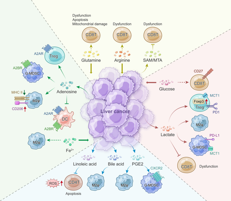
“Tumor cells often extract these nutrients from the tumor interstitial fluid (TIF) to hinder tumorolytic activities [10, 16, 96–101].”
BackgroundComprehensive and Integrative Genomic Characterization of Hepatocellular Carcinoma
Ally, A., Balasundaram, M., Carlsen, R., Chuah, E., Clarke, A., Dhalla, N., Holt, R. A., Jones, S. J. M., Lee, D., Ma, Y., et al. (2017) · Cell · doi:10.1016/j.cell.2017.05.046 · Cited by 2,490
Vogel, A., Meyer, T., Sapisochin, G., Salem, R., & Saborowski, A. (2022) · The Lancet · doi:10.1016/s0140-6736(22)01200-4 · Cited by 2,427
Understanding the Intersections between Metabolism and Cancer Biology
Heiden, M. G. V., & DeBerardinis, R. J. (2017) · Cell · doi:10.1016/j.cell.2016.12.039 · Cited by 2,369 · cited 1×
“Significant alterations in metabolic genes, including ALB , APOB , and IDH1/2 , also induce metabolic changes in PLC [77, 91].”
BackgroundOne-Carbon Metabolism in Health and Disease
Ducker, G. S., & Rabinowitz, J. D. (2017) · Cell Metabolism · doi:10.1016/j.cmet.2016.08.009 · Cited by 2,239 · cited 1×
“Folate-mediated one-carbon (1C) metabolism contributes to the availability of various building blocks for tumor cell proliferation [67–70], and the expression of central enzymes involved in 1C metabolism is largely dysregulated in PLC [71, 72].”
BackgroundObesity-induced gut microbial metabolite promotes liver cancer through senescence secretome
Yoshimoto, S., Loo, T. M., Atarashi, K., Kanda, H., Sato, S., Oyadomari, S., Iwakura, Y., Oshima, K., Morita, H., Hattori, M., et al. (2013) · Nature · doi:10.1038/nature12347 · Cited by 2,219 · cited 2×
“This leads to an immunosuppressive TME through PGE2 production in obesity-associated HCC mouse models [198, 224], potentially causing ICB resistance [225, 226].”
BackgroundLDHA-Associated Lactic Acid Production Blunts Tumor Immunosurveillance by T and NK Cells
Brand, A., Singer, K., Koehl, G. E., Kolitzus, M., Schoenhammer, G., Thiel, A., Matos, C., Bruss, C., Klobuch, S., Peter, K., et al. (2016) · Cell Metabolism · doi:10.1016/j.cmet.2016.08.011 · Cited by 2,039
Inhibitory effect of tumor cell-derived lactic acid on human T cells
Fischer, K., Hoffmann, P., Voelkl, S., Meidenbauer, N., Ammer, J., Edinger, M., Gottfried, E., Schwarz, S., Rothe, G., Hoves, S., et al. (2007) · Blood · doi:10.1182/blood-2006-07-035972 · Cited by 1,946 · cited 1×
“Oncometabolites, such as lactic acid [102–114], kynurenine [115–117], adenosine [117], 2-HG [118, 119], and prostaglandin E2 (PGE2) [120], typically antagonize the anti-tumor immune response and/or promote the immunosuppressive activities of TME components, ultimately leading to immune evasion.”
BackgroundSchulze, K., Imbeaud, S., Letouzé, E., Alexandrov, L. B., Calderaro, J., Rebouissou, S., Couchy, G., Meiller, C., Shinde, J., Soysouvanh, F., et al. (2015) · Nature Genetics · doi:10.1038/ng.3252 · Cited by 1,803 · cited 2×
“Apart from MASLD, HCC patients with etiologies of HCV infections or alcohol abuse also harbor unique genetic variations, which may impact the metabolic reprogramming and the TME [75, 158].”
BackgroundL-Arginine Modulates T Cell Metabolism and Enhances Survival and Anti-tumor Activity
Geiger, R., Rieckmann, J. C., Wolf, T., Basso, C., Feng, Y., Fuhrer, T., Kogadeeva, M., Picotti, P., Meissner, F., Mann, M., et al. (2016) · Cell · doi:10.1016/j.cell.2016.09.031 · Cited by 1,775 · cited 1×
“Intracellular arginine concentrations directly determine the metabolic fitness and functionality of activated T cells [100].”
BackgroundSerine, glycine and one-carbon units: cancer metabolism in full circle
Locasale, J. W. (2013) · Nature reviews. Cancer · doi:10.1038/nrc3557 · Cited by 1,727 · cited 1×
“Folate-mediated one-carbon (1C) metabolism contributes to the availability of various building blocks for tumor cell proliferation [67–70], and the expression of central enzymes involved in 1C metabolism is largely dysregulated in PLC [71, 72].”
BackgroundAn Interaction between Kynurenine and the Aryl Hydrocarbon Receptor Can Generate Regulatory T Cells
Mezrich, J. D., Fechner, J. H., Zhang, X., Johnson, B. P., Burlingham, W. J., & Bradfield, C. A. (2010) · The Journal of Immunology · doi:10.4049/jimmunol.0903670 · Cited by 1,674
Targeting lactate-fueled respiration selectively kills hypoxic tumor cells in mice
Sonveaux, P., Végran, F., Schroeder, T., Wergin, M., Verrax, J., Rabbani, Z. N., Saedeleer, C. J. D., Kennedy, K. M., Diepart, C., Jordan, B. F., et al. (2008) · Journal of Clinical Investigation · doi:10.1172/jci36843 · Cited by 1,635 · cited 1×
“On the other hand, excessive lactate generated by aerobic glycolysis also fuels neighboring oxygenated cells, leading to a metabolic symbiosis between glycolytic and oxidative metabolism [11–13].”
BackgroundGut microbiome–mediated bile acid metabolism regulates liver cancer via NKT cells
Ma, C., Han, M., Heinrich, B., Fu, Q., Zhang, Q., Sandhu, M., Agdashian, D., Terabe, M., Berzofsky, J. A., Fako, V., et al. (2018) · Science · doi:10.1126/science.aan5931 · Cited by 1,508 · cited 1×
“Secondary BA suppresses the recruitment of CXCR6 + tumorolytic NKT cells into the liver by reducing CXCL16 expression in liver sinusoidal endothelial cells (LSECs) [211–213].”
BackgroundMetabolic pathways promoting cancer cell survival and growth
Boroughs, L. K., & DeBerardinis, R. J. (2015) · Nature Cell Biology · doi:10.1038/ncb3124 · Cited by 1,499 · cited 1×
“In addition to glucose metabolism, other central metabolic pathways are often reprogrammed, leading to dysregulated nutrient depletion, oncometabolite accumulation, and signaling pathway perturbations in the TME [15].”
BackgroundPhosphoenolpyruvate Is a Metabolic Checkpoint of Anti-tumor T Cell Responses
Ho, P.-C., Bihuniak, J. D., Macintyre, A. N., Staron, M., Liu, X., Amezquita, R., Tsui, Y.-C., Cui, G., Micevic, G., Perales, J. C., et al. (2015) · Cell · doi:10.1016/j.cell.2015.08.012 · Cited by 1,462 · cited 1×
“Tumor cells often extract these nutrients from the tumor interstitial fluid (TIF) to hinder tumorolytic activities [10, 16, 96–101].”
BackgroundBile acid metabolites control TH17 and Treg cell differentiation
Hang, S., Paik, D., Yao, L., Kim, E., Trinath, J., Lu, J., Ha, S., Nelson, B. N., Kelly, S. P., Wu, L., et al. (2019) · Nature · doi:10.1038/s41586-019-1785-z · Cited by 1,457 · cited 1×
“Bile acid metabolites such as isoalloLCA and isoDCA promote Treg cell differentiation through FOXP3 induction, while 3-oxoLCA and isoLCA inhibit Th17 cell differentiation by targeting RORγt in vitro and in vivo [215–217].”
The reverse Warburg effect: Aerobic glycolysis in cancer associated fibroblasts and the tumor stroma
Pavlides, S., Whitaker-Menezes, D., Castello-Cros, R., Flomenberg, N., Witkiewicz, A. K., Frank, P. G., Casimiro, M. C., Wang, C., Fortina, P., Addya, S., et al. (2009) · Cell Cycle · doi:10.4161/cc.8.23.10238 · Cited by 1,391
Metabolic support of tumour-infiltrating regulatory T cells by lactic acid
Watson, M. J., Vignali, P., Mullett, S. J., Overacre-Delgoffe, A. E., Peralta, R. M., Grebinoski, S., Menk, A. V., Rittenhouse, N., DePeaux, K., Whetstone, R. D., et al. (2021) · Nature · doi:10.1038/s41586-020-03045-2 · Cited by 1,319
The cancer metabolic reprogramming and immune response
Xia, L., Oyang, L., Lin, J., Tan, S., Han, Y., Wu, N., Yi, P., Tang, L., Pan, Q., Rao, S., et al. (2021) · Molecular Cancer · doi:10.1186/s12943-021-01316-8 · Cited by 1,312 · cited 3×
“Mean-while, the aberrant consumption of macromolecules and metabolic substances leads to the production of numerous byproducts, and some of them could be harmful to immunosurveillance [10].”
BackgroundSelf-citationGenomic spectra of biliary tract cancer
Nakamura, H. K., Arai, Y., Totoki, Y., Shirota, T., Elzawahry, A., Kato, M., Hama, N., Hosoda, F., Urushidate, T., Ohashi, S., et al. (2015) · Nature Genetics · doi:10.1038/ng.3375 · Cited by 1,306
NASH limits anti-tumour surveillance in immunotherapy-treated HCC
Pfister, D., Núñez, N. G., Pinyol, R., Govaere, O., Pinter, M., Szydlowska, M., Gupta, R., Qiu, M., Deczkowska, A., Weiner, A., et al. (2021) · Nature · doi:10.1038/s41586-021-03362-0 · Cited by 1,294 · cited 1×
“Following anti-PD1 treatment, MASH also promoted aberrant activation of PD-1 + CD8 + T cells, leading to tissue damage, immune anergy, and reduced response to immunotherapy in pre-clinical HCC models [181].”
BackgroundDurvalumab plus Gemcitabine and Cisplatin in Advanced Biliary Tract Cancer
Oh, D.-Y., Ruth He, A., Qin, S., Chen, L.-T., Okusaka, T., Vogel, A., Kim, J. W., Suksombooncharoen, T., Ah Lee, M., Kitano, M., et al. (2022) · NEJM Evidence · doi:10.1056/evidoa2200015 · Cited by 1,217 · cited 1×
“The ICB-based regimen has also obtained impressive survival benefits in two recent phase 3 trials for the treat-ment of advanced biliary tract cancer, including iCCA [3, 4].”
BackgroundTargeting cancer metabolism in the era of precision oncology
Stine, Z. E., Schug, Z. T., Salvino, J. M., & Dang, C. V. (2022) · Nature Reviews Drug Discovery · doi:10.1038/s41573-021-00339-6 · Cited by 1,189 · cited 2×
“In recent decades, there has been limited progress in the use of compounds targeting specific metabolic alterations in the treatment of PLC, likely due to metabolic plasticity, drug specificity, and the heterogeneous metabolic microenvironment [127].”
BackgroundAbou‐Alfa, G. K., Macarulla, T. M., Javle, M. M., Kelley, R. K., Lubner, S. J., Adeva, J., Cleary, J. M., Catenacci, D. V. T., Borad, M. J., Bridgewater, J. A., et al. (2020) · The Lancet Oncology · doi:10.1016/s1470-2045(20)30157-1 · Cited by 1,159 · cited 1×
“In a phase 3 clinical trial for mIDH1 iCCA, Ivosidenib/AG120 was found to significantly improve the progression-free survival (PFS) of patients compared with placebo, although the absolute improvement in the median PFS appeared modest [166, 167] (Table 3).”
BackgroundCyclooxygenase-Dependent Tumor Growth through Evasion of Immunity
Zelenay, S., van der Veen, A. G., Böttcher, J. P., Snelgrove, K. J., Rogers, N. C., Acton, S. E., Chakravarty, P., Girotti, M. R., Marais, R., Quezada, S. A., et al. (2015) · Cell · doi:10.1016/j.cell.2015.08.015 · Cited by 1,146 · cited 1×
“Oncometabolites, such as lactic acid [102–114], kynurenine [115–117], adenosine [117], 2-HG [118, 119], and prostaglandin E2 (PGE2) [120], typically antagonize the anti-tumor immune response and/or promote the immunosuppressive activities of TME components, ultimately leading to immune evasion.”
BackgroundShort chain fatty acids in human gut and metabolic health
Blaak, E. E., Canfora, E. E., Theis, S., Frost, G., Groen, A. K., Mithieux, G., Nauta, A., Scott, K., Stahl, B., van Harsselaar, J., et al. (2020) · Beneficial Microbes · doi:10.3920/bm2020.0057 · Cited by 1,103 · cited 1×
“Butyrate is mainly produced by the gut microbiota during fermentation and is subsequently absorbed by the liver via the gut–liver axis [201].”
BackgroundIndoleamine 2,3-dioxygenase and tumor-induced tolerance
Munn, D. H., & Mellor, A. L. (2007) · Journal of Clinical Investigation · doi:10.1172/jci31178 · Cited by 1,093 · cited 1×
“Emerging evidence indicates that metabolic alterations in tumor cells affect the composition and function of surrounding cells [16, 17].”
BackgroundmTOR signalling and cellular metabolism are mutual determinants in cancer
Mossmann, D., Park, S., & Hall, M. N. (2018) · Nature reviews. Cancer · doi:10.1038/s41568-018-0074-8 · Cited by 1,088 · cited 1×
“Furthermore, the “Warburg effect” is promoted by TP53 mutations and PI3K/AKT/mTOR pathway activation in PLCs through upregulating related glycolytic enzymes [86, 87].”
BackgroundImmune surveillance by the liver
Jenne, C. N., & Kubes, P. (2013) · Nature Immunology · doi:10.1038/ni.2691 · Cited by 1,047 · cited 1×
“The liver immune system precludes gut-derived microbes and corresponding metabolites without evoking a systemic immune response, thus demonstrating immune privilege [209, 210].”
BackgroundT cell apoptosis by tryptophan catabolism
Fallarino, F., Grohmann, U., Vacca, C., Bianchi, R., Orabona, C., Spreca, A., Fioretti, M. C., & Puccetti, P. (2002) · Cell Death & Differentiation · doi:10.1038/sj.cdd.4401073 · Cited by 1,022 · cited 1×
“Oncometabolites, such as lactic acid [102–114], kynurenine [115–117], adenosine [117], 2-HG [118, 119], and prostaglandin E2 (PGE2) [120], typically antagonize the anti-tumor immune response and/or promote the immunosuppressive activities of TME components, ultimately leading to immune evasion.”
BackgroundThe cancer-immunity cycle: Indication, genotype, and immunotype
Mellman, I., Chen, D. S., Powles, T. B., & Turley, S. J. (2023) · Immunity · doi:10.1016/j.immuni.2023.09.011 · Cited by 1,018 · cited 1×
“Metabolic derangements within the TME are increasingly recognized as one of the most important factors halting the CIC [94].”
BackgroundLocasale, J., Grassian, A., Beroukhim, R., Meyerson, M., Wagner, G., Asara, J., Brugge, J., Heiden, M. V. V., & Cantley, L. (2012) · BMC Proceedings · doi:10.1186/1753-6561-6-s3-o15 · Cited by 976 · cited 1×
“On the other hand, excessive lactate generated by aerobic glycolysis also fuels neighboring oxygenated cells, leading to a metabolic symbiosis between glycolytic and oxidative metabolism [11–13].”
BackgroundMichelotti, G. A., Machado, M. V., & Diehl, A. M. (2013) · Nature Reviews Gastroenterology & Hepatology · doi:10.1038/nrgastro.2013.183 · Cited by 974 · cited 1×
“Folate-mediated one-carbon (1C) metabolism contributes to the availability of various building blocks for tumor cell proliferation [67–70], and the expression of central enzymes involved in 1C metabolism is largely dysregulated in PLC [71, 72].”
BackgroundProteomics identifies new therapeutic targets of early-stage hepatocellular carcinoma
Jiang, Y., Sun, A., Zhao, Y. C., Ying, W., Sun, H., Yang, X., Xing, B., Sun, W., Ren, L., Hu, B., et al. (2019) · Nature · doi:10.1038/s41586-019-0987-8 · Cited by 951 · cited 1×
“…proteomics and phosphoproteomics have revealed that targeting sterol O-acyltransferase 1 (SOAT1) to reduce cholesterol content in plasma membranes presents effective treat-ment options for early-stage HCC patients, which was further verified in a patient-derived tumor xenograft mouse model [58].”
BackgroundSelf-citationLactate modulation of immune responses in inflammatory versus tumour microenvironments
Certo, M., Tsai, C.-H., Pucino, V., Ho, P.-C., & Mauro, C. (2021) · Nature Reviews Immunology · doi:10.1038/s41577-020-0406-2 · Cited by 924 · cited 1×
“The well-known “Warburg effect” rearranges glucose metabolism to produce excessive lactic acid, which has emerged as an important regulator in promoting immune evasion in PLC [134, 135].”
BackgroundBacterial metabolism of bile acids promotes generation of peripheral regulatory T cells
Campbell, C., McKenney, P. T., Konstantinovsky, D., Isaeva, O. I., Schizas, M., Verter, J., Mai, C., Jin, W.-B., Guo, C.-J., Violante, S., et al. (2020) · Nature · doi:10.1038/s41586-020-2193-0 · Cited by 901
Integrated Proteogenomic Characterization of HBV-Related Hepatocellular Carcinoma.
Gao, Q., Zhu, H.-W., Dong, L.-Q., Shi, W.-W., Chen, R., Song, Z.-J., Huang, C., Li, J., Dong, X., Zhou, Y.-T., et al. (2019) · Cell · doi:10.1016/j.cell.2019.10.038 · Cited by 895 · cited 2×
“We and others have identified numerous putative driver genes that reshape PLC metabolism [29, 30, 75– 79] (Table 1).”
BackgroundSelf-citationKumagai, S., Koyama, S., Itahashi, K., Tanegashima, T., Lin, Y.-T., Togashi, Y., Kamada, T., Irie, T., Okumura, G., Kono, H., et al. (2022) · Cancer Cell · doi:10.1016/j.ccell.2022.01.001 · Cited by 892
Metabolism of immune cells in cancer
Leone, R. D., & Powell, J. D. (2020) · Nature Reviews Cancer · doi:10.1038/s41568-020-0273-y · Cited by 877 · cited 4×
“With the advent of immunome-tabolism in cancer treatment [18–22], greater attention should be given to the interplay of metabolism-related immune signaling.”
BackgroundInfluentialThe CASTOR Proteins Are Arginine Sensors for the mTORC1 Pathway
Chantranupong, L., Scaria, S. M., Saxton, R. A., Gygi, M. P., Shen, K., Wyant, G. A., Wang, T., Harper, J. W., Gygi, S. P., & Sabatini, D. M. (2016) · Cell · doi:10.1016/j.cell.2016.02.035 · Cited by 843
Zhang, X., Coker, O. O., Chu, E. S., Fu, K., Lau, H. C. H., Wang, Y.-X., Chan, A. W. H., Wei, H., Yang, X., Sung, J. J. Y., et al. (2021) · Gut · doi:10.1136/gutjnl-2019-319664 · Cited by 841 · cited 1×
“Using 16S rRNA sequencing and serum metabolomic analysis, another independent group found that the development of MASLD-HCC induced by a high-cholesterol diet in mice was closely correlated to dysbiosis of the gut microbiota and the resultant alterations in metabolites [199].”
MethodHusain, Z., Huang, Y., Seth, P., & Sukhatme, V. P. (2013) · The Journal of Immunology · doi:10.4049/jimmunol.1202702 · Cited by 827
Ren, Z., Li, A., Jiang, J., Zhou, L., Yu, Z., Lu, H., Xie, H., Chen, X., Shao, L., Zhang, R., et al. (2018) · Gut · doi:10.1136/gutjnl-2017-315084 · Cited by 782 · cited 1×
“Multi-omics studies incorporating 16S rRNA MiSeq sequencing in different cohorts from various regions of China have also identified gut microbes as non-invasive biomarkers for the early diagnosis of PLC [204–206].”
BackgroundTrans-ancestry mutational landscape of hepatocellular carcinoma genomes
Totoki, Y., Tatsuno, K., Covington, K. R., Ueda, H., Creighton, C. J., Kato, M., Tsuji, S., Donehower, L. A., Slagle, B. L., Nakamura, H., et al. (2014) · Nature Genetics · doi:10.1038/ng.3126 · Cited by 781 · cited 1×
“Apart from MASLD, HCC patients with etiologies of HCV infections or alcohol abuse also harbor unique genetic variations, which may impact the metabolic reprogramming and the TME [75, 158].”
BackgroundNAFLD causes selective CD4+ T lymphocyte loss and promotes hepatocarcinogenesis
Ma, C., Kesarwala, A. H., Eggert, T., Medina-Echeverz, J., Kleiner, D. E., Jin, P., Stroncek, D. F., Terabe, M., Kapoor, V., ElGindi, M., et al. (2016) · Nature · doi:10.1038/nature16969 · Cited by 766 · cited 1×
“This mediates selective loss of intrahepatic CD4 + T lymphocytes due to their great mitochondrial mass [143].”
BackgroundThe Tumor Suppressor p53 Down-Regulates Glucose Transporters GLUT1 and GLUT4 Gene Expression
Bar‐Yoseph, F., Armoni, M., & Karnieli, E. (2004) · Cancer Research · doi:10.1158/0008-5472.can-03-0846 · Cited by 758 · cited 1×
“Furthermore, the “Warburg effect” is promoted by TP53 mutations and PI3K/AKT/mTOR pathway activation in PLCs through upregulating related glycolytic enzymes [86, 87].”
BackgroundNavigating metabolic pathways to enhance antitumour immunity and immunotherapy
Li, X., Wenes, M., Romero, P. J., Huang, S. C., Fendt, S., & Ho, P. (2019) · Nature Reviews Clinical Oncology · doi:10.1038/s41571-019-0203-7 · Cited by 750 · cited 1×
“Consequently, they adapt metabolic characteristics to fulfill their duties [19].”
BackgroundMetabolic programming and immune suppression in the tumor microenvironment
Arner, E. N., & Rathmell, J. C. (2023) · Cancer Cell · doi:10.1016/j.ccell.2023.01.009 · Cited by 715 · cited 1×
“Glucose, amino acids, and fatty acids are critical energy sources not only for tumors, but also for proliferative cells, particularly anti-tumor immune cells [95].”
BackgroundMetabolomics in cancer research and emerging applications in clinical oncology
Schmidt, D. R., Patel, R., Kirsch, D. G., Lewis, C. A., Heiden, M. G. V., & Locasale, J. W. (2021) · CA A Cancer Journal for Clinicians · doi:10.3322/caac.21670 · Cited by 675 · cited 1×
“Tumor cells often exhibit a high dependence on a reprogrammed metabolic state for stress adaptation and infinite proliferation, which can be leveraged for non-invasive cancer diagnosis [6–8].”
BackgroundKim, J.-W., Gao, P., Liu, Y.-C., Semenza, G. L., & Dang, C. V. (2007) · Molecular and Cellular Biology · doi:10.1128/mcb.00440-07 · Cited by 658 · cited 1×
“High levels of GLUT1 expression are also associated with poor prognosis in both HCC and iCCA [80–84].”
BackgroundAlternative macrophage activation and metabolism
Odegaard, J. I., & Chawla, A. (2011) · Annual Review of Pathology: Mechanisms of Disease · doi:10.1146/annurev-pathol-011110-130138 · Cited by 640 · cited 1×
“On the other hand, macrophage polarization is tightly correlated with metabolic rearrangement [188].”
BackgroundThe gut microbiome and liver cancer: mechanisms and clinical translation
Yu, L.-X., & Schwabe, R. F. (2017) · Nature Reviews Gastroenterology & Hepatology · doi:10.1038/nrgastro.2017.72 · Cited by 634 · cited 1×
“Emerging studies have addressed the pivotal roles of the gut microbiota in PLC pathogenesis and anti-tumor therapy [5, 193–196].”
BackgroundYang, Z., Yan, C., Ma, J., Peng, P., Ren, X., Cai, S., Shen, X., Wu, Y. C., Zhang, S., Wang, X., et al. (2023) · Nature Metabolism · doi:10.1038/s42255-022-00710-w · Cited by 629 · cited 1×
“…tumor progression, lactate accumulation also causes lactylation of adenylate kinase 2 (AK2) at K28, compromising its kinase activity and disrupting energy homeostasis in HCC cells, thereby facilitating tumor proliferation, invasion, and metastasis as shown in several xenograft mouse models [39].”
BackgroundSelf-citationWnt–β-catenin signalling in liver development, health and disease
Perugorria, M. J., Olaizola, P., Labiano, I., Esparza-Baquer, A., Marzioni, M., Marín, J. J. G., Bujanda, L., & Bañales, J. M. (2018) · Nature Reviews Gastroenterology & Hepatology · doi:10.1038/s41575-018-0075-9 · Cited by 622
Molecular and histological correlations in liver cancer
Caldéraro, J., Ziol, M., Paradis, V., & Zucman‐Rossi, J. (2019) · Journal of Hepatology · doi:10.1016/j.jhep.2019.06.001 · Cited by 619 · cited 1×
“Consistent with the histological features [25], both iHCC1 and iHCC2 exhibit hepatocyte differentiation and maturation, whereas iHCC3 is associated with proliferation and immune activation [24].”
BackgroundCalvisi, D. F., Wang, C., Ho, C., Ladu, S., Lee, S. A., Mattu, S., Destefanis, G., Delogu, S., Zimmermann, A., Ericsson, J., et al. (2010) · Gastroenterology · doi:10.1053/j.gastro.2010.12.006 · Cited by 611 · cited 1×
“Concordantly, de novo lipogenesis gradually increases from normal liver tissue to liver tumors and is generally associated with advanced HCC and worse patient prognoses [49–52].”
BackgroundHuman gut bacteria produce ΤΗ17-modulating bile acid metabolites
Paik, D., Yao, L., Zhang, Y., Bae, S., D’Agostino, G. D., Zhang, M., Kim, E., Franzosa, E. A., Avila-Pacheco, J., Bisanz, J. E., et al. (2022) · Nature · doi:10.1038/s41586-022-04480-z · Cited by 594
Self-citationDisturbed mitochondrial dynamics in CD8+ TILs reinforce T cell exhaustion
Yu, Y.-R., Imrichova, H., Wang, H., Chao, T., Xiao, Z., Gao, M., Rincon-Restrepo, M., Franco, F., Genolet, R., Cheng, W.-C., et al. (2020) · Nature Immunology · doi:10.1038/s41590-020-0793-3 · Cited by 593
Sullivan, M. R., Danai, L. V., Lewis, C. A., Chan, S. H., Gui, D. Y., Kunchok, T., Dennstedt, E. A., Vander Heiden, M. G., & Muir, A. (2019) · eLife · doi:10.7554/elife.44235 · Cited by 588
Living in the liver: hepatic infections
Protzer, U., Maini, M. K., & Knolle, P. A. (2012) · Nature reviews. Immunology · doi:10.1038/nri3169 · Cited by 577 · cited 1×
“The liver immune system precludes gut-derived microbes and corresponding metabolites without evoking a systemic immune response, thus demonstrating immune privilege [209, 210].”
BackgroundSuppression of antitumor T cell immunity by the oncometabolite (R)-2-hydroxyglutarate
Bunse, L., Pusch, S., Bunse, T., Sahm, F., Sanghvi, K., Friedrich, M., Alansary, D., Sonner, J. K., Green, E., Deumelandt, K., et al. (2018) · Nature Medicine · doi:10.1038/s41591-018-0095-6 · Cited by 566 · cited 1×
“Oncometabolites, such as lactic acid [102–114], kynurenine [115–117], adenosine [117], 2-HG [118, 119], and prostaglandin E2 (PGE2) [120], typically antagonize the anti-tumor immune response and/or promote the immunosuppressive activities of TME components, ultimately leading to immune evasion.”
BackgroundZheng, Y., Wang, T., Tu, X., Huang, Y., Zhang, H., Tan, D., Jiang, W., Cai, S., Zhao, P., Song, R., et al. (2019) · Journal for ImmunoTherapy of Cancer · doi:10.1186/s40425-019-0650-9 · Cited by 550
Dysregulated Microbial Fermentation of Soluble Fiber Induces Cholestatic Liver Cancer
Singh, V., Yeoh, B. S., Chassaing, B., Xiao, X., Saha, P., Aguilera Olvera, R., Lapek, J. D., Zhang, L., Wang, W.-B., Hao, S., et al. (2018) · Cell · doi:10.1016/j.cell.2018.09.004 · Cited by 532 · cited 1×
“In wild-type mice, a diet rich in inulin-enriched soluble fiber leads to microbiota-dependent cholestasis, hepatocyte death, and subsequent neutrophilic inflammation in the liver, culminating in HCC [231].”
BackgroundGu, J., Zhou, J., Chen, Q., Xu, X., Gao, J., Li, X., Shao, Q., Zhou, B., Zhou, H., Wei, S., et al. (2022) · Cell Reports · doi:10.1016/j.celrep.2022.110986 · Cited by 529 · cited 1×
“Considering the critical role of Treg cells in immunotherapy, lactylation of MOESIN in Treg cells may predict the response of anti-PD-1 therapy in HCC [139].”
BackgroundOne-carbon metabolism in cancer
Newman, A. C., & Maddocks, O. D. K. (2017) · British Journal of Cancer · doi:10.1038/bjc.2017.118 · Cited by 525
Zhu, A. X., Macarulla, T. M., Javle, M. M., Kelley, R. K., Lubner, S. J., Adeva, J., Cleary, J. M., Catenacci, D. V. T., Borad, M. J., Bridgewater, J. A., et al. (2021) · JAMA Oncology · doi:10.1001/jamaoncol.2021.3836 · Cited by 524
CD8+ T cells in the cancer-immunity cycle
Giles, J. R., Globig, A.-M., Kaech, S. M., & Wherry, E. J. (2023) · Immunity · doi:10.1016/j.immuni.2023.09.005 · Cited by 507 · cited 1×
“Recent advances in cancer immunology have highlighted metabolic fuels/nutrients as a fourth signal beyond the Advanced and/ or treatment-refractory solid tumors three-signal model for effective T-cell priming and differentiation [179].”
BackgroundTumor-Expressed IDO Recruits and Activates MDSCs in a Treg-Dependent Manner
Holmgaard, R., Zamarin, D., Li, Y., Gasmi, B., Munn, D. H., Allison, J. P., Merghoub, T., & Wolchok, J. D. (2015) · Cell Reports · doi:10.1016/j.celrep.2015.08.077 · Cited by 496 · cited 1×
“Oncometabolites, such as lactic acid [102–114], kynurenine [115–117], adenosine [117], 2-HG [118, 119], and prostaglandin E2 (PGE2) [120], typically antagonize the anti-tumor immune response and/or promote the immunosuppressive activities of TME components, ultimately leading to immune evasion.”
BackgroundBehary, J., Amorim, N. M. L., Jiang, X., Raposo, A. E., Gong, L., McGovern, E., Ibrahim, R., Chu, F., Stephens, C., Jebeili, H., et al. (2021) · Nature Communications · doi:10.1038/s41467-020-20422-7 · Cited by 483 · cited 2×
“Ex vivo studies showed that gut bacteria convert dietary fiber into short-chain fatty acids in MASLD-HCC, leading to an immunosuppressive TME with an elevated CD4 + Treg cell/ CD8 + T-cell ratio [208].”
BackgroundLoo, T. M., Kamachi, F., Watanabe, Y., Yoshimoto, S., Kanda, H., Arai, Y., Nakajima-Takagi, Y., Iwama, A., Koga, T., Sugimoto, Y., et al. (2017) · Cancer Discovery · doi:10.1158/2159-8290.cd-16-0932 · Cited by 478 · cited 1×
“This leads to an immunosuppressive TME through PGE2 production in obesity-associated HCC mouse models [198, 224], potentially causing ICB resistance [225, 226].”
BackgroundChen, Y., Ramjiawan, R. R., Reiberger, T., Ng, M. R., Hato, T., Huang, Y., Ochiai, H., Kitahara, S., Unan, E. C., Reddy, T. P., et al. (2015) · Hepatology · doi:10.1002/hep.27665 · Cited by 451 · cited 1×
“Chen et al. proposed that sorafenib treatment increases intra-tumoral hypoxia, promoting immunosuppression through the stromal cell-derived factor 1α (SDF1α)-CXCR4 axis-induced Treg cells/M2φ accumulation in orthotopic HCC mouse models [152].”
BackgroundMetabolic Characterization of Hepatocellular Carcinoma Using Nontargeted Tissue Metabolomics
Huang, Q., Tan, Y., Yin, P., Ye, G., Gao, P., Lu, X., Wang, H., & Xu, G. (2013) · Cancer Research · doi:10.1158/0008-5472.can-13-0308 · Cited by 420 · cited 2×
“Moreover, numerous studies indicate enhanced amino acid metabolism in liver tumors compared to non-tumor tissues [1, 31, 37, 60, 61].”
BackgroundDong, L., Lu, D., Chen, R., Lin, Y., Zhu, H., Zhang, Z., Cai, S., Cui, P., Song, G., Rao, D., et al. (2021) · Cancer Cell · doi:10.1016/j.ccell.2021.12.006 · Cited by 410 · cited 2×
“We and others have identified numerous putative driver genes that reshape PLC metabolism [29, 30, 75– 79] (Table 1).”
BackgroundSelf-citationWang, H., Zhang, H., Wang, Y., Brown, Z. J., Xia, Y., Huang, Z., Shen, C., Hu, Z., Beane, J., Ansa-Addo, E. A., et al. (2021) · Journal of Hepatology · doi:10.1016/j.jhep.2021.07.032 · Cited by 398 · cited 1×
“In MASH-associated HCC mouse models, neutrophil extra-cellular traps (NETs) interact with naïve CD4 + T cells to facilitate its mitochondrial OXPHOS through TLR4 expression, contributing to their differentiation into immunosuppressive Treg cells [180].”
BackgroundLoeuillard, E. J., Yang, J., Buckarma, E. H., Wang, J., Liu, Y., Conboy, C. B., Pavelko, K. D., Li, Y., O’Brien, D. R., Wang, C., et al. (2020) · Journal of Clinical Investigation · doi:10.1172/jci137110 · Cited by 376
Gut microbiome in HCC – Mechanisms, diagnosis and therapy
Schwabe, R. F., & Greten, T. F. (2020) · Journal of Hepatology · doi:10.1016/j.jhep.2019.08.016 · Cited by 375 · cited 3×
“Changes in the gut microbiome disturb immune cell infiltration and function in the tumor microenvironment (TME) of PLC, potentially affecting the efficacy of current immunotherapies [5].”
BackgroundGLUT1 Expression Is Increased in Hepatocellular Carcinoma and Promotes Tumorigenesis
Amann, T., Maegdefrau, U., Hartmann, A., Agaimy, A., Marienhagen, J., Weiß, T. S., Stoeltzing, O., Warnecke, C., Schölmerich, J., Oefner, P. J., et al. (2009) · American Journal Of Pathology · doi:10.2353/ajpath.2009.080596 · Cited by 370 · cited 1×
“High levels of GLUT1 expression are also associated with poor prognosis in both HCC and iCCA [80–84].”
BackgroundElia, I., Rowe, J. H., Johnson, S. E., Joshi, S., Notarangelo, G., Kurmi, K., Weiss, S. A., Freeman, G. J., Sharpe, A. H., & Haigis, M. C. (2022) · Cell Metabolism · doi:10.1016/j.cmet.2022.06.008 · Cited by 362
The gluconeogenic enzyme PCK1 phosphorylates INSIG1/2 for lipogenesis
Xu, D., Wang, Z., Xia, Y., Shao, F., Xia, W., Wei, Y., Li, X., Qian, X., Lee, J.-H., Du, L., et al. (2020) · Nature · doi:10.1038/s41586-020-2183-2 · Cited by 360 · cited 1×
“Concordantly, de novo lipogenesis gradually increases from normal liver tissue to liver tumors and is generally associated with advanced HCC and worse patient prognoses [49–52].”
BackgroundInosine is an alternative carbon source for CD8+-T-cell function under glucose restriction
Wang, T., Gnanaprakasam, J. R., Chen, X., Kang, S., Xu, X., Sun, H., Liu, L., Rodgers, H., Miller, E., Cassel, T., et al. (2020) · Nature Metabolism · doi:10.1038/s42255-020-0219-4 · Cited by 343
Advances in PET imaging of cancer
Schwenck, J., Sonanini, D., Cotton, J. M., Rammensee, H., la Fougère, C., Zender, L., & Pichler, B. J. (2023) · Nature reviews. Cancer · doi:10.1038/s41568-023-00576-4 · Cited by 343 · cited 1×
“Due to significant glucose uptake in PLC bulk tissues, 18 F-2-deoxyglucose ( 18 FDG) positron emission tomography (PET) imaging is widely used for PLC diagnosis and progression monitoring [14].”
BackgroundHan, S., Bao, X., Zou, Y., Wang, L., Li, Y., Yang, L., Liao, A., Zhang, X., Jiang, X. M., Liang, D., et al. (2023) · Science Advances · doi:10.1126/sciadv.adg2697 · Cited by 328 · cited 1×
“Additionally, gut microbiome-derived metabolite D-lactate can shift TAMs from the M2 to M1 phenotype, remodeling the immunosuppressive TME in HCC mice [111, 228].”
BackgroundLipid alterations in chronic liver disease and liver cancer
Paul, B., Lewińska, M., & Andersen, J. B. (2022) · JHEP Reports · doi:10.1016/j.jhepr.2022.100479 · Cited by 326 · cited 1×
“High-cholesterol diets induce HCC in mice, partly through dysregulation of metabolism and calcium signaling [55–57].”
BackgroundMitochondrial fragmentation limits NK cell-based tumor immunosurveillance
Zheng, X., Qian, Y., Fu, B., Jiao, D., Jiang, Y., Chen, P., Shen, Y., Zhang, H., Sun, R., Tian, Z., et al. (2019) · Nature Immunology · doi:10.1038/s41590-019-0511-1 · Cited by 316 · cited 1×
“Another study showed that hypoxic TME drives NK cell loss and dysfunction via mTOR-GTPase dynamin-related protein 1 (Drp1) mitochondrial fragmentation, leading to HCC immune evasion in human liver cancer and mouse liver models [106].”
BackgroundSun, L., Song, L., Wan, Q., Wu, G., Li, X., Wang, Y., Wang, J., Liu, Z., Zhong, X., He, X., et al. (2015) · Cell Research · doi:10.1038/cr.2015.33 · Cited by 313 · cited 1×
“Under glucose/glu-tamine-deprived conditions, overexpressed cMYC also activates the serine biosynthesis pathway to adopt metabolic switch through transcriptionally upregulated the final rate-limiting enzyme phosphoserine phosphatase in both HCC cell lines and xenograft mouse models [85].”
BackgroundArginine reprograms metabolism in liver cancer via RBM39
Mossmann, D., Müller, C., Park, S., Ryback, B. M., Colombi, M., Ritter, N., Weißenberger, D., Dazert, E., Coto‐Llerena, M., Nuciforo, S., et al. (2023) · Cell · doi:10.1016/j.cell.2023.09.011 · Cited by 309 · cited 1×
“Targeting RBM39 instead of circulating arginine may offer a way to reverse the oncogenic pathway triggered by high arginine pools in HCC cells, thereby avoiding the adverse side effects of circulating arginine-depleting therapy [63].”
BackgroundHalford, S. E. R., Veal, G. J., Wedge, S. R., Payne, G. S., Bacon, C. M., Sloan, P., Dragoni, I., Heinzmann, K., Potter, S., Salisbury, B. M., et al. (2023) · Clinical Cancer Research · doi:10.1158/1078-0432.ccr-22-2263 · Cited by 305
NAD+ Metabolism Maintains Inducible PD-L1 Expression to Drive Tumor Immune Evasion
Lv, H., Lv, G., Chen, C., Zong, Q., Jiang, G., Ye, D., Cui, X., He, Y., Xiang, W., Han, Q. Y., et al. (2020) · Cell Metabolism · doi:10.1016/j.cmet.2020.10.021 · Cited by 304 · cited 1×
“For instance, NAD + metabolism triggers PD-L1 expression on tumor cells, impairing the cytolytic activity of PD-1 + T cells through αKG-mediated epigenetic modifications [164].”
BackgroundOncometabolite d -2HG alters T cell metabolism to impair CD8 + T cell function
Notarangelo, G., Spinelli, J. B., Perez, E. M., Baker, G. J., Kurmi, K., Elia, I., Stopka, S. A., Baquer, G., Lin, J., Golby, A. J., et al. (2022) · Science · doi:10.1126/science.abj5104 · Cited by 298 · cited 1×
“Oncometabolites, such as lactic acid [102–114], kynurenine [115–117], adenosine [117], 2-HG [118, 119], and prostaglandin E2 (PGE2) [120], typically antagonize the anti-tumor immune response and/or promote the immunosuppressive activities of TME components, ultimately leading to immune evasion.”
BackgroundSelf-citationYang, X., Lu, Y., Hang, J., Zhang, J., Zhang, T., Huo, Y., Liu, J., Lai, S., Luo, D., Wang, L., et al. (2020) · Cancer Immunology Research · doi:10.1158/2326-6066.cir-20-0111 · Cited by 289 · cited 1×
“Oncometabolites, such as lactic acid [102–114], kynurenine [115–117], adenosine [117], 2-HG [118, 119], and prostaglandin E2 (PGE2) [120], typically antagonize the anti-tumor immune response and/or promote the immunosuppressive activities of TME components, ultimately leading to immune evasion.”
BackgroundZhang, J., Zhang, Q., Lou, Y., Fu, Q., Chen, Q., Wei, T., Yang, J., Tang, J., Wang, J., Chen, Y., et al. (2018) · Hepatology · doi:10.1002/hep.29681 · Cited by 286 · cited 1×
“M2-like TAMs upregulate the glycolysis pathway under hypoxic TME and produce excessive IL1β to facilitate EMT and subsequent metastasis in HCC [190].”
BackgroundMao, J., Wang, D., Long, J., Xu, Y., Lin, J., Song, Y., Xie, F., Xun, Z., Wang, Y., Wang, Y., et al. (2021) · Journal for ImmunoTherapy of Cancer · doi:10.1136/jitc-2021-003334 · Cited by 285
Self-citationAltorki, N., Keresztes, R., Port, J., Libby, D., Korst, R., Flieder, D., Ferrara, C. A., Yankelevitz, D., Subbaramaiah, K., Pasmantier, M., et al. (2003) · Journal of Clinical Oncology · doi:10.1200/jco.2003.07.127 · Cited by 277 · cited 1×
“…isocitrate dehydrogenase IDH1/2 inhibitor Enasidenib/Ivosidenib (for 2-HG production) [124], and the cyclooxygenase COX-2 inhibitor Celecoxib (for PGE2 accumulation) [125, 126] are being evaluated or approved to target cancer metabolism, aiming to enhance the efficacies of current therapies [127].”
BackgroundRIPK3 Orchestrates Fatty Acid Metabolism in Tumor-Associated Macrophages and Hepatocarcinogenesis
Wu, L., Zhang, X., Zheng, L., Zhao, H., Yan, G., Zhang, Q., Zhou, Y. J., Lei, J., Zhang, J., Wang, J., et al. (2020) · Cancer Immunology Research · doi:10.1158/2326-6066.cir-19-0261 · Cited by 277 · cited 1×
“In mouse and human HCC, the serine/threonine kinase RIPK3 is downregulated in macrophages, leading to FAO-dominated M2φ polarization via the ROS/Cas-pase1/PPAR pathway [189].”
BackgroundGut Microbiome Directs Hepatocytes to Recruit MDSCs and Promote Cholangiocarcinoma
Zhang, Q., Ma, C., Duan, Y., Heinrich, B., Rosato, U., Diggs, L. P., Ma, L., Roy, S., Fu, Q., Brown, Z. J., et al. (2020) · Cancer Discovery · doi:10.1158/2159-8290.cd-20-0304 · Cited by 266 · cited 2×
“This induces TLR4-dependent CXCL1 expression in hepatocytes, leading to the recruitment of CXCR2 + PMN-MDSC, and ultimately promoting an immunosuppressive TME [203].”
Background(2021) · Nature Reviews Disease Primers · doi:10.1038/s41572-021-00245-6 · Cited by 265
Antagonistic Inflammatory Phenotypes Dictate Tumor Fate and Response to Immune Checkpoint Blockade
Bonavita, E., Bromley, C. P., Jonsson, G., Pelly, V. S., Sahoo, S., Walwyn-Brown, K., Mensurado, S., Moeini, A., Flanagan, E., Bell, C. R., et al. (2020) · Immunity · doi:10.1016/j.immuni.2020.10.020 · Cited by 257 · cited 1×
“This leads to an immunosuppressive TME through PGE2 production in obesity-associated HCC mouse models [198, 224], potentially causing ICB resistance [225, 226].”
BackgroundDietary supplements and disease prevention — a global overview
Rautiainen, S., Manson, J. E., Lichtenstein, A. H., & Sesso, H. D. (2016) · Nature Reviews Endocrinology · doi:10.1038/nrendo.2016.54 · Cited by 252 · cited 1×
“People often consume dietary supplements for health benifits [230].”
BackgroundMetabolic rearrangements in primary liver cancers: cause and consequences
Satriano, L., Lewińska, M., Rodrigues, P. M., Bañales, J. M., & Andersen, J. B. (2019) · Nature Reviews Gastroenterology & Hepatology · doi:10.1038/s41575-019-0217-8 · Cited by 245 · cited 5×
“These risk factors may promote the initiation and progression of PLC through metabolic reprogramming [1, 23].”
BackgroundInfluentialLiu, L., Lu, Y., Martinez, J., Bi, Y., Lian, G., Wang, T., Milasta, S., Wang, J., Yang, M., Liu, G., et al. (2016) · Proceedings of the National Academy of Sciences · doi:10.1073/pnas.1518000113 · Cited by 243
Tedeschi, P. M., Markert, E., Gounder, M., Lin, H.-X., Dolfi, S., Chan, L., Qiu, J., Hirshfield, K., Boros, L., Bertino, J., et al. (2013) · doi:10.1158/1535-7163.targ-13-c151 · Cited by 242
Budhu, A., Roessler, S. K., Zhao, X., Yu, Z., Forgues, M., Ji, J., Karoly, E. D., Qin, L., Ye, Q., Jia, H., et al. (2013) · Gastroenterology · doi:10.1053/j.gastro.2013.01.054 · Cited by 238 · cited 1×
“Moreover, numerous studies indicate enhanced amino acid metabolism in liver tumors compared to non-tumor tissues [1, 31, 37, 60, 61].”
BackgroundPelly, V. S., Moeini, A., Roelofsen, L. M., Bonavita, E., Bell, C. R., Hutton, C., Blanco‐Gómez, A., Banyard, A., Bromley, C. P., Flanagan, E., et al. (2021) · Cancer Discovery · doi:10.1158/2159-8290.cd-20-1815 · Cited by 230 · cited 1×
“This leads to an immunosuppressive TME through PGE2 production in obesity-associated HCC mouse models [198, 224], potentially causing ICB resistance [225, 226].”
BackgroundLiang, Q., Teoh, N., Xu, L., Pok, S., Li, X., Chu, E. S., Chiu, J., Dong, L., Arfianti, A., Haigh, W. G., et al. (2018) · Nature Communications · doi:10.1038/s41467-018-06931-6 · Cited by 229
Schneider, K. M., Mohs, A., Gui, W., Galvez, E. J. C., Candels, L. S., Hoenicke, L., Muthukumarasamy, U., Holland, C. H., Elfers, C., Kilic, K., et al. (2022) · Nature Communications · doi:10.1038/s41467-022-31312-5 · Cited by 225 · cited 1×
“Increasing evidence suggests that the gut microbiota modulates immune responses in the TME of PLC [5, 194, 203, 207, 208].”
BackgroundLowery, M. A., Burris, H. A., Jankú, F., Shroff, R. T., Cleary, J. M., Azad, N. S., Goyal, L., Maher, E. A., Gore, L., Hollebecque, A., et al. (2019) · The Lancet. Gastroenterology & hepatology · doi:10.1016/s2468-1253(19)30189-x · Cited by 223
Iron alters macrophage polarization status and leads to steatohepatitis and fibrogenesis
Handa, P., Thomas, S., Morgan‐Stevenson, V., Maliken, B. D., Gochanour, E. M., Boukhar, S., Yeh, M. M., & Kowdley, K. V. (2019) · Journal of Leukocyte Biology · doi:10.1002/jlb.3a0318-108r · Cited by 222 · cited 1×
“Compared to M1φ, M2φ tends to polarize under low ferrous iron levels [132].”
BackgroundBayerl, F., Meiser, P., Donakonda, S., Hirschberger, A., Lacher, S. B., Pedde, A., Hermann, C. D., Elewaut, A., Knolle, M. A., Ramsauer, L., et al. (2023) · Immunity · doi:10.1016/j.immuni.2023.05.011 · Cited by 218 · cited 1×
“PGE2, a bioactive lipid generated from arachidonic acid, has recently been implicated in immune evasion through multiple mechanisms [145].”
BackgroundContreras‐Baeza, Y., Sandoval, P. Y., Alarcón, R., Galaz, A., Cortés‐Molina, F., Alegría, K., Baeza‐Lehnert, F., Arce‐Molina, R., Guequén, A., Flores, C. A., et al. (2019) · Journal of Biological Chemistry · doi:10.1074/jbc.ra119.009093 · Cited by 218 · cited 1×
“For instance, upregulated MCT4 expression in HCC cells contributes to lactate exportation and subsequent TME acidification, ultimately leading to CD8 + T-cell exhaustion and M2φ polarization [136–138].”
BackgroundLee, P.-C., Wu, C.-J., Hung, Y.-W., Lee, C. J., Chi, C.-T., Lee, I.-C., Yu-Lun, K., Chou, S.-H., Luo, J.-C., Hou, M.-C., et al. (2022) · Journal for ImmunoTherapy of Cancer · doi:10.1136/jitc-2022-004779 · Cited by 217 · cited 1×
“Lee et al. suggested that Lachnoclostridium , along with ursodeoxycholic and ursocholic acids, produce better responses to ICB treatment in patients with unresectable HCC [229].”
BackgroundGut microbiota–derived short‐chain fatty acids regulate group 3 innate lymphoid cells in HCC
Hu, C., Xu, B., Wang, X., Wan, W., Lu, J., Kong, D., Jin, Y., You, W., Sun, H., Mu, X., et al. (2022) · Hepatology · doi:10.1002/hep.32449 · Cited by 214 · cited 1×
“Thus, combining acetate supplementation with PD-1 blockades significantly boosts anti-tumor immunity [227].”
BackgroundHu, B., Yu, M., Ma, X., Sun, J., Liu, C., Wang, C., Wu, S., Fu, P., Yang, Z. F., He, Y., et al. (2022) · Cancer Discovery · doi:10.1158/2159-8290.cd-21-1022 · Cited by 213 · cited 1×
“The consequent accumulation of glucose in the TME stimulates the expression of the costimulatory molecule CD27 via mTOR–FOXM1 signaling in CD8 + T cells, thereby reinforcing the functions of cytotoxic T cells in both immunocompetent orthotopic and spontaneous HCC models [131].”
BackgroundBidkhori, G., Benfeitas, R., Klevstig, M., Zhang, C., Nielsen, J., Uhlén, M., Borén, J., & Mardinoğlu, A. (2018) · Proceedings of the National Academy of Sciences · doi:10.1073/pnas.1807305115 · Cited by 212 · cited 2×
“…retrieved from several public datasets, genome-wide metabolic models (GEMs) stratified HCC patients into three prognostic subgroups with significant differences in dysregulated kynurenine metabolism (iHCC1), WNT/β-catenin-related lipid metabolism (iHCC2), and PI3K/AKT/mTOR signaling (iHCC3) [24].”
BackgroundQin, W.-H., Yang, Z.-S., Li, M., Chen, Y., Zhao, X.-F., Qin, Y.-Y., Song, J.-Q., Wang, B.-B., Yuan, B., Cui, X.-L., et al. (2020) · Gastroenterology · doi:10.1053/j.gastro.2020.01.028 · Cited by 209 · cited 1×
“Of note, high serum cholesterol levels drive NK cell accumulation and subsequent lipid raft formation, further enhancing the anti-tumor activity in both DEN-induced HCC mouse models and Hepa1-6 (mouse hepatoma) subcutaneous models [184].”
BackgroundHuang, H., Ren, Z., Gao, X., Hu, X., Zhou, Y. M., Jiang, J., Lu, H., Yin, S., Ji, J., Zhou, L., et al. (2020) · Genome Medicine · doi:10.1186/s13073-020-00796-5 · Cited by 200 · cited 1×
“Multi-omics studies incorporating 16S rRNA MiSeq sequencing in different cohorts from various regions of China have also identified gut microbes as non-invasive biomarkers for the early diagnosis of PLC [204–206].”
BackgroundLoss of SIRT5 promotes bile acid-induced immunosuppressive microenvironment and hepatocarcinogenesis
Sun, R., Zhang, Z., Bao, R., Guo, X., Gu, Y., Yang, W., Wei, J., Chen, X., Tong, L., Meng, J., et al. (2022) · Journal of Hepatology · doi:10.1016/j.jhep.2022.02.030 · Cited by 190 · cited 1×
“Sun et al. reported that SIRT5 expression is repressed in tumor cells, which leads to aberrant BA biosynthesis in the peroxisome and subsequent M2φ-induced immunosuppression in oncogene-induced HCC mouse models [144].”
BackgroundJia, X., Lu, S., Zeng, Z., Liu, Q., Dong, Z., Chen, Y., Zhu, Z., Hong, Z., Zhang, T., Du, G., et al. (2019) · Hepatology · doi:10.1002/hep.30852 · Cited by 184
Deng, H., Kan, A., Lyu, N., He, M. X., Huang, X., Qiao, S., Li, S., Lu, W., Xie, Q., Chen, H., et al. (2021) · Journal for ImmunoTherapy of Cancer · doi:10.1136/jitc-2020-002305 · Cited by 181 · cited 1×
“Simultaneously, tumor-derived lactate induces programmed cell death ligand 1 (PD-L1) expression in infiltrated neutrophils through the MCT1/NF-κB/COX-2 pathway, thus counteracting the efficacy of lenvatinib monotherapy in HCC mouse models [142].”
BackgroundDinardo, C. D., Stein, A. S., Stein, E. M., Fathi, A. T., Frankfurt, O., Schuh, A. C., Döhner, H., Martinelli, G., Patel, P. A., Raffoux, E., et al. (2020) · Journal of Clinical Oncology · doi:10.1200/jco.20.01632 · Cited by 180 · cited 1×
“…inhibitor Oleclumab (for adenosine conversion) [123], the isocitrate dehydrogenase IDH1/2 inhibitor Enasidenib/Ivosidenib (for 2-HG production) [124], and the cyclooxygenase COX-2 inhibitor Celecoxib (for PGE2 accumulation) [125, 126] are being evaluated or approved to target cancer…”
BackgroundMetabolic reprogramming in cholangiocarcinoma
Raggi, C., Taddei, M. L., Rae, C. J., Braconi, C., & Marra, F. (2022) · Journal of Hepatology · doi:10.1016/j.jhep.2022.04.038 · Cited by 179 · cited 1×
“Leveraging genetically engineered mouse models of iCCA, IDH mutations were shown to increase the production of D-2-hydroxyglutarate (D-2-HG), affecting α-ketoglutarate (αKG)-dependent dioxygenases involved in DNA repair and epigenetic remodeling [92].”
Lipid Metabolic Reprogramming in Hepatocellular Carcinoma
Nakagawa, H., Hayata, Y., Kawamura, S., Yamada, T., Fujiwara, N., & Koike, K. (2018) · Cancers · doi:10.3390/cancers10110447 · Cited by 176 · cited 1×
“Lipid metabolic pathways, including fatty acid synthase (FASN) and stearoyl-COA desaturase (SCD) signaling, also sustain cancer stem cells in HCC, contributing to metastasis and drug resistance [54].”
BackgroundOGDHL silencing promotes hepatocellular carcinoma by reprogramming glutamine metabolism
Dai, W., Xu, L., Yu, X., Zhang, G., Guo, H., Liu, H., Song, G., Weng, S., Dong, L., Zhu, J., et al. (2019) · Journal of Hepatology · doi:10.1016/j.jhep.2019.12.015 · Cited by 174 · cited 1×
“Glutamine, the most abundant amino acid in human blood, is a key carbon source for de novo lipogenesis in mitochondrial dysfunctional HCC [64].”
BackgroundFang, Y., Liu, W., Tang, Z., Ji, X. L., Zhou, Y., Song, S., Tian, M., Tao, C., Huang, R., Zhu, G., et al. (2022) · Hepatology · doi:10.1002/hep.32348 · Cited by 170
Chen, D., Ning, W.-R., Jiang, Z.-Z., Peng, Z.-P., Zhu, L., Zhuang, S., Kuang, D., Zheng, L., & Wu, Y. (2019) · Journal of Hepatology · doi:10.1016/j.jhep.2019.04.007 · Cited by 170 · cited 1×
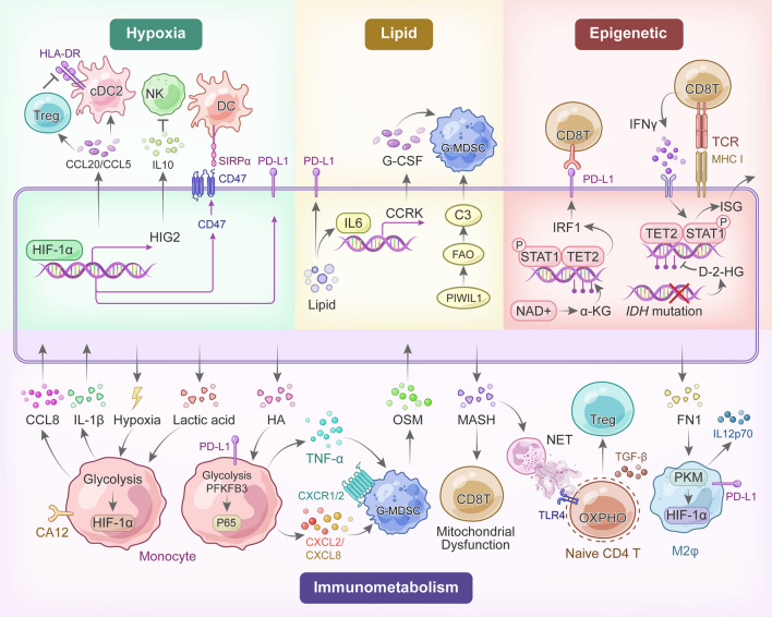
“Chen et al. proposed that tumor-derived hyaluronan (HA) fragments induce aerobic glycolysis in monocytes and upregulate PD-L1 expression through the PFKFB3-NF-κB pathway [185].”
BackgroundZhu, Q., Zhou, L., Yang, Z., Lai, M.-C., Xie, H., Wu, L., Xing, C., Zhang, F., & Zheng, S. (2011) · Medical Oncology · doi:10.1007/s12032-011-9912-1 · Cited by 168
Britten, C. D., Garrett-Mayer, E. S., Chin, S. H., Shirai, K., Öğretmen, B., Bentz, T. A., Brisendine, A., Anderton, K., Cusack, S., Maines, L. W., et al. (2017) · Clinical Cancer Research · doi:10.1158/1078-0432.ccr-16-2363 · Cited by 168
Wang, S., Wu, Q., Chen, T., Su, R., Pan, C., Qian, J., Huang, H., Yin, S., Xie, H., Zhou, L., et al. (2022) · Journal of Hepatology · doi:10.1016/j.jhep.2022.03.011 · Cited by 168 · cited 1×
“Thus, blocking CD47 on the cell surface enhances NK cell-mediated anti-tumor immunity in the hypoxic microenvironment of HCC [155].”
BackgroundLipid Remodeling in Hepatocyte Proliferation and Hepatocellular Carcinoma
Hall, Z., Chiarugi, D., Charidemou, E., Leslie, J., Scott, E., Pellegrinet, L., Allison, M., Mocciaro, G., Anstee, Q. M., Evan, G. I., et al. (2020) · Hepatology · doi:10.1002/hep.31391 · Cited by 164 · cited 1×
“High-cholesterol diets induce HCC in mice, partly through dysregulation of metabolism and calcium signaling [55–57].”
BackgroundHypoxia‐driven immunosuppression by Treg and type‐2 conventional dendritic cells in HCC
Suthen, S., Lim, C. J., Nguyen, P. H. D., Dutertre, C., Lai, H. L. H., Wasser, M., Chua, C. J. H., Lim, T. K. H., Leow, W. Q., Loh, T. J., et al. (2022) · Hepatology · doi:10.1002/hep.32419 · Cited by 161 · cited 1×
“Subsequently, in vitro assays demonstrated that the direct interaction of infiltrated Treg cells with cDC2 mediates HLA-DR loss, a critical antigen presentation molecule required for anti-tumor T-cell activation [153].”
Backgroundβ-catenin-activated hepatocellular carcinomas are addicted to fatty acids
Senni, N., Savall, M., Granados, D. C., Alves‐Guerra, M., Sartor, C., Lagoutte, I., Gougèlet, A., Terris, B., Gilgenkrantz, H., Perret, C., et al. (2018) · Gut · doi:10.1136/gutjnl-2017-315448 · Cited by 160 · cited 1×
“Wnt-β-catenin signaling is often hyperactivated, promoting PLC growth and dissemination [88]. β-catenin (encoded by CTNNB1 ) oncogenic activation in HCC cells induces FAO through the transcription factor peroxisome proliferator-activated receptor α (PPARα) [89].”
BackgroundWu, M., Shi, L., Dubrot, J., Merritt, J., Vijay, V., Wei, T.-Y., Kessler, E., Olander, K. E., Adil, R., Pankaj, A., et al. (2021) · Cancer Discovery · doi:10.1158/2159-8290.cd-21-1077 · Cited by 158 · cited 1×
“Consequently, the IDH1/2 inhibitor AG120 synergizes with ICBs for advancing immunotherapy in the treatment of mIDH1-driven genetically engineered mouse models [165].”
BackgroundAdebayo Michael, A. O., Ko, S., Tao, J., Moghe, A., Yang, H., Xu, M., Russell, J. O., Pradhan-Sundd, T., Liu, S., Singh, S., et al. (2019) · Cell Metabolism · doi:10.1016/j.cmet.2019.01.002 · Cited by 155 · cited 2×
“Glutamine addiction in glutamine synthetase (GS)-overexpressing HCC supports mTOR-dependent cell proliferation and survival in clinically relevant HCC models [65].”
BackgroundGCN2 inhibition sensitizes arginine-deprived hepatocellular carcinoma cells to senolytic treatment
Missiaen, R., Anderson, N. M., Kim, L. C., Nance, B., Burrows, M., Skuli, N., Carens, M., Riscal, R., Steensels, A., Li, F., et al. (2022) · Cell Metabolism · doi:10.1016/j.cmet.2022.06.010 · Cited by 154 · cited 1×
“Thus, combining GCN2 inhibition with senolytic agents could be an effective treatment strategy in arginine-deprived HCC cells [62].”
BackgroundIdentification of the Consistently Altered Metabolic Targets in Human Hepatocellular Carcinoma
Nwosu, Z. C., Megger, D. A., Hammad, S., Sitek, B., Roessler, S. K., Ebert, M. P. A., Meyer, C., & Dooley, S. (2017) · Cellular and Molecular Gastroenterology and Hepatology · doi:10.1016/j.jcmgh.2017.05.004 · Cited by 154 · cited 3×
“Moreover, numerous studies indicate enhanced amino acid metabolism in liver tumors compared to non-tumor tissues [1, 31, 37, 60, 61].”
BackgroundMcDonald, B., Zucoloto, A. Z., Yu, I.-L., Burkhard, R., Brown, K., Geuking, M. B., & McCoy, K. D. (2020) · Cell Host & Microbe · doi:10.1016/j.chom.2020.07.014 · Cited by 152 · cited 1×
“Additionally, gut microbiome-derived metabolite D-lactate can shift TAMs from the M2 to M1 phenotype, remodeling the immunosuppressive TME in HCC mice [111, 228].”
BackgroundWang, M., Wu, H., Fu, G., Zhang, H., Zhou, X., Tang, L., Dong, L., Qin, C., Huang, S., Zhao, L., et al. (2015) · Hepatology · doi:10.1002/hep.28415 · Cited by 149
Wabitsch, S., McCallen, J., Kamenyeva, O., Ruf, B., McVey, J. C., Kabát, J., Walz, J. S., Rotman, Y., Bauer, K. C., Craig, A. J., et al. (2022) · Journal of Hepatology · doi:10.1016/j.jhep.2022.03.010 · Cited by 147 · cited 1×
“This impairment diminishes the efficacy of ICB therapy, which could be salvaged by metformin [182].”
BackgroundXu, M., Zhou, C., Weng, J., Chen, Z., Zhou, Q., Gao, J., Shi, G., Ke, A., Ren, N., Sun, H., et al. (2022) · Journal of Experimental & Clinical Cancer Research · doi:10.1186/s13046-022-02458-3 · Cited by 145 · cited 1×
“For instance, upregulated MCT4 expression in HCC cells contributes to lactate exportation and subsequent TME acidification, ultimately leading to CD8 + T-cell exhaustion and M2φ polarization [136–138].”
BackgroundPharmaco-proteogenomic characterization of liver cancer organoids for precision oncology
Ji, S., Li, F., Fu, Z., Wu, G., Wu, Y. C., Lin, Y., Lu, D., Song, Y., Cui, P., Yang, Z., et al. (2023) · Science Translational Medicine · doi:10.1126/scitranslmed.adg3358 · Cited by 142 · cited 1×
“Glucose-6-phosphate dehydrogenase (G6PD) was identified as a potential target in the subtype with an enriched drug metabolism pathway, consistent with the previous results [32–35].”
ResultSelf-citationMurai, H., Kodama, T., Maesaka, K., Tange, S., Motooka, D., Suzuki, Y., Shigematsu, Y., Inamura, K., Mise, Y., Saiura, A., et al. (2022) · Hepatology · doi:10.1002/hep.32573 · Cited by 141 · cited 1×
“Multi-omics study showed lipid accumulation in HCC elevates PD-L1 expression, inducing an immunosuppressive TME [28].”
BackgroundMetabolic reprogramming and its clinical implication for liver cancer
Yang, F., Hilakivi-Clarke, L. A., Shaha, A., Wang, Y., Wang, X., Deng, Y., Lai, J., & Kang, N. (2023) · Hepatology · doi:10.1097/hep.0000000000000005 · Cited by 140 · cited 1×
“It has been demonstrated that liver cancer cells typically utilize glycolysis under hypoxic conditions to produce lactate via lactic dehydrogenase (LDH) [38].”
BackgroundLiu, J., Geng, W., Sun, H., Liu, C., Huang, F., Cao, J., Xia, L., Zhao, H., Zhai, J., Li, Q., et al. (2021) · Gut · doi:10.1136/gutjnl-2021-325189 · Cited by 140 · cited 1×
“This gut–liver axis executes critical functions in nutrient metabolism and bacterial metabolite clearance, mainly through the portal vein [192].”
BackgroundChaudagar, K., Hieromnimon, H. M., Khurana, R., Labadie, B., Hirz, T., Mei, S., Hasan, R., Shafran, J. S., Kelley, A., Apostolov, E., et al. (2022) · bioRxiv · doi:10.1101/2022.05.12.490223 · Cited by 138
Ding, Z., Ericksen, R. E., Escande‐Beillard, N., Lee, Q. Y., Loh, A., Denil, S. L. I. J., Steckel, M., Haegebarth, A., Ho, T. S. W., Chow, P. K. H., et al. (2019) · Journal of Hepatology · doi:10.1016/j.jhep.2019.10.026 · Cited by 138 · cited 1×
“Additionally, the glutamate-to-proline biosynthetic flux is elevated in tumor tissues, promoting HCC cell proliferation and tumor growth in both tumor models and regenerating tissues [66].”
BackgroundChu, Q. S.-C., Sangha, R. S., Spratlin, J. L., Vos, L. J., Mackey, J. R., McEwan, A. J. B., Venner, P. M., & Michelakis, E. D. (2015) · Investigational New Drugs · doi:10.1007/s10637-015-0221-y · Cited by 137
Lü, M., Lu, L., Dong, Q., Yu, G., Chen, J., Qin, L., Wang, L.-X., Zhu, W., & Jia, H. (2018) · Acta Biochimica et Biophysica Sinica · doi:10.1093/abbs/gmy009 · Cited by 134 · cited 1×
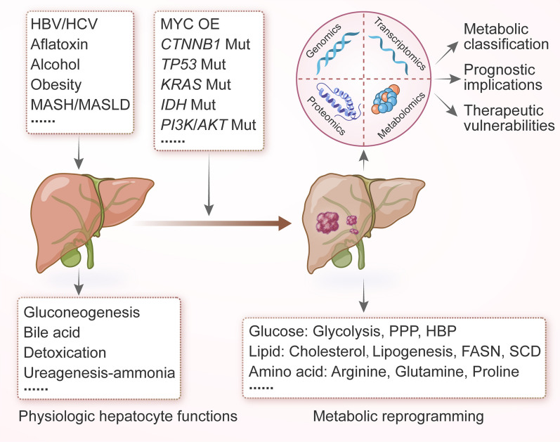
“Other glucose metabolic pathways, such as the pentose phosphate pathway (PPP) and the hexosamine biosynthetic pathway (HBP), are also more active in HCC tissue compared to normal adjacent tissues [44–47] (Fig.”
BackgroundTumor-Derived Adenosine Promotes Macrophage Proliferation in Human Hepatocellular Carcinoma
Wang, J., Wang, Y., Chu, Y., Li, Z., Yu, X., Huang, Z., Xu, J., & Zheng, L. (2021) · Journal of Hepatology · doi:10.1016/j.jhep.2020.10.021 · Cited by 133 · cited 1×
“Tumor cell-derived adeno - sine also synergizes with autocrine granulocyte–macrophage colony-stimulating factor (GM-CSF) secreted from activated TAM to promote their proliferation, thus maintaining the Mφ pools in HCC [151].”
BackgroundPeng, Z.-P., Jiang, Z.-Z., Guo, H.-F., Zhou, M., Huang, Y., Ning, W.-R., Huang, J., Zheng, L., & Wu, Y. (2020) · Journal of Hepatology · doi:10.1016/j.jhep.2020.05.004 · Cited by 128 · cited 1×
“These processes ultimately lead to the metastasis of HCC, as shown by both ex vivo and in vitro experiments [186].”
BackgroundNonalcoholic Fatty Liver Disease and the Gut-Liver Axis: Exploring an Undernutrition Perspective
Bauer, K. C., Littlejohn, P. T., Ayala, V., Creus-Cuadros, A., & Finlay, B. B. (2022) · Gastroenterology · doi:10.1053/j.gastro.2022.01.058 · Cited by 127
Lercher, A., Bhattacharya, A., Popa, A., Caldera, M., Schlapansky, M. F., Baazim, H., Agerer, B., Gürtl, B., Kosack, L., Májek, P., et al. (2019) · Immunity · doi:10.1016/j.immuni.2019.10.014 · Cited by 124 · cited 1×
“…activates hepatocyte-intrinsic type I interferon (IFN-I) responses to break the urea cycle, leading to decreased arginine/ ornithine ratios in the circulation and subsequently suppressed virus-specific CD8 + T-cell responses in chronic lymphocytic choriomeningitis virus (LCMV)-infected mice [148].”
BackgroundHong, X.-H., Song, R.-P., Song, H.-W., Zheng, T., Wang, J.-B., Liang, Y.-J., Qi, S., Lu, Z., Song, X., Jiang, H.-C., et al. (2013) · Gut · doi:10.1136/gutjnl-2013-305302 · Cited by 123
Exploring Metabolic Configurations of Single Cells within Complex Tissue Microenvironments
Miller, A., Nagy, C., Knapp, B., Laengle, J., Ponweiser, E., Gröger, M., Starkl, P. M., Bergmann, M. M., Wagner, O. F., & Haschemi, A. (2017) · Cell Metabolism · doi:10.1016/j.cmet.2017.08.014 · Cited by 123 · cited 1×
“With the advent of immunome-tabolism in cancer treatment [18–22], greater attention should be given to the interplay of metabolism-related immune signaling.”
BackgroundZhou, P., Chang, W.-Y., Gong, D.-A., Xia, J. L., Chen, W., Huang, L., Liu, R., Liu, Y., Chen, C., Wang, K., et al. (2023) · Cell Metabolism · doi:10.1016/j.cmet.2023.09.009 · Cited by 122 · cited 1×
“…mechanism showed that microbiota-derived acetate enhanced UDP-GlcNAc biosynthesis and O-GlcNAcylation, and hyper-O-GlcNAcylation of eukaryotic elongation factor 1A1 (eEF1A1) at T279 promoted the proliferation of tumor cells and HCC progression in the DEN + CCl 4 -induced HCC mouse model [200].”
BackgroundPhoomak, C., Vaeteewoottacharn, K., Silsirivanit, A., Saengboonmee, C., Seubwai, W., Sawanyawisuth, K., Wongkham, C., & Wongkham, S. (2017) · Scientific Reports · doi:10.1038/srep43842 · Cited by 122 · cited 1×
“Other glucose metabolic pathways, such as the pentose phosphate pathway (PPP) and the hexosamine biosynthetic pathway (HBP), are also more active in HCC tissue compared to normal adjacent tissues [44–47] (Fig.”
BackgroundGLUT1 and ASCT2 as Predictors for Prognosis of Hepatocellular Carcinoma
Sun, H.-W., Yu, X.-J., Wu, W., Chen, J., Shi, M., Zheng, L., & Xu, J. (2016) · PLoS ONE · doi:10.1371/journal.pone.0168907 · Cited by 121
Li, B., Li, Y., Zhou, H., Xu, Y., Cao, Y., Cheng, C., Peng, J., Li, H., Zhang, L., Su, K., et al. (2023) · Hepatology · doi:10.1097/hep.0000000000000553 · Cited by 110 · cited 1×
“Thus, HCC mouse models derived from the F1 subtype are responsive to anti-PD-1 (programmed cell death protein 1) therapy, in contrast with mouse models derived from the F2 and F3 subtypes [36].”
BackgroundHypoxia-induced macropinocytosis represents a metabolic route for liver cancer
Zhang, M. S., Cui, J. D., Lee, D., Yuen, V. W., Chiu, D. K., Goh, C. C., Cheu, J. W., Tse, A. P., Bao, M. H., Wong, B. P., et al. (2022) · Nature Communications · doi:10.1038/s41467-022-28618-9 · Cited by 109 · cited 1×
“Hypoxia also induces micropinocytosis for nutrient scavenging via the hypoxia-inducible factor (HIF)/EH domain-containing protein 2 (EHD2) pathways in several HCC cell lines and mouse models [43].”
BackgroundSelf-citationKotsiliti, E., Leone, V., Schuehle, S., Govaere, O., Li, H., Wolf, M. J., Horvatic, H., Bierwirth, S., Hundertmark, J., Inverso, D., et al. (2023) · Journal of Hepatology · doi:10.1016/j.jhep.2023.04.037 · Cited by 108 · cited 1×
“Meanwhile, gastrointestinal IgA + metabolically activated B cells can license auto-aggressive T cells, promoting HCC development in an antigen-independent manner in MASH-induced HCC mouse models [183].”
BackgroundSLC2A2 (GLUT2) as a novel prognostic factor for hepatocellular carcinoma
Kim, Y. H., Jeong, D. C., Pak, K., Han, M., Kim, J., Liu, L., Kim, H. J., Kim, T. W., Kim, T. H., Hyun, D. W., et al. (2017) · Oncotarget · doi:10.18632/oncotarget.20266 · Cited by 105
Che, Y., Chen, G., Guo, Q., Duan, Y., Feng, H., & Xia, Q. (2023) · Hepatology · doi:10.1097/hep.0000000000000047 · Cited by 105 · cited 1×
“Butyrate accumulation disrupts intracellular calcium homeostasis and induces the production of ROS, thus improving the efficacy of TKI therapy in HCC mouse models [202].”
BackgroundTap, W., Villalobos, V., Cote, G., Burris, H., Janku, F., Mir, O., Beeram, M., Wagner, A., Jiang, L.-W., Wu, B., et al. (2020) · Reactions weekly · doi:10.1007/s40278-020-83841-5 · Cited by 103
Notch-mediated lactate metabolism regulates MDSC development through the Hes1/MCT2/c-Jun axis
Zhao, J., Ye, Y., Gao, C.-C., Wang, L., Ren, K., Jiang, R., Hu, S., Liang, S.-Q., Bai, J., Liang, J.-L., et al. (2022) · Cell Reports · doi:10.1016/j.celrep.2022.110451 · Cited by 101
Jouve, J., Lecomte, T., Bouché, O., Barbier, E., Akouz, F. K., Riachi, G., Khac, É. N., Ollivier-Hourmand, I., Debette-Gratien, M., Faroux, R., et al. (2019) · Journal of Hepatology · doi:10.1016/j.jhep.2019.04.021 · Cited by 99
G6PD: A hub for metabolic reprogramming and redox signaling in cancer
Yang, H., Stern, A., & Chiu, D. T.-Y. (2020) · Biomedical Journal · doi:10.1016/j.bj.2020.08.001 · Cited by 99
Murakami, Y., Kubo, S., Tamori, A., Itami, S., Kawamura, E., Iwaisako, K., Ikeda, K., Kawada, N., Ochiya, T., & Taguchi, Y. (2015) · Scientific Reports · doi:10.1038/srep16294 · Cited by 99 · cited 1×
“Moreover, numerous studies indicate enhanced amino acid metabolism in liver tumors compared to non-tumor tissues [1, 31, 37, 60, 61].”
BackgroundLu, L.-G., Zhou, Z.-L., Wang, X.-Y., Liu, B., Lu, J.-Z., Liu, S., Zhang, G.-B., Zhan, M.-X., & Chen, Y. (2022) · Gut · doi:10.1136/gutjnl-2021-326350 · Cited by 98 · cited 1×
“Furthermore, the PKM2/HIF-1α axis in human HCC samples and syngeneic mouse models drives fibronectin 1 (FN1) production to instigate pluripotent polarization of macrophages, concurrent with anti-tumorigenic IL-12p70 production from glycolytic macrophages [191].”
BackgroundFaloppi, L., Scartozzi, M., Bianconi, M., Svegliati‐Baroni, G., Toniutto, P., Giampieri, R., Prete, M. D., Minicis, S. D., Bitetto, D., Loretelli, C., et al. (2014) · BMC Cancer · doi:10.1186/1471-2407-14-110 · Cited by 97
Kowalik, M. A., Guzzo, G., Morandi, A., Perra, A., Menegon, S., Masgras, I., Trevisan, E., Angioni, M. M., Fornari, F., Quagliata, L., et al. (2016) · Oncotarget · doi:10.18632/oncotarget.8632 · Cited by 97
Sun, H., Yang, W., Tian, Y., Zeng, X., Zhou, J., Mok, M. T. S., Tang, W., Feng, Y., Xu, L., Chan, A. W. H., et al. (2018) · Nature Communications · doi:10.1038/s41467-018-07402-8 · Cited by 93 · cited 1×
“In MASH-related HCC mouse models, the androgen receptor (AR)-driven oncogene, cell cycle-related kinase (CCRK), combined with obesity-induced IL-6/STAT3 signaling, induces lipid metabolic reprogramming and MDSC-dominated immunosuppression [161].”
BackgroundNelson, M. E., Lahiri, S., Chow, J. D. Y., Byrne, F. L., Hargett, S. R., Breen, D. S., Olzomer, E. M., Wu, L. E., Cooney, G. J., Turner, N. A., et al. (2017) · Nature Communications · doi:10.1038/ncomms14689 · Cited by 93 · cited 1×
“This discrepancy may be attributed to differences between clinical samples and preclinical models of HCC, highlighting the need for a thorough exploration of tumor-driving events and metabolic plasticity [53].”
BackgroundYang, Z.-S., Qin, W., Chen, Y., Yuan, B., Song, X.-L., Wang, B., Shen, F., Fu, J., & Wang, H.-Y. (2018) · Cancer Letters · doi:10.1016/j.canlet.2018.04.038 · Cited by 91 · cited 1×
“Conversely, high serum cholesterol levels are linked with better patient outcomes by inhibiting tumor metastasis [59], implying that cholesterol distribution and homeostasis significantly influence HCC tumorigenesis (Fig.”
BackgroundFu, Y., Mackowiak, B., Feng, D., Lu, H., Guan, Y., Lehner, T., Pan, H., Wang, X. W., He, Y., & Gao, B. (2023) · Gut · doi:10.1136/gutjnl-2022-327924 · Cited by 86 · cited 1×
“This results in adenosine-mediated immunosuppressive roles on T cells and myeloid cells [149, 150].”
BackgroundPhase Ib/II Study of Pembrolizumab and Pegylated-Interferon Alfa-2b in Advanced Melanoma
Davar, D., Wang, H., Chauvin, J.-M., Pagliano, O., Fourcade, J. J., Ka, M., Menna, C., Rose, A., Sander, C., Borhani, A. A., et al. (2018) · Journal of Clinical Oncology · doi:10.1200/jco.18.00632 · Cited by 86 · cited 1×
“The results of a recent clinical trial demonstrated the promising efficacy of combined IFNα and ICB therapy in patients with advanced melanoma [130].”
BackgroundLin, J., Dai, Y., Sang, C., Song, G., Xiang, B., Zhang, M., Dong, L., Xia, X., Ma, J., Shen, X., et al. (2022) · Journal for ImmunoTherapy of Cancer · doi:10.1136/jitc-2022-004892 · Cited by 86 · cited 1×
“This results in an immunosuppressive TME dominated by excessive neutrophil infiltration and contributes to poor prognosis in iCCA [147].”
BackgroundSelf-citationMolenaar, R. J., Coelen, R. J. S., Khurshed, M., Roos, E., Caan, M. W. A., Linde, M. E. V., Kouwenhoven, M. C. M., Bramer, J. A. M., Bovée, J. V. M. G., Mathôt, R. A. A., et al. (2017) · BMJ Open · doi:10.1136/bmjopen-2016-014961 · Cited by 83
NeuCode Proteomics Reveals Bap1 Regulation of Metabolism
Baughman, J. M., Rose, C. M., Kolumam, G., Webster, J. D., Wilkerson, E. M., Merrill, A. E., Rhoads, T. W., Noubade, R., Katavolos, P., Lesch, J., et al. (2016) · Cell Reports · doi:10.1016/j.celrep.2016.05.096 · Cited by 73 · cited 2×
“We and others have identified numerous putative driver genes that reshape PLC metabolism [29, 30, 75– 79] (Table 1).”
BackgroundLin, M., Lv, D., Zheng, Y., Wu, M., Xu, C., Zhang, Q., & Wu, L. (2018) · OncoTargets and Therapy · doi:10.2147/ott.s163266 · Cited by 73 · cited 1×
“In high-fat diet (HFD)-induced HCC, or steatohepa-titic HCC, the fatty acid oxidation (FAO) pathway tends to be downregulated to protect HCC cells from lipotox-icity [48].”
BackgroundGlycine decarboxylase maintains mitochondrial protein lipoylation to support tumor growth
Mukha, D., Fokra, M., Feldman, A., Sarvin, B., Sarvin, N., Nevo-Dinur, K., Besser, E., Hallo, E., Aizenshtein, E., Schug, Z. T., et al. (2022) · Cell Metabolism · doi:10.1016/j.cmet.2022.04.006 · Cited by 69 · cited 1×
“In HCC cells, glycine-derived 1C units support purine and pyrimidine biosynthesis and tumor progression through glycine cleavage system (GCS) flux [73].”
BackgroundThe gut–liver axis: host microbiota interactions shape hepatocarcinogenesis
Silveira, M. A. D., Bilodeau, S., Greten, T. F., Wang, X. W., & Trinchieri, G. (2022) · Trends in cancer · doi:10.1016/j.trecan.2022.02.009 · Cited by 69 · cited 1×
“Increasing evidence suggests that the gut microbiota modulates immune responses in the TME of PLC [5, 194, 203, 207, 208].”
BackgroundWang, N., Tan, H., Lu, Y., Chan, Y.-T., Wang, D., Guo, W., Xu, Y., Zhang, C., Chen, F., Tang, G., et al. (2021) · Signal Transduction and Targeted Therapy · doi:10.1038/s41392-021-00485-8 · Cited by 68 · cited 1×
“Meanwhile, PIWIL1 regulates the secretion of Complement C3 to mediate interaction between HCC cells and MDSC, promoting the immunosuppressive cytokine IL10 in the TME [162].”
BackgroundNing, W.-R., Jiang, D., Liu, X.-C., Huang, Y.-F., Peng, Z.-P., Jiang, Z.-Z., Kang, T., Zhuang, S.-M., Wu, Y., & Zheng, L. (2022) · Journal of Clinical Investigation · doi:10.1172/jci153110 · Cited by 66 · cited 1×
“This production promotes epithelial-mesenchymal transition (EMT) and HCC metastasis [187].”
BackgroundHypoxia-inducible factor orchestrates adenosine metabolism to promote liver cancer development
Cheu, J. W.-S., Chiu, D. K.-C., Kwan, K. K.-L., Yang, C., Yuen, V. W.-H., Goh, C. C., Chui, N. N.-Q., Shen, W., Law, C.-T., Li, Q., et al. (2023) · Science Advances · doi:10.1126/sciadv.ade5111 · Cited by 65 · cited 1×
“This results in adenosine-mediated immunosuppressive roles on T cells and myeloid cells [149, 150].”
BackgroundSelf-citationDietary folate drives methionine metabolism to promote cancer development by stabilizing MAT IIA
Li, J., Yang, H. Z., Lei, M., Zhu, W., Su, Y., Li, K.-Y., Zhu, W.-Y., Wang, J., Zhang, L., Qu, J., et al. (2022) · Signal Transduction and Targeted Therapy · doi:10.1038/s41392-022-01017-8 · Cited by 63 · cited 1×
“Recent findings have also highlighted the promotion of tumor development by dietary folate supplementation through the integration of methionine and 1C metabolism in the HCC mouse model induced by DEN/HFD [74].”
BackgroundProteogenomic characterization of cholangiocarcinoma
Deng, M., Ran, P., Chen, L., Wang, Y., Yu, Z., Cai, K., Feng, J., Qin, Z., Yin, Y., Tan, S., et al. (2022) · Hepatology · doi:10.1002/hep.32624 · Cited by 61
Antonarakis, E., Heath, E., Walczak, J. R., Nelson, W., Fedor, H., Marzo, A. D. D., Zahurak, M., Piantadosi, S., Dannenberg, A., Gurganus, R., et al. (2009) · Journal of Clinical Oncology · doi:10.1200/jco.2009.21.9410 · Cited by 60 · cited 1×
“…isocitrate dehydrogenase IDH1/2 inhibitor Enasidenib/Ivosidenib (for 2-HG production) [124], and the cyclooxygenase COX-2 inhibitor Celecoxib (for PGE2 accumulation) [125, 126] are being evaluated or approved to target cancer metabolism, aiming to enhance the efficacies of current therapies [127].”
BackgroundSun, J., Zhang, N., Xu, R.-C., Zhang, G., Liu, Z., Abuduwaili, W., Wang, F., Yu, X., Shi, X., Song, G., et al. (2021) · Journal of Translational Medicine · doi:10.1186/s12967-021-03034-7 · Cited by 60 · cited 1×
“This iron competition culminates in an M2-like TAM polarization in vitro [133].”
BackgroundHalford, S. E. R., Jones, P. S., Wedge, S. R., Hirschberg, S., KATUGAMPOLA, S. D., Veal, G. J., Payne, G. S., Bacon, C. M., Potter, S., Griffin, M., et al. (2017) · Journal of Clinical Oncology · doi:10.1200/jco.2017.35.15_suppl.2516 · Cited by 59 · cited 1×
“Currently, the monocarboxylate transporter MCT1 inhibitor AZD3965 (for lactate sym-porter) [121], the indoleamine 2,3-dioxygenase IDO1 inhibitor Indoximod/Epacadostat (for kynurenine synthesis) [122], the CD73 inhibitor Oleclumab (for adenosine conversion) [123], the isocitrate dehydrogenase IDH1/2…”
BackgroundXing, X., Hu, E., Ouyang, J., Zhong, X., Wang, F., Liu, K., Cai, L., Zhou, Y., Wang, Y., Chen, G., et al. (2023) · Cell Reports Medicine · doi:10.1016/j.xcrm.2023.101315 · Cited by 59 · cited 1×
“Multi-omics studies have nominated metabolic pathways as the most dramatic alterations in HCC and iCCA, compared with normal adjacent tissues [26–29].”
BackgroundWong, A. M., Ding, X., Wong, A. M. G., Xu, M., Zhang, L., Leung, H. H. W., Chan, A. W. H., Song, Q. X., Kwong, J., Chan, L. K., et al. (2022) · Journal of Hepatology · doi:10.1016/j.jhep.2022.03.015 · Cited by 58 · cited 1×
“This shift in the driver mutations results in immune exclusion through the repression of the TNF receptor superfamily member 19 (TNFRSF19)- mediated senescence-associated secretory phenotype (SASP), as shown in a syngeneic immunocompetent mouse model [157].”
BackgroundLi, Y., Mo, H., Wu, S., Liu, X., & Tu, K. (2022) · Frontiers in Cell and Developmental Biology · doi:10.3389/fcell.2021.801959 · Cited by 55 · cited 1×
“The well-known “Warburg effect” rearranges glucose metabolism to produce excessive lactic acid, which has emerged as an important regulator in promoting immune evasion in PLC [134, 135].”
Background
Research works printed but not linked: 3
- Kelley RK, et al. Pembrolizumab in combination with gemcitabine and cisplatin compared with gemcitabine and cisplatin alone for patients with advanced biliary tract cancer (KEYNOTE-966): a randomised, double-blind, placebo-controlled, phase 3 trial. Lancet. 2023;401:1853–65. https://doi.org/10.1016/S0140-6736(23)00727-4. · DOI · Paper page
- Xun X, et al. Cyclooxygenase-2 expressed hepatocellular carcinoma induces cytotoxic T lymphocytes exhaustion through M2 macrophage polarization. Am J Transl Res. 2021;13:4360–75.
- Tap WD, et al. Phase I study of the mutant IDH1 inhibitor ivosidenib: safety and clinical activity in patients with advanced chondrosarcoma. J Clin Oncol. 2020;38:1693–701. https://doi.org/10.1200/JCO.19.02492. · DOI · Paper page
Cited by
Sorted by citations
Molecular mechanisms in liver repair and regeneration: from physiology to therapeutics
Ma, X., Huang, T., Chen, X., Li, Q., Liao, M., Fu, L., Huang, J., Yuan, K., Wang, Z., & Zeng, Y. (2025) · Signal Transduction and Targeted Therapy · doi:10.1038/s41392-024-02104-8 · Cited by 133
The roles of KRAS in cancer metabolism, tumor microenvironment and clinical therapy
Ma, Q., Zhang, W., Wu, K., & Shi, L. (2025) · Molecular Cancer · doi:10.1186/s12943-024-02218-1 · Cited by 101
“Unlike normal cells, cancer cells undergo metabolic reprogramming to meet the increased demands for energy, macromolecule biosynthesis, and redox balance [16, 17].”
Zhang, M., Zheng, H., Jin, H., Zhu, X., Liu, S., Chen, Y., Zhang, H., & Zhang, S. (2025) · Bioactive Materials · doi:10.1016/j.bioactmat.2025.09.003 · Cited by 26
“Moreover, interfering glucose metabolic pathways, including OXPHOS, glycolysis and PPP, can also disrupt glucose metabolic process, thus inducing glucose dyshomeo-stasis [28,29].”
Self-citationHuang, Y., Xie, Y., Zhang, Y., Liu, Z., Jiang, W., Ye, Y., Tang, J., Li, Z., Yin, Z., & Lin, X.-J. (2025) · Molecular Cancer · doi:10.1186/s12943-025-02370-2 · Cited by 41
“Reprogramming of liver microenvironment is a key feature of hepatocarcinogenesis, which encompasses but not limited to metabolic alterations of hepatocyte and immune cells, re-emergence of fetal-associated endothelial cells and altered inflammatory milieu [10, 46, 65, 66].”
Fan, S., Wang, W., Che, W., Xu, Y., Jin, C., Dong, L., & Xia, Q. (2025) · Metabolites · doi:10.3390/metabo15030201 · Cited by 43
Metadata
- Authors
- Lin, J., Rao, D., Zhang, M., & Gao, Q.
- Affiliations
- Fudan University (CN)
- Shanghai Medical College of Fudan University (CN)
- Corresponding author
- Gao, Qiang
- Venue
- Journal of Hematology & Oncology
- Published
- 2024-01-31
- Volume
- 17
- Issue
- 1
- Pages
- 6-6
- ISSN
- 1756-8722 (ISSN-L)
- PMC
- PMC10832230
- OpenAlex
- W4391404272
- Cited by
- 251
- References
- 231
- Citations by year
- 2026: 125 · 2025: 110 · 2024: 16
- FWCI
- 12.25
- Type
- journal article
- Language
- en
- Open access
- gold
- doi.org
- Locations
- Journal of Hematology & Oncology (publishedVersion, cc-by)
- PubMed Central (publishedVersion, cc-by)
- PubMed (publishedVersion)
- DOAJ (DOAJ: Directory of Open Access Journals) (submittedVersion, cc-by-sa)
- Funders
- National Natural Science Foundation of China (82103314, 82130077)
- Jinshan Hospital Flexible Mobile Talent Research Startup Fund (RXRC-2020-1)
- Topics
- Life Sciences › Biochemistry, Genetics and Molecular Biology › Cancer Research › Cancer, Hypoxia, and Metabolism
- Life Sciences › Immunology and Microbiology › Immunology › Immune cells in cancer
- Health Sciences › Medicine › Oncology › Cancer Immunotherapy and Biomarkers
- Keywords
- metabolic reprogramming, liver cancer, tumor microenvironment, tumor immune microenvironment, cancer immunotherapy, immunotherapy resistance, gut-liver axis, immune surveillance, immune evasion
- MeSH terms
- Metabolic Reprogramming, Energy Metabolism, Glycolysis, Humans, Immunotherapy, Liver Neoplasms, Neoplasms, Tumor Microenvironment
About these checks
Registry, retraction, Cites and statistics results are computed, not judged, with one exception: a printed reference that no index lists is read once by a model before it counts as a citation error. Claims are extracted by a model and kept only when their quote appears verbatim in the text; each cited source is then checked, with a model’s help, for whether it says what the claim relies on. Those verdicts can be wrong — read the claim’s quote and the cited source before relying on one.
Statistics and arithmetic are recomputed, not judged.
Confidence bands are shown only for fields calibrated on our annotated test set. None is calibrated yet, so no band is shown.
What counts as a claim ›How we measure confidence ›How we check this ›
Are you an author, or do you see a problem with this page? Request a review
