

The DOI matches its Crossref record; 51 of the 53 references it cites were found in a registry or an index.
Digital Photoplethysmography for Assessment of Arterial Stiffness: Repeatability and Comparison with Applanation Tonometry
Open accessAbstract
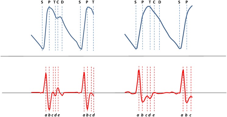
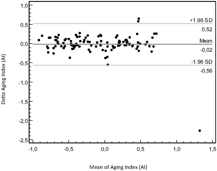
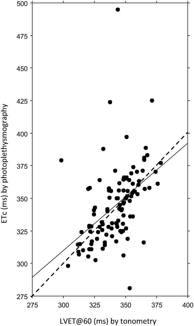
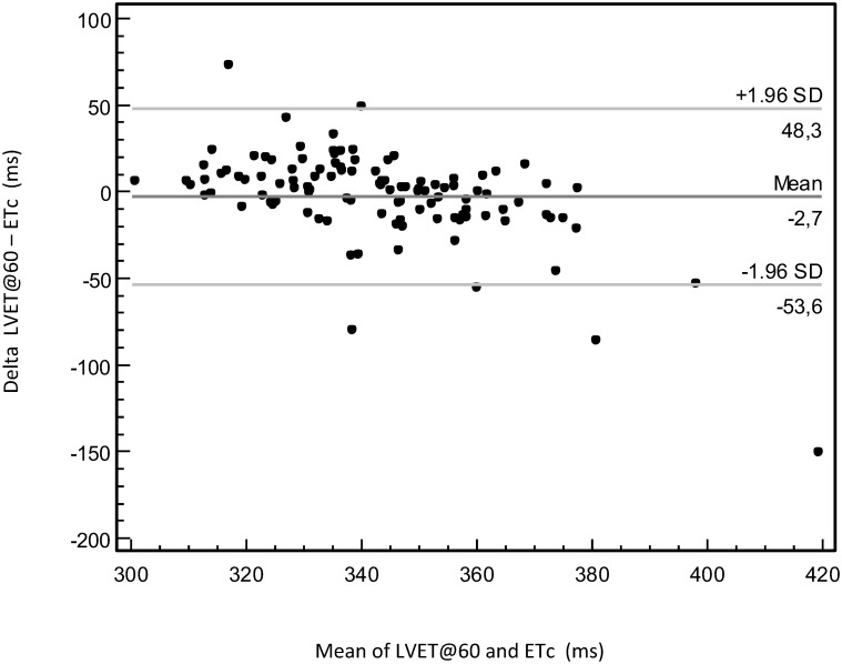
INTRODUCTION: Arterial stiffness is an independent risk factor for cardiovascular morbidity and can be assessed by applanation tonometry by measuring pulse wave velocity (PWV) and augmentation index (AIX) by pressure pulse wave analysis (PWA). As an inexpensive and operator independent alternative, photoelectric plethysmography (PPG) has been introduced with analysis of the digital volume pulse wave (DPA) and its second derivatives of wave reflections. OBJECTIVE: The objective was to investigate the repeatability of arterial stiffness parameters measured by digital pulse wave analysis (DPA) and the associations to applanation tonometry parameters. METHODS AND RESULTS: 112 pregnant and non-pregnant individuals of different ages and genders were examined with SphygmoCor arterial wall tonometry and Meridian DPA finger photoplethysmography. Coefficients of repeatability, Bland-Altman plots, intraclass correlation coefficients and correlations to heart rate (HR) and body height were calculated for DPA variables, and the DPA variables were compared to tonometry variables left ventricular ejection time (LVET), PWV and AIX. No DPA variable showed any systematic measurement error or excellent repeatability, but dicrotic index (DI), dicrotic dilatation index (DDI), cardiac ejection elasticity index (EEI), aging index (AI) and second derivatives of the crude pulse wave curve, b/a and e/a, showed good repeatability. Overall, the correlations to AIX were better than to PWV, with correlations coefficients >0.70 for EEI, AI and b/a. Considering the level of repeatability and the correlations to tonometry, the overall best DPA parameters were EEI, AI and b/a. The two pansystolic time parameters, ejection time compensated (ETc) by DPA and LVET by tonometry, showed a significant but weak correlation. CONCLUSION: For estimation of the LV function, ETc, EEI and b/a are suitable, for large artery stiffness EEI, and for small arteries DI and DDI. The only global parameter, AI, showed a high repeatability and the overall best correlations with AIX and PWV.
Overview
The claims and statistics of this paper haven’t been checked yet.
Run the check — free, no account needed. It reads the open-access text and checks the sources it cites.
Integrity signals
Registry
Cites
Statistics
Figures
Verification
Claims the paper makes
The claims and statistics of this paper haven’t been checked yet.
Run the check — free, no account needed. It reads the open-access text and checks the sources it cites.
Claims other papers make about this one
Each is checked against this paper’s own text: does it say what the citing paper says it does?
In the citing paper
“Over 30 features have been proposed for assessing the stiffness of both the large and small arteries from a single PPG”
From Wearable Photoplethysmography for Cardiovascular Monitoring (2022) · cites this paper as [172]
Self-check
Percentages, sums and reported differences re-computed from the paper’s own numbers.
The self-check needs the paper’s full text.
Citations
Cites
STATISTICAL METHODS FOR ASSESSING AGREEMENT BETWEEN TWO METHODS OF CLINICAL MEASUREMENT
Martin Bland, J., & Altman, D. (1986) · The Lancet · doi:10.1016/s0140-6736(86)90837-8 · Cited by 47,965
Measuring agreement in method comparison studies
Bland, J. M., & Altman, D. G. (1999) · Statistical Methods in Medical Research · doi:10.1177/096228029900800204 · Cited by 9,619 · cited 1×
“We described agreement between the two DPA measurements with Bland-Altman plots [36,37].”
Measuring agreement in method comparison studies
Bland, J. M., & Altman, D. G. (1999) · Statistical Methods in Medical Research · doi:10.1191/096228099673819272 · Cited by 9,619
Expert consensus document on arterial stiffness: methodological issues and clinical applications
Laurent, S., Cockcroft, J., Van Bortel, L., Boutouyrie, P., Giannattasio, C., Hayoz, D., Pannier, B., Vlachopoulos, C., Wilkinson, I., Struijker-Boudier, H., et al. (2006) · European Heart Journal · doi:10.1093/eurheartj/ehl254 · Cited by 6,077
McDonald's blood flow in arteries; theoretical, experimental and clinical principles
(1992) · General Pharmacology The Vascular System · doi:10.1016/0306-3623(92)90079-y · Cited by 2,395
Aortic Stiffness Is an Independent Predictor of Primary Coronary Events in Hypertensive Patients
Boutouyrie, P., Tropeano, A., Asmar, R. G., Gautier, I., Bénétos, A., Lacolley, P. J., & Laurent, S. (2002) · Hypertension · doi:10.1161/hy0102.099031 · Cited by 1,745 · cited 2×
“Although pressure PW analysis shows benefits in predicting cardiovascular morbidity [1,10], the impact in clinical practice is small because the tonometry method is cumbersome with a need for highly specialized operators.”
McDonald's Blood Flow in Arteries: Theoretical, Experimental and Clinical Principles
Vlachopoulos, C., O'Rourke, M., & Nichols, W. W. (2011) · doi:10.1201/b13568 · Cited by 1,613
Aortic pulse wave velocity as a marker of cardiovascular risk in hypertensive patients
Blacher, J., Asmar, R., Djane, S., London, G. M., & Safar, M. E. (1999) · Hypertension · doi:10.1161/01.hyp.33.5.1111 · Cited by 1,493 · cited 1×
“The PWV is correlated to age, blood pressure (BP) and arteriosclerosis and has a better predictive value for cardiovascular disease than traditional risk markers [3,4].”
On the Analysis of Fingertip Photoplethysmogram Signals
Elgendi, M. (2012) · Current Cardiology Reviews · doi:10.2174/157340312801215782 · Cited by 1,206 · cited 3×
“The e wave represents the dictrotic notch and corresponds to the aortic valve closure [11].”
InfluentialClinical measurement of arterial stiffness obtained from noninvasive pressure waveforms
NICHOLS, W. (2005) · American Journal of Hypertension · doi:10.1016/j.amjhyper.2004.10.009 · Cited by 1,008 · cited 1×
“AIX is influenced by both large artery stiffness and properties in the PW-reflecting peripheral arterial tree [5 – 7] and is associated with cardiovascular risk, although it is less established than the PWV as a risk factor for cardiovascular morbidity [8 – 10].”
Research works printed but not linked: 3
- R Gonzales, 2008, A computer base photoplethysmographic vascular analyser through derivatives, Comp Cardiol, 35, 177
- T Ozawa, 1978, Pattern of second derivative of volume pulse wave, the relation between non-invasive index of ventricular function and peak acceleration and effect of preloading to peak velocity, Myakuha (Pulse Wave), 8, 22
- HC Bazett, 1920, An analysis of the time-relations of electrocardiograms, Heart, 7, 35
Cited by
Sorted by citations
Self‐Powered Pulse Sensor for Antidiastole of Cardiovascular Disease
Ouyang, H., Tian, J., Sun, G., Zou, Y., Liu, Z., Li, H., Zhao, L., Shi, B., Fan, Y., Fan, Y., et al. (2017) · Advanced Materials · doi:10.1002/adma.201703456 · Cited by 470
Photoplethysmogram Analysis and Applications: An Integrative Review
Park, J., Seok, H. S., Kim, S.-S., & Shin, H. (2022) · Frontiers in Physiology · doi:10.3389/fphys.2021.808451 · Cited by 514
Sources of inaccuracy in photoplethysmography for continuous cardiovascular monitoring
Fine, J., Branan, K. L., Rodriguez, A. J., Boonya-ananta, T., Ajmal, Ramella-Roman, J. C., McShane, M. J., & Coté, G. L. (2021) · Biosensors · doi:10.3390/bios11040126 · Cited by 348
Wearable Photoplethysmography for Cardiovascular Monitoring
Charlton, P. H., Kyriacou, P. A., Mant, J., Marozas, V., Chowienczyk, P., & Alastruey, J. (2022) · Proceedings of the IEEE · doi:10.1109/jproc.2022.3149785 · Cited by 250
An optimal filter for short photoplethysmogram signals
Liang, Y., Elgendi, M., Chen, Z., & Ward, R. (2018) · Scientific Data · doi:10.1038/sdata.2018.76 · Cited by 242
A new, short-recorded photoplethysmogram dataset for blood pressure monitoring in China
Liang, Y., Chen, Z., Liu, G., & Elgendi, M. (2018) · Scientific Data · doi:10.1038/sdata.2018.20 · Cited by 215
Diagnostic Features and Potential Applications of PPG Signal in Healthcare: A Systematic Review
Almarshad, M. A., Islam, M. S., Al-Ahmadi, S. A., & BaHammam, A. S. (2022) · Healthcare · doi:10.3390/healthcare10030547 · Cited by 215
Charlton, P. H., Paliakaitė, B., Pilt, K., Bachler, M., Zanelli, S., Kulin, D., Allen, J., Hallab, M., Bianchini, E., Mayer, C. C., et al. (2021) · American Journal of Physiology-Heart and Circulatory Physiology · doi:10.1152/ajpheart.00392.2021 · Cited by 166
“For instance, coef fi cients with absolute values from 0.64 to 0.72 have been found between reference PWVs and PPG-derived parameters ( fi nger-toe PWV and pulse wave parameters) (73, 86, 94, 127, 139).”
InfluentialPhotoplethysmography signal processing and synthesis
Mejía-Mejía, E., Allen, J., Budidha, K., El-Hajj, C., Kyriacou, P. A., & Charlton, P. H. (2022) · Photoplethysmography · doi:10.1016/b978-0-12-823374-0.00015-3 · Cited by 138
“Studies have found limits of agreement for between PPG-derived and reference LVETs of -24 to 17 ms [263], -41 to 13 ms [259], and (HR corrected LVET) -70 to 67 ms [262].”
BackgroundMethodWearable Photoplethysmography for Cardiovascular Monitoring.
Charlton, P. H., Kyriacou, P. A., Mant, J., Marozas, V., Chowienczyk, P., & Alastruey, J. (2022) · KTUePubl (Repository of Kaunas University of Technology) · doi:10.17863/cam.81148 · Cited by 113
Toward Generating More Diagnostic Features from Photoplethysmogram Waveforms
Elgendi, M., Liang, Y., & Ward, R. K. (2018) · Diseases · doi:10.3390/diseases6010020 · Cited by 107
Song, Z., Li, W., Bao, Y., Wang, W., Liu, Z., Han, F., Han, D., & Niu, L. (2018) · Advanced Electronic Materials · doi:10.1002/aelm.201800252 · Cited by 90
Barvik, D., Černý, M., Penhaker, M., & Noury, N. (2021) · IEEE Reviews in Biomedical Engineering · doi:10.1109/rbme.2021.3109643 · Cited by 85
Finnegan, E., Davidson, S. M., Harford, M., Watkinson, P., Tarassenko, L., & Villarroel, M. (2023) · Scientific Reports · doi:10.1038/s41598-022-27170-2 · Cited by 87
Arterial pulse wave modeling and analysis for vascular-age studies: a review from VascAgeNet
Alastruey, J., Charlton, P. H., Bikia, V., Paliakaitė, B., Hametner, B., Bruno, R. M., Mulder, M. P., Vennin, S., Pişkin, Ş., Khir, A. W., et al. (2023) · American Journal of Physiology-Heart and Circulatory Physiology · doi:10.1152/ajpheart.00705.2022 · Cited by 87
XGBoost Regression of the Most Significant Photoplethysmogram Features for Assessing Vascular Aging
Shin, H. (2022) · IEEE Journal of Biomedical and Health Informatics · doi:10.1109/jbhi.2022.3151091 · Cited by 81
Djeldjli, D., Bousefsaf, F., Maaoui, C., Bereksi-Reguig, F., & Pruski, A. (2020) · Biomedical Signal Processing and Control · doi:10.1016/j.bspc.2020.102242 · Cited by 74
A water-resistant, ultrathin, conformable organic photodetector for vital sign monitoring
Du, B., Xiong, S., Sun, L., Tagawa, Y., Inoue, D., Hashizume, D., Wang, W., Guo, R., Yokota, T., Wang, S., et al. (2024) · Science Advances · doi:10.1126/sciadv.adp2679 · Cited by 74
Mazumder, O., Roy, D., Bhattacharya, S., Sinha, A., & Pal, A. (2019) · Annual International Conference of the IEEE Engineering in Medicine and Biology Society (EMBC) · doi:10.1109/embc.2019.8856691 · Cited by 68
Photoplethysmography for the Assessment of Arterial Stiffness
Karimpour, P., May, J. M., & Kyriacou, P. A. (2023) · Sensors · doi:10.3390/s23249882 · Cited by 52
“Wowern et al. [55] conducted an in vivo trial on people of various ages and genders, including pregnant women, using the SphygmoCor-Px System and Meridian digital pulse wave analysis (DPA) (Salcor AB, Uppsala, Sweden).”
MethodpyPPG: a Python toolbox for comprehensive photoplethysmography signal analysis
Goda, M. Á., Charlton, P. H., & Behar, J. A. (2024) · Physiological Measurement · doi:10.1088/1361-6579/ad33a2 · Cited by 59
“The u-point has been used to assess arterial stiffness [78].”
MethodPeltokangas, M., Telembeci, A. A., Verho, J., Mattila, V. M., Romsi, P., Vehkaoja, A., Lekkala, J., & Oksala, N. (2018) · IEEE Journal of Biomedical and Health Informatics · doi:10.1109/jbhi.2017.2679904 · Cited by 32
“84 reported in [27].”
Estimation of phase distortions of the photoplethysmographic signal in digital IIR filtering
Lapitan, D. G., Рогаткин, Д. А., Molchanova, E. A., & Tarasov, A. P. (2024) · Scientific Reports · doi:10.1038/s41598-024-57297-3 · Cited by 34
“However, morphological analysis of the pulse waveform extracted from the AC component of a PPG signal provides other important information about the state of blood vessels, for example, their stiffness, tone, compliance, etc. 8,9 .”
Wang, H., Wang, L., Sun, N., Yao, Y., Hao, L., Xu, L., & Greenwald, S. E. (2020) · Frontiers in Physiology · doi:10.3389/fphys.2019.01563 · Cited by 39
“The optical sensor contains an infrared-emitting diode and a phototransistor to detect the light and acts as a photoplethysmograph (Jingfan Technology, Tianjin, China) (von Wowern et al., 2015).”
MethodReddy, G. N. K., Manikandan, M. S., & Murty, N. V. L. N. (2022) · IEEE Access · doi:10.1109/access.2022.3148256 · Cited by 34
Rabow, S., Hjorth, U., Schönbeck, S., & Olofsson, P. (2018) · BMC Pregnancy and Childbirth · doi:10.1186/s12884-018-2029-1 · Cited by 29
“Lewis et al. pointed out already in 1977 that LV muscle failure and diminished preload is associated with a shortened LVET, and that a marked increase in SV would lengthen the LVET [19].”
InfluentialSelf-citationBousefsaf, F., Djeldjli, D., Ouzar, Y., Maaoui, C., & Pruski, A. (2021) · Computers in Biology and Medicine · doi:10.1016/j.compbiomed.2021.104860 · Cited by 26
Bousefsaf, F., Desquins, T., Djeldjli, D., Ouzar, Y., Maaoui, C., & Pruski, A. (2022) · Biomedical Signal Processing and Control · doi:10.1016/j.bspc.2022.103895 · Cited by 27
Inverse Relationship between Sleep Duration and Cardio-Ankle Vascular Index in Children
Morita, N., Kambayashi, I., Okuda, T., Oda, S., Takada, S., Nakajima, T., Shide, N., Shinkaiya, H., & Okita, K. (2016) · Journal of Atherosclerosis and Thrombosis · doi:10.5551/jat.36517 · Cited by 20
Mahri, N., Gan, K. B., Meswari, R., Jaáfar, M. H., & Ali, M. A. M. (2017) · Journal of Medical Engineering & Technology · doi:10.1080/03091902.2017.1299229 · Cited by 20
““d” waves correspond to the late systolic phase [25].”
BackgroundSharkey, E., Maria, C. D., Klinge, A., Murray, A., Zheng, D., O’Sullivan, J. J., & Allen, J. (2018) · Physiological Measurement · doi:10.1088/1361-6579/aac76a · Cited by 20
Hellqvist, H., Karlsson, M. K., Hoffman, J., Kahan, T., & Spaak, J. (2024) · Frontiers in Cardiovascular Medicine · doi:10.3389/fcvm.2024.1350726 · Cited by 19
“…r = − 0.75, respectively), which are stronger than what has been reported before; for the “ spring constant ” with cfPWV ( r = − 0.72) (44), the ageing index “ (b − c − d − e)/a ” with cfPWV ( r = 0.65) (31), and for the stiffness index with central PWV ( r = 0.65, r = 0.58 and r = 0.66) (45 – 47).”
MethodInfluentialSondej, T., Jannasz, I., Sieczkowski, K., Dobrowolski, A. P., Obiała, K., Targowski, T., & Olszewski, R. (2021) · Journal of Applied Biomedicine · doi:10.1016/j.bbe.2021.11.001 · Cited by 19
Weismann, C. G., Maretic, A., Grell, B. S., Åkesson, A., Hlebowicz, J., & Liuba, P. (2020) · International Journal of Cardiology · doi:10.1016/j.ijcard.2020.08.095 · Cited by 17
Zhang, T. H., Tang, X. C., Xu, L., Yan, W., Hu, Y. G., Cui, H. R., Tang, Y. Y., Chen, T., Li, C. B., Zhou, L., et al. (2021) · Schizophrenia Bulletin · doi:10.1093/schbul/sbab080 · Cited by 16
Wearable Blood Pressure Monitoring Devices: Understanding Heterogeneity in Design and Evaluation
Cheung, M. Y., Sabharwal, A., Coté, G. L., & Veeraraghavan, A. (2024) · IEEE Transactions on Biomedical Engineering · doi:10.1109/tbme.2024.3434344 · Cited by 23
“Somefeaturesare morerepeatable(multiplemeasurementsinidenticalconditions forthesamesubject),reproducible(multiplemeasurementsin different conditions for the same subject), and reliable (multiple measurements in different conditions for different subjects) [115], [137].”
Kaewboonchoo, O., Sembajwe, G. N., & Li, J. (2018) · International Journal of Environmental Research and Public Health · doi:10.3390/ijerph15040659 · Cited by 16
“Therefore, a new method of digital finger photoplethysmography has been developed in recent years to measure pulse-wave velocity [76,77] and the aging index derived from this new method has been verified to have a high repeatability and best correlations with other standard measures of pulse-wave velocity in clinical…”
MethodRabow, S., & Olofsson, P. (2016) · The Journal of Maternal-Fetal & Neonatal Medicine · doi:10.1080/14767058.2016.1186162 · Cited by 15
“The DPA has been validated against invasive aortic measurement and correlates well with radial pulse applanation tonometry [10,11].”
BackgroundMethodInfluentialSelf-citationA comparative study of photoplethysmogram and piezoelectric plethysmogram signals
Qananwah, Q. M., Dagamseh, A., Alquran, H., Ibrahim, K. S., Alodat, M. T., & Hayden, O. (2020) · Physical and Engineering Sciences in Medicine · doi:10.1007/s13246-020-00923-x · Cited by 14
Nowak, J. K., Wykrętowicz, A., Mądry, E., Krauze, T., Drzymała-Czyż, S., Krzyżanowska-Jankowska, P., Sobkowiak, P., Schneider, A., Goździk-Spychalska, J., Kurek, S., et al. (2021) · Journal of Cystic Fibrosis · doi:10.1016/j.jcf.2021.06.010 · Cited by 14
Cunningham, J. W., Achille, P. D., Morrill, V. N., Weng, L.-C. C., Choi, S. H., Khurshid, S. S., Nauffal, V. D., Pirruccello, J. P., Solomon, S. D., Batra, P., et al. (2022) · Circulation Genomic and Precision Medicine · doi:10.1161/circgen.121.003676 · Cited by 13
Rabow, S., Jonsson, H., Bro, E., & Olofsson, P. (2023) · The Journal of Maternal-Fetal & Neonatal Medicine · doi:10.1080/14767058.2023.2208252 · Cited by 14
Self-citationPulse Wave Variation during the Menstrual Cycle in Women with Menstrual Pain
Jeon, S. H., Kim, K. K., Lee, I. S., Lee, Y. T., Kim, G. C., Chi, G. Y., Cho, H. S., Kang, H. J., & Kim, J. W. (2016) · BioMed Research International · doi:10.1155/2016/1083208 · Cited by 13
Bays, H. E., Khera, A., Blaha, M. J., Budoff, M. J., & Toth, P. P. (2021) · American Journal of Preventive Cardiology · doi:10.1016/j.ajpc.2021.100176 · Cited by 13
von Wowern, E., Källén, K., & Olofsson, P. (2018) · Pregnancy Hypertension · doi:10.1016/j.preghy.2018.11.002 · Cited by 13
Self-citationAn, D., Muhammad, I. F., Li, M., Borné, Y., Sheng, C., Persson, M., Cai, R., Guo, Q., Wang, J., Engström, G., et al. (2021) · Hypertension · doi:10.1161/hypertensionaha.121.17891 · Cited by 13
Self-citationKulin, D., Antali, F., Kulin, S., Wafa, D., Lucz, K. I., Veres, D. S., & Miklós, Z. (2020) · Applied Sciences · doi:10.3390/app10227977 · Cited by 12
Alian, A. A., & Shelley, K. H. (2021) · Elsevier eBooks · doi:10.1016/b978-0-12-823374-0.00006-2 · Cited by 12
Lee, J. J., Heo, J. H., Han, J. H., Kim, B. R., Gwon, H. Y., & Yoon, Y. R. (2020) · Journal of Medical and Biological Engineering · doi:10.1007/s40846-020-00507-w · Cited by 11
Lapitan, D. G., Tarasov, A. P., & Рогаткин, Д. А. (2022) · Journal of Biomedical Photonics & Engineering · doi:10.18287/jbpe22.08.030306 · Cited by 11
Cardiovascular variations in patients with major depressive disorder versus bipolar disorder
Xie, F., Zhou, L., Hu, Q., Zeng, L., Wei, Y., Tang, X., Gao, Y., Hu, Y., Xu, L., Chen, T., et al. (2023) · Journal of Affective Disorders · doi:10.1016/j.jad.2023.08.128 · Cited by 11
Chen, Y., Yang, X., Song, R., Liu, X., & Zhang, J. (2024) · IEEE Journal of Biomedical and Health Informatics · doi:10.1109/jbhi.2024.3383234 · Cited by 11
“(cid:2) AGI: The aging index, which is defined as: AGI = ( Hb − Hc − Hd − He ) / Ha , (10) revealed a better correlation of PWV and a high reproducibility [35].”
BackgroundAminuddin, A. (2016) · Medicine & health · doi:10.17576/mh.2016.1102.11 · Cited by 9
“A study by von Wowern et al. (2015) found that b/a ratio was correlated significantly with PWV (r=0.56).”
BackgroundInfluentialWeismann, C. G., Ljungberg, S., Åkesson, A., & Hlebowicz, J. (2021) · Frontiers in Cardiovascular Medicine · doi:10.3389/fcvm.2021.643900 · Cited by 10
“decelerations of the blood flow (Acceleration Plethysmography) (13).”
BackgroundMethodInfluentialWearable photoplethysmography devices
Charlton, P. H., & Marozas, V. (2021) · Elsevier eBooks · doi:10.1016/b978-0-12-823374-0.00011-6 · Cited by 10
“Several pulse wave features have been identified as potentially indicative of arterial stiffness [102], [130], based on similar physiological mechanisms to those exploited when assessing BP from a single pulse wave.”
BackgroundNoninvasive hemodynamic indices of vascular aging: an in silico assessment
Hong, J., Nandi, M., Charlton, P. H., & Alastruey, J. (2023) · American Journal of Physiology-Heart and Circulatory Physiology · doi:10.1152/ajpheart.00454.2023 · Cited by 11
Bae, J.-H., Ku, B. U., Jeon, Y. J., Kim, H., Kim, J., Lee, H., Kim, J. U., & Kim, J. U. (2017) · Chinese Journal of Integrative Medicine · doi:10.1007/s11655-017-2972-0 · Cited by 10
“variables for investigating the characteristics of the pulse in TEAM practice.((34)) The AIr was calculated to estimate the changes in the left ventricular load and the peripheral resistance.”
MethodPhotoplethysmography signal analysis to assess obesity, age group and hypertension
Ferdinando, H., Huotari, M., & Myllyla, T. (2019) · 2019 41st Annual International Conference of the IEEE Engineering in Medicine and Biology Society (EMBC) · doi:10.1109/embc.2019.8857570 · Cited by 9
“Several attempts have been made to use PPG signals to estimate blood pressure using the PPG signals [1]–[5] and arterial stiffness [6]–[8].”
Casacanditella, L., Cosoli, G., Casaccia, S., Tomasini, E. P., & Scalise, L. (2016) · Annual International Conference of the IEEE Engineering in Medicine and Biology Society (EMBC) · doi:10.1109/embc.2016.7591499 · Cited by 9
“Recent studies have demonstrated a strong relationship between photoplethysmographic signal (PPG) and arterial tonometry measured at radial level [10].”
Backgroundvon Wowern, E., Saldeen, P., & Olofsson, P. (2018) · Hypertension in Pregnancy · doi:10.1080/10641955.2018.1516225 · Cited by 8
Self-citationPhotoplethysmography Detection of Overreaching
Bourdillon, N., Nilchian, M., & Millet, G. P. (2018) · Medicine & Science in Sports & Exercise · doi:10.1249/mss.0000000000001836 · Cited by 9
A novel method for vascular age estimation via pressure pulse wave of radial artery
Tang, Q., Tao, C., Pan, Z., Wang, G., Liu, K., Pan, Z., Liu, G., Su, B., & Liu, N. (2022) · Biomedical Signal Processing and Control · doi:10.1016/j.bspc.2022.103904 · Cited by 7
Non-Invasive Diagnostic Approach for Diabetes Using Pulse Wave Analysis and Deep Learning
Gunathilaka, H., Rajapaksha, R., Kumarika, T., Perera, D. P., Herath, U., Jayathilaka, K. M. D. C., Liyanage, J. A., & Kalingamudali, S. R. D. (2024) · Informatics · doi:10.3390/informatics11030051 · Cited by 9
Pernice, R., Parisi, A., Guarino, S., Fallica, G., Vinciguerra, V., Ferla, G., Faes, L., & Busacca, A. (2020) · IEEE Mediterranean Electrotechnical Conference (MELECON) · doi:10.1109/melecon48756.2020.9140716 · Cited by 7
“In particular, in [6] we showed the capability of extracting heart and breath rate, pulse arrival time, pulse transit time and pulse wave velocity (we refer to [24], [27] for further details) and also of estimating blood pressure, while in [25] we described a more advanced version of the combo system supporting a high…”
BackgroundTAMURA, T., & Maeda, Y. (2017) · Springer eBooks · doi:10.1007/978-3-319-69362-0_6 · Cited by 11
Analysis of myocardial infarction signals using optical technique
Mahri, N., Gan, K. B., Ali, M. A. M., Jaáfar, M. H., & Meswari, R. (2016) · Journal of Medical Engineering & Technology · doi:10.3109/03091902.2016.1153740 · Cited by 7
“[12] stated that the ‘‘d’’ wave mainly reflects the intensity of the reflected pulse wave from small peripheral arteries and the time for the reflected tidal wave to travel is proportional to the height of the individual and a prolonged a–d time might indicate vasodilation or low vascular tone.[12] Zahedi et al.”
BackgroundJanjua, G. M. W., Finlay, D., Guldenring, D., Haq, A. U., & McLaughlin, J. A. (2019) · IEEE Access · doi:10.1109/access.2019.2944212 · Cited by 6
Analysis of Cardiovascular Changes Caused by Epileptic Seizures in Human Photoplethysmogram SignalPreprint
Safavi, S. M., Valisharifabad, N., Sabino, R. C., Chen, H., HeydariGorji, A., Tran, D., Lin, J. J., Lopour, B. A., & Chou, P. H. (2019) · arXiv (Cornell University) · doi:10.48550/arxiv.1912.05083 · Cited by 6
von Wowern, E., Andersson, J., Skarping, I. D., Howie, M. T., & Olofsson, P. (2016) · The Journal of Maternal-Fetal & Neonatal Medicine · doi:10.1080/14767058.2016.1247264 · Cited by 5
“For further description of the DPA parameters, see [20] and Table 1.”
BackgroundMethodInfluentialSelf-citationAreas under peripheral pulse waves: a potential marker of atherosclerotic changes
Peltokangas, M., Verho, J. A., Mattila, V. M., Romsi, P., Vehkaoja, A. T., Lekkala, J. O., & Oksala, N. (2017) · Physiological Measurement · doi:10.1088/1361-6579/aaa46b · Cited by 5
“These include aging index (AGI) based on the second derivative of the index finger photoplethysmographic (PPG) blood volume PW (Bortolotto et al 2000), index finger PPG-derived reflection index as a ratio of diastolic and systolic peak amplitudes (Millasseau et al 2002), stiffness index inversely proportional to the t…”
MethodInfluentialPhotoplethysmographic assessment of arterial stiffness and endothelial function
Zheng, D., Yao, Y., Morrison, I., & Greenwald, S. (2021) · Elsevier eBooks · doi:10.1016/b978-0-12-823374-0.00007-4 · Cited by 7
Kock, K. D. S., da Silva, J. B. F., & Marques, J. L. B. (2019) · Jornal Vascular Brasileiro · doi:10.1590/1677-5449.180084 · Cited by 7
Shin, H. (2024) · Computers in Biology and Medicine · doi:10.1016/j.compbiomed.2024.108952 · Cited by 4
Pilt, K., & Reiu, A. (2023) · Medical & Biological Engineering & Computing · doi:10.1007/s11517-023-02992-y · Cited by 4
“6, it can be debated that the transmural pressure might affect as well the other waveform parameters used for the estimation of the arterial stiffness [33] or blood pressure [11].”
Parameter Estimation of Hemodynamic Cardiovascular Model for Synthesis of Photoplethysmogram Signal
Roy, D., Mazumder, O., Chakravarty, K., Sinha, A., Ghose, A., & Pal, A. (2020) · Annual International Conference of the IEEE Engineering in Medicine and Biology Society (EMBC) · doi:10.1109/embc44109.2020.9175352 · Cited by 5
“There have been few prototypes which are deriving PPG signals from the estimation of PPG features [8], [11] to study the impact of the Atrial Fibrillation using a hemodynamic model.”
BackgroundMethodInfluentialMuhajir, M., Aminuddin, A., Ugusman, A., Salamt, N., Asmawi, Z., Zulkefli, A. F., Azmi, M. F., Chellappan, K., & Nordin, N. A. M. M. (2018) · Sains Malaysiana · doi:10.17576/jsm-2018-4710-25 · Cited by 4
The Effect of Short-Term Breath Holding on Pulse Wave Velocity and Vascular Stiffness
Sobol, K. V., Бурдыгин, А. И., & Нестеров, В. П. (2019) · BIOPHYSICS · doi:10.1134/s0006350919060204 · Cited by 3
Zhang, M., Zhang, T., & Cheng, Y. (2021) · International Conference on Applied Machine Learning (ICAML) · doi:10.1109/icaml54311.2021.00094 · Cited by 3
Using Smart Wearables to Monitor Cardiac Ejection
Mathieu, A. J. W., Charlton, P. H., & Alastruey, J. (2018) · International Electronic Conference on Sensors and Applications (ECSA) · doi:10.3390/ecsa-5-05744 · Cited by 3
“Techniques have been previously proposed for estimating LVET from BP [6-8] and PPG [9-11] PWs.”
MethodAcute Cardiovascular Effects of Hydrotreated Vegetable Oil Exhaust
Marc-Derrien, Y., Gren, L., Dierschke, K., Albin, M. C., Gudmundsson, A., Wierzbicka, A., & Sandberg, F. (2022) · Frontiers in Physiology · doi:10.3389/fphys.2022.828311 · Cited by 2
Diabetes‐Associated Changes in the Toe Photoplethysmogram During Local Heating Test
Lapitan, D. G., Xing, X., Glazkov, A. A., Krasulina, K. A., Kovaleva, Y. A., Glazkova, P. A., Альбертович, Б. Т., Загаров, С. С., Николаевич, Л. Р., & Рогаткин, Д. А. (2025) · Journal of Biophotonics · doi:10.1002/jbio.202500190 · Cited by 2
Wang, L., Wang, L., LI, Q., Zhang, W., & Wang, Y. (2019) · Journal of Mechanics in Medicine and Biology · doi:10.1142/s0219519419400578 · Cited by 2
von Wowern, E., & Olofsson, P. (2017) · The Journal of Maternal-Fetal & Neonatal Medicine · doi:10.1080/14767058.2017.1341483 · Cited by 2
“described the physiologic background of photoplethysmographic pulse wave contour analysis and we have described and validated the different indexes [41].”
MethodSelf-citationReal-Time Pulse Waveform Profiling Algorithm for Wearable Applications
Liu, H., Fujita, D., Zhang, L., & Suzuki, A. (2018) · IEEE Access · doi:10.1109/access.2018.2875548 · Cited by 2
“However, because these parameters are not straightforward compared to the original PPG signal, the physiological characteristics behind these derived parameters are not clearly understood, and some parameters do not have acceptable repeatability [32], [33].”
BackgroundReal-time classification of blood pressure changes using photoplethysmography and deep learning
Hong, J., Jin, W., Nandi, M., & Alastruey, J. (2025) · Biomedical Signal Processing and Control · doi:10.1016/j.bspc.2025.108380 · Cited by 2
Contactless Hemodynamic Monitoring Based on Multi-Scale Gaussian Filtering via Imaging PPG
Tong, Y., Huang, Z., Qiu, F., Wu, C., Tao, X., Huang, W. F., & Qin, F. (2025) · IEEE Journal of Biomedical and Health Informatics · doi:10.1109/jbhi.2025.3605591 · Cited by 1
“The relative amplitude ratios of these points have been shown to correlate with vascular aging and arterial stiffness [34].”
Zhao, A., Chong, Y., Zhong, H., Ma, J., Zhang, H., Luo, Z., Li, G., & Luo, X. (2017) · IEEE International Conference on Bioinformatics and Bioengineering (BIBE) · doi:10.1109/bibe.2017.00008 · Cited by 1
“Although carotid-femoral PWV (cfPWV) obtained through applanation tonometry is considered as the gold standard measure of arterial stiffness, brachial-ankle PWV (baPWV) is performed more easily than cfPWV , and has been used as an effective tool for screening vascular damage and cardiovascular risk in various populati…”
MethodWang, J.-J., Liu, S.-H., Tseng, W., & Chen, W. (2020) · Sensors · doi:10.3390/s20226463 · Cited by 1
“Photoplethysmography can continuously detect change in blood volume because red blood cells have a similar light absorption coefficient [19].”
BackgroundSystem of Comfortable Live Level Improvement
Zheliazkov, Y., Globa, L. S., & Yamnenko, I. (2023) · International Conference on Information and Digital Technologies (IDT) · doi:10.1109/idt59031.2023.10194427 · Cited by 1
Sobol, C. V., Korotkov, S. M., & Нестеров, В. П. (2021) · Elsevier eBooks · doi:10.1016/b978-0-12-819265-8.00108-x · Cited by 1
Manoj, R., Kiran, V. R., Nabeel, P. M., Sivaprakasam, M., & Joseph, J. (2022) · Annual International Conference of the IEEE Engineering in Medicine and Biology Society (EMBC) · doi:10.1109/embc48229.2022.9871405 · Cited by 1
“Most of the conventions in defining these markers are referred to elsewhere [5].”
BackgroundSleep Arousal and Cardiovascular Dynamics
Shahrbabaki, S. S. (2020) · Adelaide Research & Scholarship (AR&S) (University of Adelaide) · Cited by 1
Short-term stability of combined finger and toe photoplethysmogram analysis
Peltokangas, M., Huotari, M. J., Verho, J. A., Mattila, V. M., Röning, J., Romsi, P., Lekkala, J. O., Vehkaoja, A. T., & Oksala, N. (2017) · World Congress on Medical Physics and Biomedical Engineering, September 7 - 12, 2009, Munich, Germany · doi:10.1007/978-981-10-5122-7_86
Screening Test on Metabolic Syndrome Using Electro Interstitial Scan Instrument
Norchai, P., Tharavanij, T., Suwannahitatorn, P., & Charernboon, T. (2020) · Medical Devices Evidence and Research · doi:10.2147/mder.s268948
A PPG-ECG Combo System for the Monitoring of the Aging State of Arteries
Andò, B., Baglio, S., Fallica, P. G., Marletta, V., Ambra, E., Pace, M., Garozzo, F. S., & Vinciguerra, V. (2020) · IEEE Sensors Applications Symposium (SAS) · doi:10.1109/sas48726.2020.9220015
“From the amplitude of the five peaks, it is possible to evaluate the following ratios b/a, c/a, d/a and e/a, that has been demonstrated to be correlated with aging and arterial stiffness [20].”
BackgroundPliego‐Carrillo, A. C., Vega, R., García, D. E. F., Ramírez, C. I. L., & Soto, E. (2022) · doi:10.22533/at.ed.4142221048
SISTEMA SUPERVISÓRIO E CONTROLE MIMO ATRAVÉS DE LÓGICA
Mendonça, M., Trancolin, G. M. S., dos Santos, M. R. P., Paschoalino, C. A., Finocchio, M. A. F., Scannavino, F. D. A., Fabri, J. A., Koroishi, E. H., Shiguemoto, A. L., Corrêa, C. A., et al. (2022) · doi:10.22533/at.ed.4142221044
de Oliveira, E. E. C., Júnior, P. C. D. A., de Queiroz, V. N., & Rodrigues, M. L. (2022) · doi:10.22533/at.ed.4142221041
Metadata
- Authors
- von Wowern, E., Östling, G., Nilsson, P. M., & Olofsson, P.
- Affiliations
- Lund University (SE)
- Venue
- PLOS ONE
- Publisher
- Public Library of Science (PLoS)
- Published
- 2015-08-20
- Volume
- 10
- Issue
- 8
- Pages
- e0135659
- ISSN
- 1932-6203 (ISSN-L)
- PMC
- PMC4546304
- OpenAlex
- W1784071036
- Cited by
- 134
- References
- 53
- Citations by year
- 2026: 3 · 2025: 8 · 2024: 9 · 2023: 8 · 2022: 39 · 2021: 19 · 2020: 12 · 2019: 7 · 2018: 13 · 2017: 9 · 2016: 7
- FWCI
- 7.03
- Type
- journal article
- Language
- en
- Open access
- gold
- doi.org
- Licence
- Locations
- PLoS ONE (publishedVersion, cc-by)
- Europe PMC (PubMed Central) (publishedVersion, cc-by)
- PubMed Central (publishedVersion, cc-by)
- PubMed (publishedVersion)
- RePEc: Research Papers in Economics (submittedVersion)
- DOAJ (DOAJ: Directory of Open Access Journals) (submittedVersion, cc-by-sa)
- Lund University Publications (Lund University) (submittedVersion, other-oa)
- Funders
- Lunds Universitet
- Region Skåne
- Topics
- Health Sciences › Medicine › Cardiology and Cardiovascular Medicine › Cardiovascular Health and Disease Prevention
- Physical Sciences › Engineering › Biomedical Engineering › Non-Invasive Vital Sign Monitoring
- Health Sciences › Medicine › Cardiology and Cardiovascular Medicine › Heart Rate Variability and Autonomic Control
- Keywords
- arterial stiffness, digital photoplethysmography, applanation tonometry, pulse wave analysis, pulse wave velocity, augmentation index, repeatability, aging index, left ventricular ejection time, pregnancy, cardiovascular risk
- MeSH terms
- Adult, Aged, Aged, 80 and over, Aorta, Arteries, Blood Pressure, Elasticity, Female, Heart Rate, Humans, Male, Manometry, Middle Aged, Pregnancy, Pulse, Risk Factors, Photoplethysmography, Young Adult, Vascular Stiffness, Pulse Wave Analysis
About these checks
Registry, retraction, Cites and statistics results are computed, not judged, with one exception: a printed reference that no index lists is read once by a model before it counts as a citation error. Claims are extracted by a model and kept only when their quote appears verbatim in the text; each cited source is then checked, with a model’s help, for whether it says what the claim relies on. Those verdicts can be wrong — read the claim’s quote and the cited source before relying on one.
Statistics and arithmetic are recomputed, not judged.
Confidence bands are shown only for fields calibrated on our annotated test set. None is calibrated yet, so no band is shown.
What counts as a claim ›How we measure confidence ›How we check this ›
Are you an author, or do you see a problem with this page? Request a review
