

The DOI matches its Crossref record; 7 of the 7 references it cites were found in a registry or an index.
Identification of RNA-binding proteins in exosomes capable of interacting with different types of RNA: RBP-facilitated transport of RNAs into exosomes
Overview
The claims and statistics of this paper haven’t been checked yet.
Run the check — free, no account needed. It reads the open-access text and checks the sources it cites.
Integrity signals
Registry
Cites
Statistics
Figures
Verification
Claims the paper makes
The claims and statistics of this paper haven’t been checked yet.
Run the check — free, no account needed. It reads the open-access text and checks the sources it cites.
Claims other papers make about this one
Each is checked against this paper’s own text: does it say what the citing paper says it does?
The cited source says what the claim attributes to it.
In the citing paper
“Among the differentially expressed genes, we specifically focused on RNA-binding proteins (RBPs) because of their known role as possible mediators for loading RNA into sEVs [46].”
From Differential RNA packaging into small extracellular vesicles by neurons and astrocytes (2021) · cites this paper as [46]
The cited source says what the claim attributes to it.
In the citing paper
“MVP, as a major component of multi-subunit vaults that are ubiquitously expressed in various types of cells, is associated with the transport of miRNAs into sEVs.”
From Differential RNA packaging into small extracellular vesicles by neurons and astrocytes (2021) · cites this paper as [46]
In the citing paper
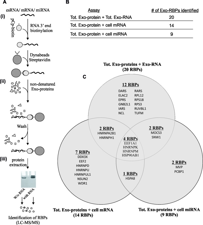
“In a previous report, 30 RBPs were identified in exosomes in complex with different RNA species in a cell-free assay, 20 of which were associated with exosomal RNAs [46].”
From Differential RNA packaging into small extracellular vesicles by neurons and astrocytes (2021) · cites this paper as [46]
In the citing paper
“A previous report showed that 23 of 30 RBPs discovered in exosomes were in a complex with cellular miRNAs and/or exosomal RNAs [46].”
From Differential RNA packaging into small extracellular vesicles by neurons and astrocytes (2021) · cites this paper as [46]
In the citing paper
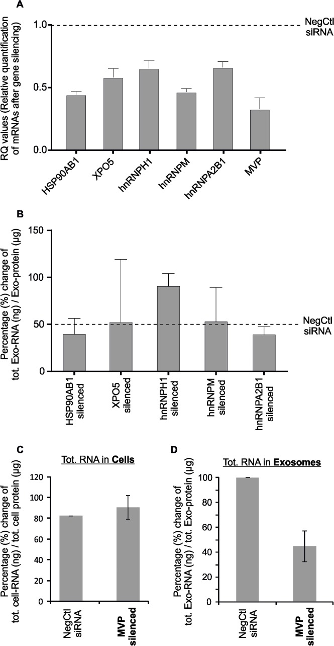
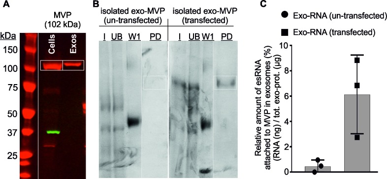
“Under normal conditions, silencing of MVP in cytoplasm resulted in 50% reduction of RNA shuttled into exosomes but the total RNA in cells remained unchanged, while introduction of MVP increased the amount of RNA [46].”
From Differential RNA packaging into small extracellular vesicles by neurons and astrocytes (2021) · cites this paper as [46]
Self-check
Percentages, sums and reported differences re-computed from the paper’s own numbers.
The self-check needs the paper’s full text.
Citations
Cites
Valadi, H., Ekström, K., Bossios, A., Sjöstrand, M., Lee, J. J., & Lötvall, J. O. (2007) · Nature Cell Biology · doi:10.1038/ncb1596 · Cited by 12,834
Self-citationTumour exosome integrins determine organotropic metastasis
Hoshino, A., Costa-Silva, B., Shen, T.-L., Rodrigues, G., Hashimoto, A., Tesic Mark, M., Molina, H., Kohsaka, S., Di Giannatale, A., Ceder, S., et al. (2015) · Nature · doi:10.1038/nature15756 · Cited by 5,101
Adipose-derived circulating miRNAs regulate gene expression in other tissues
Thomou, T., Mori, M. A., Dreyfuss, J. M., Konishi, M., Sakaguchi, M., Wolfrum, C., Rao, T. N., Winnay, J. N., Garcia-Martin, R., Grinspoon, S. K., et al. (2017) · Nature · doi:10.1038/nature21365 · Cited by 1,539
Vesiclepedia: A Compendium for Extracellular Vesicles with Continuous Community Annotation
Kalra, H., Simpson, R. J., Ji, H., Aikawa, E., Altevogt, P., Askenase, P., Bond, V. C., Borràs, F. E., Breakefield, X., Budnik, V., et al. (2012) · PLoS Biology · doi:10.1371/journal.pbio.1001450 · Cited by 1,367
Self-citationRatajczak, J., Wysoczynski, M., Hayek, F., Janowska-Wieczorek, A., & Ratajczak, M. Z. (2006) · Leukemia · doi:10.1038/sj.leu.2404296 · Cited by 1,340
Protein–RNA interactions: new genomic technologies and perspectives
König, J., Zarnack, K., Luscombe, N. M., & Ule, J. (2012) · Nature Reviews Genetics · doi:10.1038/nrg3141 · Cited by 555
Ekström, K., Valadi, H., Sjöstrand, M., Malmhäll, C., Bossios, A., Eldh, M., & Lötvall, J. (2012) · Journal of Extracellular Vesicles · doi:10.3402/jev.v1i0.18389 · Cited by 201
Self-citation
Research works printed but not linked: 85
- B Fevrier, 2004, Exosomes: endosomal-derived vesicles shipping extracellular messages, Current opinion in cell biology, 16, 415, 10.1016/j.ceb.2004.06.003 · DOI · Paper page
- M Nawaz, 2014, The emerging role of extracellular vesicles as biomarkers for urogenital cancers, Nature reviews Urology, 11, 688, 10.1038/nrurol.2014.301 · DOI
- F Fatima, 2017, Vesiculated Long Non-Coding RNAs: Offshore Packages Deciphering Trans-Regulation between Cells, Cancer Progression and Resistance to Therapies, Non-Coding RNA, 3, 10, 10.3390/ncrna3010010 · DOI
- S Mathivanan, 2010, Exosomes: extracellular organelles important in intercellular communication, Journal of proteomics, 73, 1907, 10.1016/j.jprot.2010.06.006 · DOI
- M Nawaz, 2017, Extracellular Vesicles, Tunneling Nanotubes, and Cellular Interplay: Synergies and Missing Links, Front Mol Biosci, 4, 50, 10.3389/fmolb.2017.00050 · DOI
- M Simons, 2009, Exosomes—vesicular carriers for intercellular communication, Current opinion in cell biology, 21, 575, 10.1016/j.ceb.2009.03.007 · DOI
- AK Ludwig, 2012, Exosomes: small vesicles participating in intercellular communication, The international journal of biochemistry & cell biology, 44, 11, 10.1016/j.biocel.2011.10.005 · DOI
- F Fatima, 2017, Long Distance Metabolic Regulation through Adipose-Derived Circulating Exosomal miRNAs: A Trail for RNA-Based Therapies?, Front Physiol, 8, 545, 10.3389/fphys.2017.00545 · DOI
- M Yanez-Mo, 2015, Biological properties of extracellular vesicles and their physiological functions, J Extracell Vesicles, 4, 27066, 10.3402/jev.v4.27066 · DOI · Paper page
- T Katsuda, 2013, The roles of extracellular vesicles in cancer biology: towards the development of novel cancer biomarkers, Proteomics
- SA Bellingham, 2012, Exosomes: vehicles for the transfer of toxic proteins associated with neurodegenerative diseases?, Frontiers in physiology, 3, 124, 10.3389/fphys.2012.00124 · DOI · Paper page
- EI Buzas, 2014, Emerging role of extracellular vesicles in inflammatory diseases, Nature reviews Rheumatology, 10, 356, 10.1038/nrrheum.2014.19 · DOI
- MI Ramirez, 2018, Technical challenges of working with extracellular vesicles, Nanoscale
- DJ Gibbings, 2009, Multivesicular bodies associate with components of miRNA effector complexes and modulate miRNA activity, Nat Cell Biol, 11, 1143, 10.1038/ncb1929 · DOI
- C Lasser, 2011, Human saliva, plasma and breast milk exosomes contain RNA: uptake by macrophages, J Transl Med, 9, 9, 10.1186/1479-5876-9-9 · DOI · Paper page
- SS Luo, 2009, Human villous trophoblasts express and secrete placenta-specific microRNAs into maternal circulation via exosomes, Biol Reprod, 81, 717, 10.1095/biolreprod.108.075481 · DOI · Paper page
- A Michael, 2009, Exosomes from human saliva as a source of microRNA biomarkers, Oral Dis, 16, 34, 10.1111/j.1601-0825.2009.01604.x · DOI
- KC Miranda, 2010, Nucleic acids within urinary exosomes/microvesicles are potential biomarkers for renal disease, Kidney Int, 78, 191, 10.1038/ki.2010.106 · DOI
- J Skog, 2008, Glioblastoma microvesicles transport RNA and proteins that promote tumour growth and provide diagnostic biomarkers, Nat Cell Biol, 10, 1470, 10.1038/ncb1800 · DOI · Paper page
- J Wahlgren, 2012, Plasma exosomes can deliver exogenous short interfering RNA to monocytes and lymphocytes, Nucleic Acids Res, 40, e130, 10.1093/nar/gks463 · DOI
- M Kanada, 2015, Differential fates of biomolecules delivered to target cells via extracellular vesicles, Proceedings of the National Academy of Sciences of the United States of America, 112, E1433, 10.1073/pnas.1418401112 · DOI
- DM Pegtel, 2010, Functional delivery of viral miRNAs via exosomes, Proceedings of the National Academy of Sciences of the United States of America, 107, 6328, 10.1073/pnas.0914843107 · DOI
- B Mateescu, 2017, Obstacles and opportunities in the functional analysis of extracellular vesicle RNA—an ISEV position paper, J Extracell Vesicles, 6, 1286095, 10.1080/20013078.2017.1286095 · DOI
- M Eldh, 2010, Exosomes communicate protective messages during oxidative stress; possible role of exosomal shuttle RNA, PLoS One, 5, e15353, 10.1371/journal.pone.0015353 · DOI
- SA Bellingham, 2012, Small RNA deep sequencing reveals a distinct miRNA signature released in exosomes from prion-infected neuronal cells, Nucleic Acids Res
- A Montecalvo, 2012, Mechanism of transfer of functional microRNAs between mouse dendritic cells via exosomes, Blood, 119, 756, 10.1182/blood-2011-02-338004 · DOI · Paper page
- M Ragusa, 2014, Highly skewed distribution of miRNAs and proteins between colorectal cancer cells and their exosomes following Cetuximab treatment: biomolecular, genetic and translational implications, Oncoscience, 1, 132, 10.18632/oncoscience.19 · DOI
- MJ Shurtleff, 2016, Y-box protein 1 is required to sort microRNAs into exosomes in cells and in a cell-free reaction, eLife, 5, 10.7554/eLife.19276 · DOI · Paper page
- T Glisovic, 2008, RNA-binding proteins and post-transcriptional gene regulation, FEBS Lett, 582, 1977, 10.1016/j.febslet.2008.03.004 · DOI · Paper page
- S Gerstberger, 2014, A census of human RNA-binding proteins, Nature reviews Genetics, 15, 829, 10.1038/nrg3813 · DOI · Paper page
- J Ratajczak, 2006, Embryonic stem cell-derived microvesicles reprogram hematopoietic progenitors: evidence for horizontal transfer of mRNA and protein delivery, Leukemia, 20, 847, 10.1038/sj.leu.2404132 · DOI
- JD Arroyo, 2011, Argonaute2 complexes carry a population of circulating microRNAs independent of vesicles in human plasma, Proceedings of the National Academy of Sciences of the United States of America, 108, 5003, 10.1073/pnas.1019055108 · DOI · Paper page
- A Turchinovich, 2011, Characterization of extracellular circulating microRNA, Nucleic acids research, 39, 7223, 10.1093/nar/gkr254 · DOI · Paper page
- C Gourzones, 2013, Consistent high concentration of the viral microRNA BART17 in plasma samples from nasopharyngeal carcinoma patients—evidence of non-exosomal transport, Virology journal, 10, 119, 10.1186/1743-422X-10-119 · DOI
- JG Patton, 2015, Biogenesis, delivery, and function of extracellular RNA, Journal of extracellular vesicles, 4, 27494, 10.3402/jev.v4.27494 · DOI
- J Guduric-Fuchs, 2012, Selective extracellular vesicle-mediated export of an overlapping set of microRNAs from multiple cell types, BMC genomics, 13, 357, 10.1186/1471-2164-13-357 · DOI
- JR Chevillet, 2014, Quantitative and stoichiometric analysis of the microRNA content of exosomes, Proceedings of the National Academy of Sciences of the United States of America, 111, 14888, 10.1073/pnas.1408301111 · DOI
- C Villarroya-Beltri, 2013, Sumoylated hnRNPA2B1 controls the sorting of miRNAs into exosomes through binding to specific motifs, Nature communications, 4, 2980, 10.1038/ncomms3980 · DOI · Paper page
- L Santangelo, 2016, The RNA-Binding Protein SYNCRIP Is a Component of the Hepatocyte Exosomal Machinery Controlling MicroRNA Sorting, Cell reports, 17, 799, 10.1016/j.celrep.2016.09.031 · DOI · Paper page
- MF Bolukbasi, 2012, miR-1289 and "Zipcode"-like Sequence Enrich mRNAs in Microvesicles, Mol Ther Nucleic Acids, 1, e10, 10.1038/mtna.2011.2 · DOI
- AJ McKenzie, 2016, KRAS-MEK Signaling Controls Ago2 Sorting into Exosomes, Cell reports, 15, 978, 10.1016/j.celrep.2016.03.085 · DOI
- ML Squadrito, 2014, Endogenous RNAs modulate microRNA sorting to exosomes and transfer to acceptor cells, Cell Rep, 8, 1432, 10.1016/j.celrep.2014.07.035 · DOI · Paper page
- BW van Balkom, 2015, Quantitative and qualitative analysis of small RNAs in human endothelial cells and exosomes provides insights into localized RNA processing, degradation and sorting, J Extracell Vesicles, 4, 26760, 10.3402/jev.v4.26760 · DOI
- JP Tosar, 2015, Assessment of small RNA sorting into different extracellular fractions revealed by high-throughput sequencing of breast cell lines, Nucleic Acids Res, 43, 5601, 10.1093/nar/gkv432 · DOI · Paper page
- A Iavello, 2016, Role of Alix in miRNA packaging during extracellular vesicle biogenesis, International journal of molecular medicine, 37, 958, 10.3892/ijmm.2016.2488 · DOI · Paper page
- A Marchler-Bauer, 2011, CDD: a Conserved Domain Database for the functional annotation of proteins, Nucleic Acids Res, 39, D225, 10.1093/nar/gkq1189 · DOI
- T Kampers, 1996, RNA stimulates aggregation of microtubule-associated protein tau into Alzheimer-like paired helical filaments, FEBS Lett, 399, 344, 10.1016/S0014-5793(96)01386-5 · DOI · Paper page
- D Koppers-Lalic, 2014, Nontemplated nucleotide additions distinguish the small RNA composition in cells from exosomes, Cell Rep, 8, 1649, 10.1016/j.celrep.2014.08.027 · DOI · Paper page
- M Gonzalez-Begne, 2009, Proteomic analysis of human parotid gland exosomes by multidimensional protein identification technology (MudPIT), J Proteome Res, 8, 1304, 10.1021/pr800658c · DOI
- S Mathivanan, 2010, Proteomics analysis of A33 immunoaffinity-purified exosomes released from the human colon tumor cell line LIM1215 reveals a tissue-specific protein signature, Mol Cell Proteomics, 9, 197, 10.1074/mcp.M900152-MCP200 · DOI
- C Olver, 2007, Proteomic analysis of secreted exosomes, Subcell Biochem, 43, 99, 10.1007/978-1-4020-5943-8_7 · DOI
- S Mathivanan, 2012, ExoCarta 2012: database of exosomal proteins, RNA and lipids, Nucleic Acids Res, 40, D1241, 10.1093/nar/gkr828 · DOI
- S Principe, 2013, In-depth proteomic analyses of exosomes isolated from expressed prostatic secretions in urine, Proteomics, 13, 1667, 10.1002/pmic.201200561 · DOI
- JL Welton, 2016, Proteomics analysis of vesicles isolated from plasma and urine of prostate cancer patients using a multiplex, aptamer-based protein array, J Extracell Vesicles, 5, 31209, 10.3402/jev.v5.31209 · DOI
- S Keerthikumar, 2016, ExoCarta: A Web-Based Compendium of Exosomal Cargo, Journal of molecular biology, 428, 688, 10.1016/j.jmb.2015.09.019 · DOI
- G Dreyfuss, 2002, Messenger-RNA-binding proteins and the messages they carry, Nat Rev Mol Cell Biol, 3, 195, 10.1038/nrm760 · DOI
- K Bomsztyk, 1997, Diverse molecular interactions of the hnRNP K protein, FEBS Lett, 403, 113, 10.1016/S0014-5793(97)00041-0 · DOI
- A Chaudhury, 2010, Heterogeneous nuclear ribonucleoproteins (hnRNPs) in cellular processes: Focus on hnRNP E1's multifunctional regulatory roles, RNA, 16, 1449, 10.1261/rna.2254110 · DOI
- AB Shyu, 2000, The double lives of shuttling mRNA binding proteins, Cell, 102, 135, 10.1016/S0092-8674(00)00018-0 · DOI
- N Volk, 2011, Versatility of MicroRNA biogenesis, PLoS One, 6, e19391, 10.1371/journal.pone.0019391 · DOI
- YS Lee, 2009, Silencing by small RNAs is linked to endosomal trafficking, Nat Cell Biol, 11, 1150, 10.1038/ncb1930 · DOI
- S Jha, 2009, RVB1/RVB2: running rings around molecular biology, Mol Cell, 34, 521, 10.1016/j.molcel.2009.05.016 · DOI
- SG Park, 2005, Functional expansion of aminoacyl-tRNA synthetases and their interacting factors: new perspectives on housekeepers, Trends Biochem Sci, 30, 569, 10.1016/j.tibs.2005.08.004 · DOI
- GS Hussey, 2011, Identification of an mRNP complex regulating tumorigenesis at the translational elongation step, Mol Cell, 41, 419, 10.1016/j.molcel.2011.02.003 · DOI
- D Haussecker, 2010, Human tRNA-derived small RNAs in the global regulation of RNA silencing, RNA, 16, 673, 10.1261/rna.2000810 · DOI
- EN Nolte-'t Hoen, 2012, Deep sequencing of RNA from immune cell-derived vesicles uncovers the selective incorporation of small non-coding RNA biotypes with potential regulatory functions, Nucleic Acids Res
- G Jannot, 2011, The ribosomal protein RACK1 is required for microRNA function in both C. elegans and humans, EMBO Rep, 12, 581, 10.1038/embor.2011.66 · DOI
- M van Kouwenhove, 2011, MicroRNA regulation by RNA-binding proteins and its implications for cancer, Nat Rev Cancer, 11, 644, 10.1038/nrc3107 · DOI · Paper page
- S Iwasaki, 2010, Hsc70/Hsp90 chaperone machinery mediates ATP-dependent RISC loading of small RNA duplexes, Mol Cell, 39, 292, 10.1016/j.molcel.2010.05.015 · DOI
- D Koppers-Lalic, 2014, Nontemplated nucleotide additions distinguish the small RNA composition in cells from exosomes, Cell Rep, 8, 1649, 10.1016/j.celrep.2014.08.027 · DOI · Paper page
- S Wani, 2018, Cancer cells govern miR-2909 exosomal recruitment through its 3'-end post-transcriptional modification, Cell Biochem Funct, 36, 106, 10.1002/cbf.3323 · DOI
- J Li, 2016, Identification and Characterization of 293T Cell-Derived Exosomes by Profiling the Protein, mRNA and MicroRNA Components, PLoS One, 11, e0163043, 10.1371/journal.pone.0163043 · DOI
- SA Melo, 2014, Cancer exosomes perform cell-independent microRNA biogenesis and promote tumorigenesis, Cancer Cell, 26, 707, 10.1016/j.ccell.2014.09.005 · DOI
- VN Kim, 2006, Genomics of microRNA, Trends in Genetics, 22, 165, 10.1016/j.tig.2006.01.003 · DOI
- P Bouvet, 2001, Recognition of pre-formed and flexible elements of an RNA stem-loop by nucleolin, J Mol Biol, 309, 763, 10.1006/jmbi.2001.4691 · DOI
- M Srivastava, 1999, Molecular dissection of nucleolin's role in growth and cell proliferation: new insights, FASEB J, 13, 1911, 10.1096/fasebj.13.14.1911 · DOI
- K Tominaga, 2011, Competitive regulation of nucleolin expression by HuR and miR-494, Mol Cell Biol, 31, 4219, 10.1128/MCB.05955-11 · DOI
- C Yang, 2002, Identification of nucleolin and nucleophosmin as genotoxic stress-responsive RNA-binding proteins, Nucleic Acids Res, 30, 2251, 10.1093/nar/30.10.2251 · DOI
- P Folk, 2004, Transcriptional coregulator SNW/SKIP: the concealed tie of dissimilar pathways, Cell Mol Life Sci, 61, 629, 10.1007/s00018-003-3215-4 · DOI
- EN Nolte-'t Hoen, 2012, Deep sequencing of RNA from immune cell-derived vesicles uncovers the selective incorporation of small non-coding RNA biotypes with potential regulatory functions, Nucleic acids research, 40, 9272, 10.1093/nar/gks658 · DOI
- F Fatima, 2017, Non-coding RNAs in Mesenchymal Stem Cell-Derived Extracellular Vesicles: Deciphering Regulatory Roles in Stem Cell Potency, Inflammatory Resolve, and Tissue Regeneration, Front Genet, 8, 161, 10.3389/fgene.2017.00161 · DOI
- Y Teng, 2017, MVP-mediated exosomal sorting of miR-193a promotes colon cancer progression, Nature communications, 8, 14448, 10.1038/ncomms14448 · DOI · Paper page
- SD Speese, 2012, Nuclear Envelope Budding Enables Large Ribonucleoprotein Particle Export during Synaptic Wnt Signaling (vol 149, pg 832, 2012), Cell, 151, 687, 10.1016/j.cell.2012.10.003 · DOI
- A Parchure, 2017, Getting mRNA-Containing Ribonucleoprotein Granules Out of a Nuclear Back Door, Neuron, 96, 604, 10.1016/j.neuron.2017.10.020 · DOI
- B Fahrenkrog, 2003, The nuclear pore complex: nucleocytoplasmic transport and beyond, Nat Rev Mol Cell Biol, 4, 757, 10.1038/nrm1230 · DOI
Cited by
Sorted by citations
Gene regulation by long non-coding RNAs and its biological functions
Statello, L., Guo, C.-J., Chen, L.-L., & Huarte, M. (2021) · Nature Reviews Molecular Cell Biology · doi:10.1038/s41580-020-00315-9 · Cited by 4,991
Self-citationRNA delivery by extracellular vesicles in mammalian cells and its applications
O’Brien, K., Breyne, K., Ughetto, S., Laurent, L. C., & Breakefield, X. O. (2020) · Nature Reviews Molecular Cell Biology · doi:10.1038/s41580-020-0251-y · Cited by 1,811
Lin, Z., Wu, Y., Xu, Y., Li, G., Li, Z., & Liu, T. (2022) · Molecular Cancer · doi:10.1186/s12943-022-01650-5 · Cited by 297
Current Knowledge on Exosome Biogenesis, Cargo-Sorting Mechanism and Therapeutic Implications
Xie, S., Zhang, Q., & Jiang, L. (2022) · Membranes · doi:10.3390/membranes12050498 · Cited by 262
Differential RNA packaging into small extracellular vesicles by neurons and astrocytes
Luo, X., Jean-Toussaint, R., Sacan, A., & Ajit, S. K. (2021) · Cell Communication and Signaling · doi:10.1186/s12964-021-00757-4 · Cited by 32
Metadata
- Authors
- Statello, L., Maugeri, M., Garre, E., Nawaz, M., Wahlgren, J., Papadimitriou, A., Lundqvist, C., Lindfors, L., Collén, A., Sunnerhagen, P., Ragusa, M., Purrello, M., Di Pietro, C., Tigue, N., & Valadi, H.
- Venue
- PLOS ONE
- Publisher
- Public Library of Science (PLoS)
- Published
- 2018-04-24
- Volume
- 13
- Issue
- 4
- Pages
- e0195969
- ISSN
- 1932-6203
- PMC
- PMC5918169
- Cited by
- 242
- References
- 7
- Language
- en
- Funders
- Vetenskapsrådet
- Stiftelsen för Strategisk Forskning (IRC15-0065)
- Aroseniusfonden
- Magnus Bergvalls Stiftelse
- IngaBritt och Arne Lundbergs Forskningsstiftelse
- Fundação de Amparo à Pesquisa do Estado de São Paulo (12/24574-3)
- Coordenação de Aperfeiçoamento de Pessoal de Nível Superior (99999.007057/2015-06)
About these checks
Registry, retraction, Cites and statistics results are computed, not judged, with one exception: a printed reference that no index lists is read once by a model before it counts as a citation error. Claims are extracted by a model and kept only when their quote appears verbatim in the text; each cited source is then checked, with a model’s help, for whether it says what the claim relies on. Those verdicts can be wrong — read the claim’s quote and the cited source before relying on one.
Statistics and arithmetic are recomputed, not judged.
Confidence bands are shown only for fields calibrated on our annotated test set. None is calibrated yet, so no band is shown.
What counts as a claim ›How we measure confidence ›How we check this ›
Are you an author, or do you see a problem with this page? Request a review
