

The DOI matches its Crossref record; 61 of the 62 references it cites were found in a registry or an index.
A Novel Co-Crystal Structure Affords the Design of Gain-of-Function Lentiviral Integrase Mutants in the Presence of Modified PSIP1/LEDGF/p75
Open accessAbstract
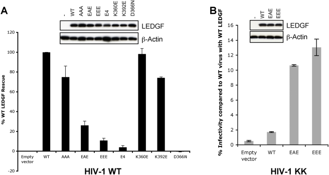
Lens epithelium derived growth factor (LEDGF), also known as PC4 and SFRS1 interacting protein 1 (PSIP1) and transcriptional co-activator p75, is the cellular binding partner of lentiviral integrase (IN) proteins. LEDGF accounts for the characteristic propensity of Lentivirus to integrate within active transcription units and is required for efficient viral replication. We now present a crystal structure containing the N-terminal and catalytic core domains (NTD and CCD) of HIV-2 IN in complex with the IN binding domain (IBD) of LEDGF. The structure extends the known IN-LEDGF interface, elucidating primarily charge-charge interactions between the NTD of IN and the IBD. A constellation of acidic residues on the NTD is characteristic of lentiviral INs, and mutations of the positively charged residues on the IBD severely affect interaction with all lentiviral INs tested. We show that the novel NTD-IBD contacts are critical for stimulation of concerted lentiviral DNA integration by LEDGF in vitro and for its function during the early steps of HIV-1 replication. Furthermore, the new structural details enabled us to engineer a mutant of HIV-1 IN that primarily functions only when presented with a complementary LEDGF mutant. These findings provide structural basis for the high affinity lentiviral IN-LEDGF interaction and pave the way for development of LEDGF-based targeting technologies for gene therapy.
Overview
The claims and statistics of this paper haven’t been checked yet.
Run the check — free, no account needed. It reads the open-access text and checks the sources it cites.
Integrity signals
Registry
Cites
Statistics
Figures
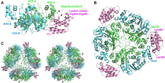
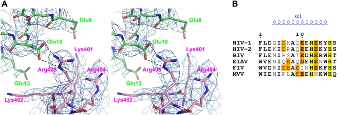
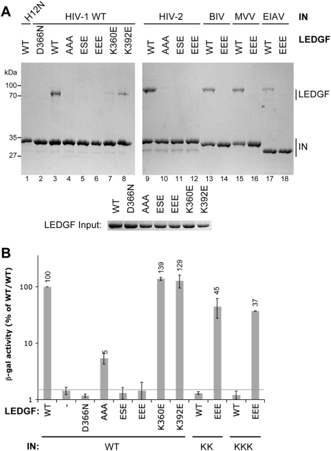
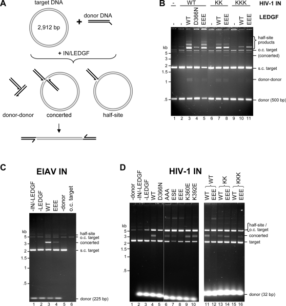
Verification
Claims the paper makes
The claims and statistics of this paper haven’t been checked yet.
Run the check — free, no account needed. It reads the open-access text and checks the sources it cites.
Claims other papers make about this one
Each is checked against this paper’s own text: does it say what the citing paper says it does?
The source backs part of the claim, not all of it — or backs it for a different population, condition or comparison.
In the citing paper
“Residues Ile356 and Asp366 located on the loop between α-helices 1 and 2 mediate interactions with both IN monomers of the CCD dimer while electropositive residues on the outward face of α-helix 4 interface with electronegative residues of IN NTD α-helix 1 (REF.107,109).”
From Structure and function of retroviral integrase (2022) · cites this paper as [107]
Experimental — accuracy not yet measuredThe cited source says what the claim attributes to it.
In the citing paper
“The CCD is the main IN binding determinant, with the NTD contributing secondary interactions for full binding affinity106,107.”
From Structure and function of retroviral integrase (2022) · cites this paper as [107]
Self-check
Percentages, sums and reported differences re-computed from the paper’s own numbers.
The self-check needs the paper’s full text.
Citations
Cites
Coot : model-building tools for molecular graphics
Emsley, P., & Cowtan, K. (2004) · Acta Crystallographica Section D Biological Crystallography · doi:10.1107/s0907444904019158 · Cited by 30,571
Phaser crystallographic software
McCoy, A. J., Grosse-Kunstleve, R. W., Adams, P. D., Winn, M. D., Storoni, L. C., & Read, R. J. (2007) · Journal of Applied Crystallography · doi:10.1107/s0021889807021206 · Cited by 20,612
The CCP4 suite: programs for protein crystallography
Number Collaborative Computational Project (1994) · Acta Crystallographica Section D Biological Crystallography · doi:10.1107/s0907444994003112 · Cited by 17,317
Refinement of Macromolecular Structures by the Maximum-Likelihood Method
Murshudov, G. N., Vagin, A. A., & Dodson, E. J. (1997) · Acta Crystallographica Section D Biological Crystallography · doi:10.1107/s0907444996012255 · Cited by 14,796
MOLREP : an Automated Program for Molecular Replacement
Vagin, A. A., & Teplyakov, A. (1997) · Journal of Applied Crystallography · doi:10.1107/s0021889897006766 · Cited by 4,756
PHENIX : building new software for automated crystallographic structure determination
Adams, P. D., Grosse-Kunstleve, R. W., Hung, L.-W., Ioerger, T. R., McCoy, A. J., Moriarty, N. W., Read, R. J., Sacchettini, J. C., Sauter, N. K., & Terwilliger, T. C. (2002) · Acta Crystallographica Section D Biological Crystallography · doi:10.1107/s0907444902016657 · Cited by 4,361
LMO2 -Associated Clonal T Cell Proliferation in Two Patients after Gene Therapy for SCID-X1
Hacein-Bey-Abina, S., Von Kalle, C., Schmidt, M., McCormack, M. P., Wulffraat, N., Leboulch, P., Lim, A., Osborne, C. S., Pawliuk, R., Morillon, E., et al. (2003) · Science · doi:10.1126/science.1088547 · Cited by 3,587
Dyda, F., Hickman, A. B., Jenkins, T. M., Engelman, A., Craigie, R., & Davies, D. R. (1994) · Science · doi:10.1126/science.7801124 · Cited by 810 · cited 1×
“The CCD spans the most conserved region of IN and bears close structural homology to prokaryotic transposases [6].”
BackgroundSelf-citationMossessova, E., & Lima, C. D. (2000) · Molecular Cell · doi:10.1016/s1097-2765(00)80326-3 · Cited by 704
HIV-1 Integrase Forms Stable Tetramers and Associates with LEDGF/p75 Protein in Human Cells
Cherepanov, P., Maertens, G., Proost, P., Devreese, B., Van Beeumen, J., Engelborghs, Y., De Clercq, E., & Debyser, Z. (2003) · Journal of Biological Chemistry · doi:10.1074/jbc.m209278200 · Cited by 704
Self-citation
Research works printed but not linked: 2
- AGW Leslie, 1992, Recent changes to the MOSFLM package for processing film and image plate data., Joint CCP4+ESF-EAMCB Newsletter on Protein Crystallography, 26
- PR Evans, 1993, Data reduction., Proceedings of CCP4 Study Weekend, 1993, on Data Collection & Processing, 114
Cited by
Sorted by citations
Integrase-mediated spacer acquisition during CRISPR–Cas adaptive immunity
Nuñez, J. K., Lee, A. S. Y., Engelman, A., & Doudna, J. A. (2015) · Nature · doi:10.1038/nature14237 · Cited by 347
Self-citationCraigie, R., & Bushman, F. D. (2012) · Cold Spring Harbor Perspectives in Medicine · doi:10.1101/cshperspect.a006890 · Cited by 327
Lesbats, P., Engelman, A., & Cherepanov, P. (2016) · Chemical Reviews · doi:10.1021/acs.chemrev.6b00125 · Cited by 250
Self-citationProtein delivery using engineered virus-like particles
Kaczmarczyk, S. J., Sitaraman, K., Young, H. A., Hughes, S. H., & Chatterjee, D. K. (2011) · Proceedings of the National Academy of Sciences · doi:10.1073/pnas.1101874108 · Cited by 220
Structural basis for high-affinity binding of LEDGF PWWP to mononucleosomes
Eidahl, J. O., Crowe, B. L., North, J. A., McKee, C. J., Shkriabai, N., Feng, L., Plumb, M., Graham, R. L., Gorelick, R. J., Hess, S., et al. (2013) · Nucleic Acids Research · doi:10.1093/nar/gkt074 · Cited by 207
Cryo-EM structures and atomic model of the HIV-1 strand transfer complex intasome
Passos, D. O., Li, M., Yang, R., Rebensburg, S. V., Ghirlando, R., Jeon, Y., Shkriabai, N., Kvaratskhelia, M., Craigie, R., & Lyumkis, D. (2017) · Science · doi:10.1126/science.aah5163 · Cited by 202
Multimode, Cooperative Mechanism of Action of Allosteric HIV-1 Integrase Inhibitors
Kessl, J. J., Jena, N., Koh, Y., Taskent‐Sezgin, H., Slaughter, A., Feng, L., de Silva, S., Wu, L. J., Grice, S. F. J. L., Engelman, A., et al. (2012) · Journal of Biological Chemistry · doi:10.1074/jbc.m112.354373 · Cited by 186
Self-citationLEDGF Hybrids Efficiently Retarget Lentiviral Integration Into Heterochromatin
Gijsbers, R., Ronen, K., Vets, S., Malani, N., Rijck, J. D., McNeely, M., Bushman, F. D., & Debyser, Z. (2010) · Molecular Therapy · doi:10.1038/mt.2010.36 · Cited by 161
Novel approaches to inhibiting HIV-1 replication
Adamson, C. S., & Freed, E. O. (2009) · Antiviral Research · doi:10.1016/j.antiviral.2009.09.009 · Cited by 149
Lens epithelium-derived growth factor fusion proteins redirect HIV-1 DNA integration
Ferris, A. L., Wu, X., Hughes, C. M., Stewart, C., Smith, S. J., Milne, T. A., Wang, G. G., Shun, M.-C., Allis, C. D., Engelman, A., et al. (2010) · Proceedings of the National Academy of Sciences · doi:10.1073/pnas.0914142107 · Cited by 146
Self-citationMolecular mechanisms of retroviral integration site selection
Kvaratskhelia, M., Sharma, A., Larue, R. C., Serrao, E., & Engelman, A. (2014) · Nucleic Acids Research · doi:10.1093/nar/gku769 · Cited by 150
Self-citationLe Rouzic, E., Bonnard, D., Chasset, S., Bruneau, J.-M., Chevreuil, F., Le Strat, F., Nguyen, J., Beauvoir, R., Amadori, C., Brias, J., et al. (2013) · Retrovirology · doi:10.1186/1742-4690-10-144 · Cited by 127
Structure and function of retroviral integrase
Maertens, G. N., Engelman, A. N., & Cherepanov, P. (2022) · Nature Reviews Microbiology · doi:10.1038/s41579-021-00586-9 · Cited by 124
Self-citationA New Class of Multimerization Selective Inhibitors of HIV-1 Integrase
Sharma, A., Slaughter, A., Jena, N., Feng, L., Kessl, J. J., Fadel, H. J., Malani, N., Male, F., Wu, L., Poeschla, E., et al. (2014) · PLoS Pathogens · doi:10.1371/journal.ppat.1004171 · Cited by 124
Structural Basis for Functional Tetramerization of Lentiviral Integrase
Hare, S., Di Nunzio, F., Labeja, A., Wang, J., Engelman, A., & Cherepanov, P. (2009) · PLoS Pathogens · doi:10.1371/journal.ppat.1000515 · Cited by 122
Self-citationStructural biology of retroviral DNA integration
Li, X., Krishnan, L., Cherepanov, P., & Engelman, A. (2011) · Virology · doi:10.1016/j.virol.2010.12.008 · Cited by 116
Self-citationStructural basis of second-generation HIV integrase inhibitor action and viral resistance
Cook, N. J., Li, W., Berta, D., Badaoui, M., Ballandras-Colas, A., Nans, A., Kotecha, A., Rosta, E., Engelman, A. N., & Cherepanov, P. (2020) · Science · doi:10.1126/science.aay4919 · Cited by 108
Self-citationAntiretroviral therapy and drug resistance in human immunodeficiency virus type 2 infection
Menéndez‐Arias, L., & Álvarez, M. (2013) · Antiviral Research · doi:10.1016/j.antiviral.2013.12.001 · Cited by 110
Wang, H., Jurado, K. A., Wu, X., Shun, M.-C., Li, X., Ferris, A. L., Smith, S. J., Patel, P. A., Fuchs, J. R., Cherepanov, P., et al. (2012) · Nucleic Acids Research · doi:10.1093/nar/gks913 · Cited by 101
Self-citationCryo-EM reveals a novel octameric integrase structure for betaretroviral intasome function
Ballandras-Colas, A., Brown, M., Cook, N. J., Dewdney, T. G., Demeler, B., Cherepanov, P., Lyumkis, D., & Engelman, A. N. (2016) · Nature · doi:10.1038/nature16955 · Cited by 98
Self-citationRetroviral Integrase Proteins and HIV-1 DNA Integration
Krishnan, L., & Engelman, A. (2012) · Journal of Biological Chemistry · doi:10.1074/jbc.r112.397760 · Cited by 112
Self-citationStructural basis for HIV‐1 DNA integration in the human genome, role of the LEDGF/P75 cofactor
Michel, F., Crucifix, C., Granger, F., Eiler, S., Mouscadet, J. F., Korolev, S. V., Agapkina, J., Ziganshin, R., Gottikh, M., Nazabal, A., et al. (2009) · The EMBO Journal · doi:10.1038/emboj.2009.41 · Cited by 114
Retroviral Integrase Structure and DNA Recombination Mechanism
Engelman, A., & Cherepanov, P. (2014) · Microbiology Spectrum · doi:10.1128/microbiolspec.mdna3-0024-2014 · Cited by 101
Self-citationHost proteins involved in HIV infection: New therapeutic targets
Arhel, N. J., & Kirchhoff, F. (2009) · Biochimica et Biophysica Acta (BBA) - Molecular Basis of Disease · doi:10.1016/j.bbadis.2009.12.003 · Cited by 99
Cellular and molecular mechanisms of HIV-1 integration targeting
Engelman, A., & Singh, P. K. (2018) · Cellular and Molecular Life Sciences · doi:10.1007/s00018-018-2772-5 · Cited by 102
Self-citationFeng, L., Sharma, A., Slaughter, A., Jena, N., Koh, Y., Shkriabai, N., Larue, R. C., Patel, P. A., Mitsuya, H., Kessl, J., et al. (2013) · Journal of Biological Chemistry · doi:10.1074/jbc.m112.443390 · Cited by 99
Self-citationLow, A., Prada, N., Topper, M. B., Vaida, F., Castor, D., Mohri, H., Hazuda, D. J., Muesing, M. A., & Markowitz, M. H. (2009) · Antimicrobial Agents and Chemotherapy · doi:10.1128/aac.00397-09 · Cited by 89
Piecing together the structure of retroviral integrase, an important target in AIDS therapy
Jaskólski, M., Alexandratos, J. N., Bujacz, G., & Wlodawer, A. (2009) · FEBS Journal · doi:10.1111/j.1742-4658.2009.07009.x · Cited by 89
Allosteric Inhibition of Human Immunodeficiency Virus Integrase
Gupta, K., Brady, T., Dyer, B. M., Malani, N., Hwang, Y., Male, F., Nolte, R. T., Wang, L., Velthuisen, E., Jeffrey, J., et al. (2014) · Journal of Biological Chemistry · doi:10.1074/jbc.m114.551119 · Cited by 81
Multifaceted HIV integrase functionalities and therapeutic strategies for their inhibition
Engelman, A. (2019) · Journal of Biological Chemistry · doi:10.1074/jbc.rev119.006901 · Cited by 83
Self-citationImmunoregulation in human American leishmaniasis: balancing pathology and protection
Gollob, K. J., Viana, A. G., & Dutra, W. O. (2014) · Parasite Immunology · doi:10.1111/pim.12100 · Cited by 77
Multiple cellular proteins interact with LEDGF/p75 through a conserved unstructured consensus motif
Tesina, P., Čermáková, K., Hořejší, M., Procházková, K., Fábry, M., Sharma, S., Christ, F., Demeulemeester, J., Debyser, Z., Rijck, J. D., et al. (2015) · Nature Communications · doi:10.1038/ncomms8968 · Cited by 74
Demeulemeester, J., De Rijck, J., Gijsbers, R., & Debyser, Z. (2015) · BioEssays · doi:10.1002/bies.201500051 · Cited by 71
Fadel, H. J., Morrison, J. H., Saenz, D. T., Fuchs, J. R., Kvaratskhelia, M., Ekker, S. C., & Poeschla, E. M. (2014) · Journal of Virology · doi:10.1128/jvi.01397-14 · Cited by 70
Olifiers, N., Jansen, A. M., Herrera, H. M., Bianchi, R. D. C., D’Andrea, P. S., Mourão, G. M., & Gompper, M. E. (2015) · PLoS ONE · doi:10.1371/journal.pone.0143997 · Cited by 70
LEDGF/p75 Proteins with Alternative Chromatin Tethers Are Functional HIV-1 Cofactors
Meehan, A. M., Saenz, D. T., Morrison, J. H., Garcia-Rivera, J. A., Peretz, M., Llano, M., & Poeschla, E. M. (2009) · PLoS Pathogens · doi:10.1371/journal.ppat.1000522 · Cited by 71
Hughes, S., Jenkins, V. A., Dar, M. J., Engelman, A., & Cherepanov, P. (2009) · Journal of Biological Chemistry · doi:10.1074/jbc.m109.036491 · Cited by 69
Self-citationStructural Basis for Inhibitor-Induced Aggregation of HIV Integrase
Gupta, K., Turkki, V., Sherrill-Mix, S., Hwang, Y., Eilers, G., Taylor, L., McDanal, C., Wang, P., Temelkoff, D., Nolte, R. T., et al. (2016) · PLOS Biology · doi:10.1371/journal.pbio.1002584 · Cited by 68
Serrao, E., Krishnan, L., Shun, M.-C., Li, X., Cherepanov, P., Engelman, A., & Maertens, G. N. (2014) · Nucleic Acids Research · doi:10.1093/nar/gku136 · Cited by 59
Self-citationSmall molecules targeting the interaction between HIV-1 integrase and LEDGF/p75 cofactor
Luca, L. D., Ferro, S., Gitto, R., Barreca, M. L., Agnello, S., Christ, F., Debyser, Z., & Chimirri, A. (2010) · Bioorganic & Medicinal Chemistry · doi:10.1016/j.bmc.2010.08.051 · Cited by 59
Hendrix, J., Gijsbers, R., Rijck, J. D., Voet, A. R. D., Hotta, J., McNeely, M., Hofkens, J., Debyser, Z., & Engelborghs, Y. (2010) · Nucleic Acids Research · doi:10.1093/nar/gkq933 · Cited by 61
Engelman, A., & Cherepanov, P. (2017) · Current Opinion in Structural Biology · doi:10.1016/j.sbi.2017.04.005 · Cited by 58
Self-citationPeat, T. S., Rhodes, D. I., Vandegraaff, N., Le, G. T., Smith, J. A., Clark, L. J., Jones, E. D., Coates, J. A. V., Thienthong, N., Newman, J. M., et al. (2012) · PLoS ONE · doi:10.1371/journal.pone.0040147 · Cited by 52
Phylogenetic history demonstrates two different lineages of dengue type 1 virus in Colombia
Méndez, J. A., Usme-Ciro, J. A., Domingo, C., Rey-Benito, G. J., Sánchez, J. A., Tenório, A., & Gallego-Gómez, J. C. (2010) · Virology Journal · doi:10.1186/1743-422x-7-226 · Cited by 52
Kessl, J. J., Li, M., Ignatov, M. E., Shkriabai, N., Eidahl, J. O., Feng, L., Musier‐Forsyth, K., Craigie, R., & Kvaratskhelia, M. (2011) · Nucleic Acids Research · doi:10.1093/nar/gkr581 · Cited by 55
Phylogenetic reconstruction of dengue virus type 2 in Colombia
Méndez, J. A., Usme-Ciro, J. A., Domingo, C., Rey-Benito, G. J., Sánchez, J. A., Tenório, A., & Gallego-Gómez, J. C. (2012) · Virology Journal · doi:10.1186/1743-422x-9-64 · Cited by 51
An Allosteric Mechanism for Inhibiting HIV-1 Integrase with a Small Molecule
Kessl, J. J., Eidahl, J. O., Shkriabai, N., Zhao, Z., McKee, C. J., Hess, S., Burke, T. R., & Kvaratskhelia, M. (2009) · Molecular Pharmacology · doi:10.1124/mol.109.058883 · Cited by 50
Inflammation, Immunity, and Vaccines for Helicobacter pylori
D’Elios, M. M., & Andersen, L. P. (2009) · Helicobacter · doi:10.1111/j.1523-5378.2009.00698.x · Cited by 48
Intasome architecture and chromatin density modulate retroviral integration into nucleosome
Benleulmi, M. S., Matysiak, J., Henriquez, D. R., Vaillant, C., Lesbats, P., Calmels, C., Naughtin, M., Leon, O., Skalka, A. M., Ruff, M., et al. (2015) · Retrovirology · doi:10.1186/s12977-015-0145-9 · Cited by 47
HIV-1 Integrase Multimerization as a Therapeutic Target
Feng, L., Larue, R. C., Slaughter, A., Kessl, J. J., & Kvaratskhelia, M. (2015) · Current topics in microbiology and immunology · doi:10.1007/82_2015_439 · Cited by 59
Tsiang, M., Jones, G. S., Hung, M., Mukund, S., Han, B., Liu, X., Babaoglu, K., Lansdon, E. B., Chen, X., Todd, J., et al. (2009) · Journal of Biological Chemistry · doi:10.1074/jbc.m109.040121 · Cited by 46
Host Factors in Retroviral Integration and the Selection of Integration Target Sites
Craigie, R., & Bushman, F. D. (2014) · Microbiology Spectrum · doi:10.1128/microbiolspec.mdna3-0026-2014 · Cited by 49
Johnson, B. C., Métifiot, M., Pommier, Y. G., & Hughes, S. H. (2011) · Antimicrobial Agents and Chemotherapy · doi:10.1128/aac.05292-11 · Cited by 46
Factors that mold the nuclear landscape of HIV-1 integration
Bedwell, G. J., & Engelman, A. (2020) · Nucleic Acids Research · doi:10.1093/nar/gkaa1207 · Cited by 42
Self-citationEngineered Hyperactive Integrase for Concerted HIV-1 DNA Integration
Li, M., Jurado, K. A., Lin, S., Engelman, A., & Craigie, R. (2014) · PLoS ONE · doi:10.1371/journal.pone.0105078 · Cited by 45
Self-citationWielens, J., Headey, S. J., Jeevarajah, D., Rhodes, D. I., Deadman, J. J., Chalmers, D. K., Scanlon, M. J., & Parker, M. W. (2010) · FEBS Letters · doi:10.1016/j.febslet.2010.03.016 · Cited by 42
Debyser, Z., Vansant, G., Bruggemans, A., Janssens, J., & Christ, F. (2018) · Viruses · doi:10.3390/v11010012 · Cited by 41
The HIV-1 Integrase Monomer Induces a Specific Interaction with LTR DNA for Concerted Integration
Pandey, K. K., Bera, S., & Grandgenett, D. P. (2011) · Biochemistry · doi:10.1021/bi201247f · Cited by 41
Slaughter, A., Jurado, K. A., Deng, N., Feng, L., Kessl, J. J., Shkriabai, N., Larue, R. C., Fadel, H. J., Patel, P. A., Jena, N., et al. (2014) · Retrovirology · doi:10.1186/s12977-014-0100-1 · Cited by 38
Self-citationStructural basis of host protein hijacking in human T-cell leukemia virus integration
Bhatt, V., Shi, K., Salamango, D. J., Moeller, N. H., Pandey, K. K., Bera, S., Bohl, H. O., Kurniawan, F., Orellana, K., Zhang, W., et al. (2020) · Nature Communications · doi:10.1038/s41467-020-16963-6 · Cited by 39
Mutations affecting interaction of integrase with TNPO3 do not prevent HIV-1 cDNA nuclear import
Cribier, A., Ségéral, E., Delelis, O., Parissi, V., Simon, A., Ruff, M., Bénarous, R., & Emiliani, S. (2011) · Retrovirology · doi:10.1186/1742-4690-8-104 · Cited by 48
Biochemical and virological analysis of the 18-residue C-terminal tail of HIV-1 integrase
Dar, M. J., Monel, B., Krishnan, L., Shun, M.-C., Nunzio, F. D., Helland, D. E., & Engelman, A. (2009) · Retrovirology · doi:10.1186/1742-4690-6-94 · Cited by 39
Self-citationFeng, L., Dharmarajan, V., Serrao, E., Hoyte, A., Larue, R. C., Slaughter, A., Sharma, A., Plumb, M. R., Kessl, J. J., Fuchs, J. R., et al. (2016) · ACS Chemical Biology · doi:10.1021/acschembio.6b00167 · Cited by 37
Self-citationMeehan, A. M., Saenz, D. T., Morrison, J. H., Hu, C., Peretz, M., & Poeschla, E. M. (2011) · Journal of Virology · doi:10.1128/jvi.01295-10 · Cited by 33
Inhibitors of the Interactions between HIV‐1 IN and the Cofactor LEDGF/p75
Luca, L. D., Ferro, S., Morreale, F. E., Grazia, S. D., & Chimirri, A. (2011) · ChemMedChem · doi:10.1002/cmdc.201100071 · Cited by 36
Structural and Functional Role of INI1 and LEDGF in the HIV-1 Preintegration Complex
Maillot, B., Lévy, N., Eiler, S., Crucifix, C., Granger, F., Richert, L., Didier, P., Godet, J., Pradeau‐Aubreton, K., Emiliani, S., et al. (2013) · PLoS ONE · doi:10.1371/journal.pone.0060734 · Cited by 52
Alpharetroviral Vectors: From a Cancer-Causing Agent to a Useful Tool for Human Gene Therapy
Suerth, J. D., Labenski, V., & Schambach, A. (2014) · Viruses · doi:10.3390/v6124811 · Cited by 35
Virological and Cellular Roles of the Transcriptional Coactivator LEDGF/p75
Llano, M., Morrison, J. H., & Poeschla, E. M. (2009) · Current topics in microbiology and immunology · doi:10.1007/978-3-642-02175-6_7 · Cited by 35
Rhodes, D. I., Peat, T. S., Vandegraaff, N., Jeevarajah, D., Newman, J. M., Martyn, J. C., Coates, J. A. V., Ede, N. J., Rea, P., & Deadman, J. J. (2011) · ChemBioChem · doi:10.1002/cbic.201100350 · Cited by 33
In Vitro DNA Tethering of HIV-1 Integrase by the Transcriptional Coactivator LEDGF/p75
McNeely, M., Hendrix, J., Busschots, K., Boons, E., Deleersnijder, A., Gérard, M., Christ, F., & Debyser, Z. (2011) · Journal of Molecular Biology · doi:10.1016/j.jmb.2011.03.073 · Cited by 34
Spatial and Genomic Correlates of HIV-1 Integration Site Targeting
Singh, P. K., Bedwell, G. J., & Engelman, A. (2022) · Cells · doi:10.3390/cells11040655 · Cited by 36
Self-citationBedwell, G. J., Jang, S., Li, W., Singh, P. K., & Engelman, A. (2021) · Nucleic Acids Research · doi:10.1093/nar/gkab514 · Cited by 28
Self-citationAnti-HIV Drug Development Through Computational Methods
Gu, W.-G., Zhang, X., & Yuan, J. (2014) · The AAPS Journal · doi:10.1208/s12248-014-9604-9 · Cited by 31
Influence of mosquito genotype on transcriptional response to dengue virus infection
Behura, S. K., Gomez-Machorro, C., deBruyn, B. S., Lovin, D. D., Harker, B. W., Romero-Severson, J., Mori, A., & Severson, D. W. (2014) · Functional & Integrative Genomics · doi:10.1007/s10142-014-0376-1 · Cited by 31
Shkriabai, N., Dharmarajan, V., Slaughter, A., Kessl, J. J., Larue, R. C., Feng, L., Fuchs, J. R., Griffin, P. R., & Kvaratskhelia, M. (2014) · Journal of Biological Chemistry · doi:10.1074/jbc.m114.589572 · Cited by 30
Feline Central Retinal Degeneration
Bellhorn, R. W., & Fischer, C. A. (1970) · Journal of the American Veterinary Medical Association · doi:10.2460/javma.1970.157.06.842 · Cited by 45
Naughtin, M., Haftek-Terreau, Z., Xavier, J., Meyer, S., Silvain, M., Jaszczyszyn, Y., Levy, N., Miele, V., Benleulmi, M. S., Ruff, M., et al. (2015) · PLoS ONE · doi:10.1371/journal.pone.0129427 · Cited by 34
Sites of retroviral DNA integration: From basic research to clinical applications
Serrao, E., & Engelman, A. (2015) · Critical Reviews in Biochemistry and Molecular Biology · doi:10.3109/10409238.2015.1102859 · Cited by 32
Self-citationDiscovery of novel inhibitors of LEDGF/p75-IN protein–protein interactions
Sanchez, T. W., Debnath, B., Christ, F., Otake, H., Debyser, Z., & Neamati, N. (2012) · Bioorganic & Medicinal Chemistry · doi:10.1016/j.bmc.2012.12.012 · Cited by 27
Carayon, K., Leh, H., Henry, E., Simon, F., Mouscadet, J., & Deprez, E. (2010) · Nucleic Acids Research · doi:10.1093/nar/gkq087 · Cited by 31
Interrogating HIV integrase for compounds that bind- a SAMPL challenge
Peat, T. S., Dolezal, O., Newman, J. M., Mobley, D. L., & Deadman, J. J. (2014) · Journal of Computer-Aided Molecular Design · doi:10.1007/s10822-014-9721-7 · Cited by 26
HIV-1 Integrase-DNA Recognition Mechanisms
Kessl, J. J., McKee, C. J., Eidahl, J. O., Shkriabai, N., Katz, A., & Kvaratskhelia, M. (2009) · Viruses · doi:10.3390/v1030713 · Cited by 27
Structural Properties of HIV Integrase·Lens Epithelium-derived Growth Factor Oligomers
Gupta, K., Diamond, T. L., Hwang, Y. S., Bushman, F. D., & Duyne, G. D. V. (2010) · Journal of Biological Chemistry · doi:10.1074/jbc.m110.114413 · Cited by 24
Li, X., Koh, Y., & Engelman, A. (2012) · Journal of Virology · doi:10.1128/jvi.06386-11 · Cited by 24
Self-citationThe Interaction Between Lentiviral Integrase and LEDGF: Structural and Functional Insights
Hare, S., & Cherepanov, P. (2009) · Viruses · doi:10.3390/v1030780 · Cited by 26
Self-citationChromatin tethering and retroviral integration: Recent discoveries and parallels with DNA viruses
Meehan, A. M., & Poeschla, E. M. (2009) · Biochimica et Biophysica Acta (BBA) - Gene Regulatory Mechanisms · doi:10.1016/j.bbagrm.2009.10.001 · Cited by 24
Impact of Chromatin on HIV Replication
Agosto, L. M., Gagné, M., & Henderson, A. J. (2015) · Genes · doi:10.3390/genes6040957 · Cited by 23
Garcia-Rivera, J. A., Bueno, M. T. D., Morales, E., Kugelman, J. R., Rodríguez, D. F., & Llano, M. (2009) · Journal of Virology · doi:10.1128/jvi.01043-09 · Cited by 23
Tintori, C., Veljković, N., Veljković, V., & Botta, M. (2010) · Proteins Structure Function and Bioinformatics · doi:10.1002/prot.22847 · Cited by 23
Diketoacid chelating ligands as dual inhibitors of HIV-1 integration process
Rogolino, D., Carcelli, M., Compari, C., Luca, L. D., Ferro, S., Fisicaro, E., Rispoli, G., Neamati, N., Debyser, Z., Christ, F., et al. (2014) · European Journal of Medicinal Chemistry · doi:10.1016/j.ejmech.2014.03.070 · Cited by 21
Tsiang, M., Jones, G. S., Hung, M., Samuel, D., Novikov, N. Y., Mukund, S., Brendza, K. M., Niedziela‐Majka, A., Jin, D., Liu, X., et al. (2011) · Biochemistry · doi:10.1021/bi101504w · Cited by 22
Vets, S., Kimpel, J., Volk, A., Rijck, J. D., Schrijvers, R., Verbinnen, B., Maes, W., von Laer, D., Debyser, Z., & Gijsbers, R. (2012) · Molecular Therapy · doi:10.1038/mt.2012.6 · Cited by 19
Retroviral integrase protein and intasome nucleoprotein complex structures
Grawenhoff, J., & Engelman, A. (2017) · World Journal of Biological Chemistry · doi:10.4331/wjbc.v8.i1.32 · Cited by 21
Self-citationDahal, P., Singh-Phulgenda, S., Olliaro, P., & Guerin, P. J. (2021) · PLoS neglected tropical diseases · doi:10.1371/journal.pntd.0009204 · Cited by 20
Multivalent interactions essential for lentiviral integrase function
Ballandras-Colas, A., Chivukula, V., Gruszka, D. T., Shan, Z., Singh, P. K., Pye, V. E., McLean, R. K., Bedwell, G. J., Li, W., Nans, A., et al. (2022) · Nature Communications · doi:10.1038/s41467-022-29928-8 · Cited by 23
Self-citationGupta, K., Allen, A., Giraldo, C., Eilers, G., Sharp, R., Hwang, Y., Murali, H., Cruz, K., Janmey, P., Bushman, F., et al. (2021) · Structure · doi:10.1016/j.str.2020.12.001 · Cited by 19
Integration Site and Clonal Expansion in Human Chronic Retroviral Infection and Gene Therapy
Niederer, H. A., & Bangham, C. R. M. (2014) · Viruses · doi:10.3390/v6114140 · Cited by 23
Nucleoprotein Intermediates in HIV-1 DNA Integration: Structure and Function of HIV-1 Intasomes
Craigie, R. (2018) · Sub-cellular biochemistry/Subcellular biochemistry · doi:10.1007/978-981-10-8456-0_9 · Cited by 19
Biochemical Characterization of Novel Retroviral Integrase Proteins
Ballandras-Colas, A., Naraharisetty, H., Li, X., Serrao, E., & Engelman, A. (2013) · PLoS ONE · doi:10.1371/journal.pone.0076638 · Cited by 18
Self-citationLi, M., Chen, X., Wang, H., Jurado, K. A., Engelman, A. N., & Craigie, R. (2020) · Journal of Molecular Biology · doi:10.1016/j.jmb.2020.01.040 · Cited by 18
Self-citation
Metadata
- Authors
- Hare, S., Shun, M.-C., Gupta, S. S., Valkov, E., Engelman, A., & Cherepanov, P.
- Affiliations
- Imperial College London (GB)
- Dana-Farber Cancer Institute (US)
- Corresponding author
- Engelman, Alan
- Cherepanov, Peter
- Venue
- PLoS Pathogens
- Publisher
- Public Library of Science (PLoS)
- Published
- 2009-01-09
- Volume
- 5
- Issue
- 1
- Pages
- e1000259
- ISSN
- 1553-7366 (ISSN-L)
- 1553-7374
- PMC
- PMC2606027
- OpenAlex
- W2032297596
- Cited by
- 166
- References
- 62
- Citations by year
- 2026: 3 · 2025: 2 · 2024: 2 · 2023: 3 · 2022: 6 · 2021: 6 · 2020: 6 · 2019: 4 · 2018: 6 · 2017: 6 · 2016: 7 · 2015: 15 · 2014: 26 · 2013: 11 · 2012: 13
- FWCI
- 12.09
- Type
- journal article
- Language
- en
- Open access
- gold
- doi.org
- Locations
- PLoS Pathogens (publishedVersion, cc-by)
- PubMed Central (publishedVersion, cc-by)
- PubMed (publishedVersion)
- Sussex Research Online (University of Sussex) (submittedVersion, cc-by)
- DOAJ (DOAJ: Directory of Open Access Journals) (submittedVersion)
- Figshare (submittedVersion, cc-by)
- Funders
- University College London
- European Synchrotron Radiation Facility (ID23‐1)
- National Institutes of Health
- Medical Research Council
- National Institute of Allergy and Infectious Diseases (AI07245, AI70042, AI39394, R01AI039394, R01AI070042, R37AI039394, T32AI007245)
- Topics
- Life Sciences › Biochemistry, Genetics and Molecular Biology › Genetics › Virus-based gene therapy research
- Life Sciences › Immunology and Microbiology › Virology › HIV Research and Treatment
- Health Sciences › Medicine › Infectious Diseases › HIV/AIDS drug development and treatment
- Keywords
- LEDGF, PSIP1, p75, HIV-1 integrase, crystal structure, gain-of-function mutants, charge-charge interactions, HIV-1 replication
- MeSH terms
- Humans, Mutation, Protein Binding, Transcription Factors, Virus Replication, HIV-2, Lentivirus, Crystallography, X-Ray, HIV Integrase, Adaptor Proteins, Signal Transducing, Human Immunodeficiency Virus Proteins
About these checks
Registry, retraction, Cites and statistics results are computed, not judged, with one exception: a printed reference that no index lists is read once by a model before it counts as a citation error. Claims are extracted by a model and kept only when their quote appears verbatim in the text; each cited source is then checked, with a model’s help, for whether it says what the claim relies on. Those verdicts can be wrong — read the claim’s quote and the cited source before relying on one.
Statistics and arithmetic are recomputed, not judged.
Confidence bands are shown only for fields calibrated on our annotated test set. None is calibrated yet, so no band is shown.
What counts as a claim ›How we measure confidence ›How we check this ›
Are you an author, or do you see a problem with this page? Request a review
