

The DOI matches its Crossref record; 66 of the 68 references it cites were found in a registry or an index.
Effects of dopamine depletion on the morphology of medium spiny neurons in the shell and core of the rat nucleus accumbens
Open accessAbstract
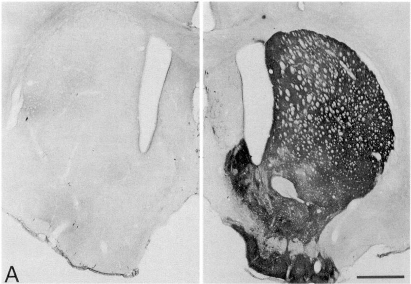
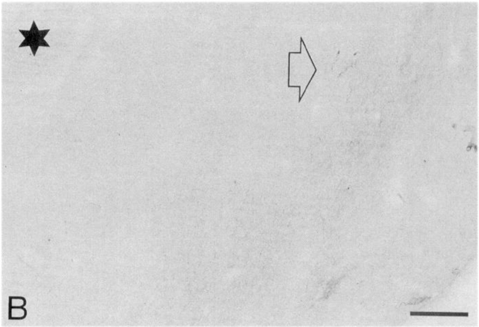
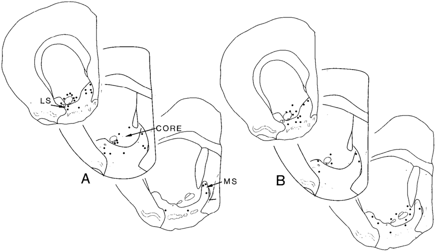
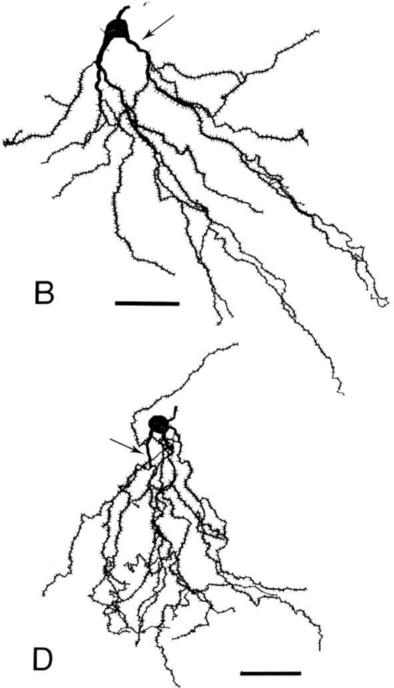
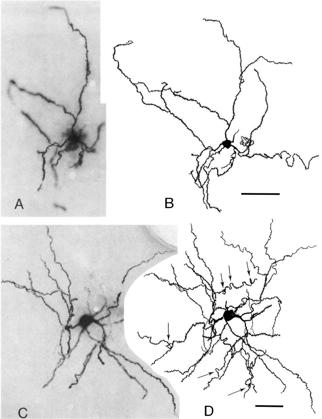
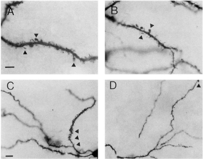
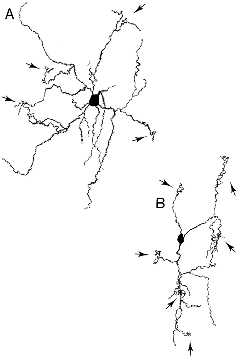
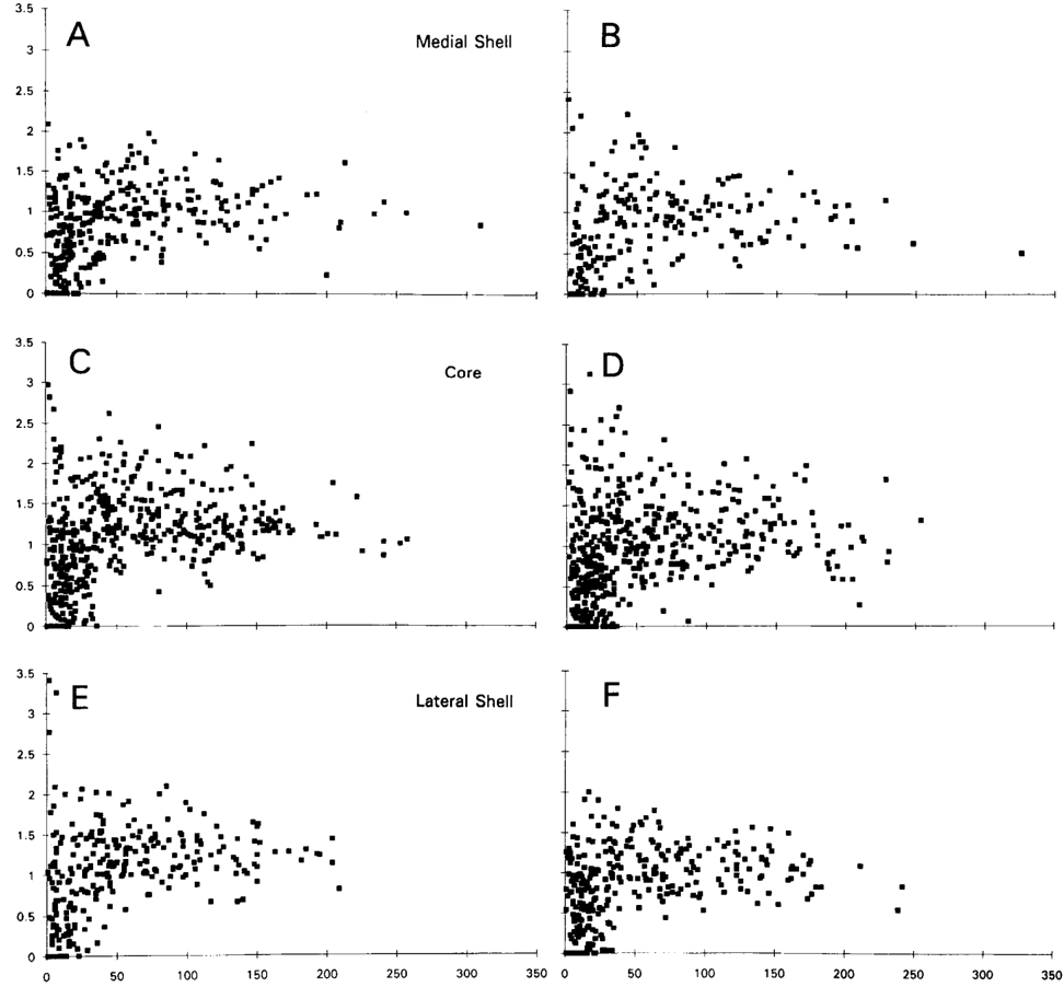
Nucleus accumbens receives a dense dopaminergic innervation which is important in regulating motivated states of behavior such as goal-directed actions, stimulus-reward associations and reinforcement of addictive substances. The shell and core territories of this nucleus each receive functionally and morphologically distinct dopaminergic inputs and lesions of the ascending pathways totally deprive the core but not the shell of dopaminergic fibers. Medium spiny neurons are the principal targets of dopaminergic terminals. The present study explored whether the loss of dopamine inputs can affect these neurons and whether cells in the shell and core would be equally susceptible to such a loss. Intracellular injection in fixed slices and neuronal reconstruction were used to analyze the dendritic trees of 62 neurons in the shell and core of animals that received a unilateral, chronic 6-hydroxydopamine lesion of the medial forebrain bundle. In the dopamine-depleted core, dendrites are significantly shorter (16% decrease) than in the intact core and in both the dopamine-depleted core and lateral shell, dendrites are less spiny than in respective control regions. Dopamine loss in the medial shell is associated with significantly more tortuous dendrites that are lower in spine density. However, the number of spines is not reduced which may mean that the increase recorded for segment length, although insignificant in tests, could be responsible for the change in spine density. These data suggest that the loss of dopamine can affect accumbal neuronal morphology and, moreover, can affect neuronal structures differentially in the shell and core.
Overview
The claims and statistics of this paper haven’t been checked yet.
Run the check — free, no account needed. It reads the open-access text and checks the sources it cites.
Integrity signals
Registry
Cites
Statistics
Figures
Verification
Claims the paper makes
The claims and statistics of this paper haven’t been checked yet.
Run the check — free, no account needed. It reads the open-access text and checks the sources it cites.
Claims other papers make about this one
Each is checked against this paper’s own text: does it say what the citing paper says it does?
In the citing paper
“Features such as spine density and terminal and branch segments appear to differ between the medial shell and the lateral shell, and between the shell and the core (Meredith et al., 1992; 1995).”
From Dopamine reward circuitry: Two projection systems from the ventral midbrain to the nucleus accumbens–olfactory tubercle complex (2007) · cites this paper as Meredith et al., 1995
Self-check
Percentages, sums and reported differences re-computed from the paper’s own numbers.
The self-check needs the paper’s full text.
Citations
Cites
Calcium-mediated neurotoxicity: relationship to specific channel types and role in ischemic damage
Choi, D. W. (1988) · Trends in Neurosciences · doi:10.1016/0166-2236(88)90200-7 · Cited by 1,726
Efferent connections of the substantia nigra and ventral tegmental area in the rat
Beckstead, R. M., Domesick, V. B., & Nauta, W. J. H. (1979) · Brain Research · doi:10.1016/0006-8993(79)91001-1 · Cited by 1,293
Specificity in the projection patterns of accumbal core and shell in the rat
Heimer, L., Zahm, D. S., Churchill, L., Kalivas, P. W., & Wohltmann, C. (1991) · Neuroscience · doi:10.1016/0306-4522(91)90202-y · Cited by 1,219
Self-citationAlheid, G. F., & Heimer, L. (1988) · Neuroscience · doi:10.1016/0306-4522(88)90217-5 · Cited by 1,217
Ascending Monoamine Neurons to the Telencephalon and Diencephalon
Andén, N. ‐., Dahlström, A. B., Fuxé, K., Larsson, K., Olson, L., & Ungerstedt, U. (1966) · Acta Physiologica Scandinavica · doi:10.1111/j.1748-1716.1966.tb03318.x · Cited by 1,214
Jitsukawa, K., Sueki, H., Sato, S., & Anzai, T. (1987) · American Journal of Dermatopathology · doi:10.1097/00000372-198704000-00003 · Cited by 1,130
Graybiel, A. M., Moratalla, R., & Robertson, H. A. (1990) · Proceedings of the National Academy of Sciences · doi:10.1073/pnas.87.17.6912 · Cited by 957
Two dopamine receptors: Biochemistry, physiology and pharmacology
Stoof, J. C., & Kebabian, J. W. (1984) · Life Sciences · doi:10.1016/0024-3205(84)90519-8 · Cited by 944
Graveland, G. A., Williams, R. S., & DiFiglia, M. (1985) · Science · doi:10.1126/science.3155875 · Cited by 612
Haydon, P. G., McCobb, D. P., & Kater, S. B. (1984) · Science · doi:10.1126/science.6093252 · Cited by 545
Research works printed but not linked: 1
- Groenewegen HJ, Berendse HW, Meredith GE, Haber SN, Voorn P, Wolters JG, Lohman AHM (1991) Functional anatomy of the ven- tral, limbic system innervated striatum. In: The mesolimbic dopamine system: from motivation to action (Willner P Scheel-Kruger J, eds), pp 19-59. Chichester: Wiley.
Cited by
Sorted by citations
Ikemoto, S. (2007) · Brain Research Reviews · doi:10.1016/j.brainresrev.2007.05.004 · Cited by 1,477
Addiction and the brain: The neurobiology of compulsion and its persistence
Hyman, S. E., & Malenka, R. C. (2001) · Nature reviews. Neuroscience · doi:10.1038/35094560 · Cited by 1,337
Addiction, Dopamine, and the Molecular Mechanisms of Memory
Berke, J. D., & Hyman, S. E. (2000) · Neuron · doi:10.1016/s0896-6273(00)81056-9 · Cited by 1,184
Functional–anatomical implications of the nucleus accumbens core and shell subterritories
ZAHM, D. S. (1999) · Annals of the New York Academy of Sciences · doi:10.1111/j.1749-6632.1999.tb09264.x · Cited by 384
Self-citationAfferents of the ventral tegmental area in the rat-anatomical substratum for integrative functions
Geisler, S., & Zahm, D. S. (2005) · The Journal of Comparative Neurology · doi:10.1002/cne.20668 · Cited by 379
Self-citationSchwarting, R. K. W., & Huston, J. P. (1996) · Progress in Neurobiology · doi:10.1016/s0301-0082(96)00015-9 · Cited by 356
Caffeine Induces Dopamine and Glutamate Release in the Shell of the Nucleus Accumbens
Solinas, M., Ferré, S., You, Z.-B., Karcz-Kubicha, M., Popoli, P., & Goldberg, S. R. (2002) · The Journal of Neuroscience · doi:10.1523/jneurosci.22-15-06321.2002 · Cited by 338
Dendritic spines: Revisiting the physiological role
Chidambaram, S. B., Rathipriya, A. G., Bolla, S. R., Bhat, A., Ray, B., Mahalakshmi, A. M., Manivasagam, T., Thenmozhi, A. J., Essa, M. M., Guillemin, G. J., et al. (2019) · Progress in Neuro-Psychopharmacology and Biological Psychiatry · doi:10.1016/j.pnpbp.2019.01.005 · Cited by 266
Usuda, I., Tanaka, K., & Chiba, T. (1998) · Brain Research · doi:10.1016/s0006-8993(98)00359-x · Cited by 248
Norrholm, S. D., Bibb, J. A., Nestler, E. J., Ouimet, C. C., Taylor, J. R., & Greengard, P. (2003) · Neuroscience · doi:10.1016/s0306-4522(02)00560-2 · Cited by 249
The Synaptic Framework for Chemical Signaling in Nucleus Accumbens
Meredith, G. E. (1999) · Annals of the New York Academy of Sciences · doi:10.1111/j.1749-6632.1999.tb09266.x · Cited by 215
Self-citationThe structural basis for mapping behavior onto the ventral striatum and its subdivisions
Meredith, G. E., Baldo, B. A., Andrezjewski, M. E., & Kelley, A. E. (2008) · Brain Structure and Function · doi:10.1007/s00429-008-0175-3 · Cited by 202
Self-citationQuantitative analysis of pre‐ and postsynaptic sex differences in the nucleus accumbens
Forlano, P. M., & Woolley, C. S. (2009) · The Journal of Comparative Neurology · doi:10.1002/cne.22279 · Cited by 137
Neuroplasticity in the Mesolimbic System Induced by Natural Reward and Subsequent Reward Abstinence
Pitchers, K. K., Balfour, M. E., Lehman, M. N., Richtand, N. M., Yu, L., & Coolen, L. M. (2009) · Biological Psychiatry · doi:10.1016/j.biopsych.2009.09.036 · Cited by 117
Jupp, B., Caprioli, D., Saigal, N., Reverte, I., Shrestha, S., Cumming, P., Everitt, B. J., Robbins, T. W., & Dalley, J. W. (2013) · European Journal of Neuroscience · doi:10.1111/ejn.12146 · Cited by 109
Dumitriu, D., LaPlant, Q. C., Grossman, Y. S., Dias, C., Janssen, W. G. M., Russo, S. J., Morrison, J. H., & Nestler, E. J. (2012) · Journal of Neuroscience · doi:10.1523/jneurosci.5718-11.2012 · Cited by 116
Gene × Disease Interaction on Orbitofrontal Gray Matter in Cocaine Addiction
Alia‐Klein, N., Parvaz, M. A., Woicik, P. A., Konova, A. B., Maloney, T., Shumay, E., Wang, R., Telang, F., Biegon, A. R., Wang, G., et al. (2011) · Archives of General Psychiatry · doi:10.1001/archgenpsychiatry.2011.10 · Cited by 107
Topographical organization of projections from the entorhinal cortex to the striatum of the rat
Totterdell, S., & Meredith, G. E. (1997) · Neuroscience · doi:10.1016/s0306-4522(96)00592-1 · Cited by 104
Self-citationActivation of dopaminergic D1 receptors promotes morphogenesis of developing striatal neurons
Schmidt, U. G., Beyer, C., Oestreicher, A. B., Reisert, I., Schilling, K., & Pilgrim, C. (1996) · Neuroscience · doi:10.1016/0306-4522(96)00201-1 · Cited by 98
Solís, Ó., Limón, I. D., Flores‐Hernández, J., & Flores, G. (2007) · Synapse · doi:10.1002/syn.20381 · Cited by 96
Darmopil, S., Muñetón‐Gómez, V. C., De Ceballos, M. L., Bernson, M., & Moratalla, R. (2008) · European Journal of Neuroscience · doi:10.1111/j.1460-9568.2008.06040.x · Cited by 91
Meredith, G. E., Farrell, T., Kellaghan, P., Tan, Y., Zahm, D. S., & Totterdell, S. (1999) · European Journal of Neuroscience · doi:10.1046/j.1460-9568.1999.00774.x · Cited by 89
Self-citationBoye, S. M., Grant, R. J., & Clarke, P. B. S. (2001) · Neuropharmacology · doi:10.1016/s0028-3908(01)00003-x · Cited by 87
Caprioli, D., Sawiak, S. J., Merlo, E., Theobald, D. E. H., Spoelder, M., Jupp, B., Voon, V., Carpenter, T. A., Everitt, B. J., Robbins, T. W., et al. (2013) · Biological Psychiatry · doi:10.1016/j.biopsych.2013.07.013 · Cited by 90
Morphine withdrawal‐induced morphological changes in the nucleus accumbens
Spiga, S., Puddu, M. C., Pisano, M., & Diana, M. (2005) · European Journal of Neuroscience · doi:10.1111/j.1460-9568.2005.04416.x · Cited by 86
Ma, Y., Zhan, M., OuYang, L., Li, Y., Chen, S. X., Wu, J., Chen, J., Luo, C., & Lei, W. (2014) · Behavioural Brain Research · doi:10.1016/j.bbr.2014.02.039 · Cited by 83
Microcircuits in nucleus accumbens’ shell and core involved in cognition and reward
Meredith, G. E., & Totterdell, S. (1999) · Psychobiology · doi:10.3758/bf03332112 · Cited by 79
Self-citationHaim, A., Sherer, M., & Leuner, B. (2014) · European Journal of Neuroscience · doi:10.1111/ejn.12752 · Cited by 76
Astroglial plasticity and glutamate function in a chronic mouse model of Parkinson's disease
Dervan, A. G., Meshul, C. K., Beales, M., McBean, G. J., Moore, C., Totterdell, S., Snyder, A. K., & Meredith, G. E. (2004) · Experimental Neurology · doi:10.1016/j.expneurol.2004.07.004 · Cited by 69
Self-citationA spiking Basal Ganglia model of synchrony, exploration and decision making
Mandali, A., Rengaswamy, M., Chakravarthy, V. S., & Moustafa, A. A. (2015) · Frontiers in Neuroscience · doi:10.3389/fnins.2015.00191 · Cited by 64
Shen, H., Sesack, S. R., Toda, S., & Kalivas, P. W. (2008) · Brain Structure and Function · doi:10.1007/s00429-008-0184-2 · Cited by 64
Lex, B., & Hauber, W. (2010) · Neurobiology of Learning and Memory · doi:10.1016/j.nlm.2009.11.002 · Cited by 61
Persistent and Reversible Morphine Withdrawal‐Induced Morphological Changes in the Nucleus Accumbens
Diana, M., Spiga, S., & Acquas, E. (2006) · Annals of the New York Academy of Sciences · doi:10.1196/annals.1369.045 · Cited by 59
David, D., Zahniser, N. R., Hoffer, B. J., & Gerhardt, G. A. (1998) · Experimental Neurology · doi:10.1006/exnr.1998.6898 · Cited by 57
Differentiative Effects of Dopamine on Striatal Neurons Involve Stimulation of the cAMP/PKA Pathway
Schmidt, U. G., Pilgrim, C., & Beyer, C. (1998) · Molecular and Cellular Neuroscience · doi:10.1006/mcne.1998.0668 · Cited by 55
Metabolic mapping of the effects of oral alcohol self-administration in rats.
Porrino, L. J., Williams‐Hemby, L., Whitlow, C. T., Bowen, C. A., & Samson, H. H. (1998) · PubMed · Cited by 55
Meredith, G. E., Souza, I. E. J. D., Hyde, T. M., Tipper, G., Wong, M. L., & Egan, M. F. (2000) · Journal of Neuroscience · doi:10.1523/jneurosci.20-20-07798.2000 · Cited by 54
Self-citationReconstructing the Three-Dimensional GABAergic Microcircuit of the Striatum
Humphries, M. D., Wood, R., & Gurney, K. R. (2010) · PLoS Computational Biology · doi:10.1371/journal.pcbi.1001011 · Cited by 53
Nicotine effects on regional cerebral blood flow in awake, resting tobacco smokers
Domino, E. F., Minoshima, S., Guthrie, S. K., Ohl, L. E., Ni, L., Koeppe, R. A., & Zubieta, J. (2000) · Synapse · doi:10.1002/1098-2396(20001201)38:3 · Cited by 104
Dall'Igna, O. P. P., da Silva, A. L., Dietrich, M. O., Hoffmann, A., de Oliveira, R. V., Souza, D. O., & Lara, D. R. (2003) · Psychopharmacology · doi:10.1007/s00213-002-1362-1 · Cited by 47
A neuroinductive biomaterial based on dopamine
Gao, J., Kim, Y. M., Coe, H., Zern, B. J., Sheppard, B. J., & Wang, Y. (2006) · Proceedings of the National Academy of Sciences · doi:10.1073/pnas.0606237103 · Cited by 45
Kasture, S. B., Vinci, S., Ibba, F., Puddu, A., Marongiu, M., Balasubramanian, M., Pisanu, A., Lecca, D., Zernig, G., & Acquas, E. (2009) · Neurotoxicity Research · doi:10.1007/s12640-009-9069-2 · Cited by 44
Phillips, G. D., Setzu, E., & Hitchcott, P. K. (2003) · Behavioral Neuroscience · doi:10.1037/0735-7044.117.4.675 · Cited by 43
Fox, M. E., Wulff, A. B., Franco, D., Choi, E. Y., Calarco, C. A., Engeln, M., Turner, M. D., Chandra, R., Rhodes, V. M., Thompson, S. M., et al. (2022) · Biological Psychiatry · doi:10.1016/j.biopsych.2022.08.023 · Cited by 40
Martin, B. J., Naughton, B. J., Rajamani, K. T., Yoon, D. J., Han, D. D., DeVries, A. C., & Gu, H. H. (2010) · Synapse · doi:10.1002/syn.20865 · Cited by 40
Temporal Rewiring of Striatal Circuits Initiated by Nicotine
Adermark, L., Morud, J., Lotfi, A., Danielsson, K., Ulenius, L., Söderpalm, B., & Ericson, M. (2016) · Neuropsychopharmacology · doi:10.1038/npp.2016.118 · Cited by 38
Learning and Memory Mechanisms Involved in Compulsive Drug Use and Relapse
Berke, J. D. (2003) · Humana Press eBooks · doi:10.1385/1-59259-358-5:75 · Cited by 36
HIV-Associated Apathy/Depression and Neurocognitive Impairments Reflect Persistent Dopamine Deficits
McLaurin, K. A., Harris, M., Madormo, V. A., Harrod, S. B., Mactutus, C. F., & Booze, R. M. (2021) · Cells · doi:10.3390/cells10082158 · Cited by 36
Anabolic-androgenic steroids decrease dendritic spine density in the nucleus accumbens of male rats
Wallin-Miller, K. G., Li, G., Kelishani, D., & Wood, R. I. (2016) · Neuroscience · doi:10.1016/j.neuroscience.2016.05.045 · Cited by 33
Guirado, S., D�vila, J. C., Real, M. A., & Medina, L. M. (1999) · The Journal of Comparative Neurology · doi:10.1002/(sici)1096-9861(19990301)405:1 · Cited by 33
Neuroengineering Models of Brain Disease
Finkel, L. H. (2000) · Annual Review of Biomedical Engineering · doi:10.1146/annurev.bioeng.2.1.577 · Cited by 32
Vadakkan, K. I. (2015) · SpringerPlus · doi:10.1186/s40064-015-1283-1 · Cited by 32
Karadurmus, D., Rial, D., Backer, J. D., Communi, D., d’Exaerde, A. D. K., & Schiffmann, S. N. (2019) · Journal of Neuroscience · doi:10.1523/jneurosci.2454-18.2019 · Cited by 30
Transfer of Neuroplasticity from Nucleus Accumbens Core to Shell Is Required for Cocaine Reward
Marie, N., Canestrelli, C., & Noble, F. (2012) · PLoS ONE · doi:10.1371/journal.pone.0030241 · Cited by 29
Küppers, E., Sabolek, M., Anders, U., Pilgrim, C., & Beyer, C. (2000) · Molecular Brain Research · doi:10.1016/s0169-328x(00)00156-x · Cited by 30
BDNF heterozygous mice demonstrate age-related changes in striatal and nigral gene expression
Saylor, A. J., Meredith, G. E., Vercillo, M. S., Zahm, D. S., & McGinty, J. F. (2006) · Experimental Neurology · doi:10.1016/j.expneurol.2006.01.004 · Cited by 29
Self-citationBubser, M., Scruggs, J. L., Young, C. D., & Deutch, A. Y. (2000) · European Journal of Neuroscience · doi:10.1046/j.1460-9568.2000.00052.x · Cited by 29
Tan, Y., Brog, J. S., Williams, E. S., & Zahm, D. S. (1995) · Brain Research · doi:10.1016/0006-8993(95)00556-6 · Cited by 29
Self-citationSubramaniam, S., Marcotte, E. R., & Srivastava, L. K. (2001) · Brain Research · doi:10.1016/s0006-8993(01)02347-2 · Cited by 28
Metabolic Mapping of the Effects of Oral Alcohol Self‐Administration in Rats
Porrino, L. J., Williams‐Hemby, L., Whitlow, C. T., Bowen, C. A., & Samson, H. H. (1998) · Alcoholism Clinical and Experimental Research · doi:10.1111/j.1530-0277.1998.tb03635.x · Cited by 27
Gruß, M., Appenroth, D., Flubacher, A., Enzensperger, C., Bock, J., Fleck, C., Gille, G., & Braun, K. (2012) · Journal of Neurochemistry · doi:10.1111/j.1471-4159.2012.07713.x · Cited by 26
Waselus, M., Flagel, S. B., Jedynak, J., Akil, H., Robinson, T. E., & Watson, S. J. (2013) · Neuroscience · doi:10.1016/j.neuroscience.2013.06.042 · Cited by 25
Cho, J., Duke, D. C., Manzino, L., Sonsalla, P. K., & West, M. O. (2002) · The Journal of Comparative Neurology · doi:10.1002/cne.10349 · Cited by 26
Tan, Y., Williams, E. S., & Zahm, D. S. (1999) · Brain Research · doi:10.1016/s0006-8993(99)01890-9 · Cited by 25
Self-citationThe Central Effects of Androgenic-anabolic Steroid Use
Mędraś, M., Brona, A., & Jóźków, P. (2018) · Journal of Addiction Medicine · doi:10.1097/adm.0000000000000395 · Cited by 26
Hatip‐Al‐Khatib, I., 三島, 健., Iwasaki, K., & Fujiwara, M. (2001) · Brain Research · doi:10.1016/s0006-8993(01)02382-4 · Cited by 23
Nelson, A. J. D., Thur, K. E., Horsley, R. R., Spicer, C., Marsden, C. A., & Cassaday, H. J. (2010) · Pharmacology Biochemistry and Behavior · doi:10.1016/j.pbb.2010.11.025 · Cited by 20
Striatal mechanisms in Parkinson’s disease: new insights from computer modeling
Kötter, R., & Wickens, J. R. (1998) · Artificial Intelligence in Medicine · doi:10.1016/s0933-3657(98)00003-7 · Cited by 21
Kim, B., Weerasinghe-Mudiyanselage, P. D. E., Ang, M. J., Lee, J., Kang, S., Kim, J.-C., Kim, S., Kim, J., Jung, C., Shin, T., et al. (2022) · International Neurourology Journal · doi:10.5213/inj.2244252.126 · Cited by 19
Lee, H., & Sawatari, A. (2011) · European Journal of Neuroscience · doi:10.1111/j.1460-9568.2011.07852.x · Cited by 20
Muma, N. A., Lee, J. M., Gorman, L. K., Heidenreich, B. A., Mitrovic, I., & Napier, T. C. (2001) · Experimental Neurology · doi:10.1006/exnr.2000.7582 · Cited by 18
Kobrin, K. L., Arena, D. T., Heinrichs, S. C., Nguyen, O. H., & Kaplan, G. B. (2017) · Behavioural Brain Research · doi:10.1016/j.bbr.2017.01.011 · Cited by 17
Acute MPTP Treatment Impairs Dendritic Spine Density in the Mouse Hippocampus
Weerasinghe-Mudiyanselage, P. D. E., Ang, M. J., Wada, M., Kim, S., Shin, T., Yang, M., & Moon, C. (2021) · Brain Sciences · doi:10.3390/brainsci11070833 · Cited by 15
Caravaggio, F., Chung, J. K., Plitman, E., Boileau, I., Gerretsen, P., Kim, J., Iwata, Y., Patel, R., Chakravarty, M. M., Remington, G. J., et al. (2017) · Human Brain Mapping · doi:10.1002/hbm.23744 · Cited by 15
Coincident signaling in mesolimbic structures underlying alcohol reinforcement
Zhang, T. A., Maldve, R. E., & Morrisett, R. A. (2006) · Biochemical Pharmacology · doi:10.1016/j.bcp.2006.04.022 · Cited by 15
Section II. The Dopamine System
Smythies, J. (2005) · International review of neurobiology · doi:10.1016/s0074-7742(05)64002-0 · Cited by 15
Hara, Y., & Pickel, V. M. (2007) · Neuroscience · doi:10.1016/j.neuroscience.2007.04.005 · Cited by 15
Intact risk-based decision making in rats with prefrontal or accumbens dopamine depletion
Mai, B., & Hauber, W. (2012) · Cognitive Affective & Behavioral Neuroscience · doi:10.3758/s13415-012-0115-9 · Cited by 14
Pezzi, S., Checa, N., & Alberch, J. (2005) · International Journal of Developmental Neuroscience · doi:10.1016/j.ijdevneu.2004.12.007 · Cited by 14
Lithium treatment impacts nucleus accumbens shape in bipolar disorder
Vecchio, D., Piras, F., Piras, F., Banaj, N., Janiri, D., Simonetti, A., Sani, G., & Spalletta, G. (2020) · NeuroImage Clinical · doi:10.1016/j.nicl.2020.102167 · Cited by 13
Haim, A., Albin-Brooks, C., Brothers, H. M., Breach, M. R., & Leuner, B. (2025) · Scientific Reports · doi:10.1038/s41598-024-84043-6 · Cited by 15
Lucifer Yellow filling of area X-projecting neurons in the high vocal center of female canaries
Benton, S., Cardin, J. A., & DeVoogd, T. J. (1998) · Brain Research · doi:10.1016/s0006-8993(98)00485-5 · Cited by 11
Shortall, S. E., Green, A. R., Fone, K. C. F., & King, M. V. (2016) · Journal of Psychopharmacology · doi:10.1177/0269881116650408 · Cited by 11
Maria-Rios, C. E., Murphy, G. G., & Morrow, J. D. (2023) · eNeuro · doi:10.1523/eneuro.0432-22.2023 · Cited by 8
Huston-Lyons, D., & Porrino, L. J. (1997) · Brain Research · doi:10.1016/s0006-8993(96)01493-x · Cited by 8
Imaging phosphodiesterase‐10a availability in cocaine use disorder with [11C]IMA107 and PET
Tollefson, S., Gertler, J., Himes, M. L., Paris, J., Kendro, S., Lopresti, B. J., Mason, N. S., & Narendran, R. (2018) · Synapse · doi:10.1002/syn.22070 · Cited by 7
Ma, J., Ye, N., & Cohen, B. M. (2006) · Neuroscience · doi:10.1016/j.neuroscience.2006.05.013 · Cited by 7
Funamizu, Y., Nishijima, H., Ueno, T., Ueno, S., Mizukami, H., Yagihashi, S., & Tomiyama, M. (2017) · Neuroscience Research · doi:10.1016/j.neures.2017.03.010 · Cited by 8
Dopaminergic Innervation of the Monkey Caudal Nucleus Accumbens
Ikemoto, K. (1997) · Brain Research Bulletin · doi:10.1016/s0361-9230(97)00028-2 · Cited by 5
González‐Burgos, I., García-Martínez, S., Velázquez-Zamora, D. A., & Ponce‐Rolón, R. (2010) · International Journal of Developmental Neuroscience · doi:10.1016/j.ijdevneu.2010.06.005 · Cited by 4
Lancia, A. J., Williams, E. A., McKnight, L. V., & Zahm, D. S. (2003) · Brain Research · doi:10.1016/j.brainres.2003.10.044 · Cited by 4
Self-citationThe Ventral Pallidum as a Limbic Pleasure Generator.
Ho, C. (2010) · Deep Blue (University of Michigan) · Cited by 4
Antón, B., Calva, J. J., Valdez, A. D. P., Acevedo, R., Morales, A., Medécigo, M., & Leff, P. (2000) · Redalyc (Universidad Autónoma del Estado de México) · Cited by 2
Dervan, A. G., Totterdell, S., Lau, Y.-S., & Meredith, G. E. (2003) · Annals of the New York Academy of Sciences · doi:10.1111/j.1749-6632.2003.tb07487.x · Cited by 2
Self-citationFox, M. E., Wulff, A. B., Franco, D., Choi, E. Y., Calarco, C. A., Engeln, M., Turner, M. D., Chandra, R., Rhodes, V. M., Thompson, S. M., et al. (2022) · bioRxiv (Cold Spring Harbor Laboratory) · doi:10.1101/2022.05.15.491856 · Cited by 2
Morphology of Human Nucleus Accumbens Neurons Based on the Immunohistochemical Expression of Gad67
Sazdanovic, М., Mitrović, S., Todorović, M., Vulović, M., Jeremić, D., Milosavljević, Z., Sazdanovic, P. S., & Ognjanović, N. (2016) · Serbian Journal of Experimental and Clinical Research · doi:10.1515/sjecr-2016-0041 · Cited by 1
Kirazlı, Ö., Özkan, M., Verimli, U., Aker, R. G., Arman, A., & Sehirli, U. S. (2024) · Anatomical Science International · doi:10.1007/s12565-024-00790-6 · Cited by 1
Colom, M., Kraev, I., Stramek, A. K., Loza, I. B., Rostron, C. L., Heath, C. J., Dommett, E. J., & Singer, B. F. (2024) · European Journal of Neuroscience · doi:10.1111/ejn.16513 · Cited by 2
Zhang, T. (2005) · Texas ScholarWorks (Texas Digital Library) · Cited by 1
Colom, M., Kraev, I., Stramek, A. K., Loza, I. B., Rostron, C. L., Heath, C. J., Dommett, E. J., & Singer, B. F. (2023) · doi:10.22541/au.168927128.84529083/v1 · Cited by 1
Metadata
- Authors
- Meredith, G., Ypma, P., & Zahm, D.
- Affiliations
- Vrije Universiteit Amsterdam (NL)
- Venue
- The Journal of Neuroscience
- Publisher
- Society for Neuroscience
- Published
- 1995-05-01
- Volume
- 15
- Issue
- 5
- Pages
- 3808-3820
- ISSN
- 0270-6474 (ISSN-L)
- 1529-2401
- PMC
- PMC6578201
- OpenAlex
- W1589481227
- Cited by
- 112
- References
- 68
- Citations by year
- 2025: 1 · 2024: 3 · 2023: 2 · 2022: 3 · 2021: 2 · 2020: 2 · 2019: 2 · 2018: 2 · 2017: 3 · 2016: 4 · 2015: 2 · 2014: 3 · 2013: 3 · 2012: 5
- FWCI
- 1.87
- Type
- journal article
- Language
- en
- Open access
- hybrid
- doi.org
- Licence
- Locations
- Journal of Neuroscience (publishedVersion, cc-by-nc-sa)
- PubMed Central (publishedVersion)
- PubMed (publishedVersion)
- Funders
- U.S. Public Health Service
- National Institute of Neurological Disorders and Stroke (NS23805, R01NS023805)
- Topics
- Life Sciences › Neuroscience › Cellular and Molecular Neuroscience › Neurotransmitter Receptor Influence on Behavior
- Life Sciences › Neuroscience › Cellular and Molecular Neuroscience › Neuroscience and Neuropharmacology Research
- Health Sciences › Medicine › Clinical Neurology › Deep Brain Stimulation and Movement Disorders
- Keywords
- dopamine depletion, medium spiny neurons, nucleus accumbens, dendritic morphology, dendritic spines, dendritic spine density, 6-hydroxydopamine lesion, medial forebrain bundle, dopaminergic innervation, neuron reconstruction
- MeSH terms
- Animals, Dendrites, Dopamine, Male, Nerve Endings, Neurons, Nucleus Accumbens, Reference Values, Tyrosine 3-Monooxygenase, Prosencephalon, Oxidopamine, Rats, Wistar, Rats
About these checks
Registry, retraction, Cites and statistics results are computed, not judged, with one exception: a printed reference that no index lists is read once by a model before it counts as a citation error. Claims are extracted by a model and kept only when their quote appears verbatim in the text; each cited source is then checked, with a model’s help, for whether it says what the claim relies on. Those verdicts can be wrong — read the claim’s quote and the cited source before relying on one.
Statistics and arithmetic are recomputed, not judged.
Confidence bands are shown only for fields calibrated on our annotated test set. None is calibrated yet, so no band is shown.
What counts as a claim ›How we measure confidence ›How we check this ›
Are you an author, or do you see a problem with this page? Request a review
