

The DOI matches its Crossref record; 9 of the 9 references it cites were found in a registry or an index.
Presynaptic D1Dopamine Receptors in Primate Prefrontal Cortex: Target-Specific Expression in the Glutamatergic Synapse
Abstract
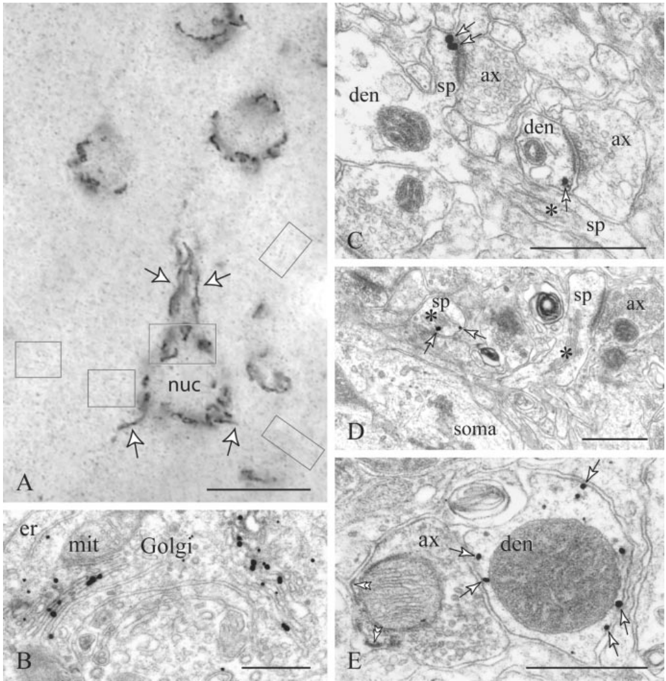
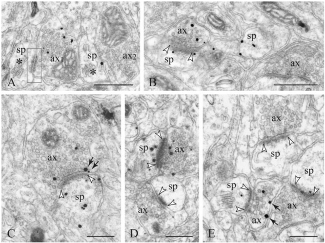
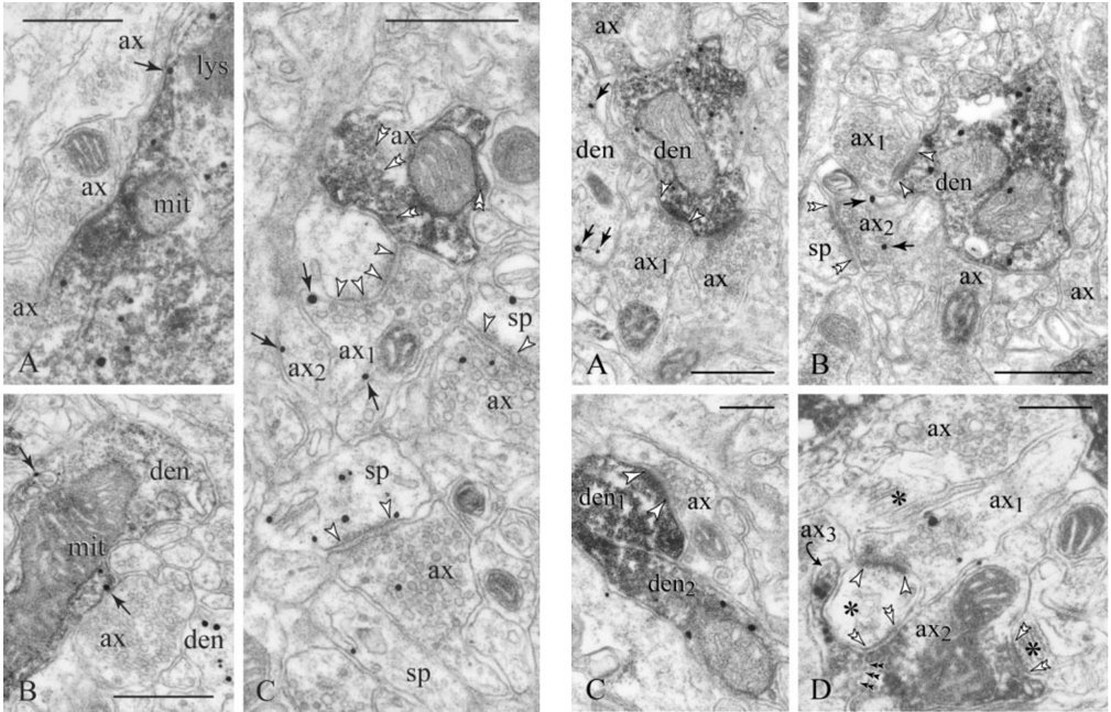
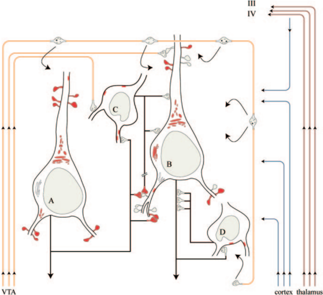
Dopaminergic modulation of glutamate neurotransmission in prefrontal cortex (PFC) microcircuits is commonly perceived as a basis for cognitive operations. Yet it appears that although the control of recurrent excitation between deep-layer prefrontal pyramids may involve presynaptic and postsynaptic D 1 receptor (D 1 R) mechanisms, pyramid-to-interneuron communication will engage a postsynaptic D 1 R component. The substrate underlying such target-specific neuromodulatory patterns was investigated in the infragranular PFC with immunoelectron microscopy for D 1 R and parvalbumin, a marker for fast-spiking interneurons. In addition to their proverbial postsynaptic expression, gold-labeled D 1 Rs were distinctly distributed on perisynaptic/extrasynaptic membranes and the axoplasm of 13% of excitatory-like, presumably glutamatergic varicosities. Most importantly, presynaptic D 1 Rs were highly specific with regard to the cellular compartment and neurochemical identity of the postsynaptic neuron, being present in spine-targeting varicosities but distinctly absent from those synapsing with parvalbumin profiles often coexpressing D 1 Rs. We define therein an axonal D 1 heteroreceptor component, apparently mediating volume neurotransmission, yet strategically positioned to convey target cell-specific modulation of the glutamatergic drive. We also indicate that presynaptic D 1 R mechanisms may indeed be associated with recurrent excitation in prefrontal microcircuits, consistent with physiological evidence for a role of these receptors in modulating the persistent activity-profile of neurons essential for working memory.
Overview
The claims and statistics of this paper haven’t been checked yet.
Run the check — free, no account needed. It reads the open-access text and checks the sources it cites.
Integrity signals
Registry
Cites
Statistics
Figures
Verification
Claims the paper makes
The claims and statistics of this paper haven’t been checked yet.
Run the check — free, no account needed. It reads the open-access text and checks the sources it cites.
Claims other papers make about this one
Each is checked against this paper’s own text: does it say what the citing paper says it does?
In the citing paper
“In addition to increasing extracellular dopamine and norepinephrine, stimulants also have direct and indirect effects on striatal and cortical glutamate critical to optimal PFC function (Paspalas and Goldman-Rakic, 2005).”
From Lisdexamfetamine Effects on Executive Activation and Neurochemistry in Menopausal Women with Executive Function Difficulties (2017) · cites this paper as Paspalas and Goldman-Rakic, 2005
Self-check
Percentages, sums and reported differences re-computed from the paper’s own numbers.
The self-check needs the paper’s full text.
Citations
Cites
AMPA Receptor Trafficking and Synaptic Plasticity
Malinow, R., & Malenka, R. C. (2002) · Annual Review of Neuroscience · doi:10.1146/annurev.neuro.25.112701.142758 · Cited by 2,588
Circuitry of Primate Prefrontal Cortex and Regulation of Behavior by Representational Memory
Goldman‐Rakic, P. S. (1987) · Comprehensive Physiology · doi:10.1002/cphy.cp010509 · Cited by 1,960
Modulation of memory fields by dopamine Dl receptors in prefrontal cortex
Williams, G. V., & Goldman-Rakic, P. S. (1995) · Nature · doi:10.1038/376572a0 · Cited by 1,593 · cited 1×
“…(D1-LR) subfamily (i.e., D1R and D5R subtypes) (Missale et al., 1998) modulate excitatory transmission in prefrontal microcircuits that generate stimulus-independent persistent activity, a property essential for working memory (Sawaguchi and GoldmanRakic, 1991; Williams and Goldman-Rakic, 1995).”
BackgroundD1 Dopamine Receptors in Prefrontal Cortex: Involvement in Working Memory
Sawaguchi, T., & Goldman-Rakic, P. S. (1991) · Science · doi:10.1126/science.1825731 · Cited by 1,364
DeFelipe, J. (1997) · Journal of Chemical Neuroanatomy · doi:10.1016/s0891-0618(97)10013-8 · Cited by 550
Targeting the dopamine D1 receptor in schizophrenia: insights for cognitive dysfunction
Goldman-Rakic, P., Castner, S., Svensson, T., Siever, L., & Williams, G. (2004) · Psychopharmacology · doi:10.1007/s00213-004-1793-y · Cited by 544
Gorelova, N., Seamans, J. K., & Yang, C. R. (2002) · Journal of Neurophysiology · doi:10.1152/jn.00335.2002 · Cited by 302
Presynaptic regulation of recurrent excitation by D1 receptors in prefrontal circuits
Gao, W.-J., Krimer, L. S., & Goldman-Rakic, P. S. (2001) · Proceedings of the National Academy of Sciences · doi:10.1073/pnas.98.1.295 · Cited by 257
Lewis, D., Volk, D., & Hashimoto, T. (2004) · Psychopharmacology · doi:10.1007/s00213-003-1673-x · Cited by 253
Research works printed but not linked: 60
- 10.1016/0306-4522(95)00350-R · DOI
- 1995, J Neurosci, 15, 7821, 10.1523/JNEUROSCI.15-12-07821.1995 · DOI
- 10.1016/S0165-6147(99)01360-7 · DOI
- 10.1093/cercor/bhg096 · DOI
- 10.1146/annurev.pharmtox.41.1.237 · DOI
- 10.1002/cne.903410109 · DOI
- 10.1016/S0306-4522(01)00591-7 · DOI · Paper page
- 10.1016/0301-0082(92)90015-7 · DOI · Paper page
- 10.1016/0006-8993(91)91184-3 · DOI
- 10.1002/cne.20084 · DOI
- 10.1016/S0896-6273(03)00423-9 · DOI
- 10.1002/(SICI)1096-9861(19960122)364:4<567::AID-CNE1>3.0.CO;2-1 · DOI
- 10.1073/pnas.262796399 · DOI · Paper page
- 10.1073/pnas.011524298 · DOI · Paper page
- 10.1002/cne.902770204 · DOI
- 1987, Handbook of physiology: the nervous system, 5, 373
- 10.1016/0896-6273(95)90304-6 · DOI · Paper page
- 10.1073/pnas.93.24.13473 · DOI
- Goldman-Rakic PS, Bergson C, Krimer LS, Lidow MS, Williams SM, Williams GV (1999) The primate mesocortical dopamine system. In: Handbook of chemical neuroanatomy: the primate nervous system, Pt III (Bloom FE, Björklund A, Hökfelt T, eds), pp 403-428. Amsterdam: Elsevier. · DOI
- 10.1016/S0031-9384(02)00940-X · DOI
- 2003, J Neurosci, 23, 867, 10.1523/JNEUROSCI.23-03-00867.2003 · DOI
- 1997, J Neurosci, 17, 5271, 10.1523/JNEUROSCI.17-14-05271.1997 · DOI
- 1989, Exp Brain Res, 76, 467
- 2000, J Neurophysiol, 84, 2799, 10.1152/jn.2000.84.6.2799 · DOI
- 10.1016/S0014-5793(03)01431-5 · DOI
- 1984, Cerebral cortex: functional properties of cortical cells, 2, 63, 10.1007/978-1-4615-6610-6_3 · DOI
- 1987, J Biol Chem, 262, 7314, 10.1016/S0021-9258(18)48238-5 · DOI
- 10.1016/S0306-4522(97)00685-4 · DOI
- 10.1073/pnas.95.13.7731 · DOI · Paper page
- 10.1002/cne.903590109 · DOI · Paper page
- 10.1196/annals.1300.063 · DOI · Paper page
- 10.1016/S0169-328X(98)00118-1 · DOI
- 10.1073/pnas.90.19.8861 · DOI
- 10.1002/cne.903380304 · DOI
- 10.1016/S0006-3223(99)00061-X · DOI
- 2004, Psychopharmacology, 174, 143
- 10.1016/S0165-6147(98)01186-9 · DOI
- 10.1002/(SICI)1096-9861(19980112)390:2<211::AID-CNE4>3.0.CO;2-4 · DOI
- 10.1002/(SICI)1096-9861(19990524)408:1<11::AID-CNE2>3.0.CO;2-T · DOI
- 1998, Physiol Rev, 78, 189, 10.1152/physrev.1998.78.1.189 · DOI · Paper page
- 10.1038/381245a0 · DOI · Paper page
- 1998, J Neurosci, 18, 10553, 10.1523/JNEUROSCI.18-24-10553.1998 · DOI
- 10.1177/28.9.7410815 · DOI
- 2002, Soc Neurosci Abstr, 28, 336
- 10.1523/JNEUROSCI.0195-04.2004 · DOI
- 2004, Soc Neurosci Abstr, 30, 277
- Peters A, Palay SL, Webster HD (1991) The fine structure of the nervous system: neurons and their supporting cells. New York: Oxford UP.
- 2000, Prog Brain Res, 125, 267, 10.1016/S0079-6123(00)25016-4 · DOI
- 10.1002/(SICI)1096-9861(19961223)376:4<614::AID-CNE9>3.0.CO;2-4 · DOI
- 2003, Soc Neurosci Abstr, 29, 160
- 10.1016/j.pneurobio.2004.05.006 · DOI · Paper page
- 10.1073/pnas.011518798 · DOI
- 10.1093/cercor/8.7.614 · DOI
- 10.1196/annals.1300.066 · DOI
- 10.1038/381523a0 · DOI · Paper page
- 10.1073/pnas.91.12.5720 · DOI
- 10.1073/pnas.94.2.719 · DOI
- 10.1113/jphysiol.2001.015024 · DOI
- 10.1002/cne.903200307 · DOI
- 10.1016/S0165-6147(99)01343-7 · DOI
Cited by
Sorted by citations
Yan, Z., & Rein, B. (2022) · Molecular Psychiatry · doi:10.1038/s41380-021-01092-3 · Cited by 300
“Dopamine also reduces recurrent excitation in layer V pyramidal neurons [50], which may involve the target-speci fi c expression of presynaptic and postsynaptic D1Rs [50, 51].”
Floresco, S. B., & Tse, M. T. (2007) · The Journal of Neuroscience · doi:10.1523/jneurosci.5474-06.2007 · Cited by 204
Del Arco, A., & Mora, F. (2008) · Pharmacology Biochemistry and Behavior · doi:10.1016/j.pbb.2008.04.011 · Cited by 163
Dopamine’s Actions in Primate Prefrontal Cortex: Challenges for Treating Cognitive Disorders
Arnsten, A. F. T., Wang, M., & Paspalas, C. D. (2015) · Pharmacological Reviews · doi:10.1124/pr.115.010512 · Cited by 149
Self-citationGalvin, V. C., Yang, S. T., Paspalas, C. D., Yang, Y., Jin, L. E., Datta, D., Morozov, Y. M., Lightbourne, T. C., Lowet, A. S., Rakic, P., et al. (2020) · Neuron · doi:10.1016/j.neuron.2020.02.030 · Cited by 79
Self-citationWang, H., Kim, S. S., & Zhuo, M. (2010) · Journal of Biological Chemistry · doi:10.1074/jbc.m110.116293 · Cited by 52
Shanmugan, S., Loughead, J., Nanga, R. P. R., Elliott, M., Hariharan, H., Appleby, D., Kim, D., Ruparel, K., Reddy, R., Brown, T. E., et al. (2017) · Neuropsychopharmacology · doi:10.1038/npp.2016.162 · Cited by 33
Metadata
- Authors
- Paspalas, C. D., & Goldman-Rakic, P. S.
- Venue
- The Journal of Neuroscience
- Publisher
- Society for Neuroscience
- Published
- 2005-02-02
- Volume
- 25
- Issue
- 5
- Pages
- 1260-1267
- ISSN
- 0270-6474
- 1529-2401
- Cited by
- 94
- References
- 9
- Language
- en
About these checks
Registry, retraction, Cites and statistics results are computed, not judged, with one exception: a printed reference that no index lists is read once by a model before it counts as a citation error. Claims are extracted by a model and kept only when their quote appears verbatim in the text; each cited source is then checked, with a model’s help, for whether it says what the claim relies on. Those verdicts can be wrong — read the claim’s quote and the cited source before relying on one.
Statistics and arithmetic are recomputed, not judged.
Confidence bands are shown only for fields calibrated on our annotated test set. None is calibrated yet, so no band is shown.
What counts as a claim ›How we measure confidence ›How we check this ›
Are you an author, or do you see a problem with this page? Request a review
