

The DOI matches its Crossref record; 3 of the 3 references it cites were found in a registry or an index.
Plasticity in Tumor-Promoting Inflammation: Impairment of Macrophage Recruitment Evokes a Compensatory Neutrophil Response
Overview
The claims and statistics of this paper haven’t been checked yet.
Run the check — free, no account needed. It reads the open-access text and checks the sources it cites.
Integrity signals
Registry
Cites
Statistics
Figures
Verification
Claims the paper makes
The claims and statistics of this paper haven’t been checked yet.
Run the check — free, no account needed. It reads the open-access text and checks the sources it cites.
Claims other papers make about this one
Each is checked against this paper’s own text: does it say what the citing paper says it does?
In the citing paper
“B-cell responses in this case were shown to aggravate the condition to chronic inflammation and to promote tumour progression due to extracellular matrix deposited by E6/E7 expressing cells [365,366,367].”
From The Not-So-Good, the Bad and the Ugly: HPV E5, E6 and E7 Oncoproteins in the Orchestration of Carcinogenesis (2021) · cites this paper as [365]
In the citing paper
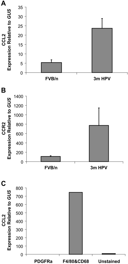
“Using an HPV-16 transgenic mice model, it has been shown that macrophage recruitment at the lesion site was dependent on CC chemokine ligand-2 (CCL2) and its receptor CCR2.”
From The Not-So-Good, the Bad and the Ugly: HPV E5, E6 and E7 Oncoproteins in the Orchestration of Carcinogenesis (2021) · cites this paper as [365]
Self-check
Percentages, sums and reported differences re-computed from the paper’s own numbers.
The self-check needs the paper’s full text.
Citations
Cites
MMP-9 Supplied by Bone Marrow–Derived Cells Contributes to Skin Carcinogenesis
Coussens, L. M., Tinkle, C. L., Hanahan, D., & Werb, Z. (2000) · Cell · doi:10.1016/s0092-8674(00)00139-2 · Cited by 1,306
Self-citationInflammatory mast cells up-regulate angiogenesis during squamous epithelial carcinogenesis
Coussens, L. M., Raymond, W. W., Bergers, G., Laig-Webster, M., Behrendtsen, O., Werb, Z., Caughey, G. H., & Hanahan, D. (1999) · Genes & Development · doi:10.1101/gad.13.11.1382 · Cited by 946 · cited 1×
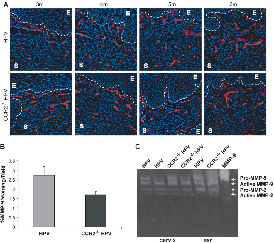
“Substrate Zymography Tissues were processed and analyzed as previously described [24].”
MethodSelf-citationCSF-1 regulation of the wandering macrophage: complexity in action
Pixley, F. J., & Stanley, E. R. (2004) · Trends in Cell Biology · doi:10.1016/j.tcb.2004.09.016 · Cited by 807
Research works printed but not linked: 38
- Waggoner, 2003, Cervical cancer, Lancet, 361, 2217, 10.1016/S0140-6736(03)13778-6 · DOI
- Bosch, 2003, Chapter 1: Human papillomavirus and cervical cancer—burden and assessment of causality, J Natl Cancer Inst Monogr, 31, 3, 10.1093/oxfordjournals.jncimonographs.a003479 · DOI
- Coussens, 1996, Genetic predisposition and parameters of malignant progression in K14-HPV16 transgenic mice, Am J Pathol, 149, 1899
- Arbeit, 1996, Chronic estrogen-induced cervical and vaginal squamous carcinogenesis in human papillomavirus type 16 transgenic mice, Proc Natl Acad Sci USA, 93, 2930, 10.1073/pnas.93.7.2930 · DOI
- Smith-McCune, 1997, Cross-species comparison of angiogenesis during the premalignant stages of squamous carcinogenesis in the human cervix and K14-HPV16 transgenic mice, Cancer Res, 57, 1294
- Bingle, 2002, The role of tumour-associated macrophages in tumour progression: implications for new anticancer therapies, J Pathol, 196, 254, 10.1002/path.1027 · DOI · Paper page
- Lewis, 2006, Distinct role of macrophages in different tumor microenviroments, Cancer Res, 66, 605, 10.1158/0008-5472.CAN-05-4005 · DOI · Paper page
- Lin, 2001, Colony-stimulating factor 1 promotes progression of mammary tumors to malignancy, J Exp Med, 193, 727, 10.1084/jem.193.6.727 · DOI · Paper page
- Lin, 2006, Macrophages regulate the angiogenic switch in a mouse model of breast cancer, Cancer Res, 66, 11238, 10.1158/0008-5472.CAN-06-1278 · DOI · Paper page
- Giraudo, 2004, An amino-bisphosphonate targets MMP-9-expressing macrophages and angiogenesis to impair cervical carcinogenesis, J Clin Invest, 114, 623, 10.1172/JCI200422087 · DOI · Paper page
- Balkwill, 2003, Chemokine biology in cancer, Semin Immunol, 15, 49, 10.1016/S1044-5323(02)00127-6 · DOI
- Audran, 1996, Adhesion molecule expression and response to chemotactic agents of human monocyte-derived macrophages, Clin Exp Immunol, 103, 155, 10.1046/j.1365-2249.1996.d01-4.x · DOI
- Robinson, 2002, Chemokine stimulation of monocyte matrix metalloproteinase-9 requires endogenous TNF-alpha, Eur J Immunol, 32, 404, 10.1002/1521-4141(200202)32:2<404::AID-IMMU404>3.0.CO;2-X · DOI
- Koide, 2004, Significance of macrophage chemoattractant protein-1 expression and macrophage infiltration in squamous cell carcinoma of the esophagus, Am J Gastroenterol, 99, 1667, 10.1111/j.1572-0241.2004.30733.x · DOI
- Ueno, 2000, Significance of macrophage chemoattractant protein-1 in macrophage recruitment, angiogenesis, and survival in human breast cancer, Clin Cancer Res, 6, 3282
- Amann, 1998, Urinary levels of monocyte chemo-attractant protein-1 correlate with tumour stage and grade in patients with bladder cancer, Br J Urol, 82, 118, 10.1046/j.1464-410x.1998.00675.x · DOI
- Kleine-Lowinski, 1999, Monocytechemo-attractant-protein-1 (MCP-1)-gene expression in cervical intra-epithelial neoplasias and cervical carcinomas, Int J Cancer, 82, 6, 10.1002/(SICI)1097-0215(19990702)82:1<6::AID-IJC2>3.0.CO;2-3 · DOI
- Zijlmans, 2006, The absence of CCL2 expression in cervical carcinoma is associated with increased survival and loss of heterozygosity at 17q11.2, J Pathol, 208, 507, 10.1002/path.1918 · DOI
- Boring, 1997, Impaired monocyte migration and reduced type 1 (Th1) cytokine responses in C-C chemokine receptor 2 knockout mice, J Clin Invest, 100, 2552, 10.1172/JCI119798 · DOI · Paper page
- Elson, 2000, Sensitivity of the cervical transformation zone to estrogen-induced squamous carcinogenesis, Cancer Res, 60, 1267
- Behrendtsen, 1992, Metalloproteinases mediate extracellular matrix degradation by cells from mouse blastocyst outgrowths, Development, 114, 447, 10.1242/dev.114.2.447 · DOI
- Riley, 2003, Dissection of human papillomavirus E6 and E7 function in transgenic mouse models of cervical carcinogenesis, Cancer Res, 63, 4862
- Bergers, 2003, Benefits of targeting both pericytes and endothelial cells in the tumor vasculature with kinase inhibitors, J Clin Invest, 111, 1287, 10.1172/JCI200317929 · DOI
- Weber, 1999, Expression of CCR2 by endothelial cells: implications for MCP-1 mediated wound injury repair and in vivo inflammatory activation of endothelium, Arterioscler Thromb Vasc Biol, 19, 2085, 10.1161/01.ATV.19.9.2085 · DOI
- Nozawa, 2006, Infiltrating neutrophils mediate the initial angiogenic switch in a mouse model of multistage carcinogenesis, Proc Natl Acad Sci USA, 103, 12493, 10.1073/pnas.0601807103 · DOI
- Henderson, 2003, Rapid recruitment of inflammatory monocytes is independent of neutrophil migration, Blood, 102, 328, 10.1182/blood-2002-10-3228 · DOI
- Bingle, 2006, Macrophages promote angiogenesis in human breast tumour spheroids in vivo, Br J Cancer, 94, 101, 10.1038/sj.bjc.6602901 · DOI
- Davidson, 1999, Expression of matrix metalloproteinase-9 in squamous cell carcinoma of the uterine cervix—clinicopathologic study using immunohistochemistry and mRNA in situ hybridization, Gynecol Oncol, 72, 380, 10.1006/gyno.1998.5285 · DOI
- Haslett, 1992, Resolution of acute inflammation and the role of apoptosis in the tissue fate of granulocytes, Clin Sci, 83, 639, 10.1042/cs0830639 · DOI
- Meszaros, 2000, Macrophage-induced neutrophil apoptosis, J Immunol, 165, 435, 10.4049/jimmunol.165.1.435 · DOI · Paper page
- Li, 2005, Blocking of monocytes chemoattractant protein-1 during tubulointerstitial nephritis resulted in delayed neutrophil clearance, Am J Pathol, 167, 637, 10.1016/S0002-9440(10)62039-1 · DOI
- Kuziel, 1997, Severe reduction in leukocyte adhesion and monocyte extravasation in mice deficient in CC chemokine receptor 2, Proc Natl Acad Sci USA, 94, 12053, 10.1073/pnas.94.22.12053 · DOI
- Jinnouchi, 2003, Impaired hepatic granuloma formation in mice deficient in C-C chemokine receptor 2, J Pathol, 200, 406, 10.1002/path.1362 · DOI
- Wareing, 2007, Chemokine regulation of the inflammatory response to a low-dose influenza infection in CCR2-/- mice, J Leukoc Biol, 81, 793, 10.1189/jlb.0506299 · DOI
- Guleria, 2001, Aberrant macrophage and neutrophil population dynamics and impaired Th1 response to Listeria monocytogenes in colony-stimulating factor 1-deficient mice, Infect Immun, 69, 1795, 10.1128/IAI.69.3.1795-1807.2001 · DOI
- Lin, 2003, Progression to malignancy in the polyoma middle Toncoprotein mouse breast cancer model provides a reliable model for human diseases, Am J Pathol, 163, 2113, 10.1016/S0002-9440(10)63568-7 · DOI
- Wyckoff, 2004, A paracrine loop between tumor cells and macrophages is required for tumor cell migration in mammary tumors, Cancer Res, 64, 7022, 10.1158/0008-5472.CAN-04-1449 · DOI
- Geissmann, 2003, Blood monocytes consist of two principal subsets with distinct migratory properties, Immunity, 19, 71, 10.1016/S1074-7613(03)00174-2 · DOI
Cited by
Accessories to the Crime: Functions of Cells Recruited to the Tumor Microenvironment
Hanahan, D., & Coussens, L. M. (2012) · Cancer Cell · doi:10.1016/j.ccr.2012.02.022 · Cited by 4,632
Self-citationMicroenvironmental regulation of tumour angiogenesis
De Palma, M., Biziato, D., & Petrova, T. V. (2017) · Nature Reviews Cancer · doi:10.1038/nrc.2017.51 · Cited by 1,847
Neutrophils in cancer: Neutral no more
Coffelt, S. B., Wellenstein, M. D., & de Visser, K. E. (2016) · Nature reviews. Cancer · doi:10.1038/nrc.2016.52 · Cited by 1,830
The role of myeloid cells in the promotion of tumour angiogenesis
Murdoch, C., Muthana, M., Coffelt, S. B., & Lewis, C. E. (2008) · Nature Reviews Cancer · doi:10.1038/nrc2444 · Cited by 1,642
Self-citationTherapeutic siRNA silencing in inflammatory monocytes in mice
Leuschner, F., Dutta, P., Gorbatov, R., Novobrantseva, T. I., Donahoe, J. S., Courties, G., Lee, K. M., Kim, J. I., Markmann, J. F., Marinelli, B., et al. (2011) · Nature Biotechnology · doi:10.1038/nbt.1989 · Cited by 839
Anti-angiogenic therapy: current challenges and future perspectives
Lopes-Coelho, F., Martins, F., Pereira, S. A., & Serpa, J. (2021) · International Journal of Molecular Sciences · doi:10.3390/ijms22073765 · Cited by 416
Neutrophils: important contributors to tumor progression and metastasis
Swierczak, A., Mouchemore, K. A., Hamilton, J. A., & Anderson, R. L. (2015) · Cancer and Metastasis Reviews · doi:10.1007/s10555-015-9594-9 · Cited by 220
Basukala, O., & Banks, L. (2021) · Viruses · doi:10.3390/v13101892 · Cited by 137
Epithelial and stromal co-evolution and complicity in pancreatic cancer
Hingorani, S. R. (2023) · Nature Reviews Cancer · doi:10.1038/s41568-022-00530-w · Cited by 108
Metadata
- Authors
- Pahler, J. C., Tazzyman, S., Erez, N., Chen, Y.-Y., Murdoch, C., Nozawa, H., Lewis, C. E., & Hanahan, D.
- Venue
- Neoplasia
- Publisher
- Elsevier BV
- Published
- 2008-04
- Volume
- 10
- Issue
- 4
- Pages
- 329-IN2
- ISSN
- 1476-5586
- Cited by
- 184
- References
- 3
- Language
- en
About these checks
Registry, retraction, Cites and statistics results are computed, not judged, with one exception: a printed reference that no index lists is read once by a model before it counts as a citation error. Claims are extracted by a model and kept only when their quote appears verbatim in the text; each cited source is then checked, with a model’s help, for whether it says what the claim relies on. Those verdicts can be wrong — read the claim’s quote and the cited source before relying on one.
Statistics and arithmetic are recomputed, not judged.
Confidence bands are shown only for fields calibrated on our annotated test set. None is calibrated yet, so no band is shown.
What counts as a claim ›How we measure confidence ›How we check this ›
Are you an author, or do you see a problem with this page? Request a review
